SCUOLA DOTTORALE in BIOLOGIA
Sezione SCIENZE BIOMOLECOLARI e CELLULARI
(Ph.D. in Biology)
XXIII Ciclo
Characterization of transgenic murine model
overexpressing spermine oxidase (SMO)
Caratterizzazione del modello murino transgenico
sovraesprimente la spermina ossidasi (SMO)
Dottoranda
Gabriella Bellavia
A.A. 2010/2011
Docente Guida: Prof. Paolo Mariottini
Coordinatore: Prof. Paolo Mariottini
TABLE OF CONTENT
Abstract
1
1.
Introduction
2
1.1.POLYAMINES 3
1.1.1. Role of polyamines in mammals 3
1.1.2. PA metabolism in mammals 4
PA synthesis 5
PA degradation 6
PA uptake 7
1.2.PAs AND BRAIN: PHYSIOLOGICAL CONDITIONS 8 1.2.1. Effect of PAs on ion channels 8 PA interaction with ionotropic Glutamate Receptors 8 AMPA and Kainate Receptors 9
NMDA Receptor 11
PA interaction with Kir channels 14 PA interaction with voltage-gated Na+ channels 15
1.2.2. PA transport in the brain 16
1.3.PAs AND BRAIN: PATHOLOGICAL CONDITIONS 17
1.4.KA-MEDIATED-EXCITOTOXICITY 19
1.4.1. Definition of excitotoxicity 19 1.4.2. The excitatory aminoacid: kainite 19 1.4.3. Molecular mechanisms in excitotoxicity 19 1.4.4. Oxidative stress mediated by KA 21 1.4.5. KA induces Apoptotic Neuronal Cell Death 21 1.4.6. Glial cell activation after KA induced-injury 23 1.4.7. PA metabolism and excitotoxicity 23
1.5.ORGANOTYPIC SLICE CULTURES 25
1.5.1. General characteristics 25
1.5.2. Applications 26
1.5.3. Model for study of excitotoxic neurodegeneration 27 1.5.4. Advantages and critical aspects of the method 27 1.5.5. Corticostriatal slice cultures 29
2.
State of Art
30
2.1. SPERMINE OXIDASE (SMO) 31
2.2. SMO discovery 31
2.3. General characteristics 31
2.4. MDL72,527: a well characterized SMO inhibitor 32 2.5. SMO activity and cellular response to PA analogues 34 2.6. Induction of SMO by various stimuli associated with 35
human pathologies
Ischemia reperfusion injury 35
Infection 35
Inflammatory cytokines 36
Prostate cancer 36
3.
Aim of research
38
4.
Results
41
4.1.EFFECTS OF KA TREATMENT ON DACH::SMO 42
TRANSGENIC MOUSE LINE
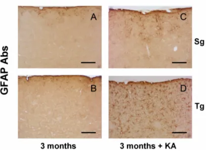
4.1.1. Behavioural evaluation 42 4.1.2. Immunoistochemical analysis 44 SMO 44 NeuN 45 GFAP 47 Iba1 48
4.1.3. Analysis of enzymatic activity of the key enzymes 49 of PA metabolism SMO 49 APAO 50 SSAT 51 ODC 52 4.1.4. Analysis of PA content 53
4.2.ORGANOTYPIC SLICE CULTURES AS A TOOL 54
FOR STUDYING THE LINK BETWEEN PA CATABOLISM AND KA EXCITOTOXICITY
4.2.1. Corticostriatal slice cultures 54 4.2.2. Effect of MDL 72,527 (SMO inhibitor) on KA 55
excitotoxicity evaluated by propidium iodide (PI) uptake and toluidine blue (TB)
PI 55
5.
Discussion and Further Perspectives
63
A
A
B
B
S
S
T
T
R
R
A
A
C
C
T
T
Natural polyamine (PAs), putrescine (Put), spermidine (Spd) and spermine (Spm) are positively charged aliphatic amines present in all tissues of almost all living organisms.
The notion that PAs are absolute required for several cell functions has led to the study of their metabolism as a strategy for therapeutic interventions. Recently, efforts are addressed to understand the link between PAs and brain physiology. This interest originates from growing data indicating that PA metabolism is affected in several neurodegenerative disorders (Alzheimer’s disease, Huntington’s disease, Parkinson’s disease, amyotrofic lateral sclerosis) or after neurotrama and cerebral ischemia.
Spermine oxidase (SMO) is an enzyme involved in PA catabolic pathway; it oxidises Spm to produce Spd and two cytotoxic compounds: 3-aminopropanal and hydrogen peroxide. The SMO gene has been found to be highly expressed in the brain organ in physiological conditions, on the other hand its altered expression in some human organs is often associated to pathological disorders. Therefore, in this study I investigated whether SMO enzyme could play a role in the pathological status such as excitotoxic condition, found to be a common mechanism in neurodegenerative disorders.
To this end, I exploited Dach::SMO mouse line which overexpresses SMO specifically in the brain cortex. Trangenic mice treated with kainic acid (KA) were more vulnerable to this neurotoxin and showed a higher neuronal cell death and gliosis and alteration of PA metabolism compared to syngenic controls.
Moreover, in vitro study performed on corticostriatal slice cultures has suggested that SMO inhibition in excitotoxic condition could be partially neuroprotective.
These data point out that SMO enzyme can be considered as a potential and additional therapeutic target against neurodegeneration induced by excitotoxicity.
1
1
I
I
n
n
t
t
r
r
o
o
d
d
u
u
c
c
t
t
i
i
o
o
n
n
This section describes polyamine metabolism focusing especially on the brain. It is here reviewed the link between this metabolism and neurodegenerative diseases. In particular, the mechanism of KA excitotocity is discussed.
Moreover, organotypic brain slice cultures were described as a useful tool in order to study the mechanisms undergoing in pathological conditions.
1.1 POLYAMINES
1.1.1. ROLE OF POLYAMINES IN MAMMALS
Natural polyamines (PAs), putrescine (Put), spermidine (Spd) and spermine (Spm) are positively charged aliphatic amines present in all tissues of almost all species (Fig. 1.1) (Wallace, 2000; Wallace et al., 2003).
The total intracellular concentration of the PAs is in the milllimolar range; however, free PA concentrations are considerably lower as they are mostly ionically bound to various anions in the cell including DNA, RNA, proteins and phopholipids (Casero & Marton, 2007). The major source of PAs in the majority of mammalian cells is via de novo biosynthesis, with the uptake by diet playing a significant, but lesser, role. The smallest contribution to intracellular PA pools is made by the gut microflora (Wallace, 2009).
The notion that PAs are absolute required for mammalian cellular functions has been already largely demonstrated. Infact, it is well-known that they play important roles in cell growth and proliferation, signal transduction, modulation of transcription and translation, as well as in the regulation of neurotransmitter receptors (Fig. 1.2) (Tabor & Tabor, 1984; Cohen, 1998; Thomas & Thomas, 2003; Williams, 1997; Wallace, 2003; Oredsson, 2003).
FIG.1.1STRUCTURE OF THE ENDOGENOUS PAS.
FIG.1.2FUNCTIONS OF PAS.
Due to their positive charge distributed along whole structure, PAs can interact with several molecules with opposite charge in the cell (nucleic acids and phospholipids). They are also involved in important processes which are required for cell functions.
1.1.2. PA METABOLISM IN MAMMALS
PA homeostasis in mammalian cells is achieved by a complex network of regulatory mechanisms concerning synthesis and degradation, as well as by membrane transport of PAs (Persson, 2009). It is clear that the cellular PAs have to be kept within certain levels for normal cell function. Infact, if the induction of cell growth is usually associated with an increase of cellular synthesis and PAs levels, on the other hand, too high concentrations of these molecules may be toxic to the cells, inducing cell death or apoptosis. Thus, cellular PA concentrations are usually maintained within rather narrow limits. PA metabolism is outlined in figure 1.3 (Wallace et al., 2003).
FIG.1.3 PATHWAY OF PA METABOLISM.
Biosynthetic and catabolic pathways of PAs are represented with blu and red arrows respectively. Acetylated PAs as well as Put can be exported from the cell. Together these mechanisms cooperate to PAs homeostasis (from Wallace, 2003).
PA synthesis
The PA biosynthetic pathway includes four different enzymes, ODC (ornithine decarboxylase), SAMDC (S-adenosylmethionine decarboxylase; also referred as AdoMetDC), spermidine synthase and spermine synthase.
ODC catalyzes the production of Put by decarboxylation of ornithine. ODC expression is tightly regulated at several levels from transcription to post-translational modification (Pegg, 2006).
Two aminopropyl groups are then added consecutively to Put to form Spd, and to Spd to produce Spm. The aminopropyl groups are provided by decarboxylated S-adenosylmethionine (dcSAM), which is itself produced from the activity of SAMDC. Spermidine synthase and spermine synthase catalyze the aminopropyltransferase reactions. These synthases are stable enzymes that
are expressed constitutively with little inducibility (Ikeguchi et al., 2006). This is in contrast with the expression of both ODC and SAMDC enzymes, which have extremely high turnover rates in the cell (Hayashi et al., 1996; Coffino, 2001; Yerlikaya & Stanley, 2004), and that are readily induced by a range of agents. The rate-limiting step in biosynthesis of PAs appears to be either the decarboxylation of ornithine or the decarboxylation of S-adenosylmethionine (SAM).
Owing to the fast turnover of ODC and SAMDC, the cellular enzyme levels and thus the corresponding activities are rapidly altered when the synthesis and/or degradation of the enzymes are changed. Both enzymes are subject to a strong feedback control by PAs. ODC and SAMDC are rapidly up-regulated when cells become depleted of their PA content. On the other hand, both enzymes are down-regulated when cells are exposed to an excess of PAs.
PA degradation
Spd and Spm may be degraded to Put and Spd respectively, in a two-step process usually referred to as ‘the PA interconversion pathway’ (Seiler, 2004). The first step in the interconversion process is the acetylation of the N1-nitrogen of Spd and Spm producing N1-acetylspermidine or N1-acetylspermine (N1-acetylSpd, N1-acetylSpm). This reaction is catalysed by the enzyme spermidine/spermine N1-acetyltransferase (SSAT).
The second step is the oxidation of acetylated PAs producing Put and Spd respectively. It is catalyzed by a FAD-dependent peroxisomal PA oxidase that is nowadays referred to as the N1-acetylpolyamine oxidase (APAO), to distinguish it from the lastly discovered spermine oxidase (SMO). Infact, towards the end of 2002, an oxidase was cloned that converts directly Spm back into Spd, without the need for an acetylation step and producing 3-aminopropanal (3AP) and hydrogen peroxide (Wang et al., 2001; Vujcic et al., 2002). This enzyme has been termed SMO.
Like APAO, SMO is a FAD-dependent oxidase but, unlike the former enzyme, SMO has a high specificity for Spm as a substrate. In great contrast with APAO, SMO is highly induced by several PA analogues, indicating an important role in PA homoeostasis (Wang et al., 2001). However, the regulation of SMO appears to be mainly at the level of mRNA transcription-stability, rather than translation/post-translation (Wang et al., 2005).
The products of APAO activity are Put and Spd respectively, as well as 3-acetamidopropanal and hydrogen peroxide. Put may be further oxidized by diamine oxidase, whereas Spd may undergo another round in the interconversion pathway. The acetylated derivatives of Spd and Spm may also be excreted from the cells. The exact mechanisms by which the acetylated PAs
The rate-limiting step in the PA interconversion pathway is catalyzed by SSAT. Like ODC and SAMDC, SSAT has a very fast turnover with a half-life as short as 15 min, whereas APAO is a stable enzyme (Casero & Pegg, 1993). The rapid turnover of SSAT is mediated by the 26S proteasome and is dependent on ubiquitination of the protein (Coleman & Pegg, 2001). SSAT is strongly induced by a variety of stimuli, including various toxins and hormones. Thus, similar to ODC and SAMDC, SSAT plays an important role in PAs homoeostasis.
PA uptake
In addition to regulating PA levels by synthesis, degradation and efflux, cells are equipped with an active transport system for the uptake of PAs (Mitchell et al., 2007). Large amounts of PAs are found in the food. Bacteria in the intestinal system produce and excrete considerable quantities of PAs. It is conceivable that a large fraction of these exogenous PAs are absorbed from the intestines and later taken up and used by cells in the body. However, to what extent cells rely upon endogenous compared with exogenous PAs is not yet clear. Nevertheless, in situations when the PA biosynthetic machinery is insufficient, cells would certainly be more dependent on extracellularly derived PAs. The exact mechanisms and the proteins involved in PA transport are still not identified. Whether there are individual transport systems for the various PAs or only a single transporter, capable of transporting all of the PAs, is not clear. Results obtained indicate that uptake of PAs by mammalian cells, at least partly, occurs by a mechanism involving cell-surface heparin sulfate proteoglycans and endocytosis (Welch et al., 2008). The polysaccharides of the proteoglycans are negatively charged and may interact with the positively charged PAs with affinities even stronger than the interaction between DNA and PAs. Moreover, it was recently demonstrated that PA uptake in human colon cancer cells follows a dynamin-dependent and clathrin-independent endocytic route, which is negatively regulated by caveolin-1 (Roy et al., 2008). A cell surface protein, capable of transporting both Put and Spd, has been cloned from the protozoan pathogen Leishmania major (Hasne & Ullman, 2005).
Mitchell et al., (2007) have shown that the activity of the PA transporter is partly regulated by cellular PA levels. Cellular depletion of PAs results in a marked increase in the cellular uptake of exogenous PAs. On the other hand, the PA transporter is down-regulated when cells are exposed to an excess of PAs. This feedback regulation is dependent on protein synthesis and involves a protein with a very fast turnover. Interestingly, antizyme (Az), which is an inhibitor of ODC enzyme, induced by an excess of PAs, also appears to negatively regulate the cellular PA transporter (Mitchell et al., 2007). Cells in
which Az is expressed to high levels exhibit a marked reduction in PA uptake. All three different forms of Az (Az1–Az3) have been shown to effectively down-regulate PA transport. However, the mechanism by which Az affects PA uptake is so far unknown.
1.2. PAs AND BRAIN: PHYSIOLOGICAL CONDITIONS
The functional role of the natural PAs in the normal and pathological states of the brain is under active research.
Several studies have led research to suppose a particular role of PAs in mammalian brain. It is intriguing to imagine PAs could play a peculiar function in the central nervous system (CNS) in addition to an universal role in the regulation of cell proliferation and growth, as seen in other organs.
Infact, endogenous PAs exert a number of key regulatory functions in the CNS ranging from cell proliferation to interactions with ion channels (Paschen, 1992; Scott et al., 1993; Kauppinen & Alhonen, 1995; Seiler et al., 1996; Williams, 1997). While the higher PAs Spd and Spm, are present at submillimolar total concentrations (300-500 µmol/kg), Put is expressed in the brain only following stimulatory factors as a consequence of ODC activation (Lukkarinen et al., 1998).
1.2.1. EFFECT OF PAs ON ION CHANNELS
In the last decade it has been demonstrated that PAs play important roles in the regulation of ion channels. Infact specific interactions of PAs with a number of distinct types of cation channels has been described. PAs have multiple modulatory effects on ionotropic glutamate receptors (iGluRs), they also interact with inwardly rectifying potassium channels (Kir) as well as some other channels that affect intracellular Ca++ signaling or Na+ transport (Williams, 1997; Fleidervish et al., 2008).
PA interaction with ionotropic Glutamate Receptors (iGluR)
iGluRs are major mediators of rapid excitatory neurotransmission in the mammalian CNS.
These receptors comprise a family of ligand-gated ion channels that include the amino-3-hydroxy-5-methyl-4-isoxazolepropionic acid (AMPA), kainate (KA), and N-methyl-D-aspartate (NMDA) receptors (Fig 1.4).
subunits themselves share a common membrane topology, with three transmembrane domains (termed M1, M3, and M4) and a reentrant loop similar to the P loop found in potassium channels (termed M2).
PAs influence GluRs mediating both fast responses at excitatory synapses, such as AMPA receptors and KA receptors and slow voltage-dependent responses such as NMDA receptors (Dingledine et al., 1999).
FIG.1.4 CLASSIFICATION OF IONOTROPIC GLUTAMATE RECEPTORS (IGLURS).
Ionotropic Glutamate Receptors (iGluR) are divided in three main subfamilies: NMDA, AMPA and Kainate receptors. Each group is characterized by different glutamate receptor subunits (from: http://www.bris.ac.uk/synaptic/receptors).
AMPA and Kainate Receptors
Recent studies have shown that intracellular PAs have a profound effect on some subtypes of AMPA and KA receptors, where PAs control rectification of these receptor channels.
Native AMPA and KA receptors are heterooligomers composed of combinations of GluR and KA subunits.
AMPA receptors are ligand-gated channels composed of four possible subunits (GluR1–4) and a large number of them have been cloned (Hollmann & Heinemann, 1994). They are responsible for fast excitatory neurotransmission in the CNS.
KA receptors are formed of the GluR5-7 and KA1-2 subunits (Hollmann & Heinemann, 1994). GluR5-7 form the channel where KA1-2 are peptides that alter the biophysical characteristic of the KA channel complex. They are similar to AMPA receptors but have unique roles in synaptic function in
sensing pain, neuronal development and synaptogenesis and in supporting plasticity processes that are the cellular bases of learning and memory.
Most native AMPA and KA receptors gate Na+, but are relatively impermeable to Ca2+ and have I-V relationships that are close to linear. However, a subset of receptors show a high Ca2+ permeability and pronounced inward rectification (Ilno et al., 1990; Burnashev et al., 1992).
The intracellular PAs are able to interact with the subset of AMPA and KA receptors that gate Ca2+. They act by blocking the pore of the these receptors to prevent the flux and Ca2+ as the membrane potential is depolarized (Fig.1.5) (Bowie & Mayer, 1995).
The sensitivity of AMPA and KA receptors to PAs depends on their subunit composition and on RNA editing of the GluR subunits. Infact, the diversity of GluR subunits is extended by alternative splicing of their mRNAs and by editing of the pre-mRNA (Hollmann & Heinemann, 1994).
The RNA editing occurs at a so-called ‘glutamine/arginine (Q/R) site’ of the GluR2 (AMPA subunit) and GluR6 (KA subunit) mRNAs (Kohler et al., 1993; Seeburg, 1996). It is a crucial event because it controls the ionic property of these channels. The presence of an arginine residue (R) in the pore channel results in receptors that have low permeability to Ca2+, whereas the presence of a glutamine (Q) residue leads to receptors with higher Ca2+ permeability. Therefore, AMPA receptors, those lacking the GluR2 subunit or with GluR2 (Q) unedited form, are permeable to Ca2+ions and are subject to a block by endogenous intracellular PAs (predominantly Spm). Binding of Spm occurs within the pore region of the channel and is voltage dependent. Infact, at depolarized potential Spm blocks the channel, and as the cells is hyperpolarized, it unbinds and returns to the cytoplasm. At extreme depolarized potentials (more positive than + 50 mV), Spm can permeate the ion channel and can pass through to the extracellular side. This interaction confers inward rectification at these channels.
Likewise, the GluR6 subunit of KA receptors can also be edited and this determines the extent of KA receptors that allow the permeation of Ca2+ ions and that are sensible to the blockage by intracellular PAs at positive potential. The degree of RNA editing for GluR6 is developmentally regulated. In the early embryonic rat brain, the GluR6 (Q) form is found exclusively, and GluR6 (R) is co-expressed (≅ 50% of GluR6) at birth. In adult rats, the edited GluR6 (R) represents 70-85% of GluR6 in the hippocampus (Bernard & Khrestchatisky, 1994). Thus KA receptors in the foetal and neonatal nervous system may be particularly sensitive to PAs, which could play a role in synapse formation, plasticity, and elimination during development.
Therefore, PAs may regulate the amount of Ca2+ flux and the excitability threshold at synapses by the interaction with containing PA-sensitive AMPA/KA receptors.
FIG.1.5 PA INTERACTION WITH AMPA/KA RECEPTORS.
Intracellular Spm (dark rectangles) can block AMPA and KA receptors at depolarized potentials and, as the cell is hyperpolarized, Spm unbinds and returns to the cytoplasm (solid arrow). At extreme depolarized potentials (more positive than +50mV), Spm can permeate the ion channels and pass through to the extracellular side (broken arrow) (from Williams, 1997, modified).
NMDA Receptor
The NMDA receptors are involved in synaptic plasticity and may also play a role in seizure activity (Collingridge & Lester, 1989; McBain & Mayer, 1994). NMDA receptors have a number of characteristics that distinguish them from other GluRs. Activation of NMDA receptors requires binding not only of the agonist glutamate but also of a co-agonist, glycine, at a separate site on the receptor. NMDA channels are blocked in a voltage-dependent manner by extracellular Mg2+, leading to a decreased conductance at negative potentials, with little or no current flow below about -70 mV.
Two families of NMDA receptor subunits, termed NR1 and NR2, have been cloned. The NR1 subunit is the product of a single gene that is transcribed as eight alternatively spliced mRNAs. The NR2 subunits, NR2A-NR2D, are distinct gene products with different regional and temporal patterns of expression (Hollmann & Heinemann, 1994). Native NMDA receptors are probably hetero-oligomers composed of combinations of NR1 and NR2 subunits.
NMDA receptors may contain two copies of the NR1 subunit in each receptor complex (Behé et al., 1995), and at least some NMDA receptors contain two different types of NR2 subunit (e.g. NR2A and NR2B) within a single hetero-oligomer (Luo et al., 1997; Wafford et al., 1993). Most studies of recombinant
NMDA receptors have focused on ‘binary’ receptors containing NR1 and one type of NR2 subunit (e.g. NR1/NR2B).
Modulation of NMDA receptors by PAs was first reported in 1988 by Ransom and Stec (Ransom & Stec, 1988) who showed that Spm and Spd increased the binding of [3 H] MK801 (an open-channel blocker of NMDA receptors) to the brain membranes. In electrophysiological studies, extracellular Spm was found to enhance NMDA-induced whole-cell currents in cultured neurons (Williams et al., 1990; Benveniste & Mayer, 1993).
Multiple and sometimes opposing effects of extracellular Spm on these channels have now been described (Williams, 1997).
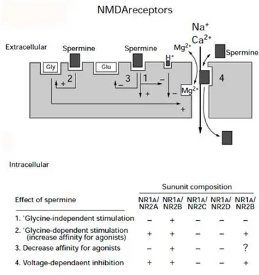
Spm has four macroscopic effects that are differentially controlled by alternative splicing of exon 5 in the NR1 subunit and by the various NR2 subunits.
One is termed “glycine independent stimulation” because Spm potentiates NMDA currents in the presence of saturating concentrations of glycine. It involves an increase in the frequency of channel opening and a decrease in the desensitization of NMDA receptors (Benveniste & Mayer, 1993, Lerma, 1992; Rock & Macdonald, 1992).
The second effect is a “glycine-dependent stimulation” which produces an increase in the affinity of the receptor for glycine.
At physiologic pH, both stimulatory effects are seen at NR1/NR2B receptors but not at NR1/NR2A receptors (Han et al, 2008).
The third effect is a voltage-dependent inhibition. Spm inhibition of NMDA receptor is strongly voltage-dependent, being more pronounced at hyperpolarized than at depolarized membrane potentials. It occurs via interactions in the outer vestibule of the channel pore. Mutations affecting this interaction have been mapped (Jin et al., 2008).
It is conceptually similar to the block of AMPA channels and Kir channels (see below) by intracellular PAs, except that, in the case of NMDA receptors, the channels are blocked by extracellular Spm, although the block is very weak compared with the block of Kir and AMPA channels by intracellular Spm. The fourth effect is a decrease affinity for agonist glutamate; it has been observed at some recombinant NMDA receptors (Williams, 1994).
The mechanism underlying this effect is not known, but it may reflect an increased rate of dissociation of glutamate from the receptor in the presence of Spm. In this case, Spm could have a marked effect on the time course of NMDA responses at the synapse, where the duration of the response is dependent on the rate of unbinding of glutamate from the receptors (Clements et al.,1992; Lester et al., 1990).
All these effects of PAs on NMDA receptors activity are schematized in figure1.6.
(Harman & Shaw, 1981; Fage et al., 1992). Thus it is possible that PAs are released from neurons or glia in the brain and can reach concentrations in the synaptic cleft that are sufficient to influence the activation of NMDA receptors, although there is as yet no direct evidence that this happens in vivo.
Excessive activation of NMDA receptors leads to neurodegeneration, and it is conceivable that excessive release of PAs, for example from injured cells, could exacerbate neuronal injury by potentiating the activity of NMDA receptors.
FIG.1.6MODULATIONSOFNMDARECEPTORSBYEXTRACELLULARPAS.
Extracellular Spm has multiple effects on NMDA receptors ranging from stimulation to inhibition according to the subunit composition. Spm can block NMDA channels at negative membrane potentials (solid arrow), and at extreme negative potentials Spm can permeate the NMDA receptor to pass into the cell (broken arrow) (from Williams, 1997, modified).
PA interaction with Kir channels
Different forms of K+-selective ion channels have been identified, based on their biophysical, pharmacological and molecular properties. These include inward rectifier potassium channels (Kir) (Doupnik et al., 1995).
The term ‘inward rectifier’ refers to the ability of the channel to conduct ions in the inward direction at negative membrane potentials, but to show a greatly decreased outward conductance at membrane potentials positive to the potassium equilibrium potential (Ek ;Fig. 1.7). Kir channels, which are present in both excitable and non-excitable cells, are crucial for maintaining the resting membrane potential close to Ek (Doupnik et al., 1995).
The Kir gene family consists of 7 subfamilies (Kir1-7). These channels are tetramers of pore-forming subunits with two transmembrane domains (M1 and M2) separated by a P-region. The P-loop and a segment of the M2 domain form the transmembrane pore. There is also a cytoplasmic pore containing the binding site for the ligands and other regulators and controlling access to the transmembrane pore.
Voltage-dependent block by intracellular PAs is the common mechanism underlying the inward rectification in all the Kir channels. All of the natural PAs can bind and have some effects in experimental conditions but the affinity increases from Put to Spd to Spm (Stanfield et al., 2003; Guo et al., 2003). This block has been most intensively studied in some strongly inward rectifying channels such as Kir2.1 and Kir6.2 (Yan et al., 2005; Kurata et al., 2008). Evidence for different states in these channels with low and high affinity blocks has been published (Ishihara et al., 2007). Two different regions with negatively charged micro-environments are critical for binding PAs and determining inward rectification characteristics; one is in the cytoplasmic pore and the other in the transmembrane pore (Guo et al., 2003; Kurata et al., 2007). Models in which pre-positioning of PAs at the cytoplasmic pore then facilitates entry of PAs into a deeper binding site located within the membrane pore, are consistent with the experimental data using PAs and experimental analogs (Kurata et al., 2007, 2008).
Mice with a deletion in the spermine synthase gene (also referred as Gyro mice), have no Spm, show elevated Spd levels, and are also totally deaf. These mice have an almost complete loss of endocochlear potential. This phenotype may be explained by effects of the PAs imbalance on the cochlear lateral wall-specific Kir4.1 channel, which is known to play a critical role in the maintenance of this potential (Becerra-Solano et al., 2009).
FIG.1.7BLOCKAGEOFKIRCHANNELSBYPAS.
Two molecules of intracellular Spm can enter deep within the ion-channel pore and may block the channel simultaneously (from Williams, 1997, modified).
PA interaction with voltage-gated Na+ channels
Voltage-gated Na+ channels are critical elements of action potential initiation and propagation in excitable cells because they are responsible for the initial depolarization of the membrane.
They are formed by a single long polypeptide that have four domains (I-IV). The four domains join together to form an acqueous pore of the channel. The membrane excitability and input/output properties of neurons are largely determined by the spatial distribution and availability of their voltage-gated Na+ channels. In neocortical pyramidal cells, Na+ channels are present not only in the axon, where voltage threshold for action potential (AP) generation is lowest, but also in soma and dendrites.
Recently, it has also been reported a novel neuromodulatory mechanism that links the availability of Na+ channels to metabolism of PAs in the cerebral cortex. Fleidervish and collegues (2008) have demonstrated that PAs, which are normally present in the intracellular and extracellular compartments, are endogenous blockers of Na+ channels in layer 5 pyramidal cells. Because the blockade is activity-dependent, it is particularly effective against Na+ channels which fail to inactivate rapidly and thus underlie the persistent Na+ current. These data suggest that changes in PA levels, whether associated with normal brain states or pathological conditions, profoundly modify the Na+ availability
and thereby shape the integrative behaviour of single neurons and neocortical circuits (Fleidervish et al., 2008).
1.2.2. PA transport in the brain
Since PAs have the ability to modulate the activity of iGluRs and thus synaptic transmission in vivo, it is reasonable suppose that they can be released from neurons or glia and rapidly reincorporate into those cells in the brain. However, little is known about mechanisms underlying transport, uptake and release of PAs in neurons and glia.
It has been demonstrated PAs are taken up in cerebral cortex slices (Harman and Shaw, 1981), cultured cerebellar astrocytes (Dot et al., 2000) and synaptosomes (Gilad and Gilad, 1991). Masuko et al., (2003) characterized PA transport systems in synaptic vesicles, synaptosomes and glial cells, as well as the release of spermine from hippocampal slices. They have suggested that PAs transporters have broad spectra of substrate specificity and recognize agmatine, histidine and histamine as well as PAs (Spm, Spd and Put) in synaptosomes and glial cells. By contrast, PA transport system is rather selective for histamine only, beside PAs, in synaptic vesicles (Masuko et al., 2003). Moreover, another report has shown that agmatine can be transported by a PA transporter in NIH3T3 cells (Satriano et al., 2001).
A speculation regarding the possible traffic and action of PAs at synapsis was suggested by Takano and collegues (2005). They have hypothisized PAs could be released into synaptic clefts from neurons to facilitate the opening of NMDA receptor channels permeable to Ca2+ ions in response to particular pathological stimuli. On the other hand, intracellular PAs may inhibit the influx of cations across AMPA and KA receptor channels through plugging of ion channel pores at the intracellular domains. Then, extracellular PAs would be incorporated into intracellular spaces through particular transporters expressed by neurons, astrocytes and/or microglia. (Fig. 1.8) (Takano et al., 2005).
FIG.1.8MODELOFPAACTIONATSYNAPTICCLEFT.
PAs could be release into synaptic clefts from neurons to facilitate the opening of NMDA receptor channel permeable to Ca+2 ions in response to particular pathological stimuli. Intracellular PAs may inhibit the influx of cations across AMPA and KA receptor channels through plugging of ion channel pores at the intracellular domains. Extracellular PAs woul be incorporated into intracellular spaces through particular transporters expressed by neurons, astrocytes and/or microglia (from Takano et al., 2005).
1.3. PAs AND BRAIN: PATHOLOGICAL CONDITIONS
In different neurodegenerative disease it has been found a change in PA metabolism. It often consists in an alteration of the delicate equilibrium among the three PAs. The mechanism of homeostasis is affected, and maybe it cannot work to restore the proper PA content in the cell.
Considering the several and additional roles that PAs play in neurons, a misregulation of this pathway can affect dramatically neuron functioning. PAs concentrations increase in the brain of neurodegenerative disorders, such as Alzheimer’s disease (AD) and ischemia (Morrison and Kish, 1995; Paschen et al., 1987). Elevated levels of immunoreactive ODC protein are demonstrated in neocortex of AD patients (Bernstein and Muller, 1995). The β-amyloid induces ODC activity and also stimulates PA uptake (Yatin et al., 1999). Recently, it has been reported the accumulation of AZ inhibitor 2 (AZIN2) and consequently ODC activation in neurons of hippocampus of AD patients (Makitie et al., 2010). Morrison et al. (2003), have shown an increased activity
of SAMDC, the key rate-limiting enzyme of Spd and Spm byosynthesis, in autopsied brains of patients with AD.
One of the most widely used animal models of Huntington’s disease (HD) is based on the intrastriatal administration of quinolinic acid (QA). Spm treatment protects against the impairment of recognition memory and prevents the astrogliosis but not neuronal damage in the striatum of rat model of HD (Velloso et al., 2009).
In addition PAs are also implicated in the pathogenesis of ischemic brain damage (Zhang et al., 1994; Harman and Shaw, 1981; Bergeron et al., 1996; Glantz et al., 1996; Anderson et al., 1994).
PA biosynthesis is increased after the onset of cerebral ischemia, for example due to an ischemia-mediated induction of ODC (Kindy et al., 1994; Marton and Pegg, 1995; Lovkvist-Wallstrom et al., 1995, Pegg et al., 1994; Paschen, 1992). The administration of inhibitors of ODC prevents the development of ischemic brain damage, suggesting that the accumulation of PAs plays an important role in the pathogenesis of stroke in the ischemic brain (Kindy et al., 1994).
Several data have also contributed to point the attention on PA catabolism as potential target of a neuroprotective strategy.
It has been shown in the normal brain that approximately 70% of Put is derived from PA interconversion pathway, whereas only approximately 30% is formed by de novo synthesis from ornithine (Seiler, 1995).
Treatment of rats with inhibitors of PA oxidases prevents the production of 3-aminopropanal (3 AP), and significantly protects against the development of ischemic brain damage in vivo. 3 AP, as toxic product of APAO activity, has been proposed as a mediator of the brain damaging sequelae of cerebral ischemia, which can be therapeutically modulated (Ivanova et al., 1998). It is worthy to underline that now it is known that another enzyme, SMO, contributes to the production of 3 AP as well as hydrogen peroxide (H2O2). It has also found a relationship between PA catabolism and traumatic brain injury (TBI). Inhibition of PA oxidase with MDL 72,527 is neuroprotective against edema formation and necrotic cavitation after TBI (Dogan et al., 1999). PA back-conversion is enhanced after TBI; SSAT and SMO expression increased in brain damaged as well as Spd, Put and acetylated Spd content. In particular they have observed a late induction of SMO (from 3 to 7 days post-injury) which correlates very well with Spd increases, suggesting that SMO activity may be elevated at later times post-injury. Thus oxidation of essential PAs may also be considered a source of secondary tissue damage, increased inflammation, and apoptotic cell death in the injured brain.
A possible mechanism through which enhanced PA back-conversion may contribute to cerebral injury is through production of toxic metabolites. Increases of aminoaldehyde and pro-oxidant metabolites, such as H2O2 and 3-acetyl-aminopropanal, are damaging to vulnerable brain tissue (Li et al., 2003;
2007). Therefore, the onset of PA back-conversion and PA synthesis after brain injury, together with concomitant elevations of oxidative metabolites and acetylated PAs, likely instigate secondary tissue injury. Thus, treatments that retard PA catabolism and reduce these cytotoxic oxidative byproducts may be expected to reduce secondary tissue damage in brain regions that demonstrate enhanced PA catabolism after TBI (Zahedi et al., 2010).
1.4. KA-MEDIATED EXCITOTOXICITY
1.4.1. Definition of excitotoxicity
In 1957, Lucas and Newhouse first described the neurotoxic effect of monosodium glutamate on retina of the mouse. Twenty years later, the explanation for this effect was formalised in the concept of excitotoxicity by John Olney (1978). Excitotoxicity refers to a process of neuronal death caused by excessive or prolonged activation of excitatory amino acid receptors. Infact in normal synaptic functioning, activation of these receptors is a transitory event. However, if, for any reason, receptor activation becomes excessive or prolonged, the target neurons become damaged and eventually die.
1.4.2. The excitatory amino acid: kainate
Kainic acid (KA) (2-carboxy-4-isopropenylpyrrolidin-3-ylacetic acid), also known as alga kaininso, is isolated from Digenea, a red alga found in tropical and subtropical waters (Coley et al., 1987). KA has been used for centuries as an anthelminthic compound for removal of worms in the gut. Subsequent studies indicated KA as a nondegradable analog of glutamate and it is 30-fold more potent in neurotoxicity than glutamate (Bleakman & Lodge, 1998). This neuroexcitant can bind to the AMPA/KA receptors in the brain (Bleakman & Lodge, 1998). Activation of KA receptor has been shown to elicit a number of cellular events, including the increase in intracellular Ca2+, production of ROS, and other biochemical events leading to neuronal cell death (Sun et al., 1992; Candelario-Jalil et al., 2001). In recent years, neurodegeneration caused by systemic injection of KA has been widely used in studies to investigate mechanisms of excitotoxicity (Wang et al., 2005).
1.4.3. Molecular mechanisms in excitotoxicity
Excessive stimulation by excitatory amino acid (EAAs) can cause death of the postsynaptic neuron. Addition of an EAAs, such as KA, stimulates both
AMPARs and KARs, which mediates fast synaptic potentials, and results in opening of cation channels permeable to Na+, K+ and sometimes Ca2+ depending on the subunit composition (Arundine & Tymianski, 2003; Wang et al., 2005).
Thus, the initial response is a large influx of Na+ escorted by water and Cl -which makes neurons appear swelled (Choi, 1992).
Continuous stimulation leads to an increase of the intracellular Ca2+ concentration. Active AMPA and KA receptors can enable Ca2+ to flow into the cell, however, the Na+ influx is substantial to generate depolarization of the cell, which is a prerequisite for activation of the slow potential NMDA receptors (Standaert et al., 1999). In this conditions, NMDA receptors release their magnesium block and allowing Ca2+ to flow through the channel. NMDA activation represents the major source of calcium entry into the cell.
Ca2+ overload can activate a series of enzymes including proteases, endonucleases and phospholipases that are Ca2+-dependent and when triggered contribute to protein breakdown and DNA fragmentation (Wang et al., 2005; Sanchez et al., 2008). Additionally, energy metabolism in the mitochondria is compromised, and the resulting lack of ATP hamperes ATP-dependent ion pumps in the plasma membrane, which causes destruction of the electrochemical gradient across the cell membrane. Formation of reactive oxygen species (ROS) increases and furthermore, nitric oxide synthase (NOS) is activated. In collaboration with ROS, NOS generate reactive nitrogen oxide species (RNOS) that cause destruction of cell membrane integrity and attack membrane lipids, proteins and DNA (Rego & Oliveira, 2003; Wang et al., 2005a; Estrada Sanchez et al., 2008).
These conditions might lead to neuronal death by apoptosis or necrosis, with further release of glutamate as a consequence. Then glutamate may act on neighboring cells by same mechanism. Contrary to KA, glutamate induces all types of iGluRs and mGluRs (metabotropic glutamate receptors) in the postsynaptic neuron, which additionally contribute to an increase in intracellular Ca2+ concentration (Choi, 1992). This self-propagating pattern can thus cause extensive neuronal death.
However, an increase in intracellular Ca2+ concentration does not always denote neuronal death. It appears that a combination of the severity of an insult and the increase in Ca2+ concentration determines the fate of the neuron. Thus, a thin line separates the outcome for the neuron and might also decide if death is caused by apoptosis or necrosis (Choi, 1995). Therefore, changes in glutamate concentration, shifting receptor-density in the cells, variations in subunit composition of GluRs and signaling properties, may all be cellular events that alter the severity of the insults and affect the outcome of excessive EAA stimulation.
1.4.4. Oxidative Stress Mediated by KA
Several studies linking oxidative stress to KA-mediated neurotoxicity.
Studies have demonstrated the production of free radicals after KA administration in vivo (Sun et al, 1992) and in neuron cells in vitro (Chen & Sun, 1994). KA also stimulated the release of lactate dehydrogenase (LDH), an indication of loss of cell membrane integrity, and a decrease in 3-(4, 5-dimethylthiazole-2-yl)-2, 5-diphenyl tetrazolium bromide (MTT), suggesting a decrease in mitochondrial function.
Exposure of rat brain homogenates to KA can significantly increase the production of malondialdehyde and 4-hydroxy-alkenals, suggesting an increase in lipid peroxidation (Candelario-Jalil & Sonia Leon 2003). In addition to the increase in lipid peroxidation, systemic administration of KA also caused a decrease in glutathione (GSH) levels in the hippocampus, cerebellum, and amygdala/piriform cortex (Wang et al, 2004). The increase in superoxide production and oxidative DNA damage following KA administration are indications of KA induced mitochondrial and oxidative damage (Patel & Li, 2003). Injection of KA, also, causes the increase in nitric oxide synthase (NOS) in neurons (Yasuda et al, 2001).
1.4.5. KA Induces Apoptotic Neuronal Cell Death
The opening of Ca+2-AMPA/KA receptor channels in the postsynaptic terminal permits rapid Ca2+ influx and stimulates oxidative pathways resulting in the generation of ROS. There is sufficient evidence that ROS generation could lead to mitochondrial dysfunction and subsequent apoptotic or necrotic cell death pathways. Apoptotic pathways can be triggered as a result of the collapse of the mitochondrial membrane potential (m∆ψ) and the opening of mitochondrial permeability transition pores (MPT) that allow the release of cytochrome-c into the cytoplasm. In turn, cytochrome-c in cytoplasm is coupled with the apoptotic-inducing factor (AIF), which subsequently leads to activation of the caspase cascade (Weiss & Sensi, 2000). Rats treated with KA showed degenerating neurons in the hippocampus. Neurons were intensely stained with in situ nick end labelling (ISNEL) and displayed pathological features suggesting both necrosis and apoptosis (Nishiyama et al., 1996). Intraventricular infusion of KA into adult mouse brain also caused neuronal morphological changes, with condensed nuclei reflecting of apoptosis in the pyramidal layer of the hippocampal formation (Osaka et al., 1999). In organotypic hippocampal slice cultures, KA-mediated neuronal damage was associated with complete reduction of rhodamine 123 fluorescence, an indication of mitochondrial membrane potentialdissipation, and increased
levels of cytochrome-c and caspase-3 in the cytosol. Cyclosporin A, an inhibitor of MPT opening, partially prevented cytochrome-c release, caspase activation, and neuronal death. Inhibition of caspase-3 activity by an inhibitor, z-VAD, also partially protected neurons from KA-induced cytotoxicity (Liu et al., 2001).
FIG.1.9NEURONALCELLDEATHPATHWAYINDUCEDBYKA.
KA stimulates the AMPA/KA receptors, leading to rapid Ca2+ entry; (2) activation of Ca2+-dependent enzymes and generation of ROS; (3) excessive Ca2+ and ROS lead to collapse of mitochondrial membrane potential (m∆Ψ) and opening of mitochondrial permeability transition pores (MPT); (4) release of mitochondrial factors (e.g., cytochrome-c and apoptotic-inducing factor (AIF); (5) cytochrome-c binding to Apaf-1 and caspase-9 to form apoptosome complex and activation of caspase-3 pathway; (6) nuclear condensation and DNA fragmentation. Alternatively, intense Ca2+ overload could directly cause mitochondrial swelling and damage, decrease in ATP, and increase in ROS, which oxidize protein, lipid, and DNA, causing acute neuronal necrosis (from Wang et al., 2005).
1.4.6. Glial Cell Activation After KA-Induced Injury
Activation or recruitment of glial cells (astrocytes and microglial cells) is a common event associated with neuronal injury. Astrocytes comprise the major cell type in the brain and they are known to play multiple functional roles in support of neurons (Aschner, 1998; Zhang et al., 2000). Both astrocytes and microglial cells are immune active and become activated under pathological conditions. Reactive astrogliosis and microgliosis are intimately associated with many neurodegeneration processes and contribute to the increase in inflammatory factors and ROS. Astrocytes are able to respond to pro-inflammatory cytokines, which stimulate transcription factors and cause induction of a number of genes, including inducible nitric oxide synthase (iNOS) (Li et al., 1999; Akama & Van Eldik, 2000), cyclooxygenase-2 (COX-2) (Kyrkanides et al., 200(COX-2), and secretory phospholipase A2 (sPLA(COX-2) (Li et al., 1999; Dorandeu et al., 1998; Wang & Sun 2000). Increase in PLA2 has been regarded as an important factor underlying a number of neurodegenerative diseases, and PLA2 inhibitors have been shown to protect against neurotoxicity induced by oxidative stressors (Xu et al., 2002; Farooqui et al., 1997). Systemic injection of KA to rats was shown to enhance cytosolic PLA2 immunoreactivity in the hippocampal area (Farooqui et al., 2004). Systemic injection of KA to rats resulted not only in neuronal cell death in the hippocampal area but also a large increase in reactive astrocytes and microglial cells (Wang et al, 2004).
These results further demonstrate the intimate relation between glial cell activation and neuron cell death in excitotoxic injury in the brain.
1.4.7. PA metabolism and excitotoxicity
PA property to interact with iGluR lead to hypothesize a link between these molecules and excitotoxicity phenomenon. However if it is known the cascade of events occurring during excitotoxicity it has not been understood the mechanism that may correlate excitotoxicity and PAs.
PA interconversion pathway is rapidly activated in limbic area following kainate-induced seizures activity. This was demonstrated both by an increase in SSAT activity, as well as in actetylated PA levels following the inhibition of PA oxidase. The results suggested that the increase in Put levels reported in several types of insults are due not only to the increased expression of ODC and the biosynthetic pathway but also to the stimulation of the interconversion via. As the catabolic pathway generates H2O2 it is conceivable that activation of this pathway contributes to the pathological manifestations of seizure activity,
such as neuronal damage and glial proliferation, as oxygen radicals have been suggested to be involved in the excitotoxicity (Baudry & Najm, 1994).
An overshoot of Put after different kind of injuries has been documentated. For instance, a concurrence between increases in Put concentrations and hippocampal or frontal cortex damage was observed in rats treated with GluR agonists in order to induce seizures. This relationship supports the proposed role of Put as a biochemical marker of acute brain damage (Camon et al., 2001; de Vera et al., 2002). It is interesting to underline that KA increases brain Put levels whereas picrotoxinin or pentilenetetrazol-induced seizures did not modify PA levels, suggesting that epileptic seizures per se do not induce Put increases (Martinez et al., 1990).
1.5. ORGANOTYPIC SLICE CULTURES
1.5.1. General characteristics
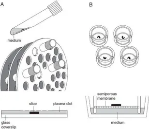
The culturing of slices of developing brain tissue dates back to over two decades. In 1981 Gahwiler (Gahwiler, 1981) introduced the roller drum technique to culture a number of different brain areas (Gahwiler et al., 1992). This method required that slice cultures were embedded in either a plasma clot or in a collagen matrix on a glass coverslips, placed inside tubes. The tubes contained a small amount of culture medium and were placed in a slowly rotating drum that periodically immersed the cultures in medium.
Slice performed by the roller drum method flattened to almost a monolayer (Gähwiler et al., 1997; Gähwiler et al., 1999).
Later a relatively simple method of culturing hippocampal slices on semiporous membranes was introducted by Stoppini (Stoppini et al., 1991).
Cultures grow on semi-porous membranes as an intermediate between medium and humidified air (Stoppini et al., 1991; Gähwiler et al., 1999).
In contrary to the roller drum method, the cultures remain multilayered demonstrating a higher degree of 3-dimensional organization (Stoppini et al., 1991; Gähwiler et al., 1997; Gähwiler et al., 1999). Illustrations of both techniques are given in figure1.10.
The characteristic of preserving the cytoarchitecture as well as the physiological properties of the tissue of origin justifies the term “organotypic”. Thus, organotypic slice cultures, maintaining the network of synaptic connections and the interactions between neurons and glia cells, mimick closely the in vivo situations. They represent a good intermediate between in vitro cell cultures and in vivo models.
The most commonly used donors of brain tissue for organotypic slice culturing have been rats and mice, lately including transgenic mice (Teter et al., 1999; Olsson et al, 2004), but also rabbits (Savas et al., 2001), pigs (Meyer et al., 2000) and human fetal brain tissue (Bauer et al., 2001; Walsh et al., 2005) have been used. Cultures are usually derived from early post-natal (P0-P7) animals, although attempts to culture adolescent or adult rat brain tissue have been recently made (Xiang et al., 2000; Hassen et al., 2004). It has also been reported that neurons and glial cells can survive for weeks in slice cultures prepared from adult human brains with a postmortem delay of maximally 8 hours (Verwer et al., 2002).
Hippocampal slice cultures are well characterized with regard to their neuronal and connective organization and electrophysiological properties (Gahwiler, 1984, 1988; Gahwiler et al., 1997; Zimmer and Gahwiler, 1984, 1987; Frotscher et al., 1990; Finsen et al., 1992; Torp et al., 1992), but also
cortex-striatum slice cultures are well studied (Ostergaard, 1993; Ostergaard et al., 1995; Plenz and Aertsen 1996; Plenz and Kitai, 1996, 1998).
FIG.1.10 ILLUSTRATION OF THE TWO PRINCIPLE METHODS OF GROWING BRAIN SLICES.
The roller drum method (a) utilizes slices placed in a plasma cloth in glass tubes added medium (slices are drawn side-view as bold back line). The tubes rotate, thus slices are covered half of the time in media and half of the time in air. In the interface method (b) slices are grown on semi-porous membranes inserted in culture trays with permanent access to humidified air on top of the slice and medium to the bottom of the slice (from Gähwiler et al., 1999)
1.5.2. Applications
Organotypic brain slice cultures are increasingly been used as models to investigate mechanisms and treatment strategies for neurodegenerative disorders like stroke (Newell et al., 1995; Strasser and Fischer 1995; Bonde et al., 2002; Bonde et al., 2005; Montero et al., 2007; Cui et al., 2009) or direct exposure to excitotoxins (Vornov et al., 1991; Zimmer et al., 2000; Kristensen et al., 2001; Noraberg, 2004; Aguirre & Baudry 2009), Alzheimer’s disease (Bruce et al., 1996; Lambert et al., 1998; Selkoe, 2008; Wei et al., 2010), Parkinson’s disease (Madsen et al, 2003; Jakobsen et al., 2005; Larsen et al., 2008), Huntington’s disease (Storgaard et al., 2000; Zhang et al., 2005), amyotrophic lateral sclerosis (Rothstein et al., 1993; Elliott, 1999; Birgbauer et
2002; Albus et al., 2008) for review see (Noraberg et al., 2005; Sundstrom et al., 2005; Cho et al., 2007; Cimarosti & Henley 2008; Lossi et al., 2009). The slice cultures are also used in studies of non-excitotoxic neurotoxic compounds (Noraberg et al., 1998; Noraberg & Zimmer 1998; Kristensen et al. 2003; Barron et al., 2008), HIV neurotoxicity (Brana et al., 1999; Prendergast et al., 2002; Self et al., 2004), meningities (Stringaris et al., 2002; Gianinazzi et al., 2005), traumatic brain injury (TBI) (Adamchik et al., 2000; Morrison et al., 2005) and neurogenesis (Raineteau et al., 2004; Poulsen et al., 2005; Lossi et al., 2009).
1.5.3. Model for study of excitotoxic neurodegeneration
Several studies are focusing their attention on discovery of neuroprotective compounds against neuronal cell death occurring in excitotoxic conditions (Choi, 1992). Infact the mechanism of neurodegeneration seems to be common to many neurodegenerative diseases such as Alzheimer’s, Huntington’s, Parkinson’s disease as well as amyotrofic lateral sclerosis, multiple sclerosis and HIV-associated dementia.
Organotypic slice cultures are become a useful model to study excitotoxic neurodegeneration. It has been shown that both hippocampal and corticostriatal cultures constitute a feasible test system for studies of the neurotoxic and neuroprotective effects of glutamate receptor agonists (AMPA, KA and NMDA) and antagonists. Futhermore, the slice cultures have responded with more in vivo-like patterns of excitotoxicity than primary neuronal cultures (Kristensen et al., 1999; 2001).
This method has been proposed as a valuable alternative model for the screening of neuroprotectans, which would provide to significantly limit the use of in vivo tests in animal (Ring et al., 2010). For instance, FK506, an inhibitor of calcineurin, has been found to protect against KA-excitotoxicity in organotypic hippocampal cultures (Lee et al., 2010). The AMPA antagonist PNQX (9-methyl-amino-6-nitro-hexahydro-benzo(F)quinoxalinedione) has been demonstrated to have a neuroprotective effect in mouse hippocampal slice cultures subjected to oxygen and glucose deprivation (OGD) (Montero et al., 2007).
1.5.4. Advantages and critical aspects of the method
Organotypic slice cultures satisfy several needs in the study of neurodegenerative disease.
This method represents a useful tool to grow brain slices for several weeks. It enables to study the effects of compounds on CNS lesions in a complex in vitro
system over a period of days to weeks in a context that reflects organ characteristics. It means, also for the first time, the possibility to monitor neuronal circuits with “long-term” live-imaging studies (Gogolla et al., 2006). The unique property to preserve synaptic interactions as they are in vivo, makes possible to use electrophysiological approaches to study the neuronal netwok activity in these brain slices to gain valuable information on the structure- activity relationship. Organotypic systems have been often used with multi-electrode arrays, where the culture are simply placed on top of a grid of electrodes (De Bouard et al., 2002).
Another interesting aspect is that the explantation of the tissue of interest allows addition to the culture medium of drugs that would not cross the blood brain barrier. It means the possibility to study the effects of such compounds on signalling pathways.
Several groups have taken advantage of the potential for growing organotypic cultures from transgenic mice, effectively producing a model knockout system for functional genomic studies (Duff et al., 2002) or using transgenic mice expressing fluorescent proteins in subpopulation in neurons or glial cells as donors (Noraberg et al., 2007). Recent studies have also shown that brain slice cultures can be relatively easily transfected using either biolistic (Wirth & Wahle, 2003) or viral vectors (Glover et al, 2002).
There is growing demands for models which can replace or reduce animal experiments (Prieto et al. 2006). This is particularly important in the screening of neurotoxic and gliotoxic compounds. Infact a very significant sparing of animals is achieved as ∼ 30 slices that can be produced from a single donor animal, allowing the generation of multiple data points from cultures derived from a single animal.
However, organotypic brain slice cultures are not without limitations. Brain slice cultures, for instance, can currently be produced only from juvenile donor animals (typically up to 12 days postnatal), and it is known that juvenile animals are more resistant to ischaemic damage than adults (Towfighi et al., 1997).
Another disadvantage is that not all areas of the brain are amenable to culture. The organotypic method is ideal for brain regions with a lamellar structure that can be aligned parallel to the plane of slicing, such as the rat striatum, cerebellum, hippocampus and cortex as well as various brain nuclei. The production of slice cultures from other regions with significantly out-of-plane projections, such as the nigro-striatal pathway, remains challenging. Lastly, organotypic brain slice cultures do not have a functional vascular compartment. Therefore, the effects of drugs that act on vascular or systemic components may not be accurately modelled in these systems, although this can be used as an advantage when trying to dissociate direct actions on neuronal tissue from indirect actions on the cardiovascular system (Sundstrom et al., 2005).
1.5.5. Corticostriatal slice cultures
Corticostriatal brain slices are well studied (Ostergaard, 1993; Ostergaard et al., 1995; Plenz and Aertsen 1996; Plenz and Kitai, 1996, 1998) for characterization of their basic cellular, connective and functional organization, or for experimental manipulation, including application of neurotrophic factors or potentially toxic compounds (Zimmer et al., 2000).
Cultured slices of neocortex seem to preserve a laminar organization both cultured together with striatum in co-culture and when cultured alone (Vogt Weisenhorn et al., 1996; Plenz & Aertsen, 1996; Petersen, 1997). The corticostriatal border is easy to see and the cortical tissue usually flattens much less than the striatal tissue. The corticostriatal projection which develop in the coculture system are thought to share common features with the projection system in vivo (Plenz & Aertsen, 1996).
In addition, a sustained function of corticostriatal pathway in such co-culture of cortex and striatum have been demonstrated by patch-clamp technique (Thomas
et al., 1998). However, for cholinergic neurons (ChAT-ir) and parvalbumin-ir
neurons there were slight differences in electric conduction (Plenz & Aertsen, 1996b, a). Still these experiments suggest that there exists a strong resemblance between cortico-striatal brain slice cultures and the corresponding tissues in
vivo.
Because slice cultures are derived from neonatal brain, the tissue has not achieved yet its final degree of maturation and development at the time it is explantated. In neonatal brain, neurons are still elaborating their dendritic trees, axons are growing and synaptogenesis is under way, cells may be migrating, and (in some brain regions) cells may still be undergoing in mitosis. The degree to which the culture retains its “organotypic” organization is therefore determined primarily by the age and maturity of the tissue at the time of the explantation: older slices generally attain a more organotypic state (Pitkanen et al., 2006). Corticostriatal cultures achieve their maturity after 3 weeks in culture.
Furthermore, expression of KA and AMPA receptors subunits (GluR1-7 and KA1-2) begins at an embryonic stage and continues through development and into adulthood at varying levels (Lilliu et al., 2002). By RT-PCR analysis all KA receptor and AMPA receptor subunits were found to be transcribed in medium-high and high levels (medium-high: 57%-75%, high: 76%-100% of the maximal level) in rat striatum at P0, with the exception of GluR6 that was expressed at medium-low levels (26-50% of maximal level) (Lilliu et al., 2002).
2
2
S
S
t
t
a
a
t
t
e
e
o
o
f
f
a
a
r
r
t
t
This section gives more information about spermine oxidase (SMO), protein of interest of this project.
2.1. SPERMINE OXIDASE (SMO)
SMO enzyme is involved in the recycling pathway of PAs. Infact, it mediates the back-conversion of the longest PA Spm to Spd, and producing 3-aminopropanal(3AP) and hydrogen peroxide at the same time (Fig.2.1).
FIG.2.1 SMO CATALITIC REACTION.
SMO enzyme oxidises spermine (Spm) to produce spermidine (Spd), 3-aminopropanal (3AP) and hydrogen peroxide.
2.2. SMO discovery
SMO discovery as an additional member of PA catabolism was made fortuitously during an attempt to clone APAO, in fact Wang and colleagues found a PA oxidise, initially called as PAOh1, able to oxidise preferentially Spm instead of the corresponding acetylated form. Subsequently, Vujcic et al. (2002) confirmed the preference of PAOh1 for Spm and renamed it as spermine oxidase utilizing the acronym SMO (Wang et al., 2001; Vujcic et al., 2002).
2.3. General characteristics
The single copy mammalian SMO gene encodes for many splice variants, both in human and mouse (Murray-Stewart et al., 2002; Cervelli et al., 2004). In particular, in mouse, among nine isoforms isolated, the SMO isoforms, alfa (SMOα) and mu (SMOµ), are the only splice variants with catalytic activity. Interestingly, these two proteins have different subcellular localization, SMOα localizes in the cytoplasm, SMOµ proved to be also nuclear localized (Cervelli et al., 2004).
However, the structural requirements for nuclear translocation is controversial (Bianchi et al., 2005; Murray-Stewart et al., 2008).
The predominant human splice variant, SMO1 (SMO/PAOh1), codes for a 61 kD protein containing 555 amino acids. The purified, recombinant protein shows a Km for Spm of 8 µM and a Kcat of 7.2 s−1 (Wang et al., 2003). It has also been characterized the murine SMO with a calculated Mr of 61,8523 (Cervelli et al., 2003). Amino acid sequence alignment between mSMO and SMO/PAOh1 has revealed that they share a 95.1% sequence identity (Wang et al., 2001). SMO expression appears to be regulated predominantly at the level of transcription and somewhat by transcript stabilization (Wang et al., 2005).
During the last 25 years, much efforts has been invested in synthesizing PA analogues and derivatives in order to modulate PA metabolism as a possible therapeutic target. The availability of selective inhibitors for SMO would allow investigation of the role of this enzyme in PA metabolism.
2.4. MDL 72,527: a well characterized SMO inhibitor
MDL 72,527 (N1,N4-bis(2,3-butadienyl)-1,4-butanediamine) is a Spm analogue lacking terminal amino groups (Fig. 2.2).
It represents the most studied mammalian SMO and APAO inhibitor. In fact, it inhibits both SMO and APAO activities, showing a comparable Ki value, of 6.3 x 10⁻⁵ M and 2.1 x 10⁻⁵ M for SMO and APAO, respectively (Bianchi et al., 2006).
Some studies have shown that MDL 72,527 could have a lysosomotropic effect in some cell lines, as shown in baby hamster kidney (BHK) and CaCo-2 cells (Brunton et al., 1991; Seiler et al., 2000). In the course of these investigations became clear that the cytotoxic effect of this inhibitor was independent of its ability to inactivate APAO and SMO (Seiler et al., 2005).
Dai et al. (1999) demonstrated also an high sensitivity of transformed haematopoietic cells to MDL 72,527. Vacuole formation and an induction of apoptosis was also shown for colon carcinoma-derived SW 480 and SW 620 cell lines (Seiler et al., 2005; Duranton et al., 2002) and MDR LoVo human colon adenocarcinoma (Agostinelli et al., 2006) and M14 melanoma cells, when they are pretreated with MDL 72,527.
However, in contrast with the latter observation on LoVo and M14 cells, vacuole formation in leukemia cells was not reversible in the presence of MDL 72,527 (Dai et al., 1999).
Moreover, in neuroblastoma cell line the dosage requested to inhibit APAO and SMO activities did not influence cell survival (Amendola et al., 2005).