Lungworms and gastrointestinal parasites of domestic cats: a European
perspective
Alessio Giannelli
a, Gioia Capelli
b, Anja Joachim
c, Barbara Hinney
c, Bertrand Losson
d,
Zvezdelina Kirkova
e, Magalie René-Martellet
f, Elias Papadopoulos
g, Róbert Farkas
h, Ettore Napoli
i,
Emanuele Brianti
i, Claudia Tamponi
j, Antonio Varcasia
j, Ana Margarida Alho
k,
Luís Madeira de Carvalho
k, Luís Cardoso
l, Carla Maia
m, Viorica Mircean
n, Andrei Daniel Mihalca
n,
Guadalupe Miró
o, Manuela Schnyder
p, Cinzia Cantacessi
q, Vito Colella
a, Maria Alfonsa Cavalera
a,
Maria Stefania Latrofa
a, Giada Annoscia
a, Martin Knaus
r, Lénaïg Halos
s, Frederic Beugnet
s,
Domenico Otranto
a,⇑a
Dipartimento di Medicina Veterinaria, Università di Bari, 70010 Valenzano, Bari, Italy
bIstituto Zooprofilattico Sperimentale delle Venezie, 35020 Legnaro, Padova, Italy
cInstitute of Parasitology, Department of Pathobiology, University of Veterinary Medicine, A-1210 Vienna, Austria d
Faculty of Veterinary Medicine, University of Liège, B-4000 Liège, Belgium
e
Department of Parasitology, Trakia University, 6000 Stara Zagora, Bulgaria
f
Laboratoirede Parasitologie et Maladies Parasitaires, VetAgro Sup campus ve´te´rinaire de Lyon, UR 0346 EPIA, INRA, 69280 Marcy l’Etoile, France
g
Laboratory of Parasitology and Parasitic Diseases, Aristotle University, 54124 Thessaloniki, Greece
h
Department of Parasitology and Zoology, University of Veterinary Medicine, 1078 Budapest, Hungary
iDipartimento di Scienze Veterinarie, Università di Messina, 98168 Messina, Italy jDipartimento di Medicina Veterinaria, Università di Sassari, 07100 Sassari, Italy k
Interdisciplinary Animal Health Research Centre (CIISA), Faculty of Veterinary Medicine, University of Lisbon, 1300-477 Lisboa, Portugal
l
Department of Veterinary Sciences, University of Trás-os-Montes e Alto Douro (UTAD), 5001-801 Vila Real, Portugal
m
Global Health and Tropical Medicine, GHTM, Instituto de Higiene e Medicina Tropical, IHMT, Universidade Nova de Lisboa, UNL, 1349-008 Lisboa, Portugal
n
Department of Parasitology, University of Agricultural Sciences and Veterinary Medicine, 400372 Cluj-Napoca, Romania
o
Departamento de Sanidad Animal, Facultad de Veterinaria, Universidad Complutense de Madrid, 28040 Madrid, Spain
pInstitute of Parasitology University of Zurich, 8006 Zürich, Switzerland
qDepartment of Veterinary Medicine, University of Cambridge, CB3 0ES Cambridge, United Kingdom r
Merial GmbH, Kathrinenhof Research Center, 83101 Rohrdorf, Germany
s
Merial SAS, 29 Avenue Tony Garnier, 69007 Lyon, France
a r t i c l e i n f o
Article history:
Received 17 December 2016
Received in revised form 23 February 2017 Accepted 24 February 2017
Available online 26 April 2017
Keywords: Cats Lungworms Gastrointestinal parasites Epidemiology Treatment Diagnosis BroadlineÒ
a b s t r a c t
With the exception of Aelurostrongylus abstrusus, feline lungworms have been poorly studied. Information on their distribution is patchy and mostly limited to case reports. In this study, the occurrence of feline lungworms and co-infecting gastrointestinal parasites has been investigated in 12 European countries (i.e. Austria, Belgium, Bulgaria, France, Greece, Hungary, Italy, Portugal, Romania, Spain, Switzerland and the United Kingdom). An average of 10 domestic cats, with regular outdoor access, was sampled each month for 12 months, and freshly passed faeces were collected. Stools were processed using a McMaster assay and a quantitative Baermann-Wetzel method. Animals positive for lungworms and/or gastrointesti-nal parasites were treated with a formulation containing fipronil, (S)-methoprene, eprinomectin, and praziquantel (BroadlineÒ, Merial), and re-sampled 28 days post-treatment. The association between lungworm infection and risk factors was analysed using statistical medians/means and the efficacy of the treatment against each lungworm species was assessed. Of 1990 cats sampled, 613 (30.8%) were pos-itive for at least one parasite, while 210 (10.6%) were infected by lungworms. The prevalence of lung-worm infection varied between the sampled sites, with the highest recorded in Bulgaria (35.8%) and the lowest in Switzerland (0.8%). None of the cats from Austria or the United Kingdom were infected by lungworms. Aelurostrongylus abstrusus was the species most frequently detected (78.1%), followed
http://dx.doi.org/10.1016/j.ijpara.2017.02.003
0020-7519/Ó 2017 The Author(s). Published by Elsevier Ltd on behalf of Australian Society for Parasitology. This is an open access article under the CC BY-NC-ND license (http://creativecommons.org/licenses/by-nc-nd/4.0/).
⇑ Corresponding author.
E-mail address:[email protected](D. Otranto).
Contents lists available atScienceDirect
International Journal for Parasitology
j o u r n a l h o m e p a g e : w w w . e l s e v i e r . c o m / l o c a t e / i j p a r aby Troglostrongylus brevior (19.5%), Eucoleus aerophilus (14.8%) and Oslerus rostratus (3.8%). The overall efficacy of the treatment was 99% for A. abstrusus and 100% for T. brevior, O. rostratus and E. aerophilus. Data presented provide a comprehensive account of the diagnosis, epidemiology and treatment of feline lungworms in Europe, as well as of the occurrence of co-infections by gastrointestinal parasites. Ó 2017 The Author(s). Published by Elsevier Ltd on behalf of Australian Society for Parasitology. This is an
open access article under the CC BY-NC-ND license (http://creativecommons.org/licenses/by-nc-nd/4.0/).
1. Introduction
Nematodes are amongst the most important etiological para-sitic agents of gastrointestinal and respiratory alterations in cats (Deplazes et al., 2016; Taylor et al., 2016). It has been estimated that these pathogens affect 20–40% of cats in Europe, with varying prevalence rates, depending upon the pathogen species, the animal population size, the sampling area and the diagnostic procedures (Mircean et al., 2010; Barutzki and Schaper, 2011; Capári et al., 2013; Beugnet et al., 2014; Knaus et al., 2014a; Elsheikha et al., 2016). For instance, studies on lungworms are mostly limited to case reports on clinically affected cats and to a few epidemiological surveys (Traversa et al., 2010). Consequently, the clinical and epi-demiological relevance of these nematodes in feline populations has been largely underestimated. In addition, limitations in current diagnostic techniques and inherent difficulties in the morphologi-cal identification of species may have hindered progress in this area (Traversa and Di Cesare, 2013; Otranto et al., 2013; Otranto, 2015).
The metastrongyloid Aelurostrongylus abstrusus (Strongylida, Angiostrongylidae) (Deplazes et al., 2016) and the trichuroid Euco-leus aerophilus (Trichurida, Trichinellidae, syn. Capillaria aerophila) (Deplazes et al., 2016) are considered the most important causes of parasite-induced respiratory infection in felids (Anderson, 2000;
Traversa and Di Cesare, 2013). Nonetheless, other lungworms
may induce verminous bronchopneumonia. These include species of Troglostrongylus (Strongylida, Crenosomatidae) (Deplazes et al., 2016), of which increasing reports (Jefferies et al., 2010; Brianti et al., 2012, 2014a) have contributed to raise questions about their actual distribution and clinical relevance in veterinary medicine. Indeed, knowledge of the fundamental biology and epidemiology of this parasite genus is limited, most likely due to the morpholog-ical similarity between L1s of Troglostrongylus brevior and those of the better-known A. abstrusus (Otranto et al., 2013; Traversa and Di Cesare, 2013). In spite of the different anatomical localizations of the adult parasites (with A. abstrusus in the bronchioles, alveolar ducts and alveoli, and T. brevior in the upper respiratory tract), both species inhabit the lung airways (Gerichter, 1949; Giannelli et al., 2014a), and share the same definitive hosts (i.e. domestic and wild cats, and lynxes) (Gerichter, 1949; Alic´ et al., 2015; Diakou et al., 2015; Otranto, 2015) and some species of gastropod intermediate hosts (Hobmaier and Hobmaier, 1935; Gerichter, 1949; Giannelli et al., 2014b).
Epidemiological studies of feline lungworms are primarily lim-ited to the Mediterranean area (e.g. Albania, Italy, Spain and Greece) (Jefferies et al., 2010; Knaus et al., 2011; Tamponi et al., 2014; Diakou et al., 2015; Di Cesare et al., 2015a;Giannelli et al., 2015a). Nonetheless, the intricate puzzle regarding their distribu-tion is further complicated by the occurrence of other nematode species, including Oslerus rostratus (Strongylida, Filaroididae), Troglostrongylus subcrenatus (Strongylida, Crenosomatidae) and Angiostrongylus chabaudi (Strongylida, Angiostrongylidae) in both domestic and wild cats (Anderson, 2000; Brianti et al., 2012, 2014b; Varcasia et al., 2014; Deplazes et al., 2016; Giannelli et al., 2016a).
Undoubtedly, information on the distribution of these patho-gens of felids needs to be updated and extended to geographical
areas where the occurrence of these parasites has been rarely investigated. Therefore, in the present study, a detailed analysis of the distribution of lungworm species, focusing on metastrongy-loids, has been assessed in cats living in 12 European countries, together with data on co-infecting gastrointestinal parasites. 2. Materials and methods
2.1. Study locations
From March 2015 to February 2016, 15 veterinary academic institutions contributed to a multicentric field study: one each in Austria (Vienna), Belgium (Liege), Bulgaria (Stara Zagora), Greece (Thessaloniki), Hungary (Budapest), Romania (Cluj-Napoca), Spain (Madrid), Switzerland (Zurich), and the United Kingdom (Cam-bridge); three each in Italy (Bari, Messina, Sassari) and in Portugal (northern, central and southern areas, including Lisbon). In addi-tion, in France (Lyon), samples were collected from June 2015 to May 2016. The same procedures were applied across all institutions.
2.2. Inclusion criteria
Approximately 10 cats referred to the Faculty Hospital at each academic institution or to local practitioners were sampled each month. Informed consent and agreement were obtained from the owners of the cats before enrolment. The examination of cats was conducted with regards to animal welfare and the protocol was approved by each local ethics committee. All cats had a history of regular outdoor access (i.e. they were exposed to external envi-ronments) and were included in the study only if in good general health condition and if not treated with any anthelmintic drugs at least over the month prior to sample collection. At the first screening, a complete anamnesis, which included animal age, gen-der, weight class (i.e. less or more than 2.5 kg), geo-referenced locality, lifestyle (i.e. owned, stray or cattery) and occurrence of respiratory alterations were recorded, together with freshly passed faeces (6–10 g), which were collected and processed according to standardized procedures (see Section2.3).
2.3. Faecal examinations
At each institution, faecal samples were analysed using a stan-dard McMaster technique and a quantitative Baermann-Wetzel method, for the detection of parasites shedding eggs/oocysts and metastrongyloid larvae, respectively.
2.3.1. McMaster assay
Two g of faeces, either fresh or refrigerated at 4°C, were pro-cessed within 24 h. Samples were weighed into a mortar and homogenized with 28 ml of zinc sulphate solution (ZnSO4, specific
gravity = 1.2), until a final volume of 30 ml was reached. The sus-pension was mixed and filtered through double-layered gauze into a clean beaker cup. McMaster chambers were filled with the faecal suspension using a pipette. After 1–2 min, floating parasitic elements were counted, the number of eggs/oocysts per gram (EPG/OPG) was calculated, and the parasite identified at group
level (Soulsby, 1965; Euzeby, 1981; Zajac, 1994). At the Institute of Parasitology (University of Zurich, Switzerland), the McMaster technique was performed using an oversaturated sodium chloride solution (NaCl, specific gravity = 1.195–1.205).
2.3.2. Baermann-Wetzel method
This assay was performed as described in Giannelli et al. (2015a). Briefly, 1 g of faeces was placed on double-layered gauze, secured with a wire; the sample was settled into a Baermann fun-nel, filled with 50 ml of tap water, and examined after 24 h. The solution was poured into a tube and centrifuged at 600 g for 5 min; the supernatant was removed and the sediment placed on a microscope slide for examination. Larvae were identified at the genus level, and counted, thus assessing the number of L1s per gram (LPG).
2.4. Treatment and follow-ups
Animals positive for gastrointestinal helminths and/or lung-worms were treated by practitioners with a topical application of the combination of fipronil/ (S)-methoprene/eprinomectin/prazi quantel (BroadlineÒ, Merial, France). The product, administered according to the manufacturer’s instructions, was delivered directly onto the skin in the midline of the neck, between the base of the skull and the shoulder blades in a single spot. Cats weighing 0.8–2.5 kg received one 0.3 ml application, while those of 2.5– 7.5 kg body weight received one 0.9 ml application, as indicated in the label dose. Animals were observed for health problems (i.e. skin reactions, drooling or neurological signs) and adverse events were recorded.
Cats scoring positive for lungworm L1s/eggs were followed up 28 days after the treatment. Their faeces were collected and exam-ined as previously described. In the case of further occurrence of lungworm eggs/L1s, an additional treatment was administered and the animal was again examined for the presence of parasites 15 ± 2 days after the second treatment.
2.5. Parasite identification
Following the primary identifications of parasites at each site (see Section2.3), the sediment of the Baermann-Wetzel test and the supernatant of the flotation solutions, collected from cats pos-itive for lungworms, were transferred to the University of Bari, Italy, where lungworms were morphologically and molecularly identified at species level. Each sample was divided into two ali-quots of 1 ml each and preserved in 70% ethanol or 10% formalin, respectively.
2.5.1. Morphological examination and identification of larvae Larvae and eggs stored in formalin were photographed, mea-sured by an optical microscope (LeicaÒ, DLMB2), analysed with LAS AF 4.1 software, and morphologically identified to species according to key descriptions (Gerichter, 1949; Anderson, 2000; Traversa et al., 2011; Brianti et al., 2012, 2014b; Giannelli et al., 2014b). In the case of metastrongyloids, measurements of larval body length, position of the oral opening and tail morphology (i.e. occurrence of dorsal kinks, depth of incisures and features of terminal endings) were the main features considered and com-pared with the larval length intervals reported by Gerichter (1949), commonly used for A. abstrusus, T. brevior and O. rostratus differentiation (i.e. 360–390mm, 300–310 mm and 300–320 mm, respectively).
2.5.2. Molecular confirmation of lungworm larvae
Genomic DNA was extracted from larvae isolated from the Baer-mann sediment and preserved in 70% ethanol, using a commercial
kit (QIAampÒFast DNA Stool Mini Kit, Qiagen, GmbH, Hilden, Ger-many) in accordance with the manufacturer’s instructions. A duplex-PCR targeting the ribosomal internal transcribed spacer 2 region (ITS 2) was used for the discrimination of A. abstrusus and/or T. brevior larvae (Annoscia et al., 2014). Conversely, in sam-ples scoring negative for the previous reaction and/or that con-tained specimens not identifiable according to their morphology, the 18S rRNA gene was amplified, according to the protocol by
Patterson-Kane et al. (2009), and sequenced. Amplicons were
resolved in ethidium bromide-stained agarose (Gellyphor, Euro-Clone, Milan, Italy) gels (2%) and sized by comparison with Gene RulerTM 100-bp DNA Ladder (MBI Fermentas, Vilnius, Lithuania),
as a molecular marker. Gels were photographed using Gel Doc 2000 (Bio-Rad, Hercules, CA, USA). Sequences were determined from both strands, using the same primers individually as for the PCR, and the electropherograms were verified manually. The nucleotide sequence was conceptually translated into the amino acid sequence, using the invertebrate mitochondrial code MEGA5 software. Sequences were compared with those available in the GenBankTMdatabase by using the Basic Local Alignment Search Tool
(BLAST).
2.6. Statistical analysis
Prevalence values were calculated as the proportion of positive animals to the total number of examined animals, whereas the rel-ative prevalence of each parasite species was calculated as the pro-portion of cats infected by a given nematode species within the total number of positive results. The association between lung-worm prevalence and cat data (i.e. location, gender, age, lifestyle and co-infection with other parasites) was investigated using a
v
2 test. The animals were categorized into four age classes (i.e.<6, 6–12, 12–24 and >24 months). The seasonality of lungworm diagnosis was evaluated according to the climatic region (Mediter-ranean area: Italy, Spain, Portugal and Greece; continental area: Bulgaria, France, Switzerland, Belgium, Hungary and Romania). 2.7. Evaluation of the treatment efficacy
The geometric means for each species of lungworm LPG/EPG were calculated after transformation of the data (count +1) into the natural logarithm. The efficacy of the treatment was assessed as a percentage, using the formula: efficacy (%) = 100[(C T)/C], where C was the geometric mean of larvae before the treatment and T was the geometric mean following the first administration of the product. The efficacy of the treatment was estimated by an ANOVA, followed by a Tukey test for pairwise comparisons. All analyses were two-sided and the significance level was set at P < 0.05. The software used was SPSS for Windows, version 13.0. 2.8. Data accessibility
Raw data on animal location, age, co-infections, and positivity for lungworms are available on Mendeley data (Doi: http:// dx.doi.org/10.17632/y6krk94vdp.1).
3. Results
3.1. Sampled population
A total of 1990 domestic cats (i.e. 935 males, 1043 females, 12 gender not reported), recruited from 424 different localities, were sampled (Table 1). Animals were aged from 30 days to 24 years (mean 3.2 years, median 2.2 months). Of these, 322 (16.2%) were less than 6 months old, 462 (23.2%) between 6 months and 1 year old, 372 (18.7%) between 1 and 2 years old, and 815 (41.0%) were
over 2 years old. Data on the age of the remaining 19 cats was unavailable. Most of the animals (1448, 72.8%) were owned (i.e. they lived indoors but had daily outdoor access), whereas 301 (15.1%) and 238 (12.0%) cats were previously strays or housed in catteries, respectively. No data was available for the remaining three animals.
A total of 613 cats (30.8%, 95% Confidence Interval (CI): 29–33%) were infected by at least one parasitic pathogen; ascarids were the most prevalent parasites (16.5%, 95% CI: 15–18%), followed by lungworms (10.6%, 95% CI: 9–12%), coccidia (6.5%, 95% CI: 5–8%) and hookworms (4.5%, 95% CI: 4–5%) (Table 1).
3.2. Distribution of cats infected by lungworms
Lungworms were recorded in 88/424 (20.8%) localities from all the countries examined, with the exception of Austria and the Uni-ted Kingdom (Fig. 1). The prevalence of the infections varied among the sampled sites, with the highest recorded in Bulgaria (35.8%) and the lowest in Switzerland (0.8%) (
v
2= 188.64;P < 0.001). Amongst the lungworm-infected animals, 110 (52.4%) were co-infected by gastrointestinal parasites, with two (n = 72; 65.5%), three (n = 28; 25.5%) or four (n = 10; 9.1%) parasite species detected simultaneously (Table 2). Animals positive for lungworms (n = 210; 10.6%) were aged from 1 month to 15 years (arithmetic mean 2.1 years, median 1.5 years), with different prevalences according the risk factors assessed (Table 3). A significantly higher prevalence was found in cats younger than 2 years (
v
2= 14.562;P = 0.002) or in those co-infected by other gastrointestinal para-sites (
v
2= 17.024; P < 0.001) (Table 3).3.3. Lungworm species
Four lungworm species were detected by morphological and/or molecular analyses. In particular, 178 cats (84.8%) were positive for a single lungworm species, and 32 (15.2%) were infected by more than one species (Table 4). Aelurostrongylus abstrusus was the spe-cies most frequently diagnosed (n = 164; 78.1%; 95% CI: 72–83%), followed by T. brevior (n = 41; 19.5%, 95% CI: 15–25%), E. aerophilus (n = 31; 14.8%; 95% CI: 11–20%) and O. rostratus (n = 8; 3.8%; 95% CI: 2–7%) (Table 4). The identification of 172 samples was assessed by studying the morphology of L1s (see Section3.6), and was con-firmed by the duplex-PCR. Conversely, 30 samples (i.e. 10 from Greece, six from Italy, four from Romania, four from Spain, three from Portugal, two from Hungary, one from France) contained unidentifiable L1s. Their identification was assessed by amplifying the 18S rRNA gene and sequences showed 100% nucleotide identity with those of the same species deposited in GenBankTM(A.
abstru-sus, AJ920366 and T. brevior, JX290562). No sequences were obtained for the eight samples positive for O. rostratus.
3.4. Evaluation of larval/egg shedding, cat age and diagnosis seasonality
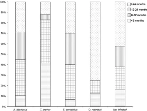
The prevalence of lungworm infections and their distribution are reported inTable 4according to the sampled areas. The infec-tion intensity (expressed as the number of LPG/EPG) varied among the lungworm species, with a mean of 8094.9 A. abstrusus LPG, 742.5 T. brevior LPG, 237.5 O. rostratus LPG and 143 E. aerophilus EPG. Cats from Portugal, Italy (Bari), Hungary and Bulgaria shed the highest numbers of A. abstrusus (mean LPG = 90,492.9, range 50–620,000), T. brevior (mean LPG = 1601, range 60–4000), O. ros-tratus (mean LPG = 441 LPG, range 102–986), and E. aerophilus EPG (mean EPG = 169.2, range 100–400), respectively. Infections by A. abstrusus and E. aerophilus were most frequently diagnosed in animals aged from 6 months to 2 years, whereas those by T. bre-vior in cats younger than 1 year of age (Fig. 2); however, these
cor-Table 1 Overall number and local prevalence (%) of gastrointestinal and respiratory parasites (groups) detected during the examination of faecal samples from cats in each sampling area. Data are shown according to the frequency of infection by each parasite. Austria Belgium Bulgaria France Greece Hungary Italy Portugal Romania Spain Switzerland United Kingdom Total Vienna (n = 80) Liege (n = 108) Stara Zagora (n = 120) Lyon (n = 92) Thessaloniki (n = 118) Budape st (n = 120) Bari (n = 120) Sassari (n = 138) Messina (n = 170) Centre and South (n = 120) Lisbon (n = 120) North (n = 120) Cluj-Napoca (n = 120) Madrid (n = 200) Zurich (n = 124) Cambridge (n = 120) (N = 1990) Ascarids 2 (2.5) 22 (20.4) 37 (30.8) 20 (21.7) 48 (40.7) 22 (18.3) 7 (5.8) 27 (19 .6) 10 (5.9) 25 (20.8) 19 (15.8) 25 (20.8) 30 (25.0) 19 (9.5) 13 (10.5) 3 (3.6 ) 329 (16.5%) Lungworms – 2 (1.9) 43 (35.8) 7 (7.6) 13 (11.0) 30 (25 .0) 20 (16.7) 16 (11 .6) 26 (15.3) 2 (1.7) 14 (11.7) 3 (2.5) 20 (16.7) 13 (6.5) 1 (0.8) – 210 (10.6%) Coccidia 3 (3.8) 4 (3.7) 12 (10.0) 3 (3.3) 9 (7.6) – 1 (0.8) 23 (16 .7) 6 (3.5) 5 (4.2) 19 (15.8) 7 (5.8) 5 (4.2) 20 (10.0) 8 (3.2) 4 (4.8) 129 (6.5%) Hookworms 34 (28.3) – 5 (4.2) – 2 (1.7) 2 (1.5) 5 (2.9) 15 (12.5) 7 (5.8) – 1 5 (12.5) 4 (2) – – 89 (4.5%) Taenia spp. 2 (2.5) – 1 (0.8) 6 (6.5) – – – 4 (2.9) 1 (0.6) – 1 (0.8) – 2 (1.7) 1 (0.5) 4 (5.0) – 2 2 (1.1%) Strongyloides sp. – – 16 (13.3) – – – – – – – – – – – – – 1 6 (0.8%) Giardia 2 (2.5) – – – 3 (2.5) – – – – – – 1 (0.8) – 8 (4.0) – – 14 (0.7%) Joyeuxiella pasqualei – – – – 12 (10.2) – – – – – – – – – – – 1 2 (0.6%) Dipylidium caninum – – 3 (2.5) – – – 2 (1.7) – – – – – – 1 (0.5) – – 6 (0.3%) Trichuris spp. – – – – – – – – – – 1 (0.8) – 1 (0.8) 1 (0.5) – – 3 (0.2%) Hymenolepis diminuta – – – – – – – – – – – – 2 (1.7) – – – 2 (0.1%)
relations were not statistically significant. Six out of the eight O. ros-tratus-positive cats were older than 2 years. Lungworm infections were diagnosed throughout the study period, with the majority of cases reported in the spring (74/499, 14.8%) and winter of 2015 (51/422; 12.1%) (
v
2= 18.132; P < 0.01). The largest number of catsinfected by metastrongyloids was recorded during spring (41%) or winter (36.6%), in Mediterranean or continental areas, respectively. 3.5. Clinical observations
Respiratory alterations (i.e. cough, dyspnoea, and nasal dis-charge) were reported in 14 (6.7%) cats positive for lungworms. The age of clinically affected animals was 7 months (mean 11.6 months, range 3 months to 3 years). Five of these cats were infected by A. abstrusus or by T. brevior, respectively, one was
co-infected with both species and three were co-infected by A. abstrusus and E. aerophilus, with mean numbers of 772.4 (range 19–187,200 LPG) and 1345.4 (range 63–4000 LPG) for A. abstrusus and T. brevior LPG, respectively, and 50 E. aerophilus EPG. Three cats (mean age 5.5 months) infected by A. abstrusus from Lisbon (Portugal) and one co-infected by E. aerophilus from Cluj Napoca (Romania) died over the course of the study; these shed the largest number of A. abstrusus LPG recorded (mean LPG = 358,925; range = 137,500– 620,000).
3.6. Morphological identification of larvae
From 180 samples which were morphologically and molecu-larly identified, more than 3000 metastrongyloid larvae stored in Fig. 1. The geographical distribution of cats in this study which were infected by lungworms. Grey-shaded squares represent locations where cats were free of infection, and black squares represent locations where infected individuals were detected. Where cats were collected from the same location, some markers overlie each other.
Table 2
Occurrence and frequency (%) of infections by lungworms and co-infections with other parasite groups according to the animal age classes, assessed for 210 lungworm-infected cats.
Age intervals (months) Total (N = 210)
< 6 (n = 39, 18.6) 6–12 (n = 62, 29.5) 12–24 (n = 37, 17.6) >24 (n = 72, 34.3) Lungworms (LW) 18 (8.6) 24 (11.4) 19 (9.1) 39 (18.6) 100 (47.6) LW mixed infections Ascarids (A) 13 (6.2) 13 (6.2) 4 (1.9) 9 (4.3) 39 (18.6) Hookworms (HW) – 10 (4.8) 3 (1.4) 7 (3.3) 20 (9.5) Coccidia (C) 4 (1.9) 1 (0.5) 1 (0.5) 1 (0.5) 7 (3.3) Taenia spp. – 2 (4.2) – 1 (0.5) 3 (1.4) Dipylidium caninum – 2 (4.2) – – 2 (1.0) Strongyloides sp. – 1 (0.5) – – 1 (0.5) A + HW – 5 (2.4) 5 (2.4) 3 (1.4) 13 (6.2) HW + Strongyloides sp. – – 1 (0.5) 4 (1.9) 5 (2.4) A + C – 2 (1.0) 1 (0.5) – 3 (1.4) A + Taenia spp. – 1 (0.5) – 1 (0.5) 2 (1.0) HW + C – – – 1 (0.5) 1 (0.5) HW + Taenia spp. – – 1 (0.5) – 1 (0.5) A + Strongyloides sp. – 1 (0.5) – – 1 (0.5) A + Hymenolepis diminuta – – – 1 (0.5) 1 (0.5) A + Joyeuxiella pasqualei – – – 1 (0.5) 1 (0.5) HW + A + Taenia spp. 2 (1.0) – 2 (1.0) – 4 (1.9) HW + A + Strongyloides sp. 1 (0.5) – – 1 (0.5) 2 (1.0) HW + C + Strongyloides sp. 1 (0.5) – – 1 (0.5) 2 (1.0) HW + A + Trichuris spp. – – – 1 (0.5) 1 (0.5) HW + C + A – – – 1 (0.5) 1 (0.5)
formalin were measured and, of those, 1250 were processed for detailed morphological identification.
3.6.1. Body length of larvae
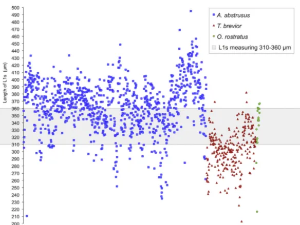
Those specimens included 967 A. abstrusus, 264 T. brevior, and 19 O. rostratus L1s, and their mean length was 364.1 ± 35.5mm (range 210.4–495.1mm), 304.1 ± 27.9 mm (range 203.2–382.2 mm) and 332.1 ± 35.9mm (range 216.6–366.8 mm), respectively. The body length of A. abstrusus L1s displayed some difference according to sampling countries (ANOVA, F = 25.233, P < 0.001) (Table 5). When compared with the larval length reference intervals used for A. abstrusus, T. brevior and O. rostratus discrimination, only 346 (35.7%), 47 (17.8%) and 10 (52.6%) L1s were, respectively, within the expected range (Fig. 3). In particular, 407 (42.1%) A. abstrusus L1s measured less than 360mm, whereas 111 (42.0%) T. brevior and 18 (94.7%) O. rostratus L1s exceeded the expected upper length limit (Fig. 3). Overall, 536 metastrongyloid L1s (42.9%) were included in the range 310–360mm.
3.6.2. Morphological features of larvae
The morphology of the anterior extremity and the position of the oral opening of L1s were consistent with the species of metas-trongyloids examined, whereas slight variations in the shape of the
caudal extremity were detected within the same species. In partic-ular, the tail shape was variable and differences in the morphology of dorsal kinks, incisure depths and terminal endings were recorded. In brief, L1s of A. abstrusus featured a slender anterior extremity, with a short and terminal oral opening leading into a narrow vestibule; the tail was bent into an S-shape, with a visible dorsal kink, distinct deep dorsal and ventral incisures, and a termi-nal knob-like extremity (Fig. 4A). Larvae of T. brevior were charac-terised by a clear and pointed anterior extremity, with a sub-terminal oral opening; the tail featured a deep dorsal incision, which divided the extremity into two appendices (a shallow ven-tral one and a slender dorsal one). The S-shape of the posterior extremity was not obvious, and the ending was straight and grad-ually tapered (Fig. 4B). Finally, O. rostratus L1s were characterized by a rounded head, with a central oral opening and a cylindrical buccal capsule; the tail was slightly undulated, with a deep ventral notch and a shallow dorsal one. The notch ended in a minute spine (Fig. 4C).
3.7. Evaluation of treatment efficacy
The treatment efficacy against lungworms was assessed in 198 cats. Twelve cats did not complete the assessment. At enrolment, Table 3
Risk factors for infection by lungworms in cats.
Parameter Class Number tested Positive (%) Chi-square Significance
Age (months) <6 322 35 (10.9) 14.562 P = 0.002
6–12 462 64 (13.9)
12–24 372 47 (12.6)
>24 815 62 (7.6)
Climatic area Continental 763 73 (9.6) 1.273 nsa
Mediterranean 1227 137 (11.2) Gender Female 1043 114 (10.9) 0.309 nsa Male 935 95 (10.2) Other infections No 1525 137 (9.0) 17.024 P < 0.001 Yes 465 73 (15.7) Lifestyle Owned 1448 159 (11.0) 2.584 ns Stray 301 33 (11.0) Cattery 238 18 (7.6) ns, not significant. a
Data was not available for 19 cats.
Table 4
Prevalence (%) of lungworm species and their distributions according to the sampled areas (see notes for information on the co-infections). Sampling areas (animals positive for lungworms, %) Lungworm species
Aelurostrongylus abstrusus Troglostrongylus brevior Eucoleus aerophilus Oslerus rostratus
Belgium (n = 2/108, 1.9%) 1 1 Bulgaria (n = 43/120, 35.8%) 20 + 7a+ 4b+ 2d 6 + 4b+ 1c+ 2d 3 + 7a+ 1c+ 2d France (n = 7/92, 7.6%) 4 1 2 Greece (n = 13/118, 11%) 8 + 2a 3 + 2a Hungary (n = 30/120, 25%) 27 3 Italy, Bari (n = 20/120, 16.7%) 2 + 4b 13 + 4b 1 Italy, Sassari (n = 16/138, 11.6%) 14 + 2b 2b Italy, Messina (n = 26/170, 15.3%) 22 3 1
Portugal, Centre and South (n = 2/120, 1.7%) 2 Portugal, Lisbon (n = 14/120, 11.7%) 14 Portugal, North (n = 3/120, 2.5%) 1 2 Romania (n = 20/120, 16.7%) 12 + 5a 3 + 5a Spain (n = 13/200, 6.5%) 5 + 5b 1 + 5b 2 Switzerland (n = 1/124, 0.8%) 1 Total 164 41 31 8 (N = 210/1990, 10.6%) (133 + 14a + 15b + 2d ) (23 + 15b + 1c + 2d ) (14 + 14a + 1c + 2d ) 8
aAelurostrongylus abstrusus and E. aerophilus. b Aelurostrongylus abstrusus and T. brevior. c Troglostrongylus brevior and E. aerophilus. d
cats shed a mean of 508.7 LPG, 742.5 LPG, and 237.5 LPG of A. abstrusus, T. brevior, and O. rostratus, respectively, and a mean of 153.3 EPG of E. aerophilus. All cats tested negative for lungworm larvae after a single treatment, with the exception of three animals from Sassari (Italy), one from Sicily (Italy), and one from France, which were positive for A. abstrusus. The overall efficacy of Broad-lineÒwas 99% for A. abstrusus and 100% for T. brevior, O. rostratus and E. aerophilus, respectively (P < 0.05). Clinical respiratory signs subsided in all treated animals. No adverse reactions related to the treatment were observed. The treatment efficacy in animals co-infected by ascarids (21 cats), hookworms, Strongyloides sp. (11 cats), Taenia spp. (eight cats), Dipylidium caninum (two cats), Hymenolepis diminuta and Joyeuxiella pasqualei (one cat) was 100% (P < 0.05).
4. Discussion
Data from the present study provides a comprehensive picture of the diagnosis, epidemiology and treatment of feline lungworms in Europe, as well as of the occurrence of co-infections by other gastrointestinal parasites. Indeed, whilst indicating that the preva-lence and composition of the endoparasitic fauna of domestic cats is extremely diversified, results also point out that ascarids are the most prevalent parasitic nematodes, as shown in previous reports (Beugnet et al., 2014; Knaus et al., 2014a; Giannelli et al., 2015a; Takeuchi-Storm et al., 2015). The cosmopolitan distribution of ascarids (mainly Toxocara cati) is most likely linked to their biology and ecology (e.g. the extreme resistance of eggs to adverse climatic conditions, lactogenic transmission), which drastically increases
Fig. 2. Distribution (as a percentage) of cats uninfected and infected by lungworms (Aelurostrongylus abstrusus, Troglostrongylus brevior, Eucoleus aerophilus, Oslerus rostratus) according to their age classes.
Table 5
Analysis of lungworm L1 length (mm) according to species and sampling area.
Lungworm species Sampling area Specimens measured (n) Mean length ± S.D. 95% CI for the mean MIN-MAX length Lower Upper
Aelurostrongylus abstrusus Italy, Bari 16 364.7a
± 32.71 347.28 382.14 305.2–406.1 Portugal, Lisbon 109 360.2b ± 42.35 352.17 368.25 210.4–448.4 Romania 31 397.3a,b,c,d,e,f ± 15.68 391.58 403.09 368.8–433.2 Bulgaria 377 350.8c ± 32.72 347.53 354.16 234.9–417.6 Italy, Sassari 166 386.5 ± 30.04 381.93 391.14 301.1–495.1 Italy, Messina 144 365.5d ± 27.06 361.08 369.99 261.9–424.0 Hungary 51 381.8 ± 37.22 371.37 392.31 296.8–456.7 Spain 66 353.4 ± 28.28 346.49 360.40 287.5–409.8 France 7 375.3f ± 24.86 352.35 398.34 326.5–395.0 Total 967 363.9 ± 35.37 361.69 366.15 210.4–495.1
Troglostrongylus brevior Italy, Bari 103 309.8g± 35.07 303.01 316.71 203.2–382.2
Italy, Sassari 6 315.2 ± 25.98 287.96 342.50 280.8–356.7 Italy, Messina 21 311.2 ± 12.71 305.49 317.07 276.9–340.5 Bulgaria 117 296.4g ± 20.86 292.58 300.22 242.9–352.7 Spain 17 308.9 ± 23.99 296.58 321.25 256.7–369.3 Total 264 304.0 ± 27.87 300.69 307.45 203.2–382.2
Oslerus rostratus Italy, Bari 7 298.7h± 38.82 262.79 334.60 216.6–331.0
Spain 12 353.2h± 9.93 346.87 359.48 336.3–366.8
Total 19 333.1 ± 35.94 315.78 350.43 216.6–366.8
Each letter (a–h) corresponds to the same significant difference. CI, confidence interval.
Fig. 3. Feline lungworm infections diagnosed during this study (scale bars = 100mm). L1s of Aelurostrongylus abstrusus (A), Troglostrongylus brevior (B), and Oslerus rostratus (C), with magnifications of the anterior (Aa, Ba, Ca) and posterior extremities (Ab, Bb, Cb). Eggs of Eucoleus aerophilus (D), with particulars of the polar plug (Da) and of the net-like surface of the egg (Db).
Fig. 4. Range of length (mm) of metastrongyloid L1s according to the species examined. The majority of Aelurostrongylus abstrusus (squares), Troglostrongylus brevior (triangles), and Oslerus rostratus (circles) specimens were within the range 310–360mm (grey shaded region).
the circulation of this nematode amongst susceptible hosts. Sur-prisingly, lungworms were the second most frequent group of nematodes diagnosed in the study population, and in half of the cases they concurred with other gastrointestinal parasites includ-ing tapeworms, hookworms and coccidia, demonstratinclud-ing a high level of parasitic co-infection in cats. The simultaneous detection of lungworms together with ascarids, Joyeuxiella pasqualei or Taenia spp. (Table 1) suggests that infected cats preyed on rodents, which may act as reservoirs for all these pathogens and therefore as a source of infection (Gerichter, 1949; Anderson, 2000; Reperant et al., 2009; Waap et al., 2014; Schuster et al., 2016). Indeed, para-tenic hosts play a relevant, but poorly studied, epidemiological role in the distribution of these parasites. Similarly, the finding of Hymenolepis diminuta in cats could be associated with the con-sumption of infected rodents harbouring mature proglottids (Owen, 2005; Schuster et al., 2016).
Based on the results reported herein, at least one out of 10 cats in this study is at risk of lungworm infection in European countries, thus suggesting that these pathogens are more widespread than previously thought (Payo-Puente et al., 2008; Traversa et al.,
2010; Beugnet et al., 2014). As previously indicated for
Angiostrongylus vasorum of canids (Morgan et al., 2009), the geo-graphical distribution of feline lungworms is patchy but stable in endemic spots, as the infection has been consistently detected in the same sampling sites, even years after its first description. Although literature reports are often not comparable due to incon-sistent sampling procedures, the number of positive cats recorded in Lisbon (i.e. 11.7%) is similar to that recorded in the same locality from June 2009 to December 2011 (i.e. 12–12.4%) (Nabais et al., 2014; Waap et al., 2014). Conversely, when prevalence data is assessed on a wider scale (e.g. at the regional or national level), comparisons are probably baseless. This explains, for instance, the difference in infection rates detected among the three Italian regions screened (i.e. Apulia, Sardinia and Sicily) both in the pre-sent study and previously (Tamponi et al., 2014; Giannelli et al., 2015a), or those assessed in Greece, Hungary, Romania and Spain in previous surveys (Miró et al., 2004; Capári et al., 2013; Mircean et al., 2010; Diakou et al., 2015). Based on these results, feline lungworms cannot be considered as emerging pathogens, as limited data is available to draw conclusions on their changing distribution. The prevalence of the infection and number of LPG/ EPG detected vary amongst the cat populations sampled and the lungworm species. Whether this is associated with climatic and ecological factors affecting parasite development and/or of their intermediate/paratenic hosts, or to regular anthelmintic treatment of cats, is yet to be determined. However, a recent survey on para-sites of domestic cats has demonstrated that the administration of regular anthelmintic treatments drastically reduces the risk of T. cati infection (Beugnet et al., 2014). Based on this knowledge, we speculate that owned cats living in Bulgaria, Hungary, Italy, Roma-nia or Portugal were treated less frequently than those from other countries, where only a few individuals were positive for lung-worms (e.g. Switzerland and Belgium) (Matos et al., 2015). In the meantime, the lack of detection of lungworm-positive cats in Aus-tria and in the United Kingdom suggests that the prevalence of feline metastrongyloids in those areas is low, as indicated by a sin-gle report of A. abstrusus in a kitten from Liverpool (Philbey et al., 2014). To the best of the authors’ knowledge, this is the first study assessing the occurrence of feline lungworms in Belgium, France and Switzerland, from where only isolated case reports were pre-viously available (Traversa et al., 2010).
The design of the study did not allow us to determine when the cats had become infected by the parasites; however, the infection was more frequently detected during winter and spring. Although this data prevents us from drawing definitive conclusions, a similar finding was recorded for A. abstrusus in Germany (Taubert et al.,
2009; Barutzki and Schaper, 2011). Due to the limited knowledge of the epidemiology of the infection in the intermediate and para-tenic hosts, the existence of seasonality in feline lungworm infec-tion is difficult to assess. In contrast, whilst analysing the seasonal trend of A. vasorum diagnosis in dogs from England, Wales and Scotland during the period 2005–2008,Morgan and colleagues (2010)concluded that the peak of canine angiostrongylosis cases in winter and spring was related to an increase in infected gastropod numbers over the previous months. One might hypothesize that the dynamic of metastrongyloid infections in cats ensues from periodic variations associated with the biology of the gastropod intermediate hosts (López et al., 2005; Je_zewski et al., 2013; Di
Cesare et al., 2013; Giannelli et al., 2014b); however, the interac-tions between snails and paratenic hosts should also be the subject of further investigations. Indeed, a number of other factors, such as the contamination of the environment with infective L3 s released via the gastropod mucus and the ‘‘intermediesis” (Giannelli et al., 2015b; Colella et al., 2015) should be taken into account when con-sidering the possibility that a definitive host comes into contact with the parasite.
The detection of up to four respiratory nematode species in cats indicates that polyspecific infections are likely to occur in felids. This is not surprising, considering the number of bronco-pulmonary nematodes of sheep and goats (i.e. Cystocaulus ocreatus, Muellerius capillaris, Protostrongylus rufescens and Dictyocaulus filar-ia) (Anderson, 2000; Alasaad et al., 2009), or the cardiopulmonary nematodes of canids, which include a main species (i.e. A. vasorum) plus others, characterised by a more limited geographical distribu-tion (i.e. Crenosoma vulpis, Oslerus osleri, Filaroides hirthi, Eucoleus boehmi, and E. aerophilus) (McGarry and Morgan, 2009; Traversa et al., 2010; Latrofa et al., 2015; Colella et al., 2016; Tolnai et al., 2015; Hodzˇic´ et al., 2016).
A similar scenario occurs in felids, with A. abstrusus represent-ing the species more frequently diagnosed, probably due to its wide host range (Giannelli et al., 2016a) and its cosmopolitan dis-tribution; this, outside Europe, includes Africa (Di Cesare et al., 2016), the Americas (Fiorello et al., 2006; Gerdin et al., 2011), Aus-tralia (Foster et al., 2004), the Middle East (Abu-Madi et al., 2007) China (Yang and Liang, 2015) and the Russian Far East (González et al., 2007). The diagnosis of this lungworm species in each cate-gory of cats analysed in this study indicates that feline aelurostrongylosis threatens almost all cats with access to outdoor environments, irrespective of their age, gender or geographical location. This is in agreement with previous studies (Beugnet et al., 2014; Giannelli et al., 2015a), but in contrast to others, in which adult (Mircean et al., 2010; Capári et al., 2013; Knaus et al., 2014a) or young (Traversa et al., 2008) animals more fre-quently scored positive. Theoretically, both age categories are at risk for the infection. The immune response of cats towards A. abstrusus infection is heterogeneous and unpredictable (Schnyder et al., 2014) and although experimental studies showed that the repeated administration of small numbers of L3s may prevent clin-ical signs and larval excretion (Hamilton, 1969), the paucity of data available on this topic, under natural conditions, does not fully clarify whether infected animals are protected against subsequent infections.
According to the results of the present survey, T. brevior is the second most frequently detected lungworm species of cats. Its presence has been confirmed in islands (i.e. Ibiza, Sicily, Sardinia and Crete) and continental areas (Italy, Greece, Bosnia and Herze-govina) of the Mediterranean basin (Traversa and Di Cesare, 2013; Brianti et al., 2014a; Alic´ et al., 2015; Diakou et al., 2015), and documented, for the first time, in domestic cats from the Balkans. Of note, the finding of this nematode species in Bulgaria supports previous data on the occurrence of this parasite in wild felids from the same geographical area (Jancˇev and Genov, 1978).
The occurrence of lungworm infections in wild felids provides interesting arguments for discussion of the alleged increases in prevalence of T. brevior across Europe, an event epidemiologically associated with a spill-over effect. This phenomenon occurs when a reservoir population, affected by high pathogen prevalence, interacts with a susceptible one (Power and Mitchell, 2004). In the case of T. brevior, this occurrence cannot be totally ruled out, considering wild and domestic cats may share habitats (Randi et al., 2001; Falsone et al., 2014). Notably, the diagnosis of T. brevior infection in most of the cats younger than 1 year of age further indicates that a direct transmission from the queen to pre-weaned kittens may occur (Brianti et al., 2013).
Eucoleus aerophilus, a respiratory nematode widespread across Europe, has been previously detected in cats from Belgium, Bulgar-ia, Greece, Italy, Portugal and Romania (see also, Mircean et al., 2010; Traversa et al., 2010; Capári et al., 2013; Knaus et al., 2014a; Di Cesare et al., 2015b). The simultaneous diagnosis of A. abstrusus in more than half of the cats positive for this capillarid species indicates that both pathogens may share the same trans-mission pathway, probably via paratenic hosts (i.e. birds, small rodents and insectivores) which previously fed on gastropods and earthworms (Woods et al., 2003). In addition, data demon-strated that cats are also infected by other capillarid nematodes, whose eggs should be morphologically and molecularly differenti-ated to achieve a correct diagnosis. Indeed, the differentiation between nematodes of the genera Eucoleus and Trichuris (e.g. Tri-churis felis, TriTri-churis campanula and TriTri-churis serrata) should be con-sidered when capillarid eggs are detected (Traversa et al., 2011; Ketzis et al., 2015a,b). However, the possibility that these eggs rep-resented pseudoparasites in the cat faeces cannot be ruled out.
Oslerus rostratus has been diagnosed in cats from Italy, Spain, Hungary and France, besides its previous detection in Israel (Gerichter, 1949), Sri Lanka (Seneviratna, 1955), the United States (Ash, 1970), Spain (Juste et al., 1992; Millán and Casanova, 2009), and Italy (Brianti et al., 2014b; Varcasia et al., 2015). Scant data on the biology, ecology and clinical features of O. rostratus (Seneviratna, 1959) currently impair a thorough assessment of its distribution and the low prevalence of the feline oslerosis in Eur-ope could also be associated with difficulties in diagnosis. Indeed, L1s are shed in the environment intermittently, and for a limited time period (Brianti et al., 2014b); in addition, when adult O. ros-tratus settle in the peri-bronchial tissues, encapsulated by a strong fibrous reaction (i.e. the so called ‘‘regressive stage” of the infec-tion), the production of larvae may cease (Brianti et al., 2014b). It should be considered that, due to the scarce information available on the morphology of O. rostratus larvae (which are similar to A. abstrusus and T. brevior L1s), its correct identification is challenging.
Approximately half of metastrongyloid larvae analysed in this study were outside their reference length range (i.e. 360–390mm, 300–310mm, 300–320 mm, for A. abstrusus, T. brevior and O. rostra-tus, respectively) (Gerichter, 1949). This finding indicates that the differentiation of feline metastrongyloids cannot be exclusively based on the evaluation of their length, but requires an apprecia-tion of the shape of anterior and posterior extremities (Gerichter, 1949; Brianti et al., 2014a; Giannelli et al., 2014b). The diagnosis of lungworm infection is, however, essential for planning appropri-ate therapeutic protocols and preventative measures (Gerdin et al., 2011; Elsheikha et al., 2016). Although most animals did not dis-play clinical signs, some presented with recurrent but transitory signs, particularly cough, dyspnoea and nasal discharge. Remark-ably, three out of 14 cats showing clinical signs most likely died as a consequence of high infection burdens of A. abstrusus (358,925 LPG) in combination with young age (7 months). This confirms that cats with high LPG have a higher probability of developing severe symptoms (Genchi et al., 2014). Although the
actual causes leading to the death of four animals were not assessed here, it is acknowledged that young, debilitated and immunosuppressed animals may succumb to feline troglostrongy-losis (Brianti et al., 2014a; Giannelli et al., 2014a), or aelurostrongylosis (Elsheikha et al., 2016), especially when it evolves into a hyper-infection syndrome (Philbey et al., 2014), or to irreversible pulmonary hypertension (Crisi et al., 2015). For this reason, a correct diagnosis followed by an efficacious therapeutic protocol is pivotal in the management of feline lungworm infec-tions. In this study, a single administration of the combination of fipronil, (S)-methoprene, eprinomectin and praziquantel (Broad-lineÒ, Merial) proved to be effective for the reduction of lungworm burdens and resolution of clinical signs, as already reported under experimental conditions (Knaus et al., 2014b, 2015) and in cats naturally exposed to these parasites (Rehbein et al., 2014; Giannelli et al., 2015a). Indeed, the overall efficacy of 99% against A. abstrusus and 100% against T. brevior, O. rostratus and E. aerophi-lus, indicates that this treatment is effective for the management of these parasitic infections.
Although this study was designed to screen a large population of animals using standardized laboratory procedures, the collec-tion of a single faecal sample from each animal might have led to an underestimation of the overall prevalence of infections, poten-tially surmountable by using a serological test (Briggs et al., 2013; Zottler et al., 2017). Therefore, additional studies are needed to investigate the occurrence of respiratory nematodes in other countries, to assess the overall prevalence of feline lungworms, and to better understand the epidemiological patterns of these par-asites. Indeed, feline lungworms represent a topic of increasing interest in the field of veterinary sciences (Giannelli et al., 2016b), as they are widespread and sometimes life-threatening parasites of domestic and wild felids. This study contributes to a better understanding of their epidemiology. Further areas of inves-tigations may include (i) the development of ecological and cli-matic models, which will allow evaluation of the impact of environmental/climatic factors on the distribution of these para-sites; (ii) surveys to assess the occurrence of lungworms in their gastropod and paratenic hosts; (iii) the development of reliable morphological keys and serological diagnostic devices; (iv) the provision of information for veterinarians and owners alike on the importance of administering correct treatments and subjecting the animals to regular follow-ups.
Acknowledgements
The authors would like to thank Maria Luisa Vitale (University of Bari, Italy), Eva-Maria Zottler (University of Zurich, Switzerland) and André Pereira (Universidade Nova de Lisboa) for their support with the laboratory procedures. This work was funded by Merial SAS (Europe). FB LH and MK are currently employees at Merial. The funders had no role in data collection, data analysis, or in the preparation of the manuscript.
References
Abu-Madi, M.A., Al-Ahbabi, D.A., Al-Mashhadani, M.M., Al-Ibrahim, R., Pal, P., Lewis, J.W., 2007. Patterns of parasitic infections in faecal samples from stray cat populations in Qatar. J. Helminthol. 81, 281–286.
Alasaad, S., Morrondo, P., Dacal-Rivas, V., Soriguer, R.C., Granados, J.E., Serrano, E., Zhu, X.Q., Rossi, L., Pérez, J.M., 2009. Bronchopulmonary nematode infection of Capra pyrenaica in the Sierra Nevada massif. Spain. Vet. Parasitol. 164, 340–343.
Alic´, A., Traversa, D., Duscher, G.G., Kadric´, M., Di Cesare, A., Hodzˇic´, A., 2015. Troglostrongylus brevior in an Eurasian lynx (Lynx lynx) from Bosnia and Herzegovina. Parasit. Vectors. 8, 653.
Anderson, R.C., 2000. The superfamily Metastrongyloidea. In: Anderson, R.C. (Ed.), Nematode Parasites of Vertebrates. Their Development and Transmission. CABI, Wallingford, UK, pp. 129–229.
Annoscia, G., Latrofa, M.S., Campbell, B.E., Giannelli, A., Ramos, R.A., Dantas-Torres, F., Brianti, E., Otranto, D., 2014. Simultaneous detection of the feline lungworms
Troglostrongylus brevior and Aelurostrongylus abstrusus by a newly developed duplex-PCR. Vet. Parasitol. 199, 172–178.
Ash, L.R., 1970. Diagnostic morphology of the third-stage larvae of Angiostrongylus cantonensis, Angiostrongylus vasorum, Aelurostrongylus abstrusus, and Anafilaroides rostratus (Nematoda: Metastrongyloidea). J. Parasitol. 56, 249–253.
Barutzki, D., Schaper, R., 2011. Results of parasitological examinations of faecal samples from cats and dogs in Germany between 2003 and 2010. Parasitol. Res. 109, S45–60.
Beugnet, F., Bourdeau, P., Chalvet-Monfray, K., Cozma, V., Farkas, R., Guillot, J., Halos, L., Joachim, A., Losson, B., Miró, G., Otranto, D., Renaud, M., Rinaldi, L., 2014. Parasites of domestic owned cats in Europe: co-infestations and risk factors. Parasit. Vectors 7, 291.
Brianti, E., Gaglio, G., Giannetto, S., Annoscia, G., Latrofa, M.S., Dantas-Torres, F., Traversa, D., Otranto, D., 2012. Troglostrongylus brevior and Troglostrongylus subcrenatus (Strongylida: Crenosomatidae) as agents of broncho-pulmonary infestation in domestic cats. Parasit. Vectors 5, 178.
Brianti, E., Gaglio, G., Napoli, E., Falsone, L., Giannelli, A., Annoscia, G., Varcasia, A., Giannetto, S., Mazzullo, G., Otranto, D., 2014b. Feline lungworm Oslerus rostratus (Strongylida: Filaridae) in Italy: first case report and histopathological findings. Parasitol. Res. 113, 3853–3857.
Brianti, E., Gaglio, G., Napoli, E., Falsone, L., Giannetto, S., Latrofa, M.S., Giannelli, A., Dantas-Torres, F., Otranto, D., 2013. Evidence for direct transmission of the cat lungworm Troglostrongylus brevior (Strongylida: Crenosomatidae). Parasitology 140, 821–824.
Brianti, E., Giannetto, S., Dantas-Torres, F., Otranto, D., 2014a. Lungworms of the genus Troglostrongylus (Strongylida: Crenosomatidae): neglected parasites for domestic cats. Vet. Parasitol. 202, 104–112.
Briggs, K.R., Yaros, J.P., Liotta, J.L., Lucio-Forster, A., Lee, A.C., Bowman, D.D., 2013. Detecting Aelurostrongylus abstrusus-specific IgG antibody using an immunofluorescence assay. J. Feline Med. Surg. 15, 1114–1118.
Capári, B., Hamel, D., Visser, M., Winter, R., Pfister, K., Rehbein, S., 2013. Parasitic infections of domestic cats, Felis catus, in western Hungary. Vet. Parasitol. 192, 33–42.
Colella, V., Giannelli, A., Brianti, E., Ramos, R.A., Cantacessi, C., Dantas-Torres, F., Otranto, D., 2015. Feline lungworms unlock a novel mode of parasite transmission. Sci. Rep. 5, 13105.
Colella, V., Mutafchiev, Y., Cavalera, M.A., Giannelli, A., Lia, R.P., Dantas-Torres, F., Otranto, D., 2016. Development of Crenosoma vulpis in the common garden snail Cornu aspersum: implications for epidemiological studies. Parasit. Vectors 9, 208.
Crisi, P.E., Traversa, D., Di Cesare, A., Luciani, A., Civitella, C., Santori, D., Boari, A., 2015. Irreversible pulmonary hypertension associated with Troglostrongylus brevior infection in a kitten. Res. Vet. Sci. 102, 223–227.
Deplazes, P., Eckert, J., Mathis, A., von Samson-Himmelstjerna, G., Zahner, H., 2016. Parasitology in Veterinary Medicine. Wageningen Academic Publishers, Wageningen.
Di Cesare, A., Crisi, P.E., Di Giulio, E., Veronesi, F., Frangipane di Regalbono, A., Talone, T., Traversa, D., 2013. Larval development of the feline lungworm Aelurostrongylus abstrusus in Helix aspersa. Parasitol. Res. 112, 3101–3108.
Di Cesare, A., Di Francesco, G., Frangipane di Regalbono, A., Eleni, C., De Liberato, C., Marruchella, G., Iorio, R., Malatesta, D., Romanucci, M.R., Bongiovanni, L., Cassini, R., Traversa, D., 2015. Retrospective study on the occurrence of the feline lungworms Aelurostrongylus abstrusus and Troglostrongylus spp. in endemic areas of Italy. Vet. J. 203, 233–238.
Di Cesare, A., Laiacona, F., Iorio, R., Marangi, M., Menegotto, A., 2016. Aelurostrongylus abstrusus in wild felids of South Africa. Parasitol. Res. 115, 3731–3735.
Di Cesare, A., Veronesi, F., Traversa, D., 2015. Felid Lungworms and Heartworms in Italy: More Questions than Answers? Trends Parasitol. 31, 665–675.
Diakou, A., Di Cesare, A., Barros, L.A., Morelli, S., Halos, L., Beugnet, F., Traversa, D., 2015. Occurrence of Aelurostrongylus abstrusus and Troglostrongylus brevior in domestic cats in Greece. Parasit. Vectors 8, 590.
Elsheikha, H.M., Schnyder, M., Traversa, D., Di Cesare, A., Wright, I., Lacher, D.W., 2016. Updates on feline aelurostrongylosis and research priorities for the next decade. Parasit. Vectors 9, 389.
Euzeby, J., 1981. Diagnostic experimental des helminthoses animals. In: Euzeby, J. (Ed.), Livre 1: Diagnostic Ante Mortem. Informations techniques des services vétérinaires. Ministère de l’Agriculture, Paris, France, pp. 303–307.
Falsone, L., Brianti, E., Gaglio, G., Napoli, E., Anile, S., Mallia, E., Giannelli, A., Poglayen, G., Giannetto, S., Otranto, D., 2014. The European wildcats (Felis silvestris silvestris) as reservoir hosts of Troglostrongylus brevior (Strongylida: Crenosomatidae) lungworms. Vet. Parasitol. 205, 193–198.
Fiorello, C.V., Robbins, R.G., Maffei, L., Wade, S.E., 2006. Parasites of free-ranging small canids and felids in the Bolivian Chaco. J. Zool. Wildl. Med. 37, 130–134.
Foster, S.F., Martin, P., Braddock, J.A., Malik, R., 2004. A retrospective analysis of feline bronchoalveolar lavage cytology and microbiology (1995–2000). J. Feline Med. Surg. 6, 189–198.
Genchi, M., Ferrari, N., Fonti, P., De Francesco, I., Piazza, C., Viglietti, A., 2014. Relation between Aelurostrongylus abstrusus larvae excretion, respiratory and radiographic signs in naturally infected cats. Vet. Parasitol. 206, 182–187.
Gerdin, J.A., Slater, M.R., Makolinski, K.V., Looney, A.L., Appel, L.D., Martin, N.M., McDonough, S.P., 2011. Post-mortem findings in 54 cases of anesthetic associated death in cats from two spay-neuter programs in New York State. J. Feline Med. Surg. 13, 959–966.
Gerichter, C.B., 1949. Studies on the nematodes parasitic in the lungs of Felidae in Palestine. Parasitology 39, 251–262.
Giannelli, A., Brianti, E., Varcasia, A., Colella, V., Tamponi, C., Di Paola, G., Knaus, M., Halos, L., Beugnet, F., Otranto, D., 2015a. Efficacy of BroadlineÒspot-on against
Aelurostrongylus abstrusus and Troglostrongylus brevior lungworms in naturally infected cats from Italy. Vet. Parasitol. 209, 273–277.
Giannelli, A., Cantacessi, C., Colella, V., Dantas-Torres, F., Otranto, D., 2016b. Gastropod-borne helminths: A look at the snail-parasite interplay. Trends Parasitol. 32, 255–264.
Giannelli, A., Colella, V., Abramo, F., do Nascimento Ramos, R.A., Falsone, L., Brianti, E., Varcasia, A., Dantas-Torres, F., Knaus, M., Fox, M.T., Otranto, D., 2015b. Release of lungworm larvae from snails in the environment: potential for alternative transmission pathways. PLoS Negl. Trop. Dis. 9, e0003722.
Giannelli, A., Kirkova, Z., Abramo, F., Latrofa, M.S., Campbell, B., Zizzo, N., Cantacessi, C., Dantas-Torres, F., Otranto, D., 2016a. Angiostrongylus chabaudi in felids: New findings and a review of the literature. Vet. Parasitol. 228, 188–192.
Giannelli, A., Passantino, G., Ramos, R.A., Lo Presti, G., Lia, R.P., Brianti, E., Dantas-Torres, F., Papadopoulos, E., Otranto, D., 2014a. Pathological and histological findings associated with the feline lungworm Troglostrongylus brevior. Vet. Parasitol. 204, 416–419.
Giannelli, A., Ramos, R.A., Annoscia, G., Di Cesare, A., Colella, V., Brianti, E., Dantas-Torres, F., Mutafchiev, Y., Otranto, D., 2014b. Development of the feline lungworms Aelurostrongylus abstrusus and Troglostrongylus brevior in Helix aspersa snails. Parasitology 141, 563–569.
González, P., Carbonell, E., Urios, V., Rozhnov, V.V., 2007. Coprology of Panthera tigris altaica and Felis bengalensis euptilurus from the Russian Far East. J. Parasitol. 93, 948–950.
Hamilton, J.M., 1969. Production of immunity in the cat against lungworm disease by administration of third-stage larvae. J. Comp. Pathol. 79, 161–165.
Hobmaier, M., Hobmaier, A., 1935. Intermediate hosts of Aelurostrongylus abstrusus of the cat. Proc. Soc. Exp. Biol. Med. 32, 1641–1647.
Hodzˇic´, A., Bruckschwaiger, P., Duscher, G.G., Glawischnig, W., Fuehrer, H.P., 2016. High prevalence of Eucoleus boehmi (syn. Capillaria boehmi) in foxes from western Austria. Parasitol. Res. 115, 3275–3278.
Jancˇev, J., Genov, T., 1978. Helminthofauna of the wild cat (Felis silvestris Schreb.) in Bulgaria. Helmintologiâ 6, 81–101.
Jefferies, R., Vrhovec, M.G., Wallner, N., Catalan, D.R., 2010. Aelurostrongylus abstrusus and Troglostrongylus sp. (Nematoda: Metastrongyloidea) infections in cats inhabiting Ibiza. Spain. Vet. Parasitol. 173, 344–348.
Je_zewski, W., Bun´kowska-Gawlik, K., Hildebrand, J., Perec-Matysiak, A., Laskowski, Z., 2013. Intermediate and paratenic hosts in the life cycle of Aelurostrongylus abstrusus in natural environment. Vet. Parasitol. 198, 401–405.
Juste, R.A., Garcia, A.L., Mencía, L., 1992. Mixed infestation of a domestic cat by Aelurostrongylus abstrusus and Oslerus rostratus. Angew. Parasitol. 33, 56–60.
Ketzis, J.K., Shell, L., Chinault, S., Pemberton, C., Pereira, M.M., 2015. The prevalence of Trichuris spp. infection in indoor and outdoor cats on St. Kitts. J. Infect. Dev. Ctries. 9, 111–113.
Ketzis, J.K., Verma, A., Burgess, G., 2015. Molecular characterization of Trichuris serrata. Parasitol. Res. 114, 1993–1995.
Knaus, M., Chester, S.T., Rosentel, J., Kühnert, A., Rehbein, S., 2014b. Efficacy of a novel topical combination of fipronil, (S)-methoprene, eprinomectin and
praziquantel against larval and adult stages of the cat lungworm,
Aelurostrongylus abstrusus. Vet. Parasitol. 202, 64–68.
Knaus, M., Kusi, I., Rapti, D., Xhaxhiu, D., Winter, R., Visser, M., Rehbein, S., 2011. Endoparasites of cats from the Tirana area and the first report on Aelurostrongylus abstrusus (Railliet, 1898) in Albania. Wien Klin Wochenschr. 123, S:31–35.
Knaus, M., Rapti, D., Shukullari, E., Kusi, I., Postoli, R., Xhaxhiu, D., Silaghi, C., Hamel, D., Visser, M., Winter, R., Rehbein, S., 2014a. Characterisation of ecto- and endoparasites in domestic cats from Tirana. Albania. Parasitol. Res. 113, 3361– 3371.
Knaus, M., Shukullari, E., Rapti, D., Rehbein, S., 2015. Efficacy of Broadline against Capillaria aerophila lungworm infection in cats. Parasitol. Res. 114, 1971–1975.
Latrofa, M.S., Lia, R.P., Giannelli, A., Colella, V., Santoro, M., D’Alessio, N., Campbell, B. E., Parisi, A., Dantas-Torres, F., Mutafchiev, Y., Veneziano, V., Otranto, D., 2015. Crenosoma vulpis in wild and domestic carnivores from Italy: a morphological and molecular study. Parasitol. Res. 114, 3611–3617.
López, C., Panadero, R., Paz, A., Sánchez-Andrade, R., Díaz, P., Díez-Baños, P., Morrondo, P., 2005. Larval development of Aelurostrongylus abstrusus (Nematoda, Angiostrongylidae) in experimentally infected Cernuella (Cernuella) virgata (Mollusca, Helicidae). Parasitol. Res. 95, 13–16.
Matos, M., Alho, A.M., Owen, S.P., Nunes, T., Madeira De Carvalho, L., 2015. Parasite control practices and public perception of parasitic diseases: A survey of dog and cat owners. Prev. Vet. Med. 122, 174–180.
McGarry, J.W., Morgan, E.R., 2009. Identification of first-stage larvae of metastrongyles from dogs. Vet. Rec. 165, 258–261.
Millán, J., Casanova, J.C., 2009. High prevalence of helminth parasites in feral cats in Majorca Island (Spain). Parasitol. Res. 106, 183–188.
Mircean, V., Titilincu, A., Vasile, C., 2010. Prevalence of endoparasites in household cat (Felis catus) populations from Transylvania (Romania) and association with risk factors. Vet. Parasitol. 171, 163–166.
Miró, G., Montoya, A., Jiménez, S., Frisuelos, C., Mateo, M., Fuentes, I., 2004. Prevalence of antibodies to Toxoplasma gondii and intestinal parasites in stray, farm and household cats in Spain. Vet. Parasitol. 126, 249–255.
Morgan, E.R., Jefferies, R., Krajewski, M., Ward, P., Shaw, S.E., 2009. Canine
pulmonary angiostrongylosis: the influence of climate on parasite
Morgan, E.R., Jefferies, R., van Otterdijk, L., McEniry, R.B., Allen, F., Bakewell, M., Shaw, S.E., 2010. Angiostrongylus vasorum infection in dogs: Presentation and risk factors. Vet. Parasitol. 173, 255–261.
Nabais, J., Alho, A.M., Gomes, L., Ferreira da Silva, J., Nunes, T., Vicente, G., Madeira de Carvalho, L., 2014. Aelurostrongylus abstrusus in cats and Angiostrongylus vasorum in dogs from Lisbon, Portugal. Acta Parasitol. Port. 20, 35–40.
Otranto, D., Brianti, E., Dantas-Torres, F., 2013. Troglostrongylus brevior and a nonexistent ’dilemma’. Trends Parasitol. 29, 517–518.
Otranto, D., 2015. Diagnostic challenges and the unwritten stories of dog and cat parasites. Vet. Parasitol. 212, 54–61.
Owen, I.L., 2005. Parasitic zoonoses in Papua New Guinea. J. Helminthol. 79, 1–14.
Patterson-Kane, J.C., Gibbons, L.M., Jefferies, R., Morgan, E.R., Wenzlow, N., Redrobe, S.P., 2009. Pneumonia from Angiostrongylus vasorum infection in a red panda (Ailurus fulgens fulgens). J. Vet. Diagn. Invest. 21, 270–273.
Payo-Puente, P., Botelho-Dinis, M., Carvaja Urueña, A.M., Payo-Puente, M., Gonzalo-Orden, J.M., Rojo-Vazquez, F.A., 2008. Prevalence study of the lungworm Aelurostrongylus abstrusus in stray cats of Portugal. J. Feline Med. Surg. 10, 242– 246.
Philbey, A.W., Krause, S., Jefferies, R., 2014. Verminous pneumonia and enteritis due to hyperinfection with Aelurostrongylus abstrusus in a kitten. J. Comp. Pathol. 150, 357–360.
Power, A.G., Mitchell, C.E., 2004. Pathogen spillover in disease epidemics. Am. Nat. 164, S79–89.
Randi, E., Pierpaoli, M., Beaumont, M., Ragni, B., Sforzi, A., 2001. Genetic identification of wild and domestic cats (Felis silvestris) and their hybrids using Bayesian clustering methods. Mol. Biol. Evol. 18, 1679–1693.
Rehbein, S., Capári, B., Duscher, G., Keidane, D., Kirkova, Z., Petkevicˇius, S., Rapti, D., Wagner, A., Wagner, T., Chester, S.T., Rosentel, J., Tielemans, E., Visser, M., Winter, R., Kley, K., Knaus, M., 2014. Efficacy against nematode and cestode infections and safety of a novel topical fipronil, (S)-methoprene, eprinomectin and praziquantel combination product in domestic cats under field conditions in Europe. Vet. Parasitol. 202, 10–17.
Reperant, L.A., Hegglin, D., Tanner, I., Fischer, C., Deplazes, P., 2009. Rodents as shared indicators for zoonotic parasites of carnivores in urban environments. Parasitology 136, 329–337.
Schnyder, M., Di Cesare, A., Basso, W., Guscetti, F., Riond, B., Glaus, T., Crisi, P., Deplazes, P., 2014. Clinical, laboratory and pathological findings in cats experimentally infected with Aelurostrongylus abstrusus. Parasitol. Res. 113, 1425–1433.
Schuster, R.K., Mustafa, M.B., Baskar, J.V., Rosentel, J., Chester, S.T., Knaus, M., 2016. Efficacy of a topical combination of fipronil, (S)-methoprene, eprinomectin and praziquantel (Broadline(Ò)) against naturally acquired infections with cestodes of the genus Joyeuxiella in cats. Parasitol. Res. 115, 2679–2684.
Seneviratna, P., 1955. Observation on helminth infection in cats in Kandy district, Ceylon. Ceylon Vet. J. 3, 54–58.
Seneviratna, P., 1959. Studies on Anafilaroides rostratus Gerichter, 1949 in cats. II. The life cycle. J. Helminthol. 33, 109–122.
Soulsby, E.J.L., 1965. Textbook of Veterinary Clinical Parasitology. Blackwell, Oxford.
Takeuchi-Storm, N., Mejer, H., Al-Sabi, M.N., Olsen, C.S., Thamsborg, S.M., Enemark, H.L., 2015. Gastrointestinal parasites of cats in Denmark assessed by necropsy and concentration McMaster technique. Vet. Parasitol. 214, 327–332.
Tamponi, C., Varcasia, A., Brianti, E., Pipia, A.P., Frau, V., Pinna Parpaglia, M.L., Sanna, G., Garippa, G., Otranto, D., Scala, A., 2014. New insights on metastrongyloid lungworms infecting cats of Sardinia, Italy. Vet. Parasitol. 203, 222–226.
Taubert, A., Pantchev, N., Vrhovec, M.G., Bauer, C., Hermosilla, C., 2009. Lungworm infections (Angiostrongylus vasorum, Crenosoma vulpis, Aelurostrongylus abstrusus) in dogs and cats in Germany and Denmark in 2003–2007. Vet. Parasitol. 159, 175–180.
Taylor, M.A., Coop, R.L., Wall, R.L., 2016. Veterinary Parasitology. Wiley Blackwell, Oxford.
Tolnai, Z., Széll, Z., Sréter, T., 2015. Environmental determinants of the spatial distribution of Angiostrongylus vasorum, Crenosoma vulpis and Eucoleus aerophilus in Hungary. Vet. Parasitol. 207, 355–358.
Traversa, D., Di Cesare, A., Conboy, G., 2010. Canine and feline cardiopulmonary parasitic nematodes in Europe: emerging and underestimated. Parasit. Vectors 3, 62.
Traversa, D., Di Cesare, A., Lia, R.P., Castagna, G., Meloni, S., Heine, J., Strube, K., Milillo, P., Otranto, D., Meckes, O., Schaper, R., 2011. New insights into morphological and biological features of Capillaria aerophila (Trichocephalida, Trichuridae). Parasitol. Res. 109, S97–104.
Traversa, D., Di Cesare, A., 2013. Feline lungworms: what a dilemma. Trends Parasitol. 29, 423–430.
Traversa, D., Lia, R.P., Iorio, R., Boari, A., Paradies, P., Capelli, G., Avorio, S., Otranto, D., 2008. Diagnosis and risk factors of Aelurostrongylus abstrusus (Nematoda, Strongylida) infection in cats from Italy. Vet. Parasitol. 153, 182–186.
Varcasia, A., Brianti, E., Tamponi, C., Pipia, A.P., Cabras, P.A., Mereu, M., Dantas-Torres, F., Scala, A., Otranto, D., 2015. Simultaneous infection by four feline lungworm species and implications for the diagnosis. Parasitol. Res. 114, 317– 321.
Varcasia, A., Tamponi, C., Brianti, E., Cabras, P.A., Boi, R., Pipia, A.P., Giannelli, A., Otranto, D., Scala, A., 2014. Angiostrongylus chabaudi Biocca, 1957: a new parasite for domestic cats? Parasit. Vectors 7, 588.
Waap, H., Gomes, J., Nunes, T., 2014. Parasite communities in stray cat populations from Lisbon, Portugal. J. Helminthol. 88, 389–395.
Woods, M., McDonald, R.A., Harris, S., 2003. Predation of wildlife by domestic cats Felis catus in Great Britain. Mamm. Rev. 33, 174–188.
Yang, Y., Liang, H., 2015. Prevalence and risk factors of intestinal parasites in cats from China. Biomed. Res. Int. 2015, 967238.
Zajac, A.M., 1994. Fecal examination in the diagnosis of parasitism. In: Sloss, M.W., Kemp, R.L., Zajac, A.M. (Eds.), Veterinary Clinical Parasitology. Iowa State University Press, Ames, IA, pp. 3–88.
Zottler, E.M., Strube, C., Schnyder, M., 2017. Detection of specific antibodies in cats infected with the lung nematode Aelurostrongylus abstrusus. Vet. Parasitol. 235, 75–82.