Edited by: Eric Vivier, INSERM U1104 Centre D’immunologie de Marseille-Luminy, France Reviewed by: Amir Horowitz, Icahn School of Medicine at Mount Sinai, United States Cai Zhang, Shandong University, China *Correspondence: Simona Sivori [email protected]
†These authors have contributed
equally to this work
Specialty section: This article was submitted to NK and Innate Lymphoid Cell Biology, a section of the journal Frontiers in Immunology Received: 12 April 2019 Accepted: 04 June 2019 Published: 19 June 2019 Citation: Vitale M, Cantoni C, Della Chiesa M, Ferlazzo G, Carlomagno S, Pende D, Falco M, Pessino A, Muccio L, De Maria A, Marcenaro E, Moretta L and Sivori S (2019) An Historical Overview: The Discovery of How NK Cells Can Kill Enemies, Recruit Defense Troops, and More. Front. Immunol. 10:1415. doi: 10.3389/fimmu.2019.01415
An Historical Overview: The
Discovery of How NK Cells Can Kill
Enemies, Recruit Defense Troops,
and More
Massimo Vitale
1†, Claudia Cantoni
2,3,4†, Mariella Della Chiesa
2,3, Guido Ferlazzo
5,
Simona Carlomagno
2, Daniela Pende
1, Michela Falco
4, Annamaria Pessino
6,
Letizia Muccio
2, Andrea De Maria
3,7,8, Emanuela Marcenaro
2,3, Lorenzo Moretta
9and
Simona Sivori
2,3*
1U.O.C. Immunologia, IRCCS Ospedale Policlinico San Martino, Genoa, Italy,2Department of Experimental Medicine,
University of Genoa, Genoa, Italy,3Centre of Excellence for Biomedical Research, University of Genoa, Genoa, Italy, 4Laboratory of Clinical and Experimental Immunology, Integrated Department of Services and Laboratories, IRCCS Istituto
Giannina Gaslini, Genoa, Italy,5Laboratory of Immunology and Biotherapy, Department of Human Pathology, University of
Messina, Messina, Italy,6Medical Oncology Unit 1, IRCCS Ospedale Policlinico San Martino, Genoa, Italy,7Dipartimento di
Scienze della Salute (DISSAL), University of Genoa, Genoa, Italy,8Clinica Malattie Infettive, IRCCS Ospedale Policlinico San
Martino, Genoa, Italy,9Laboratory of Tumor Immunology, Department of Immunology, IRCCS Ospedale Bambino Gesù,
Rome, Italy
Natural killer (NK) cells were originally defined as effector lymphocytes of innate immunity
characterized by the unique ability of killing tumor and virally infected cells without any
prior priming and expansion of specific clones. The “missing-self” theory, proposed by
Klas Karre, the seminal discovery of the first prototypic HLA class I-specific inhibitory
receptors, and, later, of the Natural Cytotoxicity Receptors (NCRs) by Alessandro
Moretta, provided the bases to understand the puzzling behavior of NK cells. Actually,
those discoveries proved crucial also for many of the achievements that, along the years,
have contributed to the modern view of these cells. Indeed, NK cells, besides killing
susceptible targets, are now known to functionally interact with different immune cells,
sense pathogens using TLR, adapt their responses to the local environment, and, even,
mount a sort of immunological memory. In this review, we will specifically focus on the
main activating NK receptors and on their crucial role in the ever-increasing number of
functions assigned to NK cells and other innate lymphoid cells (ILCs).
Keywords: human natural killer cells, innate immunity, natural cytotoxicity receptors, Toll-like receptors, activating NK receptors
INTRODUCTION
When Alessandro Moretta was appointed as Professor of Histology at the University of Genoa and
started to set up a new lab and recruit people, including most of the authors of this review, the
knowledge of how NK cells could exert their activity against tumors and viruses was very limited.
The “missing-self ” hypothesis had just been proposed by Karre and Ljunggren (
1
), but there was
no idea on the molecular mechanisms by which NK cells could spare the “good” cells and kill the
“bad” ones. Within <10 years, Moretta’s lab generated a large number of monoclonal antibodies
(mAbs) that allowed the identification and characterization of many key receptors, including,
among many others, the first-discovered Killer Ig-like receptors
(KIRs) (
2
–
4
) and the Natural Cytotoxicity Receptors (NCRs)
(
5
). These discoveries provided the mechanistic explanation of
the “missing-self ” theory. Indeed, they showed that NK cells
could kill target cells by integrating signals from activating
and inhibitory receptors, by recognizing ligands on tumor or
virus-infected cells and by sensing changes in HLA class I
expression (
6
–
9
).
Later studies indicated that NK cells, besides “killing the
enemies,” could also “incite the defense troops” by interacting
with Dendritic Cells (DCs) to induce and polarize the adaptive
immune response (
10
–
12
). A relevant role for given NK receptors
newly identified by the Moretta’s group, together with certain
Toll-like receptors (TLRs), was found also in this context (
13
–
16
). This field was then further investigated, revealing the quite
large net of interactions that NK cells can undertake with innate
(granulocytes and macrophages) and adaptive immune cells, and
even stromal and tumor cells (
17
–
24
).
After this early era of major discoveries, studies on NK cells
increased exponentially, revealing an extraordinarily complex
world, which now comprises a number of circulating or
specialized tissue-resident NK cell subsets (
25
). Some studies also
showed that NK cells can adapt their function to environmental
changes or even maintain memory of certain viral infections
(
26
–
30
). Moreover, many of the ligands for the activating NK
receptors have now been identified and demonstrated to be
variably expressed by tumor or virus-infected cells (
31
,
32
). Much
information have been added to the mechanisms that regulate the
availability and function of NK cells within tumor tissues giving
hints on the possible use of NK cells in the therapy of solid tumors
(
33
–
38
). Finally, the extensive studies of the KIR repertoire
and the “old” data on NK/DC interaction have posed the basis
for a reliable exploitation of NK cells in hematopoietic stem
cell transplantation (HSCT) to cure hematologic malignancies
(
39
–
42
), while the new findings on the immune checkpoints
regulating T and NK cell functions have reinforced the idea of
blocking HLA class I-specific NK receptors to unleash the NK cell
tumor potential. In this context, human/humanized
anti-KIR or anti-NKG2A mAbs or combinations of mAbs blocking
NKG2A and the PD-1/PD-L axis are tested in animal models and
clinics (
33
,
43
–
48
).
Alessandro Moretta, who has continued his work on NK cells
with immutable enthusiasm all over his life, also contributed to
these latter advances in the field with many key data, spanning
from the tumor escape mechanisms acting on the activating
receptor expression, to the characterization of the memory-like
NK cell subset, the role of activating KIRs, and the role of
immune checkpoints on NK cells in tumor patients. Nevertheless,
it is indubitable that the identification of the first KIRs (which
will be treated in a review aside) and of many NK activating
receptors represents his real landmark discovery and legacy to
Science. Indeed, the characterization of these receptors impressed
an acceleration of the initial research and, still now, represents the
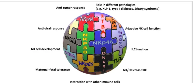
basis for many new findings on NK cells and beyond (Figure 1).
The association of different NCR splice variants with tumor
tissues or with non-pathological decidua tissues, the role of
NKp30, NKp46, and NKp80 in the NK-mediated cross-talk with
DCs, granulocytes, or monocytes, and the definition of NKp46
and NKp44 as markers of non-cytotoxic ILCs, are only some of
the indications for the involvement of these receptors in near
future studies on NK cell-based therapies against cancer, for
long-standing investigations on the maternal-fetal tolerance, and,
more extensively, on tissue homeostasis.
NATURAL CYTOTOXICITY RECEPTORS
Only few years after the identification of the first KIRs and
of CD94/NKG2A, three non-HLA class I-specific activating
receptors (namely NKp46, NKp30, and NKp44) were discovered
in Alessandro Moretta’s lab. These receptors, together with
NKG2D, turned to be crucial for the recognition of both tumor
and virus-infected cells (
5
,
49
,
50
). They were first characterized
for their functional features (i.e., their ability to induce NK
cell cytolytic activity and cytokine release) (
51
–
54
) and then
also at the molecular level, when the cDNAs coding for these
receptors were isolated (
53
,
55
,
56
) and the crystallographic
structures were solved (
57
–
60
). NKp46, NKp30, and NKp44 were
all selectively expressed on NK cells (although their expression
was differently induced during activation) and revealed, since
the initial studies, to be the main receptors responsible for
the so-called “natural cytotoxicity” of NK cells. Thus, based
on these findings, these receptors were collectively termed as
Natural Cytotoxicity Receptors (NCRs), although neither the
protein structure, nor the gene location gave indications for
their belonging to a receptor family. Their discovery paved the
way to a huge number of studies aimed at elucidating their
function in both physiological and pathological conditions and
characterizing the NCR/NCR ligand (NCR-L) interactions. As
mentioned above, NCR expression was initially thought to be
confined to NK cells, and NKp46 is still being considered a
reliable NK cell-associated marker, both in humans and in mice
(
61
,
62
). Soon thereafter it became clear that these receptors could
also be expressed in other immune cell types (
63
), extending
their role to additional biological processes. For example, the
characterization of the heterogeneous family of Innate Lymphoid
Cells (ILCs) (
25
,
64
,
65
) revealed that NKp44 is also expressed
by IFN-γ-producing intraepithelial ILC1 and by a subset of
ILC3 present at the epithelial/mucosal surfaces, in tonsils, and in
decidua tissue (
66
–
71
). Notably, NKp44
posILC3 display a unique
cytokine pattern, being able to produce IL-22 following cytokine
stimulation (
68
). In these cells, NKp44 triggering induces
TNF-α
production and activates a pro-inflammatory program (
72
),
suggesting that NKp44 could play a role in the pathogenesis
of different immune-mediated disease, including psoriasis (
73
).
In addition, NCR
pos(NKp44
pos) ILC3 have also been detected
in the lymphoid infiltrate of non-small cell lung cancer, and
have been found to release pro-inflammatory cytokines following
interaction with tumor cells and tumor-associated fibroblasts
(
34
,
67
,
74
). NKp46 expression has been detected in CD4
posT
lymphocytes derived from patients with Sézary syndrome, an
aggressive form of cutaneous T-cell lymphoma (CTCL) (
75
).
Notably, in these cells, NKp46 can act as an inhibitory
co-receptor able to decrease CD3-mediated proliferation of Sézary
FIGURE 1 | The “activating” solution of the NK cell puzzle. Different activating receptors collaborate to induce NK cell triggering in healthy and pathological conditions.
cells, and has been proposed as an additional diagnostic marker,
besides KIR3DL2, for the detection of these malignant cells (
76
).
One of the most investigated issues about NCRs is the
characterization of their ligands. Although the landscape of
NCR ligands is still incomplete, a common emerging theme is
the multiplicity and heterogeneity of NCR/NCR-L interactions
(
31
,
77
–
80
). Most NCR ligands have been shown to activate
NK cell function, while others dampen NK cell activation or
act as “decoy ligands” when released in soluble form (
81
–
85
). The panel of cellular NCR-Ls currently includes surface
glycoproteins, nuclear proteins that can be displayed at the
cell surface, soluble molecules that can be either secreted,
enzymatically shed, or conveyed through extracellular vesicles
(
82
,
85
–
92
). The expanding knowledge of NCR-Ls has opened the
possibility of targeting NCR/NCR-L interactions in the context
of cancer immunotherapy strategies. In addition, it has allowed
the identification of several mechanisms of tumor escape related
to the interaction between NK cells and malignant cells in the
tumor microenvironment (
22
,
93
–
101
). Finally, the importance
of NK cell activity, and of NCRs in particular, in the therapeutic
effect and outcome of oncolytic virotherapy has now being
appreciated (
102
–
104
). NCR-Ls are also being studied as possible
biomarkers in a variety of pathological conditions. Thus, a
soluble form of B7-H6 (sB7-H6), an NKp30 ligand, has been
demonstrated in the peritoneal fluid of ovarian cancer patients
and in patients with metastatic gastrointestinal stromal tumor
(GIST), neuroblastoma, or hepatocellular carcinoma (HCC) (
83
,
84
,
105
,
106
). The presence of soluble BAG6/BAT3 (another
NKp30-L) in the plasma of chronic lymphocytic leukemia
patients was found to correlate with advanced disease stages (
81
).
Along this line, high levels of soluble Nidogen-1, an NKp44
ligand, have been detected in the sera of patients with ovarian
or lung cancer (
107
,
108
).
Regarding the possibility of exploiting NCRs in
anti-tumor approaches, it must be considered that NKp46 and
NKp30 expression is down-regulated in NK cells derived from
patients with different types of both hematological and
non-hematological cancers (
93
,
109
–
116
). This downmodulation
leads to the impairment of NK cell anti-tumor potential and
consequently to the need to develop strategies aimed at restoring
NCR function (i.e., the use of cytokines, immunomodulatory
drugs, anti-cancer drugs, or anti-KIR mAbs) (
117
–
120
). In
addition, tumor cells themselves can become more resistant to
NK cell-mediated attack by down-regulating NCR-Ls or releasing
them in a soluble form (decoy ligands).
The role of NCRs stretches beyond cancer. B7-H6 is also
involved in the inflammatory response: its expression is induced
on monocytes following exposure to pro-inflammatory cytokines
or TLR ligands, and high levels of sB7-H6 are found in the serum
of patients with sepsis induced by Gram-negative bacteria (
121
).
NK cells, in general, have been studied in different autoimmune
disorders, including systemic lupus erythematosus, rheumatoid
arthritis, multiple sclerosis, and type I diabetes (TID) (
122
–
124
).
Focusing on NCRs, NKp46 has been shown to play a role in the
pathogenesis of TID and in the destruction of normal pancreatic
β
cells (
125
), suggesting the possibility to target this receptor
through specific anti-NKp46 mAbs (
126
).
A few years after the NCR discovery, the existence of
different splice variants of these receptors was revealed (
32
,
127
). Thus, three alternatively spliced NKp30 isoforms were
identified, characterized by distinct intracellular regions and
different functional capabilities. In GIST patients the prevalence
of NKp30c isoform has been associated to decreased NK cell
functionality and to reduced survival (
128
). Along this line, a
similar pattern of NKp30 isoform expression has been detected in
HCC patients (
106
). Notably, NKp30c isoform and sB7-H6 have
been studied in metastatic GIST patients, revealing their possible
use as predictive biomarkers of disease progression and response
to imatinib mesylate treatment (
105
). NKp44 splice variants have
been studied in different neoplastic disorders, and in particular in
acute myeloid leukemia patients, indicating a correlation between
the prevalence of the ITIM-bearing inhibitory NKp44-1 isoform
and poor survival (
129
). The induction of NKp44-1 expression
has been also observed in decidua NK cells, driven by cytokines
released in the decidua microenvironment, and could play a role
in promoting tolerance toward the fetus (
127
,
130
).
Among the NCRs, NKp44 is the main receptor involved in
the interplay between NK cells and trophoblast cells during
pregnancy (
131
,
132
), and is expressed also by a subset of ILC3
and by IFN-γ-producing ILC1-like cells found in the decidua
(
133
). Decidua NK cells represent a peculiar NK cell subset,
characterized by NKp44 expression, poor cytotoxic activity, and
contributing to decidua development, vascularization, and tissue
building/remodeling (
134
–
136
). Notably, in these cells, NKp44
triggering has been shown to induce IP10, IL-8, and VEGF
release (
132
,
137
).
ACTIVATING CO-RECEPTORS
Alessandro Moretta gave fundamental contributions also to the
identification and/or characterization of other surface receptors,
including 2B4 (
138
–
140
), NTBA (
141
,
142
), CD59 (
143
), and
NKp80 (
144
), that play a complementary or a synergistic role
with NCRs in inducing NK cell activation. Some of these
molecules received great interest because of their involvement
in NK cell function and development. 2B4 (
145
,
146
) and
NTBA (
142
), belonging to the signaling lymphocyte activation
molecule (SLAM) family, have been shown to act as co-receptors,
able to potentiate NK cell cytotoxic activity induced by the
main triggering receptors, including NKp46 (
140
,
141
). While
2B4 receptor recognizes CD48 (
146
,
147
), NTBA is involved
in homophilic interactions (
142
). Notably, 2B4 and NTBA
dysfunction was described to be associated with a severe form of
immunodeficiency, the X-linked lymphoproliferative syndrome
type 1 (XLP-1), caused by mutations in SH2D1A, the gene
encoding the signaling lymphocyte activation molecule
(SLAM)-associated protein (SAP) (
148
). Interestingly, in the absence
of SAP, the 2B4 and NTB-A co-receptors associate with the
protein tyrosine phosphatases thus delivering inhibitory, instead
of activating, signals (
141
,
149
–
151
). This immune dysfunction is
mainly responsible for the NK cell inability to kill EBV-infected B
cells (B-EBV) that express CD48, resulting in extremely severe
clinical consequences. A rapid diagnostic flowchart for XLP1,
based on a 2B4-specific functional assay, combined with
intra-cytoplasmic SAP staining, has been proposed (
152
). Moreover,
the abnormal 2B4 function also influences 2B4 cross-talk with
other NK receptors. Indeed, inhibitory 2B4 molecule selectively
blocks ITAM-dependent activating receptors, namely NCR and
CD16, while it affects neither NKG2D nor DNAM-1, which do
not transduce through ITAM (
152
). This finding explains the
selective inability, shown by NK cells, to kill B-EBV cells, which
highly express CD48 and are mainly recognized by NCRs. In
addition, in the NK cell repertoire of XLP-1 patients, NK cells
lacking any self HLA class I-specific inhibitory receptor are highly
represented and fully functional, indicating that the inhibitory
2B4 participates to NK cell education (
153
). Interestingly, a
similar role for 2B4 has been described also in particular
non-pathological processes. Indeed, at early stages of NK cell
differentiation, when HLA class I-specific inhibitory receptors are
not yet expressed, the delivery of inhibitory signals by 2B4, as
a consequence of the late SAP expression, renders self-tolerant
immature NK cells that otherwise would be autoreactive (
154
).
Another peculiar situation is represented by decidua NK cells, in
which 2B4 functions as an inhibitory receptor due to the absence
or very low levels of SAP expression (
155
).
CD59 has been found to associate to NKp46 and NKp30
receptors
and
to
enhance
NK
cell-mediated
cytotoxic
activity (
143
).
NKp80 molecule was initially described as a co-receptor,
expressed by all NK cells, and able to cooperate with triggering
receptors in the induction of natural cytotoxicity (
144
). Later,
NKp80 was found to recognize the Activation-Induced C-type
Lectin (AICL), a myeloid-specific activating receptor expressed
by monocytes, macrophages, and granulocytes (
156
).
NKp80-AICL interaction results in the secretion of pro-inflammatory
cytokines from both cell types. In addition, it has been shown
to participate in the NK cell-mediated elimination of malignant
myeloid cells (
156
). NKp80 also plays an important role in the
process of NK cell development. Indeed, it marks functionally
mature NK cells developing in secondary lymphoid tissues (SLT).
In particular, on the basis of NKp80 expression, two distinct
subsets of SLT stage 4 cells can be distinguished: an NKp80
negpopulation with both NK- and ILC3-associated features and an
NKp80
pospopulation with features similar to PB CD56
brightNK
cells (
157
).
Among the surface molecules behaving as co-receptors in
the activation of NK cell functions, a major role is assigned to
DNAX Accessory Molecule (DNAM-1 or CD226), an adhesion
molecule displaying activating function, expressed not only by
all NK cells but also by T lymphocytes and monocytes (
158
).
Alessandro Moretta’s group gave an important contribution
in this field with the identification of two different
DNAM-1 ligands, namely PVR and Nectin-2, belonging to the Nectin
family (
159
). These molecules are widely expressed on a
variety of both hematological and solid tumors (
160
,
161
),
representing suitable targets for immunotherapeutic approaches
(
162
). The role of DNAM-1 ligands in tumor cell recognition
and killing by NK cells is actually more complex, since,
besides DNAM-1, also the inhibitory receptors CD96 and TIGIT
can recognize PVR or PVR and Nectin-2, respectively (
163
,
164
). Accordingly, TIGIT and CD96 have been proposed as
immune checkpoints, and are becoming appealing targets for
the development of antibodies to be used in combination
with other immune checkpoint inhibitors with the aim of
unleashing both T and NK cell cytotoxic potential against
tumors (
165
,
166
).
ROLE OF NK CELLS IN IMMUNE
REGULATION
NK-DC Crosstalk
In the late ‘90s, it was becoming evident that innate immune
cells do not act in isolation but potentiate their efficiency by
interacting with each other, resulting even in the regulation of
adaptive immune response. In 2001 Ralph Steinman (eventually
a Nobel Laureate for the discovery of dendritic cells) visited our
laboratories in Genoa and that occasion represented a starting
point for a fruitful collaboration aimed at investigating the
cross-talk occurring between human DCs and NK cells. As always, Prof.
Moretta’s insights were pivotal in all the studies carried out in that
period, identifying which receptors and which subsets of these
two innate immune components participate in this interaction,
how this last one influences immune responses and to which
extent similar stimuli (e.g., TLR ligands) are integrated by DCs
and NK cells during innate immunity.
Until then, DCs were known for their critical role in initiating
immune responses and priming antigen-specific T cell response
(
167
), acting as sentinels in peripheral tissues, continuously
sampling the environment. The dogma also foresaw that upon
activation by danger signals, they up-regulated chemokine
receptors and co-stimulatory molecules, which allowed them
to migrate into lymph nodes and to efficiently induce T cell
responses (
167
). Thus, the idea that DCs could also act as early
activators of innate lymphocytes and, in turn, receive activating
signals by activated NK cells, was ground-breaking in the field of
innate immunity (
14
).
One of the relevant outcomes of NK/DC interaction is the
so called “editing” of DCs, a term coined by Prof. Moretta to
indicate the ability of NK cells to eliminate DCs in immature
stage, and therefore bona fide tolerogenic DCs, while sparing
activated/mature DCs able to efficiently induce the subsequent
adaptive immune response in secondary lymphoid organs (
12
,
168
,
169
). The protective mechanisms of mature DCs was
identified in the up-regulation of HLA class I molecules,
especially of the non-classical HLA-E (
170
), occurring upon
activation of DCs by either danger signals or NK cells themselves.
At the same time, also the activating receptors involved in
DC recognition by NK cells were identified (
12
,
171
). The
relevance of NKp30 receptor in NK/DC cross-talk was not
limited to the mechanisms of killing of immature DCs but
extended to the maturation process of DCs upon interaction with
NK cells (
172
).
Remarkably, this cytolytic DC editing by NK cells was
identified as a NK-mediated capability of dampening the
graft-vs.-host disease in bone marrow transplantation (
40
) and
graft rejection in solid organ transplantation (
173
,
174
). It is
noteworthy that, in case of improved skin graft rejection, NK cells
were found to home to lymph nodes where they killed allogeneic
DCs in a perforin-dependent manner (
174
).
Interestingly, and consistent with their concomitant role
during the early phase of immune responses, NK cells and DCs
are often able to sense similar stimuli in parallel. It was reported
by Moretta’s group that TLR engagement not only activates
immature DCs but also renders NK cells more prone to receive
triggering signals from pathogen-associated molecules, thus
exerting a regulatory control on the early steps of innate immune
responses against infectious agents (
16
), as more specifically
addressed in the next paragraph.
All these studies on DC/NK interactions indicate a critical
role for NK cells in the initiation and regulation of immune
responses and provide a strong rationale for a combined
targeting of NK cells and DCs in novel immunotherapeutic
strategies, harnessing this cellular cross-talk in the treatment
of patients with cancer and chronic infections resistant to
conventional therapies.
Alessandro Moretta’s contribution to the knowledge on the
molecular basis of these cellular interactions paved the way
to clinical interventions exploiting DC/NK cell cooperation.
As a matter of fact, NK cell activation by DCs is particularly
efficient, since DCs promote both effector functions and
survival/proliferation of NK cells (
169
). As a whole, these basic
discoveries, largely achieved under Prof. Moretta’s guidance,
revealed a particular translational relevance. For instance, in
the field of haplo-HSCT, a beneficial role of NK cells in
mediating graft-vs.-leukemia effects and in preventing GvHD
was highlighted. The support provided by DCs for the
proliferation/survival of NK cells is relevant also for establishing
more efficient protocols for ex vivo NK cell expansion,
given that NK cell-based immunotherapies are currently being
reconsidered in both post-transplant hematological settings and
in immunotherapy strategies for advanced solid tumors (
41
,
119
,
175
–
180
).
Finally, DCs activated by NK cells are better inducers of
the anti-tumor CTL response, at least in vitro, as compared
with the standard mature DCs currently employed in
DC-based clinical trials (
181
) and could therefore be considered in
immunization strategies for the development of next-generation
vaccines (
182
,
183
).
Expression and Function of TLRs on
Human NK Cells
Another field of research in which Prof. Moretta undoubtedly
gave important contributions is the expression and function of
TLRs in human NK cells. Indeed, in 2004 his group provided a
solid experimental evidence that pathogen-associated products,
known to strongly activate DCs and other innate immune
cells, can also act on TLRs expressed by NK cells, inducing
their activation both in terms of increased cytotoxicity and
cytokine release (
16
). Alessandro Moretta and coworkers not
only described the effect of TLR ligands on NK cell function,
but also analyzed the role of TLR in the NK/DC crosstalk. This
led to the concept of “NK cell-mediated editing of DCs,” the
“quality control” process by which NK cells select DCs that are
suited for T cell priming. The capability of TLR agonists of
potentiating NK cell function was further defined in subsequent
studies (
184
–
193
). Thus, in 2010 a peculiar cooperation between
TLR9 and KIR3DL2 in inducing triggering of NK cell function
upon treatment with CpG-ODN (TLR9 ligand) was described
(
194
,
195
). This study revealed that KIR3DL2 can bind
CpG-ODNs at the NK cell surface and shuttle them to endosomes
where TLR9 is localized, thus resulting in sharp down-regulation
of KIR3DL2 surface expression and in TLR9-mediated induction
of cytokine release. Moreover, it was demonstrated that the
KIR Ig-domain involved in the direct recognition of
CpG-ODN is represented by D0. Since this domain was hypothesized
to be expressed by the putative ancestral mammalian KIR,
these data suggested that, originally, certain KIRs could exert a
function different from recognition of HLA class I molecules.
Moreover, this newly defined functional capability of KIR3DL2
provided an important clue to understand the driving forces
that led to the conservation of the KIR3DL2-encoding gene
in all haplotypes, despite the low frequency, in the human
population, of HLA-A
∗03 or -A
∗11 alleles (i.e., the ligands of
KIR3DL2). Furthermore, in the Sézary Syndrome, in which
KIR3DL2 represents a specific marker for the assessment of
circulating tumor burden and for patient follow-up (
76
),
CpG-ODN has been shown to promote not only the internalization of
KIR3DL2 receptor but also the generation of apoptotic signals
(
196
). Thus, CpG-ODN may exert a direct anti-tumor effect
on Sézary cells through binding to KIR3DL2. In this context, a
good clinical response without major side effects was observed
upon class-B CpG-ODN subcutaneous administration in CTCL
patients (
197
). CpG-DNA and other TLR agonists have been also
explored as adjuvants for immunotherapy. Indeed, many clinical
trials based on the use of CpG-ODNs as immunotherapeutic
agents revealed that CpG-ODNs can promote Th1 immune
responses and may be used in combination with chemotherapy
to induce potent anti-tumor immune responses with relevant
clinical benefits (
186
,
198
,
199
).
NK Cell Subsets in Anti-virus Responses
Besides cancer and other diseases, NCRs also contribute to
the NK cell-mediated control of viral infections through
the recognition of virus-infected cells. Indeed, the first
characterized NCR-Ls were of viral origin, namely influenza
virus hemagglutinins (
200
,
201
). Later on, additional viral
ligands were identified and, in most cases, they were shown
to induce NK cell activation following NCR engagement
(
31
,
78
,
202
). It is of note, however, that some viral NCR-Ls
can inhibit NCR functions, representing a possible immune
evasion strategy (
203
). It has been very recently demonstrated in
mouse that NK cells may play a regulatory role during acute and
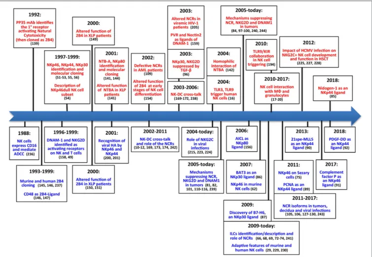
FIGURE 2 | The main steps concerning activating NK receptors/coreceptors. The timeline illustrates the main discoveries concerning NK cell activating receptors during a timespan of about 30 years. Contributions deriving from Alessandro Moretta’s research group are indicated in red (upper part), while contributions obtained by other groups are shown in blue (lower part).
chronic lymphocytic choriomeningitis virus (LCMV) infection
through the NKp46-mediated killing of LCMV-specific CD8 T
cells (
204
).
In recent years Prof. Moretta and his co-workers gave major
contributions to broaden our knowledge on NK cell diversity
and functional specialization. This occurred primarily thanks
to studies focused on NK cell-mediated responses to virus
infections. Fundamental results came from the characterization
of NK cells in patients chronically infected by HIV that
revealed a deep functional impairment of NK cells likely
determining their scarce capacity to efficiently control this
virus. In this context, the relevance of NCR contribution to
the course of HIV infection became clear when their reduced
expression on NK cells in viraemic HIV-infected patients was
demonstrated (
205
,
206
). The NCR role in anti-viral response
was also supported by the demonstration that NKp46 and
NKp30 inducibility exerted a protective role in HIV-infected
patients with excellent control not only of virus replication
but, more importantly, also of retroviral reservoir (
207
,
208
).
Outside the HIV field, the study of NCR expression on NK
cells similarly provided compelling evidence of their involvement
in the response to acute HCV infection (
209
), and in HCV
eradication in treated chronic carriers (
210
,
211
). Interestingly,
in chronically infected HIV patients the accumulation of
a dysfunctional NK cell subset, virtually absent in healthy
subjects, characterized by an aberrant CD56
negCD16
brightsurface signature (
205
,
212
,
213
) and defective DC editing was
observed (
214
). This unusual population has been subsequently
identified in several other pathological conditions including
viral infections and immune deficiencies, in which these cells
are responsible for an altered response to a chronic immune
activation (
215
–
219
).
Besides HIV, a fundamental role in shaping NK cell
repertoire and function has been described for CMV infection
(
220
–
222
). Based on the pioneering studies by M.
Lopez-Botet who first described the imprinting exerted by CMV
on NK cells (
223
,
224
), Alessandro Moretta contributed to
identify CMV infection as a key driving force promoting the
differentiation of functionally and phenotypically skewed NK
cells with several studies conducted in HSCT recipients (
225
–
228
). In this setting, CMV infection/reactivation could induce
not just NK cell maturation toward highly differentiated stages
(characterized by the expression of CD94/NKG2C or activating
KIRs), but also the unexpected acquisition of immunological
memory. Indeed, NK cells maturing in CMV-reactivating
patients share features with adaptive immune cells, such as
long-term persistence, virus-induced clonal expansion, and epigenetic
modifications (
227
,
229
–
234
).
This anti-paradigmatic concept of memory or adaptive NK
cells, to which Prof. Moretta contributed, holds important
translational promise as this NK cell population characterized
by longevity and superior ADCC ability, represents a
potential tool for novel immunotherapeutic anti-cancer
strategies, namely antibody-based tumor immunotherapies
and
generation
of
long-living
anti-tumor
CAR-NK
cells (
179
,
235
).
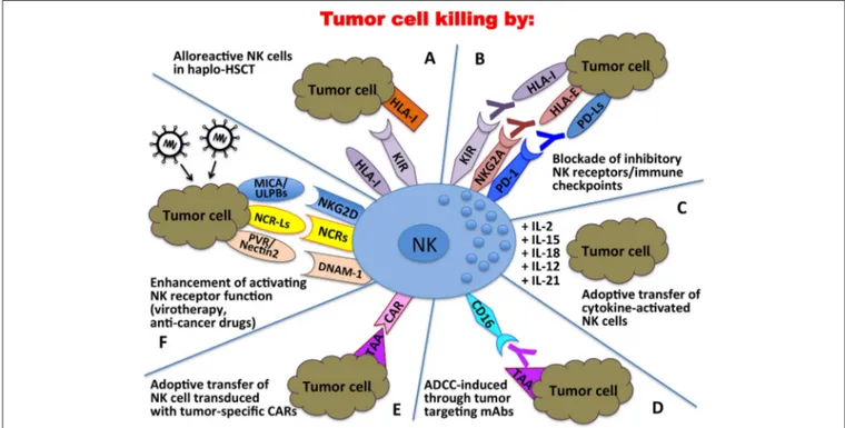
FIGURE 3 | Clinical applications of NK cells in the immunotherapy against tumors. In haplo-HSCT, alloreactive NK cells can kill residual leukemic cells (A); mAbs directed against immune checkpoints can unleash/restore NK cell anti-tumor activity (B); tumor cell killing can be enhanced by adoptive transfer of cytokine-activated NK cells (C) or NK cells transduced with tumor-specific Chimeric Antigen Receptors (CARs) (E); tumor targeting mAbs can induce NK cell-mediated ADCC (D); activating NK receptor function can be potentiated through oncolytic virotherapy or the use of anti-cancer drugs (F).
NK CELL-BASED CLINICAL
APPLICATIONS
Altogether, these discoveries in the field of NK cell biology
(Figure 2) (
236
–
243
) paved the way to the exploitation of these
cells in different anti-tumor therapeutic approaches (Figure 3).
Over the years important achievements have been obtained,
and promising novel strategies have been designed. The most
advanced clinical application exploiting the NK cell anti-tumor
potential is in the field of haplo-identical HSCT (
40
–
42
,
235
),
in which donor-derived alloreactive NK cells (i.e., unable to
recognize recipient HLA class I molecules) can exert a potent
anti-leukemia effect. Moreover, the adoptive transfer of NK cells,
in an autologous or allogeneic setting, can be pursued following
NK cell activation and expansion with cytokines (
118
–
120
).
The blockade of HLA class I-specific inhibitory receptors using
human/humanized mAbs can be used to enhance killing of HLA
class I
postumor cells. These mAbs can be used in combination
with mAbs interfering with the PD-1/PD-L axis, as PD-1 can
be expressed by human NK cells (
46
,
244
). Another clinical
approach is represented by the induction of ADCC against
tumor cells by the use of antibodies specific for tumor-associated
antigens (
119
).
More recently, the CAR technology, originally designed
for T lymphocytes, has been applied also to NK cells, with
promising results in the therapy of both hematological and solid
tumors (
118
,
120
). The ever-growing knowledge of activating NK
receptor/ligand interactions is being applied in several strategies
aimed to potentiate triggering signals through virotherapy or by
the use of anti-cancer drugs capable of enhancing the expression
of activating ligands on tumor cells and activating receptors on
NK cells (
102
,
117
). In conclusion, NK cell-based therapy used
in combination with conventional therapeutic protocols could
become more and more a powerful tool to be used in the cure
of cancer.
CONCLUDING REMARKS
By revisiting the discovery of the most important NK receptors
and considering the technical approaches available at that time,
one might have the impression that it has been simple to
obtain those results. However, experienced researchers know
that, actually, relevant pieces of information leading to a new
discovery must be selected from an initially confusing, and
often contradictory, mass of data. Alessandro had this ability,
common to many gifted scientists, but he was also endowed
with the uncommon talent of catching essential information and
rendering simple what actually is very complex. We think that
this has been the true and most important lesson for all of us and,
undoubtedly, a major legacy for Immunology and Medicine.
AUTHOR CONTRIBUTIONS
All authors listed have made a substantial, direct and
intellectual contribution to the work, and approved it
for publication.
FUNDING
Supported by the following grants: Fondazione AIRC 5X1000,
2018 Project Code 21147 (LoM and SS), Fondazione AIRC IG
2017, Project Code 19920 (LoM), Fondazione AIRC IG 2017
Project Code 20312 (SS), Fondazione AIRC IG 2015, Project
Code 16764 (DP), AIRC IG 2014 project no. 15428 (MV), and
5X1000 Min. Sal. 2013 (MV); LeM is a recipient of a fellowship
awarded by Federazione Italiana Ricerca sul Cancro (FIRC).
REFERENCES
1. Ljunggren HG, Karre K. In search of the ‘missing self ’: MHC molecules and NK cell recognition. Immunol Today. (1990) 11:237–44. doi: 10.1016/0167-5699(90)90097-S
2. Moretta A, Bottino C, Vitale M, Pende D, Biassoni R, Mingari MC, et al. Receptors for HLA class-I molecules in human natural killer cells. Annu Rev Immunol. (1996) 14:619–48. doi: 10.1146/annurev.immunol.14.1.619 3. Moretta A, Biassoni R, Bottino C, Pende D, Vitale M, Poggi A,
et al. Major histocompatibility complex class I-specific receptors on human natural killer and T lymphocytes. Immunol Rev. (1997) 155:105– 17. doi: 10.1111/j.1600-065X.1997.tb00943.x
4. Wagtmann N, Rajagopalan S, Winter CC, Peruzzi M, Long EO. Killer cell inhibitory receptors specific for HLA-C and HLA-B identified by direct binding and by functional transfer. Immunity. (1995) 3:801– 9. doi: 10.1016/1074-7613(95)90069-1
5. Moretta A, Bottino C, Vitale M, Pende D, Cantoni C, Mingari MC, et al. Activating receptors and coreceptors involved in human natural killer cell-mediated cytolysis. Annu Rev Immunol. (2001) 19:197– 223. doi: 10.1146/annurev.immunol.19.1.197
6. Long EO, Burshtyn DN, Clark WP, Peruzzi M, Rajagopalan S, Rojo S, et al. Killer cell inhibitory receptors: diversity, specificity, and function. Immunol Rev. (1997) 155:135–44. doi: 10.1111/j.1600-065X.1997.tb00946.x 7. Colonna M. Specificity and function of immunoglobulin superfamily NK
cell inhibitory and stimulatory receptors. Immunol Rev. (1997) 155:127– 33. doi: 10.1111/j.1600-065X.1997.tb00945.x
8. Parham P. MHC class I molecules and KIRs in human history, health and survival. Nat Rev Immunol. (2005) 5:201–14. doi: 10.1038/nri1570 9. Lanier LL. Natural killer cells: from no receptors to too many. Immunity.
(1997) 6:371–8. doi: 10.1016/S1074-7613(00)80280-0
10. Piccioli D, Sbrana S, Melandri E, Valiante NM. Contact-dependent stimulation and inhibition of dendritic cells by natural killer cells. J Exp Med. (2002) 195:335–41. doi: 10.1084/jem.20010934
11. Gerosa F, Baldani-Guerra B, Nisii C, Marchesini V, Carra G, Trinchieri G. Reciprocal activating interaction between natural killer cells and dendritic cells. J Exp Med. (2002) 195:327–33. doi: 10.1084/jem.20010938
12. Ferlazzo G, Tsang ML, Moretta L, Melioli G, Steinman RM, Munz C. Human dendritic cells activate resting natural killer (NK) cells and are recognized via the NKp30 receptor by activated NK cells. J Exp Med. (2002) 195:343–51. doi: 10.1084/jem.20011149
13. Sivori S, Carlomagno S, Pesce S, Moretta A, Vitale M, Marcenaro E. TLR/NCR/KIR: which one to use and when? Front Immunol. (2014) 5:105. doi: 10.3389/fimmu.2014.00105
14. Moretta A. Natural killer cells and dendritic cells: rendezvous in abused tissues. Nat Rev Immunol. (2002) 2:957–64. doi: 10.1038/nri956
15. Della Chiesa M, Sivori S, Castriconi R, Marcenaro E, Moretta A. Pathogen-induced private conversations between natural killer and dendritic cells. Trends Microbiol. (2005) 13:128–36. doi: 10.1016/j.tim.2005. 01.006
16. Sivori S, Falco M, Della Chiesa M, Carlomagno S, Vitale M, Moretta L, et al. CpG and double-stranded RNA trigger human NK cells by Toll-like receptors: induction of cytokine release and cytotoxicity against
tumors and dendritic cells. Proc Natl Acad Sci USA. (2004) 101:10116– 21. doi: 10.1073/pnas.0403744101
17. Thoren FB, Riise RE, Ousback J, Della Chiesa M, Alsterholm M, Marcenaro E, et al. Human NK Cells induce neutrophil apoptosis via an NKp46- and Fas-dependent mechanism. J Immunol. (2012) 188:1668– 74. doi: 10.4049/jimmunol.1102002
18. Riise RE, Bernson E, Aurelius J, Martner A, Pesce S, Della Chiesa M, et al. TLR-stimulated neutrophils instruct NK cells to trigger dendritic cell maturation and promote adaptive T cell responses. J Immunol. (2015) 195:1121–8. doi: 10.4049/jimmunol.1500709
19. Bellora F, Castriconi R, Dondero A, Reggiardo G, Moretta L, Mantovani A, et al. The interaction of human natural killer cells with either unpolarized or polarized macrophages results in different functional outcomes. Proc Natl Acad Sci USA. (2010) 107:21659–64. doi: 10.1073/pnas.1007654108 20. Pesce S, Thoren FB, Cantoni C, Prato C, Moretta L, Moretta A, et al.
The innate immune cross talk between NK cells and eosinophils is regulated by the interaction of natural cytotoxicity receptors with eosinophil surface ligands. Front Immunol. (2017) 8:510. doi: 10.3389/fimmu.2017. 00510
21. Mattiola I, Pesant M, Tentorio PF, Molgora M, Marcenaro E, Lugli E, et al. Priming of human resting NK cells by autologous M1 macrophages via the engagement of IL-1beta, IFN-beta, and IL-15 Pathways. J Immunol. (2015) 195:2818–28. doi: 10.4049/jimmunol.1500325
22. Vitale M, Cantoni C, Pietra G, Mingari MC, Moretta L. Effect of tumor cells and tumor microenvironment on NK-cell function. Eur J Immunol. (2014) 44:1582–92. doi: 10.1002/eji.201344272
23. Martin-Fontecha A, Thomsen LL, Brett S, Gerard C, Lipp M, Lanzavecchia A, et al. Induced recruitment of NK cells to lymph nodes provides IFN-gamma for T(H)1 priming. Nat Immunol. (2004) 5:1260–5. doi: 10.1038/ni1138
24. Ardolino M, Zingoni A, Cerboni C, Cecere F, Soriani A, Iannitto ML, et al. DNAM-1 ligand expression on Ag-stimulated T lymphocytes is mediated by ROS-dependent activation of DNA-damage response: relevance for NK-T cell interaction. Blood. (2011) 117:4778–86. doi: 10.1182/blood-2010-08-300954
25. Freud AG, Mundy-Bosse BL, Yu J, Caligiuri MA. The broad spectrum of human natural killer cell diversity. Immunity. (2017) 47:820–33. doi: 10.1016/j.immuni.2017.10.008
26. Della Chiesa M, Sivori S, Carlomagno S, Moretta L, Moretta A. Activating KIRs and NKG2C in viral infections: toward NK cell memory? Front Immunol. (2015) 6:573. doi: 10.3389/fimmu.2015. 00573
27. Della Chiesa M, Marcenaro E, Sivori S, Carlomagno S, Pesce S, Moretta A. Human NK cell response to pathogens. Semin Immunol. (2014) 26:152– 60. doi: 10.1016/j.smim.2014.02.001
28. Muntasell A, Vilches C, Angulo A, Lopez-Botet M. Adaptive reconfiguration of the human NK-cell compartment in response to cytomegalovirus: a different perspective of the host-pathogen interaction. Eur J Immunol. (2013) 43:1133–41. doi: 10.1002/eji.201243117
29. Sun JC, Beilke JN, Lanier LL. Adaptive immune features of natural killer cells. Nature. (2009) 457:557–61. doi: 10.1038/nature07665
30. Vivier E, Raulet DH, Moretta A, Caligiuri MA, Zitvogel L, Lanier LL, et al. Innate or adaptive immunity? The example of natural killer cells. Science. (2011) 331:44–9. doi: 10.1126/science.1198687
31. Kruse PH, Matta J, Ugolini S, Vivier E. Natural cytotoxicity receptors and their ligands. Immunol Cell Biol. (2014) 92:221–9. doi: 10.1038/icb. 2013.98
32. Pazina T, Shemesh A, Brusilovsky M, Porgador A, Campbell KS. Regulation of the functions of natural cytotoxicity receptors by interactions with diverse ligands and alterations in splice variant expression. Front Immunol. (2017) 8:369. doi: 10.3389/fimmu.2017.00369
33. Sivori S, Vacca P, Del Zotto G, Munari E, Mingari MC, Moretta L. Human NK cells: surface receptors, inhibitory checkpoints, and translational applications. Cell Mol Immunol. (2019) 16:430–41. doi: 10.1038/s41423-019-0206-4
34. Chiossone L, Dumas PY, Vienne M, Vivier E. Natural killer cells and other innate lymphoid cells in cancer. Nat Rev Immunol. (2018) 18:671– 88. doi: 10.1038/s41577-018-0061-z
35. Pietra G, Vitale C, Pende D, Bertaina A, Moretta F, Falco M, et al. Human natural killer cells: news in the therapy of solid tumors and high-risk leukemias. Cancer Immunol Immunother. (2016) 65:465– 76. doi: 10.1007/s00262-015-1744-y
36. Tallerico R, Garofalo C, Carbone E. A new biological feature of natural killer cells: the recognition of solid tumor-derived cancer stem cells. Front Immunol. (2016) 7:179. doi: 10.3389/fimmu.2016.00179
37. Stabile H, Fionda C, Gismondi A, Santoni A. Role of distinct natural killer cell subsets in anticancer response. Front Immunol. (2017) 8:293. doi: 10.3389/fimmu.2017.00293
38. Mehta RS, Rezvani K. Chimeric antigen receptor expressing natural killer cells for the immunotherapy of cancer. Front Immunol. (2018) 9:283. doi: 10.3389/fimmu.2018.00283
39. Moretta L, Pietra G, Montaldo E, Vacca P, Pende D, Falco M, et al. Human NK cells: from surface receptors to the therapy of leukemias and solid tumors. Front Immunol. (2014) 5:87. doi: 10.3389/fimmu.2014.00087 40. Ruggeri L, Capanni M, Urbani E, Perruccio K, Shlomchik
WD, Tosti A, et al. Effectiveness of donor natural killer cell alloreactivity in mismatched hematopoietic transplants. Science. (2002) 295:2097–100. doi: 10.1126/science.1068440
41. Pende D, Marcenaro S, Falco M, Martini S, Bernardo ME, Montagna D, et al. Anti-leukemia activity of alloreactive NK cells in KIR ligand-mismatched haploidentical HSCT for pediatric patients: evaluation of the functional role of activating KIR and redefinition of inhibitory KIR specificity. Blood. (2009) 113:3119–29. doi: 10.1182/blood-2008-06-164103
42. Cooley S, Trachtenberg E, Bergemann TL, Saeteurn K, Klein J, Le CT, et al. Donors with group B KIR haplotypes improve relapse-free survival after unrelated hematopoietic cell transplantation for acute myelogenous leukemia. Blood. (2009) 113:726–32. doi: 10.1182/blood-2008-07-171926 43. Benson DM, Jr., Bakan CE, Zhang S, Collins SM, Liang J, Srivastava S, et al.
IPH2101, a novel anti-inhibitory KIR antibody, and lenalidomide combine to enhance the natural killer cell versus multiple myeloma effect. Blood. (2011) 118:6387–91. doi: 10.1182/blood-2011-06-360255
44. Pardoll DM. The blockade of immune checkpoints in cancer immunotherapy. Nat Rev Cancer. (2012) 12:252–64. doi: 10.1038/nrc3239 45. Hughes PE, Caenepeel S, Wu LC. Targeted therapy and checkpoint
immunotherapy combinations for the treatment of cancer. Trends Immunol. (2016) 37:462–76. doi: 10.1016/j.it.2016.04.010
46. Andre P, Denis C, Soulas C, Bourbon-Caillet C, Lopez J, Arnoux T, et al. Anti-NKG2A mAb Is a checkpoint inhibitor that promotes anti-tumor immunity by unleashing both T and NK cells. Cell. (2018) 175:1731–43 e13. doi: 10.1016/j.cell.2018.10.014
47. Sanchez-Correa B, Lopez-Sejas N, Duran E, Labella F, Alonso C, Solana R, et al. Modulation of NK cells with checkpoint inhibitors in the context of cancer immunotherapy. Cancer Immunol Immunother. (2019) 68:861– 70. doi: 10.1007/s00262-019-02336-6
48. Morvan MG, Lanier LL. NK cells and cancer: you can teach innate cells new tricks. Nat Rev Cancer. (2016) 16:7–19. doi: 10.1038/nrc.2015.5
49. Bauer S, Groh V, Wu J, Steinle A, Phillips JH, Lanier LL, et al. Activation of NK cells and T cells by NKG2D, a receptor for stress-inducible MICA. Science. (1999) 285:727–9. doi: 10.1126/science.285.5428.727
50. El-Gazzar A, Groh V, Spies T. Immunobiology and conflicting roles of the human NKG2D lymphocyte receptor and its ligands in cancer. J Immunol. (2013) 191:1509–15. doi: 10.4049/jimmunol.1301071
51. Sivori S, Vitale M, Morelli L, Sanseverino L, Augugliaro R, Bottino C, et al. p46, a novel natural killer cell-specific surface molecule that mediates cell activation. J Exp Med. (1997) 186:1129–36. doi: 10.1084/jem.186.7.1129 52. Vitale M, Bottino C, Sivori S, Sanseverino L, Castriconi R, Marcenaro E,
et al. NKp44, a novel triggering surface molecule specifically expressed by activated natural killer cells, is involved in non-major histocompatibility complex-restricted tumor cell lysis. J Exp Med. (1998) 187:2065– 72. doi: 10.1084/jem.187.12.2065
53. Pende D, Parolini S, Pessino A, Sivori S, Augugliaro R, Morelli L, et al. Identification and molecular characterization of NKp30, a novel triggering receptor involved in natural cytotoxicity mediated by human natural killer cells. J Exp Med. (1999) 190:1505–16. doi: 10.1084/jem.190.10.1505 54. Sivori S, Pende D, Bottino C, Marcenaro E, Pessino A, Biassoni R, et al.
of fresh or cultured human NK cells. Correlation between surface density of NKp46 and natural cytotoxicity against autologous, allogeneic or xenogeneic target cells. Eur J Immunol. (1999) 29:1656–66.
55. Pessino A, Sivori S, Bottino C, Malaspina A, Morelli L, Moretta L, et al. Molecular cloning of NKp46: a novel member of the immunoglobulin superfamily involved in triggering of natural cytotoxicity. J Exp Med. (1998) 188:953–60. doi: 10.1084/jem.188.5.953
56. Cantoni C, Bottino C, Vitale M, Pessino A, Augugliaro R, Malaspina A, et al. NKp44, a triggering receptor involved in tumor cell lysis by activated human natural killer cells, is a novel member of the immunoglobulin superfamily. J Exp Med. (1999) 189:787–96. doi: 10.1084/jem.189.5.787
57. Cantoni C, Ponassi M, Biassoni R, Conte R, Spallarossa A, Moretta A, et al. The three-dimensional structure of the human NK cell receptor NKp44, a triggering partner in natural cytotoxicity. Structure. (2003) 11:725– 34. doi: 10.1016/S0969-2126(03)00095-9
58. Ponassi M, Cantoni C, Biassoni R, Conte R, Spallarossa A, Pesce A, et al. Structure of the human NK cell triggering receptor NKp46 ectodomain. Biochem Biophys Res Commun. (2003) 309:317–23. doi: 10.1016/j.bbrc.2003.08.007
59. Li Y, Wang Q, Mariuzza RA. Structure of the human activating natural cytotoxicity receptor NKp30 bound to its tumor cell ligand B7-H6. J Exp Med. (2011) 208:703–14. doi: 10.1084/jem.20102548
60. Joyce MG, Tran P, Zhuravleva MA, Jaw J, Colonna M, Sun PD. Crystal structure of human natural cytotoxicity receptor NKp30 and identification of its ligand binding site. Proc Natl Acad Sci USA. (2011) 108:6223– 8. doi: 10.1073/pnas.1100622108
61. Biassoni R, Pessino A, Bottino C, Pende D, Moretta L, Moretta A. The murine homologue of the human NKp46, a triggering receptor involved in the induction of natural cytotoxicity. Eur J Immunol. (1999) 29:1014–20. 62. Walzer T, Blery M, Chaix J, Fuseri N, Chasson L, Robbins SH,
et al. Identification, activation, and selective in vivo ablation of mouse NK cells via NKp46. Proc Natl Acad Sci USA. (2007) 104:3384– 9. doi: 10.1073/pnas.0609692104
63. Hudspeth K, Silva-Santos B, Mavilio D. Natural cytotoxicity receptors: broader expression patterns and functions in innate and adaptive immune cells. Front Immunol. (2013) 4:69. doi: 10.3389/fimmu.2013.00069 64. Vivier E, Artis D, Colonna M, Diefenbach A, Di Santo JP, Eberl
G, et al. Innate lymphoid cells: 10 years on. Cell. (2018) 174:1054– 66. doi: 10.1016/j.cell.2018.07.017
65. Vacca P, Munari E, Tumino N, Moretta F, Pietra G, Vitale M, et al. Human natural killer cells and other innate lymphoid cells in cancer: friends or foes? Immunol Lett. (2018) 201:14–9. doi: 10.1016/j.imlet.2018.11.004
66. Fuchs A, Vermi W, Lee JS, Lonardi S, Gilfillan S, Newberry RD, et al. Intraepithelial type 1 innate lymphoid cells are a unique subset of IL-12- and IL-15-responsive IFN-gamma-producing cells. Immunity. (2013) 38:769–81. doi: 10.1016/j.immuni.2013.02.010
67. Simoni Y, Newell EW. Toward meaningful definitions of innate-lymphoid-cell subsets. Immunity. (2017) 46:760–1. doi: 10.1016/j.immuni.2017.04.026 68. Cella M, Fuchs A, Vermi W, Facchetti F, Otero K, Lennerz JK, et al. A human
natural killer cell subset provides an innate source of IL-22 for mucosal immunity. Nature. (2009) 457:722–5. doi: 10.1038/nature07537
69. Hoorweg K, Peters CP, Cornelissen F, Aparicio-Domingo P, Papazian N, Kazemier G, et al. Functional differences between human NKp44− and NKp44+ RORC+ innate lymphoid cells. Front Immunol. (2012) 3:72. doi: 10.3389/fimmu.2012.00072
70. Killig M, Glatzer T, Romagnani C. Recognition strategies of group 3 innate lymphoid cells. Front Immunol. (2014) 5:142. doi: 10.3389/fimmu.2014.00142
71. Vacca P, Vitale C, Munari E, Cassatella MA, Mingari MC, Moretta L. Human innate lymphoid cells: their functional and cellular interactions in decidua. Front Immunol. (2018) 9:1897. doi: 10.3389/fimmu.2018.01897
72. Glatzer T, Killig M, Meisig J, Ommert I, Luetke-Eversloh M, Babic M, et al. RORγ t+innate lymphoid cells acquire a proinflammatory program upon engagement of the activating receptor NKp44. Immunity. (2013) 38:1223– 35. doi: 10.1016/j.immuni.2013.05.013
73. Villanova F, Flutter B, Tosi I, Grys K, Sreeneebus H, Perera GK, et al. Characterization of innate lymphoid cells in human skin and blood demonstrates increase of NKp44+ ILC3 in psoriasis. J Invest Dermatol. (2014) 134:984–91. doi: 10.1038/jid.2013.477
74. Carrega P, Loiacono F, Di Carlo E, Scaramuccia A, Mora M, Conte R, et al. NCR+ILC3 concentrate in human lung cancer and associate with intratumoral lymphoid structures. Nat Commun. (2015) 6:8280. doi: 10.1038/ncomms9280
75. Bensussan A, Remtoula N, Sivori S, Bagot M, Moretta A, Marie-Cardine A. Expression and function of the natural cytotoxicity receptor NKp46 on circulating malignant CD4+ T lymphocytes of Sezary syndrome patients. J Invest Dermatol. (2011) 131:969–76. doi: 10.1038/jid.2010.404
76. Bagot M, Moretta A, Sivori S, Biassoni R, Cantoni C, Bottino C, et al. CD4+ cutaneous T-cell lymphoma cells express the p140-killer cell immunoglobulin-like receptor. Blood. (2001) 97:1388–91. doi: 10.1182/blood.V97.5.1388
77. Lam RA, Chwee JY, Le Bert N, Sauer M, Pogge von Strandmann E, Gasser S. Regulation of self-ligands for activating natural killer cell receptors. Ann Med. (2013) 45:384–94. doi: 10.3109/07853890.2013.792495
78. Koch J, Steinle A, Watzl C, Mandelboim O. Activating natural cytotoxicity receptors of natural killer cells in cancer and infection. Trends Immunol. (2013) 34:182–91. doi: 10.1016/j.it.2013.01.003
79. Brusilovsky M, Radinsky O, Yossef R, Campbell KS, Porgador A. Carbohydrate-mediated modulation of NK cell receptor function: structural and functional influences of heparan sulfate moieties expressed on NK cell surface. Front Oncol. (2014) 4:185. doi: 10.3389/fonc.2014.00185
80. Horton NC, Mathew PA. NKp44 and natural cytotoxicity receptors as damage-associated molecular pattern recognition receptors. Front Immunol. (2015) 6:31. doi: 10.3389/fimmu.2015.00031
81. Reiners KS, Topolar D, Henke A, Simhadri VR, Kessler J, Sauer M, et al. Soluble ligands for NK cell receptors promote evasion of chronic lymphocytic leukemia cells from NK cell anti-tumor activity. Blood. (2013) 121:3658–65. doi: 10.1182/blood-2013-01-476606
82. Schlecker E, Fiegler N, Arnold A, Altevogt P, Rose-John S, Moldenhauer G, et al. Metalloprotease-mediated tumor cell shedding of B7-H6, the ligand of the natural killer cell-activating receptor NKp30. Cancer Res. (2014) 74:3429–40. doi: 10.1158/0008-5472.CAN-13-3017
83. Semeraro M, Rusakiewicz S, Minard-Colin V, Delahaye NF, Enot D, Vely F, et al. Clinical impact of the NKp30/B7-H6 axis in high-risk neuroblastoma patients. Sci Transl Med. (2015) 7:283ra55. doi: 10.1126/scitranslmed.aaa2327
84. Pesce S, Tabellini G, Cantoni C, Patrizi O, Coltrini D, Rampinelli F, et al. B7-H6-mediated downregulation of NKp30 in NK cells contributes to ovarian carcinoma immune escape. Oncoimmunology. (2015) 4:e1001224. doi: 10.1080/2162402X.2014.1001224
85. Gaggero S, Bruschi M, Petretto A, Parodi M, Zotto GD, Lavarello C, et al. Nidogen-1 is a novel extracellular ligand for the NKp44 activating receptor. Oncoimmunology. (2018) 7:e1470730. doi: 10.1080/2162402X.2018.14 70730
86. Pogge von Strandmann E, Simhadri VR, von Tresckow B, Sasse S, Reiners KS, Hansen HP, et al. Human leukocyte antigen-B-associated transcript 3 is released from tumor cells and engages the NKp30 receptor on natural killer cells. Immunity. (2007) 27:965–74. doi: 10.1016/j.immuni.2007.10.010 87. Brandt CS, Baratin M, Yi EC, Kennedy J, Gao Z, Fox B, et al. The B7
family member B7-H6 is a tumor cell ligand for the activating natural killer cell receptor NKp30 in humans. J Exp Med. (2009) 206:1495– 503. doi: 10.1084/jem.20090681
88. Hecht ML, Rosental B, Horlacher T, Hershkovitz O, De Paz JL, Noti C, et al. Natural cytotoxicity receptors NKp30, NKp44 and NKp46 bind to different heparan sulfate/heparin sequences. J Proteome Res. (2009) 8:712– 20. doi: 10.1021/pr800747c
89. Rosental B, Brusilovsky M, Hadad U, Oz D, Appel MY, Afergan F, et al. Proliferating cell nuclear antigen is a novel inhibitory ligand for the natural cytotoxicity receptor NKp44. J Immunol. (2011) 187:5693– 702. doi: 10.4049/jimmunol.1102267
90. Baychelier F, Sennepin A, Ermonval M, Dorgham K, Debre P, Vieillard V. Identification of a cellular ligand for the natural cytotoxicity receptor NKp44. Blood. (2013) 122:2935–42. doi: 10.1182/blood-2013-03-4 89054
91. Narni-Mancinelli E, Gauthier L, Baratin M, Guia S, Fenis A, Deghmane AE, et al. Complement factor P is a ligand for the natural killer cell-activating receptor NKp46. Sci Immunol. (2017) 2:eaam9628. doi: 10.1126/sciimmunol.aam9628
92. Barrow AD, Edeling MA, Trifonov V, Luo J, Goyal P, Bohl B, et al. Natural killer cells control tumor growth by sensing a growth factor. Cell. (2018) 172:534–48 e19. doi: 10.1016/j.cell.2017.11.037
93. Farnault L, Sanchez C, Baier C, Le Treut T, Costello RT. Hematological malignancies escape from NK cell innate immune surveillance: mechanisms and therapeutic implications. Clin Dev Immunol. (2012) 2012:421702. doi: 10.1155/2012/421702
94. Stojanovic A, Correia MP, Cerwenka A. Shaping of NK cell responses by the tumor microenvironment. Cancer Microenviron. (2013) 6:135– 46. doi: 10.1007/s12307-012-0125-8
95. Baginska J, Viry E, Paggetti J, Medves S, Berchem G, Moussay E, et al. The critical role of the tumor microenvironment in shaping natural killer cell-mediated anti-tumor immunity. Front Immunol. (2013) 4:490. doi: 10.3389/fimmu.2013.00490
96. Castriconi R, Cantoni C, Della Chiesa M, Vitale M, Marcenaro E, Conte R, et al. Transforming growth factor beta 1 inhibits expression of NKp30 and NKG2D receptors: consequences for the NK-mediated killing of dendritic cells. Proc Natl Acad Sci USA. (2003) 100:4120– 5. doi: 10.1073/pnas.0730640100
97. Della Chiesa M, Carlomagno S, Frumento G, Balsamo M, Cantoni C, Conte R, et al. The tryptophan catabolite L-kynurenine inhibits the surface expression of NKp46- and NKG2D-activating receptors and regulates NK-cell function. Blood. (2006) 108:4118–25. doi: 10.1182/blood-2006-03-006700
98. Balsamo M, Scordamaglia F, Pietra G, Manzini C, Cantoni C, Boitano M, et al. Melanoma-associated fibroblasts modulate NK cell phenotype and antitumor cytotoxicity. Proc Natl Acad Sci USA. (2009) 106:20847– 52. doi: 10.1073/pnas.0906481106
99. Pietra G, Manzini C, Rivara S, Vitale M, Cantoni C, Petretto A, et al. Melanoma cells inhibit natural killer cell function by modulating the expression of activating receptors and cytolytic activity. Cancer Res. (2012) 72:1407–15. doi: 10.1158/0008-5472.CAN-11-2544
100. Huergo-Zapico L, Parodi M, Cantoni C, Lavarello C, Fernandez-Martinez JL, Petretto A, et al. NK-cell editing mediates epithelial-to-mesenchymal transition via phenotypic and proteomic changes in melanoma cell lines. Cancer Res. (2018) 78:3913–25. doi: 10.1158/0008-5472.CAN-17-1891 101. Park A, Lee Y, Kim MS, Kang YJ, Park YJ, Jung H, et al.
Prostaglandin E2 secreted by thyroid cancer cells contributes to immune escape through the suppression of natural killer (NK) cell cytotoxicity and NK cell differentiation. Front Immunol. (2018) 9:1859. doi: 10.3389/fimmu.2018.01859
102. Alvarez-Breckenridge CA, Yu J, Price R, Wojton J, Pradarelli J, Mao H, et al. NK cells impede glioblastoma virotherapy through NKp30 and NKp46 natural cytotoxicity receptors. Nat Med. (2012) 18:1827– 34. doi: 10.1038/nm.3013
103. Yoo JY, Jaime-Ramirez AC, Bolyard C, Dai H, Nallanagulagari T, Wojton J, et al. Bortezomib Treatment Sensitizes Oncolytic HSV-1-Treated Tumors to NK Cell Immunotherapy. Clin Cancer Res. (2016) 22:5265– 76. doi: 10.1158/1078-0432.CCR-16-1003
104. J GP, Levesque S, Workenhe ST, Gujar S, Le Boeuf F, D RC, et al. Trial Watch: Oncolytic viro-immunotherapy of hematologic and solid tumors. Oncoimmunology. (2018) 7:e1503032. doi: 10.1080/2162402X.2018.1503032 105. Rusakiewicz S, Perier A, Semeraro M, Pitt JM, Pogge von Strandmann
E, Reiners KS, et al. NKp30 isoforms and NKp30 ligands are predictive biomarkers of response to imatinib mesylate in metastatic GIST patients. Oncoimmunology. (2017) 6:e1137418. doi: 10.1080/2162402X.2015.11 37418
106. Mantovani S, Oliviero B, Lombardi A, Varchetta S, Mele D, Sangiovanni A, et al. Deficient natural killer cell NKp30-mediated function and altered NCR3 splice variants in hepatocellular carcinoma. Hepatology. (2019) 69:1165–79. doi: 10.1002/hep.30235
107. Li L, Zhang Y, Li N, Feng L, Yao H, Zhang R, et al. Nidogen-1: a candidate biomarker for ovarian serous cancer. Jpn J Clin Oncol. (2015) 45:176– 82. doi: 10.1093/jjco/hyu187
108. Willumsen N, Bager CL, Leeming DJ, Bay-Jensen AC, Karsdal MA. Nidogen-1 degraded by Cathepsin S can be quantified in serum and is associated with non-small cell lung cancer. Neoplasia. (2017) 19:271– 8. doi: 10.1016/j.neo.2017.01.008
109. Costello RT, Sivori S, Marcenaro E, Lafage-Pochitaloff M, Mozziconacci MJ, Reviron D, et al. Defective expression and function of natural killer cell-triggering receptors in patients with acute myeloid leukemia. Blood. (2002) 99:3661–7. doi: 10.1182/blood.V99.10.3661
110. Fauriat C, Just-Landi S, Mallet F, Arnoulet C, Sainty D, Olive D, et al. Deficient expression of NCR in NK cells from acute myeloid leukemia: evolution during leukemia treatment and impact of leukemia cells in NCRdull phenotype induction. Blood. (2007) 109:323–30. doi: 10.1182/blood-2005-08-027979
111. Costello RT, Knoblauch B, Sanchez C, Mercier D, Le Treut T, Sebahoun G. Expression of natural killer cell activating receptors in patients with chronic lymphocytic leukaemia. Immunology. (2012) 135:151– 7. doi: 10.1111/j.1365-2567.2011.03521.x
112. Platonova S, Cherfils-Vicini J, Damotte D, Crozet L, Vieillard V, Validire P, et al. Profound coordinated alterations of intratumoral NK cell phenotype and function in lung carcinoma. Cancer Res. (2011) 71:5412– 22. doi: 10.1158/0008-5472.CAN-10-4179
113. Carrega P, Morandi B, Costa R, Frumento G, Forte G, Altavilla G, et al. Natural killer cells infiltrating human nonsmall-cell lung cancer are enriched in CD56brightCD16−cells and display an impaired capability to kill tumor cells. Cancer. (2008) 112:863–75. doi: 10.1002/cncr.23239
114. Garcia-Iglesias T, Del Arreola A, Albarran-Somoza B, Del Toro-Arreola S, Sanchez-Hernandez PE, Ramirez-Duenas MG, et al. Low NKp30, NKp46 and NKG2D expression and reduced cytotoxic activity on NK cells in cervical cancer and precursor lesions. BMC Cancer. (2009) 9:186. doi: 10.1186/1471-2407-9-186
115. Rocca YS, Roberti MP, Arriaga JM, Amat M, Bruno L, Pampena MB, et al. Altered phenotype in peripheral blood and tumor-associated NK cells from colorectal cancer patients. Innate Immun. (2013) 19:76– 85. doi: 10.1177/1753425912453187
116. Pasero C, Gravis G, Guerin M, Granjeaud S, Thomassin-Piana J, Rocchi P, et al. Inherent and tumor-driven immune tolerance in the prostate microenvironment impairs natural killer cell antitumor activity. Cancer Res. (2016) 76:2153–65. doi: 10.1158/0008-5472.CAN-15-1965
117. Cifaldi L, Locatelli F, Marasco E, Moretta L, Pistoia V. Boosting natural killer cell-based immunotherapy with anticancer drugs: a perspective. Trends Mol Med. (2017) 23:1156–75. doi: 10.1016/j.molmed.2017.10.002
118. Guillerey C, Huntington ND, Smyth MJ. Targeting natural killer cells in cancer immunotherapy. Nat Immunol. (2016) 17:1025–36. doi: 10.1038/ni.3518
119. Chiossone L, Vienne M, Kerdiles YM, Vivier E. Natural killer cell immunotherapies against cancer: checkpoint inhibitors and more. Semin Immunol. (2017) 31:55–63. doi: 10.1016/j.smim.2017.08.003
120. Souza-Fonseca-Guimaraes F, Cursons J, Huntington ND. The emergence of natural killer cells as a major target in cancer immunotherapy. Trends Immunol. (2019) 40:142–58. doi: 10.1016/j.it.2018.12.003
121. Matta J, Baratin M, Chiche L, Forel JM, Cognet C, Thomas G, et al. Induction of B7-H6, a ligand for the natural killer cell-activating receptor NKp30, in inflammatory conditions. Blood. (2013) 122:394– 404. doi: 10.1182/blood-2013-01-481705
122. Flodstrom-Tullberg M, Bryceson YT, Shi FD, Hoglund P, Ljunggren HG. Natural killer cells in human autoimmunity. Curr Opin Immunol. (2009) 21:634–40. doi: 10.1016/j.coi.2009.09.012
123. Wensveen FM, Jelencic V, Valentic S, Sestan M, Wensveen TT, Theurich S, et al. NK cells link obesity-induced adipose stress to inflammation and insulin resistance. Nat Immunol. (2015) 16:376–85. doi: 10.1038/ni.3120 124. Gianchecchi E, Delfino DV, Fierabracci A. NK cells in autoimmune diseases:
Linking innate and adaptive immune responses. Autoimmun Rev. (2018) 17:142–54. doi: 10.1016/j.autrev.2017.11.018
125. Gur C, Porgador A, Elboim M, Gazit R, Mizrahi S, Stern-Ginossar N, et al. The activating receptor NKp46 is essential for the development of type 1 diabetes. Nat Immunol. (2010) 11:121–8. doi: 10.1038/ni.1834
126. Berhani O, Glasner A, Kahlon S, Duev-Cohen A, Yamin R, Horwitz E, et al. Human anti-NKp46 antibody for studies of NKp46-dependent NK cell function and its applications for type 1 diabetes and cancer research. Eur J Immunol. (2019) 49:228–41. doi: 10.1002/eji.201847611
127. Siewiera J, Gouilly J, Hocine HR, Cartron G, Levy C, Al-Daccak R, et al. Natural cytotoxicity receptor splice variants orchestrate the distinct