I
Index
Introduction 1
Irinotecan and Curcumin in Cancer Treatment 3
Nanoparticles as Drug Delivery Systems 15
Aim of the Thesis 23
Chapter 1: PLGA/Poloxamer Nanoparticles for Intracellular Curcumin Delivery Induce a Cell Cycle Arrest in Mesothelioma Cells 25 Introduction 26
Aim of the Work 28
Materials and Methods 29
Materials 29
Preparation of Nanoparticles 30
Nanoparticle Morphology 31
Nanoparticle Size, and Potential Analyses 31
Nanoparticle Stability 31
Nanoparticle Yield and Drug Entrapment Efficacy 32
Drug Release Kinetics 32
Cell Cultures 33
Nanoparticle Cellular Uptake 34
Cell Viability/Growth 35
Cell Cycle Analysis 36
Statistical Analysis 36
Results and Discussion 38
Nanoparticle Characterization 38
Curcumin Stability Studies 41
In vitro Drug Release Kinetics 42
II
Analysis of Cell Cycle Distribution 47
Conclusions 51
Chapter 2. Nano-precipitated curcumin loaded particles: effect of carrier size and drug complexation with (2- hydroxypropyl)-β-cyclodextrin on their biological performances 52
Introduction 53
Aim of the Work 54
Materials and Methods 55
Materials 55
Preparation of Curcumin-HPCD Inclusion Complex and Nanoparticles 55
1H NMR Study of the Host-Guest Complex 57
Differential Scanning Calorimetry (DSC) 57
Characterization of Nanoparticles 58
TEM Analyses 58
Size and -Potential Analyses of Nanoparticles 58
Stability of Nanoparticles 59
Yield and Drug Loading Efficacy Nanoparticle 59
Drug Release Kinetics 60 Cell Culture 61
Cell Viability Assay 61
Cell Uptake Experiments 62
Statistical Analysis 63
Results and Discussion 64
1HNMR and DSC Analyses 64
Preparation and Characterization of Nanoparticles 66
In Vitro Biocompatibility and Cell Uptake of Nanoparticles 71
III Chapter 3. Arrangement of a Hyaluronic Acid Shell on
Irinotecan Loaded PLGA Nanoparticles to Target
CD44-Overexpressing Breast Carcinoma Cells 76
Introduction 77
Aim of the Work 83
Materials and Methods 84
Materials 84
Nanoparticle Preparation 84
Nanoparticle Characterization: Morphology, Mean Size, Size
Distribution, Yield and ζ Potential 85
Thermal Analyses 86
Drug Entrapment Efficiency 87
In Vitro Release Kinetic of Irinotecan 88 Quantification of Hyaluronic Acid 88
Cell Culture Studies 89
In Vitro Cytotoxicity 90
Statistical Analysis 91
Results and Discussion 92
Conclusions 104
1
Introduction
Cancer is one of the major causes of death worldwide. New cancer diagnoses are about 12 million each year, and about 8.2 million people in 2012 died from this disease in the world (Baban and Seymour, 1998; World Health Organization., 2015). Currently, cancer represents the second cause of death after cardiovascular diseases. According to the World Health Organization (WHO), the most lethal kinds of cancer recognized to date are the cancer of lung (1.59 million deaths), liver (745,000 deaths), stomach (723,000 deaths), colon and rectum (694,000 deaths), breast (521,000 deaths) and esophagus (400,000 deaths) (World Cancer Report., 2014). Cancer may be defined as an abnormal mass of tissue, whose growth exceeds that of normal tissue, and persists in the same excessive manner after cessation of the stimuli which caused it (Willis, 1952; Robbins, 2007). Tumors can be classified according to their biological characteristics in malignant and benign. The latters possess a structure more similar to that of healthy tissue, without invasion of the surrounding tissue and, also, do not metastasize. On the contrary, malignant tumors have an irregular shape and show a strong tendency to invade and infiltrate the surrounding tissues; in particular, metastases are the major cause of death from cancer (Figure 1).
2
Figure 1: Normal and Cancer cells
There are different possibilities for the treatment of cancer, such as surgery, radiotherapy, chemotherapy and immunotherapy; these options can also be used in combination, simultaneously or sequentially. In particular, chemotherapy is based on the use of antineoplastic drugs (such as irinotecan, cisplatin, 5-fluorouracil, anthracyclines, taxanes, just to name a few) which can be administered by different routes (intravenously, orally, etc.) and aims at killing tumor cells. Ideally, an anticancer drug should exert its cytotoxity selectively against the tumoral tissue. However, many anticancer drugs have low therapeutic indices and generally lack specificity; consequently, they are toxic to both cancer and normal cells, and cause strong non-specific side effects, such as myelosuppression, immunosuppression, gastrointestinal problems (e.g. nausea and vomiting), cardiotoxicity (myocardial ischemia, arrhythmias, more or less severe heart failure) and nephrotoxicity (Willis, 1952; Robbins, 2007). For this reason, pharmaceutical dosage forms with controlled/targeted delivery features have been attracting a great deal of interest, due to their ability to prolong the half-life of the drug, and possibly decrease the therapeutic doses, so improving the patient compliance and the
3
overall therapeutic response. In this context, the use of nanotechnologies, in particular nanoparticles, may meet this requirement, by improving the pharmacokinetic and pharmacodynamic profiles of the loaded drug(s). (Hardman et al., 2001; Torchilin, 2007; Peer et al., 2007; Gullotti and Yeo, 2009).
Irinotecan and Curcumin in Cancer Treatment
Irinotecan hydrochloride (4,11-diethyl-3,4,12,14-tetrahydro-4-hydroxy-3,14-dioxo1H-pyrano[3’,4’,6,7]-indolizino[1,2-b]quinolin–9–yl-
[1,4’bipiperidine]-1’-carboxylate, or CPT11) (Figure 2) is a semisynthetic derivative of the plant alkaloid camptothecin (isolated from the Chinese tree
Camptotheca acuminate), which exerts its antitumor activity by inhibiting
topoisomerase I enzyme.
In particular, Irinotecan is used as a first-line or second-line agent in the treatment of advanced colorectal, ovarian, cervical, and small cell lung cancer, alone or in combination with other drugs, such as 5-fluorouracil and doxorubicin (Poudel et al., 2016; Saltz, 1999; Biondi et al., 2013a). In particular, Irinotecan is the first-line treatment in combination with fluoropyrimidines for the cure of advanced colorectal cancer in patients who have not previously received a chemotherapy; alternatively, it can be administered alone if a previous treatment with 5-fluorouracil has not produced satisfactory results.
All camptothecins contain a basic structure composed of 5 rings, with a chiral center on the C-20 of the lactone terminal ring. Irinotecan is a prodrug; in
4
fact its bulky piperidino side chain, located at the C-10 position, must be cleaved enzymatically by a carboxylesterase to form the biologically active metabolite, SN-38, which is 1,000-fold more active in inhibiting topoisomerase I compared to irinotecan. Topoisomerase I acts in the S phase of the cell cycle, when the cell is actively replicating; it forms intermediate complexes with DNA by cutting one of the two strands of the double helix, then relaxing and reannealing the strand. During this process, topoisomerases stabilize the DNA break by forming a covalent bond between the enzyme (via a tyrosine hydroxyl group) and the phosphate at the break site. Camptothecins bind and stabilize the DNA-topoisomerase I complex, thus preventing the subsequent DNA repair, thereby causing accumulation of single filaments of broken DNA that interferes with the replication and thus lead to cell death (Kehrer et al., 2001; Lavergne et al, 1998; Garcia-Carbonero and Supko, 2002).
Several in vitro studies have demonstrated numerous mechanisms of resistance to topisomerase inhibitors and in particular to irinotecan. The following general mechanisms of resistance have been suggested: variable levels of the enzymes involved in the conversion of irinotecan; reduced cellular accumulation from active drug efflux; reduced levels of topoisomerase I expression and genetic alterations in its structure; alterations in the cellular response to camptothecin–topoisomerase I-DNA complex formation, which involves proteasome degradation of topoisomerase I and/or enhanced DNA repair; activation of the transcription factor nuclear factor kappa B by DNA damage and subsequent suppression of apoptosis (Xu and Villalona-Calero. 2002).
5
Figure 2. Chemical structure of irinotecan; effects due to irinotecan inhibition of
topoisomerase; metabolism of irinotecan.
Irinotecan is metabolized by carboxylesterase in the liver to SN-38, which is glucuronidated mainly by UGT1A1 and, in this form, secreted into the bile through ABCC2. A large part of 38 glucuronide (38G) is reconverted in the intestine to
SN-38 by β-glucuronidase. Intestinal SN-SN-38, once reabsorbed, is sent back to the liver (enterohepatic circulation loop), therefore increasing the possibility to develop leucopenia
6
The active drug SN-38 is, in turn, glucuronidated by the enzyme uridine diphosphate glucuronosyl 1A transferase 1 (UGT1A1); this metabolite is then eliminated into the bile. Polymorphisms of UGT1A1 are related to defects in SN-38 metabolism and are associated to an increased risk of toxicity. The major dose-limiting toxicity of irinotecan is represented by a delayed diarrhea emerging 7-10 days after the end of the treatment, with or without neutropenia. Further unwanted effects include myelosuppression, cholinergic syndrome, diaphoresis, hypersalivation, abdominal cramps, watery eyes, runny nose and bradycardia, nausea, vomiting, fatigue, vasodilatation, increased liver transaminases (Morton et al., 1999; Gupta et al., 1997). Topoisomerase I inhibitor irinotecan has been commercially available since 1996 for the treatment of colorectal cancer. Despite the fact that TOP1 inhibitors are widely used chemotherapeutic agents, limitations hamper broad clinical utility. Irinotecan and its active metabolite SN-38 have the propensity to damage healthy tissues, but also have short circulation half-lives, neither sufficiently exposing the tumor to the therapeutic agent nor adequately maintaining TOP1 clevage complexes for subsequent DNA damage.
Nanotechnology and polymer-based chemistry is transforming the field of drug delivery systems and is playing an increasingly important role in modern therapeutics. Pegylation defines the modification of a protein, peptide, or nonpeptide molecule by the linking of one or more poly(ethylene glycol) (PEG) chains. PEG is highly soluble, nontoxic, and nonimmunogenic. This technology increases the bioavailability of a pegylated drug, decreases the degradation of the drug, decreases uptake of the PEG–drug compound through the reticuloendothelial system, and decreases its immunogenicity.
7
Pegylation of certain drugs has facilitated an increase in the general therapeutic efficacy of certain biomolecules.
The first agent approved by the US Food and Drug Administration (FDA) was pegademase bovine in 1990, for the treatment of severe combined immunodeficiency disease, and 4 years later pegaspargase (pegylated L-asparaginase) was approved for the treatment of acute lymphoblastic leukemia. Since then the FDA has approved ten other drugs in which pegylation technology is used to deliver a specific therapeutic agent to treat not only certain types of malignancies, but also certain chronic infections such as chronic hepatitis C virus infection (pegylated interferon alpha), to treat macular degeneration (pegaptanib), and to stimulate erythropoiesis (peginesatide) and myelopoiesis (pegfilgrastim).
Etirinotecan pegol (pegylated irinotecan), also known as NKTR-102, is a uniquely structured, long-acting polymer conjugate that contains a large-chain polyethylene glycol (PEG) core to which four molecules of irinotecan are attached via a cleavable ester-based linker (Alemany, 2014; López-Miranda and Cortés et al., 2016). The linker slowly hydrolyses in vivo to release irinotecan, which is subsequently converted to SN-38, the active metabolite of irinotecan. The high molecular weight of etirinotecan pegol (nominal molecular weight 22 kDa) limits its ability to freely cross intact vasculature into healthy tissues but promotes extravasation through the leaky tumour microvasculature, consistent with the enhanced permeation and retention effect shown for macromolecules. Etirinotecan pegol was developed and engineered to reduce maximal exposure to SN-38 in systemic concentrations while providing continuous exposure in tumors, even when
8
administered in 14- or 21-day cycles. The half-life of SN-38, after administration of irinotecan, is approximately 12-17 h, in contrast to the half-life of SN-38 after etirinotecan pegol administration of approximately 50 days. In murine xenograft models, pharmacokinetic studies of etirinotecan pegol showed lower plasma clearance and greater exposure to irinotecan and SN-38 in comparison with irinotecan. Given its prolonged bioavailability, it was hypothesized that etirinotecan pegol would have a better antineoplastic effect. Actually, this new irinotecan formulation has shown promising activity for treatment of patient with metastatic breast cancer.
Onivyde, also known as MM-398 (pegylated liposomal irinotecan hydrochloride trihydrate), has been designed and developed as a nanoliposomal formulation of irinotecan, which employs a modified gradient-loading method using sucrose octasulfate (Ur Rehman et al., 2016; Ko, 2016). It improves the pharmacokinetics of the drug by increasing drug encapsulation and loading efficiency, protecting the drug in the active lactone configuration, prolonging circulation time, providing sustained release, rerouting the drug from sites of toxicity such as the gastrointestinal tract, increasing tumor accumulation via the EPR effect, and reducing host toxicity. Based on the encouraging data available from the phase 3 NAPOLI-1 study, Onivyde was approved by the US Food and Drug Administration (FDA) in October 2015 and by EMA in 2016 as for treatment, in combination with 5-fluorouracil and leucovorin, of patients with gemcitabine-based chemotherapy-resistant metastatic pancreatic cancer. Onivyde has been designated as an orphan medicinal product on 9 December 2011 for the treatment of this rare kind of patients.
9
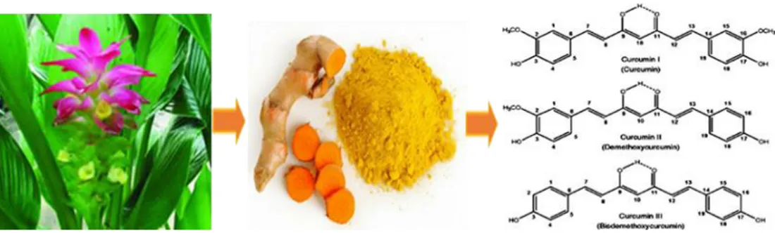
Curcumin (1,7-bis-(4-hydroxy-3-methoxyphenyl)-hepta-1,6-diene-3,5-dione) is a polyphenol extracted from the rhizome of Curcuma Longa Linn (turmeric) and has a long history as a remedy in Ayurveda, Unani, and Siddha medicine for the treatment of various diseases. Curcumin was first isolated by Vogel et al. in an impure form, then its properties were clarified by Milobedeska et al. and was synthesized chemically by Lampe et at (Vogel Pelletier., 1815; Milobedeska et al., 1910; Lampe et al., 1913). Turmeric contains a mixture of three different analogues, curcumin, demethoxycurcumin (DMC), and bisdemethoxycurcumin (BDMC). These three compounds, called curcuminoids, differ in terms of methoxy substitution on the aromatic ring. Curcumin has two symmetrical methoxy-phenol bound through the α,β-diketoneβ-unsaturated portion; also BDMC is a symmetrical molecule but lacks two o-methoxy substitutions, while DMC has an asymmetric structure with one of the phenyl rings with methoxy substitution (Anand et al., 2008; Kiuchi et al., 1993; Mehanny et al., 2016) (Figure 3).
Figure 3: Tumeric (Curcuma longa), the curcuma rhizome and the three different
10
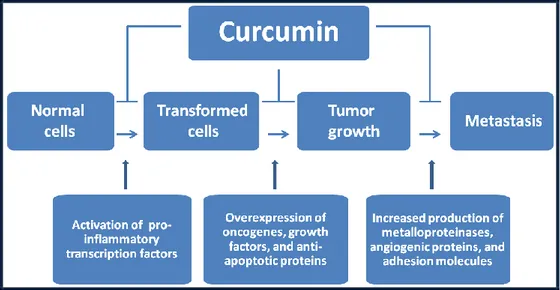
A great scientific interest has recently been focused on curcumin, since it possesses different pharmacological activities, such as antioxidant, anti-inflammatory and antimicrobial properties, and so it is effective against different diseases, such as cardiovascular (e.g. atherosclerosis, thrombosis), neurodegenerative (e.g. Parkinson and Alzheimer disease) and autoimmune (e.g. rheumatoid arthritis) diseases, gastrointestinal disorders, gastroduodenal ulcers and pulmonary illnesses, as well as viral and bacterial infections (Apetz et al., 2014; Kochi et al., 2015; Lv et al., 2014; Chiu et al., 2013; Hasan et al., 2014; Pari, et al., 2008; Kerdsakundee et al., 2015; Yadav et al., 2013; Jiang et al., 2013; Prakash et al., 2011; Srivastava et al., 1985; Shah et al., 1999; Ferreira et al., 2015; Gandapu et al., 2011). It is also well known that curcumin can be potentially used in cancer treatment taking advantage of its ability to kill tumor cells, and, more interestingly, it seems to exert a selective cytotoxic effect against tumoral cells (Jurenka et al., 2009; Sarkar et al., 2016). Not only in vitro but also in vivo preclinical studies have pointed at the ability of curcumin to block tumor progression. Cancer progression and growth are governed by multiple signaling pathways (e.g. NF-κB, Akt, MAPK, Wnt, Notch, p53) (Li et al., 2011; Sahin et al., 2016) (Figure 4).
11
Figure 4: Potential anti-cancer functions of curcumin
(see also Shanmugam et al., 2015)
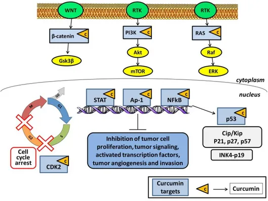
In this context, curcumin has multiple molecular targets and may intervene in different phases of the cell cycle, and for this reason a fluorishing interest of research in the medical field has been recently raised for the use of curcumin in cancer treatment. Curcumin also modulates the activities of many cell key molecules, such as kinases (e.g., EGFR, ERK, JAK, and AAPK), enzymes (e.g., MMP, iNOS, GST, and ATPase), transcription factors (e.g., NF-κB, STAT3, AP-1, NRF-2, PPAR-γ, and HIF-1), receptors (e.g., HER-2, IL-8, and CXCR-4), growth factors (e.g., EGF, NGF, HGF, and PDGF) and cytokines (e.g., TNF, IL, MIP, and MCP) (Goel et al., 2008). In particular, a main role in the anticancer activity of curcumin is ascribed to its capability to inhibit the transcription factor NF-kB, which has a key role (Figure 5).
12
Figure 5: Molecular targets of curcumin in cancer cells (see also Kasi et al., 2016)
activator protein-1 (Ap-1); CDK interacting protein/Kinase inhibitory protein (cip/kip); cyclin-dependent kinases (CDK); extracellular signal–regulated kinase (ERK); glycogen synthase kinase 3 (Gsk3); inhibitors of CDK4 (INK4); mammalian target of rapamycin (mTOR); nuclear factor kappa-light-chain-enhancer of activated B cells (NF-kB); phosphoinositide 3-kinase (PI3K); protein kinase B (Akt); receptor
tyrosine kinases RTK); signal transducer and activator of transcription (STAT)
Malignant pleural mesothelioma (MPM) arises from the mesothelial cells that line the pleural cavity, pericardium, and peritoneum and is a relatively rare but aggressive malignancy with a dismal prognosis and very limited therapeutic strategies (Bonelli et al., 2016). In fact, despite the recent advances in the combination treatment of surgery, chemotherapy, radiotherapy, immunotherapy, targeted molecular therapy and gene therapy, the prognosis is still poor, with a life expectancy of 9–12 months from time of diagnosis. Since MPM is considered to be linked at least in part to exposure to asbestos, and due to the 20-40 year delay between exposure to asbestos and the development of MPM, the incidence of the disease is
13
expected to continuously increase over the next decades. Thus, novel treatment strategies are urgently required to improve survival of these patients. Although the anticancer efficacy of curcumin was investigated in a large number of solid malignancies, there is a paucity of data about its possible effects on MPM cells. Wang investigated the biological and molecular responses of MPM cells (human H2373, H2452, H2461 and H226 cells and murine AB12 cells) to curcumin and the mechanisms involved (Wang et al. 2011). Curcumin inhibited MPM cell growth in a dose- and time-dependent manner, while pretreatment of MPM cells with curcumin enhanced cisplatin efficacy. Curcumin activated the stress-activated p38 kinase, caspases 9 and 3, caused elevated levels of proapoptotic proteins Bax, stimulated PARP cleavage, and apoptosis. In addition, curcumin treatments stimulated expression of novel transducers of cell growth suppression. Furthermore in female BALB/c mice oral administration of curcumin inhibited growth of murine MPM cell-derived tumors in part by stimulating apoptosis. Thus, curcumin targets cell cycle and promotes apoptosis to suppress MPM growth in vitro and in vivo. Yamauchi examined the effects of curcumin on ACC-MESO-1, a human derived mesothelioma cell line (Yamauchi et al., 2012). Curcumin could reduce cell viability in a dose-dependent manner, but did not induce apoptosis. The authors suggested that induction of autophagy is one of the mechanisms in the reduction of cell viability by curcumin, as demonstrated by the increased LC3B-II/LC3B-I expression and the formation of autophagosomes on el MPM ectron microscopy. More recently, Miller demonstrated that curcumin may have a double effect on malignant mesothelioma human and mouse cells, through
14
induction of pyroptosis and subsequently protecting cells against inflammation (Miller et al., 2014). Using in vitro models with mouse and human malignant mesothelioma cells, curcumin was shown to induce pyroptosis through activation of caspase-1 and increased release of the high-mobility group box 1 protein; furthermore curcumin significantly downregulated levels of inflammasome-related gene expression involved in inflammation, such as NF-κB and IL-1β. Finally, Mahajanakatti analyzed the inhibitory properties of curcumin towards virulent proteins for various cancers by computer aided virtual screening (Mahajanakatti et al., 2014). Curcumin showed good towards the virulent protein for mesothelioma platelet-derived growth factor alpha (PDGFA), compared with its natural ligands. Moreover, curcumin has a relatively low cost and a low toxic profile if compared to standard chemotherapeutic drugs (Wang et al., 2011). Unfortunately, the pharmacological potential of curcumin is severely restricted by its low water solubility/absorption, low stability and short half-life, resulting in an extremely poor bioavailability. To overcome these issues, nanotechnologies, especially those based on nanoparticles, may help to improve the bioavailability of curcumin and, thus, its anticancer effects. Curcumin activity is limited by its poor bioavailability and some possible adverse effects. Although still in its early stages, the development of curcumin formulations in the form of nanosystems (nanoparticles, liposomes, micelles, phospholipid complexes, etc.) represent an interesting and useful approach to enhance its bioavailability and efficacy (Chen et al., 2015; Yallapu et al., 2015; Shome et al., 2016). Just a few examples of curcumin loaded nanosystems tested for anticancer activity are listed below: polymeric
15
nanoparticles (50 nm) for pancreatic cancer (Bisht et al., 2007), liposomes (80-90 nm) for skin cancer (Chen et al., 2012), solid lipid nanoparticles (190-200nm) for breast cancer (Mulik et al, 2010), nanogels (74 nm) for cervical cancer (Gonçalves et al., 2012), metallic nanoparticles (10.5 nm) for pancreatic cancer (Yallapu et al., 2013), dendrimers (150 nm) for breast cancer (Falconieri et al., 2016), quantum dots (100 nm) for colon cancer (Some et al., 2014), cyclodextrin inclusions (60–65 nm) for cervical cancer (Sawant et al., 2014), superparamagnetic iron oxide nanoparticles (~ 200 nm) for leukaemia (Dilnawaz et al., 2012).
Nanoparticles as Drug Delivery Systems
Nanotechnology is an emerging branch of science in the design, fabrication and application of nanostructures or nanomaterials that include devices with a sub-micron size. The definition by the American physicist Richard Feynman, in a famous lecture given in December 1959 at the California Institute of Technology and entitled "There's plenty of room at the bottom", suggested that he could build many devices able to direct the arrangement of atoms in the matter. The modern term "nanotechnology" was first, however, invented in 1974 by Norio Taniguchi, a researcher at the University of Tokyo, to describe the precise manipulation of atoms and molecules for the production of new materials. According to Eric Drexler, in his book "Engines of Creation: the coming era of nanotechnology" (1986), nanotechnology was defined as a molecular technology allowing the positiong of every single
16
atom in a desired position (Cao, 2004; Feynman, 1960; Whatmore, 2006). Recent advances in nanotechnology have revolutionized various research fields such as engineering, medicine and pharmaceutical technology. Actually, in recent years, nanotechnology has emerged as one of the greatest challenges of modern medicine and, in particular, has been focusing on the production of nanoscale devices for the controlled release of drugs. In 1995, less than 1% of scientific publications were focussed on the study of nanoparticles. In less than a decade, this proportion rose to 16%. In 2002, moreover, the global market for nanotechnology has a turnover of 406 million dollars, with an increase of 28% over the previous year (Freitas, 2005). The administration of drugs with the aid of nanotechnology-based devices is finding practical application in the biomedical field. In this context, the term "nanomedicine" refers to the use of nanotechnology aiming to improve medical diagnosis and drug therapy. In particular, in the therapeutic field, polymeric nanocarriers can be used to achieve alternative approaches to the targeting of drugs in specific cells and tissues. In fact, it must be considered that standard chemotherapeutic drugs generally generate strong unwanted side effects and, for this reason, there is a strong need to selectively attack tumor cells (Ehdaie, 2007). Nanoparticles are also able to protect the unreleased drug from degradation and thus early inactivation, thereby helping to improve drug efficacy. Moreover, properly designed NPs also allow the controlled release of active molecules by different mechanisms (e.g. sustained, delayed or pulsatile delivery), thereby in principle being able to modulate drug release in both time and space. All these properties of nanoparticles allow to improve the efficacy and reduce the toxic side effects
17
of highly potent drugs, such as chemotherapeutics (Mohanraj and Chen, 2006; De Jong and Borm, 2008; Aziz, 1996).
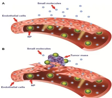
Nanoparticles can be carried to tumors by both passive and/or active targeting. Passively targeted nanoparticles exploit the well known enhanced permeability and retention (EPR) effect of tumors, which consists of leaky vessels feeding the tumor mass along with a hampered lymphatic drainage. Nanoparticles exposing a hydrophilic surface can thwart serum protein adsorption and possess stealth properties, being able to circulate in the bloodstream for prolonged times. This, in principle, increases the chances of a preferential device accumulation in solid tumor tissue (Figure 6).
Figure 6: Passive targeting strategy of nanoparticles and Enhanced Permeability and Retention (EPR) effect (from Bozzuto and Molinari, 2015). Vessel endothelium
in normal tissue (A) presents tight junctions between cells avoiding nanoparticle
diffusion from blood to tissue. Conversely, endothelium of tumor vessels (B) presents large fenestrations so that nanoparticles can reach the tumoral tissue (EPR
effect)
On the other hand, active targeting can be obtained if nanoparticle surface is functionalized by suitable modifications with specific ligands for the tumors,
18
such as nucleic acids, proteins, ligands and antibodies. (Zhang et al., 2016; Jee et al., 2012).
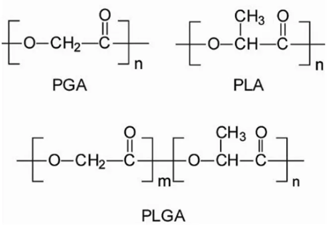
Among the materials used to produce nanoparticles, the ones approved by Food and Drug Administration (FDA) play a prominent role. In particular, polylactic-co-glycolic acid (PLGA) is a biodegradable polyester widely employed to produce nanoparticles. PLGA is a synthetic copolymer of lactic acid and glycolic acid, and its chain is formed by a hydrophilic (the polyglycolic acid, PGA) and a hydrophobic portion (the polylactic acid, PLA, unit) (Figure 7).
Figure 7: Chemical structure of Poly(glycolic acid) (PGA), Poly(lactic acid) (PLA)
and poly(lactic-co-glycolic acid) (PLGA)
The wide PLGA use has been prompted by its well known biocompatibilty, biodegradabilty and non-toxicity, along with its mechanical strength. Properly formulated PLGA-based nanoparticles are able to control the release of drugs over time (days to weeks/months) (Jain, 2000; Peresa et al., 2016; Doty et al., 2016). Drugs encapsulated in biodegradable PLGA nanoparticles
19
may remain in the bloodstream for prolonged times and, therefore, their half-life, resistance to metabolic degradation and release times can be improved. It must be underlined that nanoparticle pharmacokinetics are strongly affected by the control of their size, surface charge and hydrophilicity. For example, if a hydrophilic polymer such as poly(ethylene oxide) (PEO) is superficially exposed on nanoparticles, the adsorption of opsonins is hampered, thus conferring the desired stealth properties to nanoparticles (Mainardes et al., 2009).
Other polymeric material of pharmaceutical interest are poloxamers, which are amphiphilic tri-block copolymers of poly(ethylene oxide) - poly(propylene oxide) - poly(ethylene oxide) (PEO-PPO-PEO). In particular, PEO segments are hydrophilic, while the amphiphilic features are due to the central PPO portion of the polymer (Santander-Ortega et al., 2006) (Figure 8).
Figure 8: Chemical structure of a poloxamer (PEO-PPO-PEO)
Poloxamers form micelles in aqueous solution, with a lipophilic core, and a predominantly hydrophilic shell. When their molecular weight is < 15 kDa, they are generally eliminated by kidneys into the urine. Among poloxamers,
20
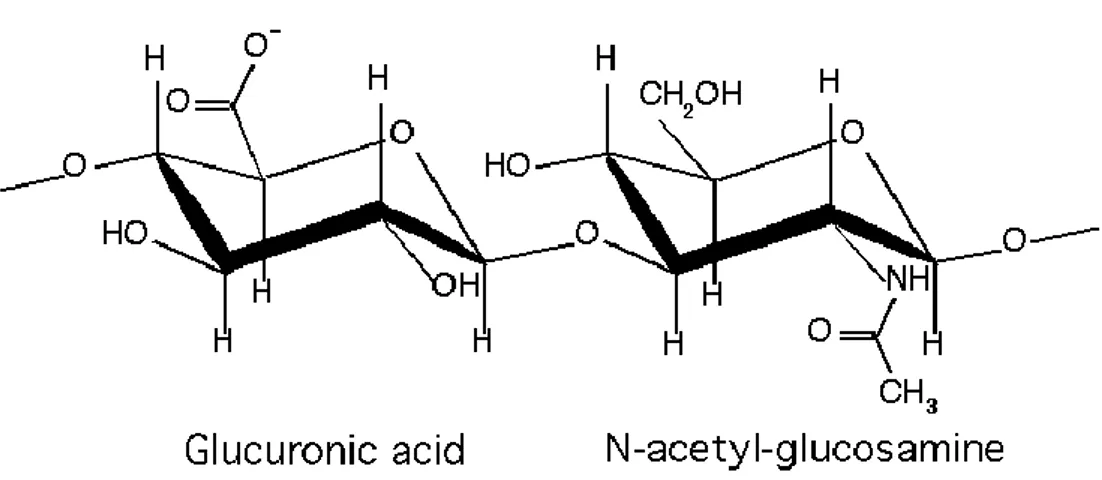
Pluronic F127 (also known as poloxamer 407) has been approved by FDA as a pharmaceutical excipient for medical use (FDA., 2013; Rao et al., 2014). Another polymer which has drawn great scientific interest in the biomedical field is hyaluronic acid (HA). HA is known for its ease of chemical modification and, in its native state, possesses unique properties such as biocompatibility, nonimmunogenicity and biodegradability. HA is a natural polysaccharide composed of repeating disaccharide units of D-glucuronic acid and N-acetyl-D-glucosamine. Discovered in 1934 by Karl Meyer and John Palmer in the vitreous body in bovine eyes (Meyer and Palmer, 1934), it is ubiquitously present in the extracellular matrix of connective tissues of mammals (Weissman and Meyer, 1954) (Figure 9).
Figure 9: Chemical structure of Hyaluronic Acid
HA is an important structural element in the skin and is also present at high concentration in synovial fluid, vitreous humor, hyaline cartilage, disc core and umbilical cord (Monheit and Coleman, 2006; Borzacchiello et al., 2000; Barbucci et al., 2000; Barbucci et al., 2002). The unique biological as well as
21
chemico-physical properties of HA are attracting a great deal of interest among researchers and, to date, HA is being used in various biomedical applications such as regenerative medicine and drug delivery (Mironov et al., 2005).
HA actively influences cell differentiation, proliferation and migration by binding to cells with specific interactions. In fact, these processes are mediated by proteins, called hyaladherins, which act as cellular receptors for HA. Examples of HA receptors are the cluster determinant 44 (CD44), the receptor for hyaluronan-mediated motility (RHAMM), the HA receptor for endocytosis (HARE), and the lymphatic vessel endothelial hyaluronan receptor-1 (LYVE-1), capable of recognizing and selectively bind HA (Alho and Underhill, 1989; Aruffo et al., 1990; Entwistle et al.,1996; Asayama et al., 1998; Takei et al. 2004; Schledzewski et al., 2006). In particular, CD44 and RHAMM receptors are being increasingly investigated because involved in tumor metastasis. CD44 is the best characterized transmembrane HA receptor because it is expressed on the surface of several cells, such as leucocytes, fibroblasts, keratinocytes and epithelial and endothelial cells, and it also involved in many cellular processes, such as cell adhesion, migration, proliferation and activation, as well as HA degradation and uptake (Ahrens et al., 2001; Noble, 2002; Toole et al., 2002; Isacke and Yarwood, 2002). In tumor therapy, there is a strong interest on the employment of HA due to its tumor targeting ability, since it shows a strong tropism toward many tumor cells which naturally overexpress CD44 receptor. Therefore, nanoparticles which superficially expose HA may be better endocytosed by target tumor cells through receptor-mediated pathways and, hence, can be endowed with
22
active targeting capability. In this panorama, HA-decorated nanoparticles can be considered as interesting candidate devices for drug targeting to tumors.
23
Aim of the Thesis
The present research work has been focused on the design of amphiphilic biodegradable nanoparticles for optimization of drug delivery and targeting in cancer treatment. The drugs that we have taken into consideration are the natural compound curcumin (Chapter 1 and 2) and the synthetic chemotherapic drug irinotecan (Chapter 3). Curcumin has many pharmacological properties and a recognized anti-tumor activity, but its pharmacological potential is severely limited due to its low water solubility, short half-life and poor bioavailability. The topoisomerase I inhibitor irinotecan is used, as a first-line or second-line agent, in the treatment of advanced colorectal, ovarian, cervical, and small cell lung cancer, alone or in combination with other drugs, such as 5-fluorouracil and doxorubicin. Unfortunately cancer cells can present and develop resistance to irinotecan through numerous mechanisms. To this aim, biodegradable PLGA-based nanoparticles for the release of curcumin and irinotecan have been produced. In particular, curcumin has been loaded in nanoparticles made of an amphiphilic blend of poloxamers and PLGA, produced by a double emulsion and nanoprecipitation technique, so as to confer stealth properties to the nanoparticles and take advantage of the EPR effect. The produced nanoparticles were tested for their stability and investigated for their in vitro ability to be internalized by mesothelioma cells and affect their growth. A development of this formulation was performed by targeting it to tumor cells by HA moieties on the surface of PLGA-based nanoparticles loaded with irinotecan. This has been accomplished by directing HA arrangement on
24
nanoparticle surfaceby means of a lipophilicity gradient between the oil and water phases of the emulsion used to produce the nanoparticles, and using poloxamers as a bridging molecule between PLGA and HA. The obtained devices were characterized for their technological and thermodynamic features as well as by ELISA tests to support the hypothesis of polymer assembly in nanoparticle formulations. In vitro biologic studies were carried out on CD44-overexpressing breast carcinoma cells (HS578T) to verify nanoparticle ability to target CD44 receptor.
25
Chapter 1
PLGA/Poloxamer Nanoparticles for
Intracellular Curcumin Delivery Induce a
26
Introduction
Curcumin (CURC) is a polyphenol extracted from the rhizome of Curcuma
longa Linn, possessing numerous pharmacological activities, such as
anti-inflammatory, antioxidant and antimicrobial (Hatcher et al., 2008;). CURC can be potentially used in cancer treatment taking advantage of its ability to block the proliferation of many tumor cells (Bhattacharyya et al., 2007a; Bhattacharyya et al., 2007b). Even more interestingly, CURC seems to induce a selective cytotoxic effect mainly towards cancer cells, even in the presence of healthy cells. This activity was correlated with both a downregulation of the nuclear factor NF-B and to its ability to react with thioredoxin reductase, which is an enzyme overexpressed in tumor cells. This interaction leads to an increased production of H2O2 in tumor, thus promoting
cancer cell cytotoxicity (Fang et al., 2005). Furthermore, CURC triggers both downregulation of AP-1, cyclin D1, cyclin E and upregulation of p21, p27 and p53 proteins, thus repressing tumor cell proliferation/migration and promoting cell cycle arrest and apoptosis (Balasubramanian and Eckert, 2007; Banerjee et al., 2010). Another very attractive feature of CURC is its ability to overcome the multidrug resistance (MDR) of cancer cells, most likely due to its ability to down-regulate the expression of P-glycoprotein (P-gp), multidrug resistance associated protein-1 (MRP-1) and mitoxantrone resistance protein (ABCG2), which are three major proteins responsible for the high drug efflux in multidrug-resistant cancer cells (Limtrakul et al., 2007). Unfortunately, the pharmacological potential of CURC is severely restricted, mainly owing to its low water solubility/absorption, short half-life
27
and extremely poor bioavailability (Aggarwal and Sung, 2009; Anand et al., 2007). To overcome solubility problems, CURC has been complexed with different cyclodextrins (CDs), which are cyclic oligosaccharides with a hydrophilic outer surface and a lipophilic cavity that can incorporate hydrophobic drugs/compounds (Yadav VR et al., 2010; Yallapu et al., 2010). Moreover, CURC encapsulation in biodegradable nanoparticles (NPs) made up of poly(lactic-co-glycolic acid) (PLGA) allows to increase its circulation times/half life, cell permeability and resistance to metabolic degradation, and also promotes controlled release and targeting (Anand et al., 2010; Farokhzad et al., 2008; Shaikh, et al., 2009; Tsai et al., 2011). Drug-loaded PLGA NPs generally possess low toxicity and also a high specificity and tolerability compared to the free drug. It must also be underlined that size, surface charge and hydrophilicity of the NPs in which CURC is loaded strongly affect its bioavailability. For example, depending on their surface properties, the NPs can be either short-circulating to reach the macrophages in liver and spleen, or long-circulating to passively accumulate in tumor tissues, which are normally characterized by the enhanced vascular permeability and retention (EPR) effect (Fang et al., 2011). For these reasons, the hydrophobic surface of PLGA NPs causes short circulation times after e.v. injection due to a rapid in vivo opsonization while, if a hydrophilic polymer such as poly(ethylene oxide) (PEO) is superficially exposed, it hampers the adsorption of opsonin proteins, thus conferring stealth properties to NPs and increasing their probability of reaching tumor sites (Mainardes et al., 2009; Owens and Peppas., 2006).
28
Aim of the Work
The aim of the work reported in this first chapter was to study the possibility to formulate PLGA–based NPs coated with hydrophilic PEO moieties without a chemical reaction between the two polymers, therefore avoiding the presence of chemical reaction solvents and wastes. In particular, the basic idea of this work was to formulate stealth NPs by a double emulsion-solvent evaporation technique, in which the organic phase is composed of a blend of PLGA and poloxamers instead of employing them as surfactants in the external aqueous phase, as is usually performed. Poloxamers (Polox) are triblock copolymers made up of poly(ethylene oxide)-poly(propylene oxide)- poly(ethylene oxide) (PEO–PPO–PEO), displaying amphiphilic properties taking advantage of the presence of hydrophilic EO and hydrophobic PO segments on polymer backbone (Mayol et al., 2011; Ranall et al., 2011). The obtained NPs have been loaded with CURC which, for the solubility problems mentioned above, was preliminarily complexed with hydroxylpropyl--CDs (HPCDs). The obtained NPs have been characterized in terms of size, morphology, zeta potential, yield and drug entrapment efficacy. NP stability was evaluated by measuring their size over time at 4 °C in aqueous media (i.e. storage condition) and at 37 °C in serum (i.e. the same conditions of in vitro cell experiments). Moreover, in vitro drug release studies have been carried out to assess the capacity of the formulated NPs to prolong CURC release. Furthermore, malignant mesothelioma cell line MSTO-211H was used to exploit the biological efficacy of CURC-loaded NPs by investigating NP cellular uptake and CURC-CURC-loaded NP effect on cell proliferation and cell cycle progression.
29
Materials and Methods
Materials
Poloxamers (PEOa-PPOb-PEOa) are a group of amphiphilic triblock
polymers, possessing variable numbers of oxyethylene (a) and oxypropylene (b) units. Poloxamer F127 (a = 100 and b = 65) and F68 (a = 76 and b = 29) were obtained from Lutrol (Basf, Germany). Equimolar uncapped poly(D,L-lactide-coglycolide) (PLGA) (Resomer RG504H, Mw: 40 kDa, inherent viscosity: 0.16-0.24 dL g-1 in acetone at 25 °C) was purchased from
Boehringer Ingelheim (Ingelheim, Germany); Curcumin ((E,E)-1,7-bis(4-hydroxy-3-methoxyphenyl)-1,6-heptadiene-3,5-dione) (purity > 90%) from Cayman Chemical Company, USA), ultrapure sucrose from Riedel-deHaen (Germany) and potassium chloride (KCl) from Carlo Erba (Italy) were used. Polyvinyl alcohol (PVA, Mowiol® 40-88), 2-Hydroxypropyl--cyclodextrin (HPCD; CD in the text), ethanol (EtOH), acetone, dimethylsulfoxide (DMSO), Tween-80, dibasic sodium phosphate (Na2HPO4), sodium chloride
(NaCl), 1,6-diphenyl-1,3,5-hexatriene (DPH), Trypan blue, propidium iodide (PI) and RNase were obtained from Sigma-Aldrich (USA). Ascorbic and citric acid from J-Baker (USA) were used. Human mesothelioma (MSTO-211H) cell line was obtained from the American Type Culture Collection (Rockville, MD, USA); Roswell Park Memorial Institute (RPMI-1640) medium, Fetal Bovine Serum (FBS), penicillin 50 UI/mL, streptomycin 0.05 mg/mL, penicillin/streptomycin 10 UI/mL, trypsin-ethylenediamine tetra-acetic acid (Trypsin-EDTA) 1mM, sodium pyruvate and 4-(2-hydroxyethyl)-1-piperazineethanesulfonic acid (HEPES) from Euroclone (Italy) were
30
employed. All chemicals and media were used as received without any further purification.
Preparation of Nanoparticles
Prior to NP preparation, to enhance CURC solubility and allow a higher drug loading in NPs, CD-CURC complex was prepared by dissolving CURC in EtOH (1 mg/mL) and adding CD (8 mg/mL). The resulting solution was placed under magnetic stirring for 20 min at 80 °C until complete evaporation of the solvent. The obtained solid was found to be soluble at > 50 g/mL in PBS, i.e. at a higher concentration than CURC solubility (about 10 g/mL), thus proving the formation of the CD-CURC complex. Blank and CURC-loaded NPs were prepared by a modified double emulsion-solvent evaporation technique. Briefly, the internal phase, composed by 640 L of EtOH, or the same volume of a 1.4 mg/mL solution of CD-CURC in EtOH, was emulsified with 5 mL of a 30 mg/mL PLGA or PLGA/poloxamer (50/50 mass ratio) solution in acetone (NP formulations have been named P or PP, correspondingly). The resulting emulsion was sonicated for 5 min at 4 °C (Branson 3510 ultrasonic bath, 100% power, operating at 42 kHz ± 6%). The resulting water-in-oil emulsion was immediately poured into 18 mL of 1% w/v aqueous PVA and further sonicated (5 min, 4 °C). The resulting double emulsion was stirred overnight for complete acetone evaporation. The obtained NP suspension was washed three times by centrifugation (Hettich Zentrifugen, Germany; 13,000 rpm, 30 min) and, once the supernatant was eliminated, NPs were stored at -80 °C. Fluorescent NPs were also prepared
31
by adding 10 mg of the fluorescent dye 1,6-diphenyl-1,3,5-hexatriene (DPH) to the organic phase containing PLGA or PLGA/poloxamer blend. DPH was chosen since it is fluorescent only in a hydrophobic environment with an intensity related to the hydrophobicity of the solution (Ranall et al., 2011).
Nanoparticle Morphology
NP morphology was studied by using a transmission electron microscope (TEM, EM208S, Philips, The Netherlands). TEM observations were carried out by spraying 100 L of ultradiluted NP suspensions in water on a copper TEM grid (300 meshes, 3 mm diameter).
Nanoparticle Size, and -Potential Analyses
NP mean size, size distribution and -potential were determined by laser light scattering (ZetaSizer Nano ZS, Malvern Instruments, Malvern, UK) on a 0.1 mg/mL suspension of NPs in water (12 runs each sample).
Nanoparticle Stability
The evolution of the hydrodynamic diameters and -potential of P and PP NPs was monitored at 4 °C up to 30 days in bidistilled water (i.e. in storage conditions) and in RPMI-1640 cell culture medium supplemented with 10% FBS at 37 °C up to 72 hours, i.e. the period of exposure used for in vitro cell
32
uptake/growth experiments. Size measurements were also performed on bare cell culture medium to verify possible self-aggregation. Results were averaged on at least five measurements.
Nanoparticle Yield and Drug Entrapment Efficacy
NP yield and CD-CURC entrapment efficiency were calculated from previously freeze-dried NPs (0.01 atm, 24 hours; Modulyo, Edwards, UK). In particular, NP yield was gravimetrically obtained from the entire mass of recovered freeze-dried NPs. For the loading efficiency, lyophilized NPs were dissolved in acetone (0.1% w/v) under gentle agitation for 1 h at room temperature to completely leach out CD-CURC from NPs. The obtained solution was centrifuged for 15 minutes at 13,000 rpm and CD-CURC quantified by spectrophotometric assay (UV-1800, Shimadzu Laboratory World, Japan) at 420 nm. The linearity of the response was verified over the concentration range 0.2 - 50 g/mL (r2 > 0.99). Results are expressed as mean ± standard deviation (SD) of values collected from three different batches.
Drug Release Kinetics
The release of CD-CURC from NPs was determined by loading P and PP NP suspension (at 15.74 mg/mL and 6.60 mg/mL, respectively) in a dialysis membrane (Spectra/Por® Biotech Cellular ester; molecular cut-off 12 kDa)
33
and placing them in 50 mL of a release medium composed by PBS 90% v/v, ethanol 10% v/v, Tween 80 0.5% w/v, acetic acid 0.01 % w/v and ascorbic acid 0.01% w/v; the pH was adjusted to 7.4. Acetic and ascorbic acid were added to the release medium to improve CD-CURC stability over time. Indeed, prior to the release experiments, CD-CURC stability was assessed by spectrophotometric assay at = 428 nm at 37 °C in the dark with or without the antioxidant agents. Regarding the release kinetics, at predetermined time intervals, aliquots of medium were withdrawn and replaced with fresh medium and the CD-CURC content determined by reverse phase high performance liquid chromatography (RP-HPLC). The chromatograph was equipped with a HPLC LC-10AD pump (Shimadzu, Milano, Italy), a 7725i injection valve (Rheodyne), a SPV-10A UV-Vis detector (Shimadzu) set at the wavelength of 420 nm and a C-R6A integrator (Shimadzu). Analyses were performed on a Phenomenex Luna C18 (2) 100Å column (250 mm × 4.6 mm, 5 m). The mobile phase was a mixture of methanol, acetonitrile and bidistilled water (40:40:20 v/v). The flow rate was 1 mL/min, and the run time was set at 7 min. The experiments were performed in triplicate and under sink conditions.
Cell Cultures
Human mesothelioma MSTO-211H cells were cultured as monolayer in culture dishes using RPMI-1640 medium, supplemented with 10% FBS, 50 UI/mL penicillin and 0.05 mg/mL streptomycin, 1 mM sodium pyruvate and 10 mM HEPES. Cell cultures were maintained in a humidified atmosphere
34
containing 5% CO2 at 37 °C. Prior to the experiments, cells were seeded in
complete growth medium for 16 hours, in order to allow cell attachment but not cell doubling. Afterwards, cells were treated with free or NP-encapsulated drug. Different treatment times were used according to the experimental conditions. Untreated cells were used as a control.
Nanoparticle Cellular Uptake
NP cellular uptake was followed by means of a fluorescence microscope (Leica Microsystems, Germany). Fluorescent P and PP NPs were formulated as described in the Preparation of Nanoparticles section. NP cellular uptake by MSTO-211H cells was analyzed by treating for 6, 12, 24 and 48 hours 2 x 10⁵ cells/well in a 6-well plate with 1 mg/mL of fluorescent P and PP NPs. At the end of the treatment, cells were rinsed with PBS and fixed with 95% ethanol for 20 min. The nuclei were stained using 2.5 µg/mL PI for 5 min. Images of stained cells were acquired and DPH blue fluorescence was monitored at = 428 nm, while PI fluorescent red signal was followed at = 610 nm. P and PP NP fluorescence intensity was quantified by Image J software on 10 cells/picture. Fluorescence intensity was normalized to the cytoplasmic area after subtracting the nucleus area from the total. Three pictures for each sample were analyzed. Experiments were performed in triplicate.
35
Cell Viability/Growth
To evaluate the cell viability, 1 x 10⁶ MSTO-211H cells per well were seeded
in 100 mm diameter culture dish for 16 hours. In a preliminary series of experiments, cells were treated for 12, 24, 48 and 72 hours with free CURC ranging from 10 to 100 M, in order to find out the best working concentration of the drug in terms of cell viability. Free CURC at 20 M decreases up to 50% cell viability after 72 h of exposure. So, in the subsequent experiments, cells were exposed for 12, 24, 48 and 72 hours to 20 M of free CD-CURC or CD-CURC-loaded NPs or with the same amount of void NPs to assess NP biocompatibility and CURC-loaded NP biological effectiveness. In all experiments untreated cells were used as a control. Experiments were performed in triplicate.
At predetermined time points, the Crystal Violet (CV) staining method was performed to assess cell viability by culturing 2x10⁴ cells/well in a 24-well plate and treating them as above reported. CV is a triphenylmethane dye (4-[(4-dimethylaminophenyl)-phenyl-methyl]-N,N-dimethyl-aniline), also known as gentian violet (or hexamethyl pararosaniline chloride). CV stains DNA and, upon solubilization, the amount of dye taken-up by the monolayer and the intensity of the color produced are proportional to cell number. Samples were washed in PBS after the treatment and fixed with 10% v/v formaline solution for 10 min at room temperature. Afterwards, cells were washed twice in bidistilled water and stained with 0.05% v/v CV aqueous solution for 20 minutes at room temperature. Cells were further washed twice with water and incubated for 15 minutes with 10% acetic acid solution to
36
dissolve the stain. Colorimetric assay, carried out in triplicate, was quantified measuring OD595 by using a TECAN instrument (Switzerland).
Cell Cycle Analysis.
Evaluation of the cell cycle distribution was performed by analyzing the DNA content in each cycle phase. Analyses were carried out by treating 1.5 x 10⁶ cells for 12, 24, 48 or 72 hours with free or NP-encapsulated CD-CURC at a final concentration of 20 M. After the treatment, cells were harvested and washed with cold PBS and centrifuged at 400 rpm for 8 min at 4 °C. The treated cells were fixed in 1 mL chilled 70% v/v EtOH and incubated for 30 minutes at 4 °C. Cells were centrifuged again (400 rpm, 8 min) and washed twice with cold PBS. Pellets were redissolved in 500 µL of cold PBS and incubated with 100 µg/mL RNaseA for 30 minutes at 37 °C. PI was added to a final concentration of 20 µg/mL and the cells were incubated on ice for 30 minutes in the dark. Flow cytometry was performed using a FACScan™ flow cytometry system (Becton Dickinson, San Jose, CA). For each sample, 5x10⁴ events were recorded and the cells fractions in each different phase of the cell cycle calculated.
Statistical Analysis.
Data were reported as mean value ± SD. Results were analyzed by using GraphPad Prism 5.0H statistical software (GraphPad Software Inc., La Jolla, CA). Paired t test was used for comparison of two paired groups. Results
37
were analyzed by one way or two way analysis of variance (ANOVA), followed by Bonferroni adjustment.
38
Results and Discussion
Nanoparticle Characterization
NPs made up of PLGA alone (P) or blended with poloxamers (PP) were prepared by the double emulsion–solvent evaporation technique. In a previous work, PP micro/nanoparticles for the controlled release of growth factors in tissue engineering were produced, aiming to protect the encapsulated protein from acidic degradation products of PLGA (Parajo et al., 2010). Here, we have employed the PP blend aiming to establish a rapid method to provide NPs with a hydrophilic surface. In figure 1.1, representative TEM images of NPs are reported while in table 1.1 the technological features of NPs are displayed. As it can be noted from Figures 1.1A and 1.1B, both P and PP NPs are spherical in shape with a regular surface.
39
As for NP diameters, NP yield and -potential, no significant effect of poloxamer presence could be evidenced, as displayed in Table 1.1.
Forms Particle mean
diameter (nm) Polydispersity Index (PDI) Zeta potential (mV) Entrapment efficiency (%) Yield (%) P 189 ± 37 0.258 ± 0.02 -15.7 ± 0.9 72.1 ± 3.2 79.6 ± 1.9 PP 160 ± 31 0.671 ± 0.03 -18.7 ± 1.3 90.0 ± 2.1 76.9 ± 1.4
Table 1.1: Size, polydispersity index (PDI), zeta potential entrapment efficiency and
yield of CD-CURC– loaded NPs
A high entrapment efficiency was found in both formulations, in particular for PP NPs, which showed about 90% of encapsulated drug. This suggests a slightly higher affinity of the CD-CURC complex for the polymeric PLGA-poloxamers blend compared to the bare PLGA solution. In order to investigate NP storage condition, size distribution and -potential of P and PP NPs were monitored as a function of time, up to 30 days, at 4 °C in double distilled water while, to reproduce the time frame of cellular experiments, after the washing step, NPs were placed in RPMI-1640 medium supplemented with 10% FBS at 37 °C up to 72 hours. Results are reported in Table 1.2, where it can be observed that the presence of poloxamers in the organic blend does not substantially affect the stability of NP suspension in comparisons with P formulation in water.
40
Forms.
Water, 4 °C RPMI, 37 °C
Day 0 Day 10 Day 20 Day 30 Day 1 Day 2 Day 3
dP 189 ± 37 233 ± 11 243 ± 76 265 ± 36 226 ± 9 221 ± 19 234 ± 15
dPP 160 ± 31 167 ± 17 153 ± 13 180 ± 24 161 ± 4 157 ± 13 164 ± 11
Table 1.2: Time evolution of NP diameter in water at 4 °C and in cell culture
medium (RPMI) at 37 °C
On the contrary, when facing serum proteins, the presence of poloxamers on the NP surface does make the difference. Actually, while the diameters of P NPs rapidly increased to about 230 nm in only 8 hours (data not shown), thus indicating a strong interaction with serum protein due to their hydrophobic surface, PP NPs diameters were basically stable within the time frame of the experiment. The interaction of NPs with serum proteins is a critical issue since it is known to strongly affect the pharmacokinetics of devices and hence their in vivo fate/activity, since long circulating NPs weakly interact with serum proteins (Fang et al., 2011). Actually, the strong difference in the NP/protein interaction indicates different surface properties between P and PP formulations. This can be reasonably ascribed to a spontaneous interaction between PLGA and poloxamers in the organic phase of the emulsion used for NP preparation. In particular, it can be easily grasped that
41
the hydrophobic PPO segments of the poloxamer dissolve in the organic phase of the emulsion together with PLGA, while the most of hydrophilic PEO segments orient themselves towards the external aqueous phase. By this way, a spontaneous cover of hydrophilic PEO is expected to arrange themselves on PLGA NP surface. Therefore, the use of an amphiphilic polymeric blend in the organic phase of the emulsion seems to be a promising alternative method to obtain stealth NPs without the economical and purification issues related to chemical reaction used for standard PEGylation.
Curcumin Stability Studies
It is well known that CURC is highly unstable at 37 °C at both neutral and neutral/basic pH. Actually, CURC undergoes extensive degradation within 24 hours and the mechanism is believed to be mainly oxidative (Subhashini et al., 2013; Pan et al., 1999). For this reason, prior to release experiments, CD-CURC stability has been assessed by measuring the absorbance of a solution of CD-CURC complex in the release medium at 25 g/mL and 37 °C (Figure 1.2). We found that CD-CURC absorbance in the release medium dropped down of approximately 90% within only 24 hours. For this reason, we added ascorbic and citric acid (0.01% w/v for both substances) as anti-oxidant agents. In these conditions, the measured absorbance was found to be stable for at least 5 days.
42 0 0.1 0.2 0.3 0 50 100 150 AA 0.01% w/v + AC 0.01% w/v AA 0.01% w/v + AC 0.02% w/v CURC AB S Time [hours]
In Vitro Drug Release Kinetics
In vitro CD-CURC release profiles from P and PP NPs, at 37 °C and
physiological conditions are shown in Figures 1.2A and 1.2B.
0 20 40 60 80 100 120 0 50 100 150 200 P PP %CD -CU RC r eleas ed Time [hours] A 0 20 40 60 80 100 0 2 4 6 8 10 P 7.4 PP 7.4 y = -18.1 + 15.0x R= 0.992 y = -10.5 + 13.1x R= 0.988 %CD-C U RC r ele ased Sqrt (time) [hours1/2]
B
Figures 1.2: CD-CURC release kinetics. A) Release profile of CD-CURC from P
and PP NPs at 37 °C and pH=7.4, and CURC stability in the release medium at 37 °C in the dark with and without antioxidants (i.e. acetic acid (AA) and ascorbic acid (AC)). B) Percentage of released CD-CURC as a function of square root of time ( ○
y = -18.1 + 15.0x R= 0.992; ■ y = -10.5 + 13.1x R= 0.988).
Release profiles were found to be reproducible and evidenced the ability of NPs to control and sustain CD-CURC release. A 24 h burst, in which about
43
60% of drug was released, followed by a slower release phase was found. The release was completed within approximately 4 days with no significant difference between the two formulation. To investigate the mechanisms of NP unloading, CD-CURC release profiles were plotted as a function of 12
t
(Fig. 1.2B). In all cases, after the initial burst, a second linear region followed, with a slope relatively constant and basically independent on the formulation, thus indicating that the release is mainly driven by drug diffusion (Biondi et al., 2013b).
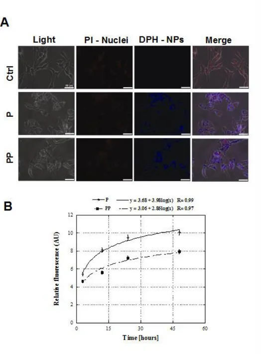
Nanoparticle Cellular Uptake
P and PP NP cellular uptake results are reported in Figures 1.3. Representative pictures, after 48 hours of treatment, are shown in Figure 1.3A. Untreated MSTO-211H cells were used as a control to assess the absence of any background fluorescence. Results indicated that both P and PP formulations are able to enter the cells. In particular, the fluorescent blue signal of DPH is localized throughout the cytoplasm, prevalently in the perinuclear space. As depicted in Figure 1.3B, a time-dependent increase of the fluorescent signal of DPH was observed, which is directly related to the total amount of internalized NPs and is qualitatively the same for both P and PP NPs. On the contrary, from a quantitative point of view, P NP accumulation in the cytoplasm was found to be significantly higher compared to PP NPs in the time frame of the experiment. A fast uptake of both formulations was found in the first 24 hours of treatment, while no significant differences in fluorescence intensity were found for each formulation from 24
44
to 48 hours, thus suggesting a saturation uptake. Actually, NP cellular uptake is a process mediated by endocytosis rather than by passive diffusion (Rosen and Abribat, 2005), thus resulting into a saturable process (Li et al., 2010). Since internalization is an active process involving the interaction of membrane proteins with NP surface, the lower internalization of PP NPs compared to P NPs is indicative of different surface properties between the two formulations and, in particular, of a lower affinity between the PP NP surface and the cellular membrane. This further confirms the presence of hydrophilic moieties on NP surface.
45
Figure 1.3. NP cellular uptake. A) Representative fluorescent microscopy images of
NP uptake by MSTO-211H cells after 48h of treatment. Control untreated cells (Ctrl) did not show any background signal referred to the blue DPH emission, while P and PP NPs labeled with DPH were located in the perinuclear region as evidenced by the merged images from PI and DPH labeling. Scale bar represents 50 µm. B)
Relative fluorescence intensity referred to cellular uptake of fluorescent NPs in MSTO-211H cells, from 3 to 48 hours after the treatment showing that the fluorescent signal from the NPs increased during time until the process is saturated.
46
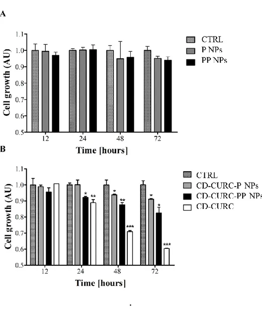
Preliminary dose-response experiments using free CD-CURC were performed to assess the best working concentration of drug, as it has been mentioned above. CD-CURC 20 M treatment has been chosen to perform all the subsequent experiments since this concentration showed ~ 50% of cell viability after 72 hours of treatment (data not shown). CD-CURC concentration higher than 20 M caused massive cell death within 72 hours. Cell growth was followed at regular time intervals up to 72 hours of treatment with free CD-CURC and CURC-loaded P and PP NPs, using unloaded NPs as a control. Results are reported in Figures 1.4. No statistically significant differences on cell growth were found between control and unloaded NPs (Figure 1.4A), thus indicating the biocompatibility and the non-toxicity of both the formulations. On the other hand, interesting results were obtained by using free or entrapped CD-CURC (Figure 1.4B). In particular, free CD-CURC was found to inhibit cell growth after only 24 hours of treatment and, after 72 hours, the inhibition was about 40% compared to control. CD-CURC-loaded NPs showed a statistically significant lower inhibition effect compared to the free drug, probably due to the sustained release of CD-CURC from NPs. In particular, the inhibition was less pronounced in the case of the P NPs than the PP NPs, especially between 24 and 48 hours of treatment demonstrating the improved bioactivity of the novel CURC-loaded PP NPs compared to the bare PLGA NPs.
47
.
Figure 1.4: Cell growth inhibition induced by CURC-loaded NPs. A) MSTO-211H
cell growth after treatment with placebo NPs. No statistically significant difference among the treatments was found. B) MSTO-211H cell growth effects induced by 20
µM of free or encapsulated CURC. Results are expressed as Arbitrary Units (AU) and the control group (untreated MSTO-211H cells, CTRL) is fixed as 1.0 cell
growth at each time point. Data represent the mean ± SD of three different experiments (*P < 0.05, **P < 0.01 and ***P < 0.001 are referred to the different
treatments in comparison to CTRL).
Analysis of Cell Cycle Distribution
The anti-proliferative activity of CD-CURC is related to its ability to interact with several transcription factors and signaling proteins, such as the tumor suppressor p53, the tumor necrosis factor, inflammatory enzymes, cyclines
48
and protein kinases (Shishodia et al., 2005). In particular, it is known that an effect of CD-CURC is the modification of cell cycle phases distribution (Sa and Das, 2008). Therefore, we performed a FACS analysis to test the response of the PI-positive MSTO-211H cells to free CURC or CD-CURC-loaded PP NPs (Figure 1.5).
Figure 1.5. CD-CURC and CD-CURC loaded NPs treatment induces cell block. A)
MSTO-211H cells were exposed to 20 µM of free CD-CURC or CD-CURC loaded in NPs and cell cycle was analyzed by flow cytometry analysis from 12 hours to 72
hours. Cells treated with naked NPs (PP) were considered as controls. B) Data summary of the cell cycle arrest. Data were performed on three independent
experiments with comparable results and standard error was less than 3%. 12 h PP PPC CUR 0 20 40 60 80 100 G0/G1 S G2/M Ce ll s p e r c e n ta g e 24 h PP PPC CUR 0 20 40 60 80 100 G0/G1 S G2/M Ce ll s p e r c e n ta g e 48 h PP PPC CUR 0 20 40 60 80 100 G0/G1 S G2/M Ce ll s p e r c e n ta g e 72 h PP PPC CUR 0 20 40 60 80 100 G0/G1 S G2/M Ce ll s p e r c e n ta g e CD-CURC CD-CURC loaded NPs PP