Università degli Studi di Messina
Dipartimento di Medicina Clinica e Sperimentale
Dottorato XXX ciclo in
Scienze Biomediche Cliniche e Sperimentali
____________________________________________________________________
Left atrial function: early target of chemotherapy related
cardiac damage
Tesi di Dottorato di
Maria Chiara Todaro Coordinatore:
Chiar.mo Prof Edoardo Spina
Tutor: Chiar.mo Prof. Scipione Carerj S.S.D Med 11
1. Introduction to cardioncology ………... 2. Cardiotoxicity: from molecular damage to clinical manifestations... 3. Subclinical cardiac dysfunction:
the role of speckle tracking echocardiography………... 3.1 Phasic LA function and atrial-ventricular coupling ...…...……..
4. Background of the study ...………....
5. Methods ………. 5.1 Standard echocardiographic evaluation………...
5.2 Speckle-tracking echocardiography………
6. Follow-up ……….. 7. Statistics………. 8. Results ………... 9. Discussion …...……….. 10. Limitations of the study ………..………. 11. Conclusions ………... 12. Tables ……… 13. Figures ………... 14. References ………... 3 4 10 12 14 15 17 19 21 22 23 25 30 30 32 35 39
3
1. Introduction to cardioncology
The improvement of antineoplastic treatment regimens as well as the advances achieved in surgical strategies have enormously increased the average survival of cancer patients, indirectly encouraging the clinical manifestation of collateral effects of chemotherapeutic agents in an increasing number of patients. Moreover, most of patients affected by cancer also have multiple cardiovascular (CV) risk factors, such as obesity, alcohol abuse and smoking that make even more likely that cancer and CV disease may both be diagnosed in the same patient.1
As a consequence, a new public health problem emerged over the last 20 years that induced cardiologists and oncologists to intensively cooperate for the interest of patients’ global good care, leading to the birth of a relatively new medical field: cardioncology.2
Cardioncology deals with all the possible damages directly or indirectly induced by chemotherapy with the aim to prevent irreversible CV
4
detrimental effects of chemotherapeutic agents, without reducing their therapeutic efficacy.3
2. Cardiotoxicity: from molecular damage to clinical
manifestations
The most common CV complications of antineoplastic therapies include vasospastic and thromboembolic ischemia, arterial hypertension, dysrhythmia, and left ventricular (LV) dysfunction, leading to heart failure (HF).4 Special emphasis is placed on LV dysfunction and HF, in consideration of its clinical and social burden.
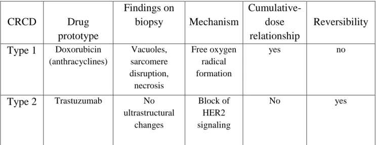
Chemotherapy related cardiac dysfunction (CRCD): Two distinct forms
of myocardial dysfunction are identifiable. Type I CRDC typically anthracycline (ANTs)-induced, is due, at least in part, to oxidative stress on cardiac muscle resulting in free radical formation and cell death; it is irreversible and typically associated with significant ultrastructural
5
changes at biopsy.5 In contrast, Type II CRCD (trastuzumab-induced) is associated with reversible myocardial dysfunction rather than structural damage, is highly reversible (up to 79%) and generally is not dose-related.6 However, a synergistic effect between ANTs and trastuzumab has been demonstrated, especially when the two compounds are administered over a short period of time. Moreover, the two types of drug-induced cardiac dysfunction can exist in the same patient.1 Table 1
Type 1 CRCD: ANTs are widely used and effective antineoplastic drugs, indicated for the therapy of many kinds of cancers including lymphomas, leukemias, and sarcomas, and for both early and advanced breast cancer. Cardiac dysfunction caused by ANTs has long been known as the main form of anti-cancer drug-induced cardiotoxicity, with production of reactive oxygen species (ROS) and reactive nitrogen species (RNS) being considered main cytotoxic agents that may be either direct or indirect inducers of the cardiac injury.7 In addition, severity of
6
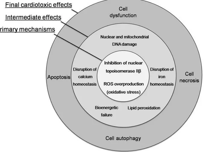
oxidative/nitrosative stress often does not correspond well with the severity of the injury and this may offer an explanation for poor efficacy of different antioxidants used to reduce Ant- induced cardiotoxicity.8 Recent data suggest that ANT-induced cardiotoxicity is also strictly related to ANTs interaction with topoisomerase 2 (Top2)-β in cardiomyocytes.7 Two types of Top2 enzymes are targeted by ANTs: Top2-α and Top2-β. While the former is present in rapidly dividing cells, such as cancer cells, inducing cell apoptosis, Top2-β is present in human cardiomyocytes, forming the Top2β-doxorubicin-DNA complex, which causes DNA double-strand breaks and transcriptome modifications, also leading to cell apoptosis.9 Figure 1
The mitochondrial pathway is an additional mechanism responsible for cellular intrinsic apoptosis: due to a high affinity of ANTs for the mitochondrial phospholipid cardiolipin, it can negatively affects its function, stimulating ROS/RNS production, inhibiting oxidative
7
phosphorylation, and causing mitochondrial DNA damage, with a consequent progressive reduction of energy production leading to cell dysfunction.7
Acute cardiotoxicity occurs in 1% of patients and does not track development of chronic HF later in life, can be asymptomatic or present with transient, and usually reversible, depression of myocardial contractility.5 Chronic ANTs induced cardiotoxicity occurs in 1.6–2.1% of patients during therapy or within the first year after treatment, with a peak incidence between the first and third months, and generally is not reversible. Late-onset chronic LV dysfunction occurs in 1.6–5% of patients at least 1 year until 10 to 30 years after completion of therapy from the first dose of treatment.10-11 An increase in cardiac damage later in life was demonstrated in 23% of children treated with ANTs and documented chronic LV dysfunction.12-13 Moreover a strong correlation between chronic LV dysfunction and cumulative doses of ANTs is widely
8
demonstrated.14 The associated incidence of HF is about 3–5% with a doxorubicin cumulative dose of 400 mg/m2, 7–26% with a cumulative dose of 550 mg/m2 and 18–48% with a cumulative dose of 700 mg/m2.5 For this reason, a cumulative doxorubicin dose of 450–550 mg/m2 has been empirically recognized as the highest allowed in clinical practice, even if subclinical cardiac dysfunction can occur in patients treated with a median ANTs dose of 240 mg/m2 (range 100–490).1 Epirubicin-induced cardiotoxicity is less common (0.9–3.3%), allowing for administration of major cumulative doses (900–1000 mg/m2). The incidence of HF induced by ANTs analogs, such as idarubicin, ranges from 5 to 18%.15
Type II CRCD: Trastuzumab is a humanized monoclonal antibody specific for the extracellular region of human epidermal growth factor receptors 2
(HER2). The reported incidence of cardiac dysfunction ranges from 2 to 8% in patients receiving trastuzumab alone, from 2 to 13% in patients
9
treated with paclitaxel plus trastuzumab and 27% (16% in Class III/IV) when trastuzumab is administered concomitantly with ANTs.16
Most patients present with asymptomatic decreased LV ejection fraction (LVEF) or, less frequently, Class III–IV congestive HF (4.5% of patients). Old age and concurrent or prior exposure to ANTs at a cumulative dose of 300 mg/m2 are independent risk factors for trastuzumab induced cardiotoxicity. Patients who develop cardiotoxicity generally improve once trastuzumab is discontinued, and mean time to recovery is reported as 1.5 months after discontinuation of the drug.17
Patients can be retreated with trastuzumab while continuing HF therapy with a low incidence of LV dysfunction recurrence.18
Drug-associated cardiotoxicity is defined as one or more of the following: 1) cardiomyopathy, in terms of a reduction in LVEF, either global or more severe in the septum; 2) symptoms associated HF; 3) signs associated with HF, such as S3 gallop, tachycardia, or both; 4) reduction in LVEF from
10
baseline that is in the range of less than or equal to 5% to less than 55% with accompanying signs or symptoms of HF, or a reduction in LVEF in the range of equal to or greater than 10% to less than 55%, without accompanying signs or symptoms.19
This definition does not include subclinical cardiovascular damage that may occur early in response to some chemotherapeutic agents.
3. Subclinical cardiac dysfunction: the role of speckle tracking
echocardiography
Serial noninvasive assessment of cardiotoxicity in patients receiving ANTs has centered for decades on the echocardiographic assessment of LV systolic function using ejection-phase indices, namely, fractional shortening and EF. However, changes in these indices are most commonly late clinical findings and often lack in the early identification of subclinical myocardial dysfunction.20 Doppler echocardiography has
11
demonstrated diastolic filling abnormalities in patients after ANTs administration: changes in mitral inflow Doppler with increased isovolumic relaxation time, decreased early filling velocity, increased late diastolic filling velocity, and decreased E/A ratio have been reported extensively in pediatric and adult studies.21-22 In addition, Doppler measures of global LV function, including the myocardial performance index, have also been reported to demonstrate significant detrimental changes in global ventricular performance in the acute and chronic setting of ANTs administration.23 The biplane method of disks (modified Simpson’s rule) is the currently recommended 2D method to assess LVEF. However, all of these parameters are load dependent and significantly influenced by changes in preload and afterload.24
Strain and strain rate (SR) imaging have been proposed as novel non-invasive echocardiographic techniques to quantify regional myocardial function independent of loading conditions. Strain represents myocardial
12
deformation, defined as the change in length of an object within a certain direction relative to its baseline length whereas SR represents the speed at which myocardial deformation occurs (expressed in s⁻1).25
Two-dimensional speckle tracking echocardiography (2DSTE) is a relatively new diagnostic tool to evaluate LV and recently also left atrial (LA) function regardless of loading conditions, representing an objective index of intrinsic myocardial performance. Global Longitudinal Strain (GLS) is the most commonly used strain-based measure of LV global systolic function; peak GLS describes the relative length change of the LV myocardium between end-diastole and end-systole.26
Recently 2DSTE has been applied to study LA phasic function during all the phases of cardiac cycle, using the software dedicated to the LV.27-28
13
Left atrium (LA) and left ventricle work in tandem as a unique functional entity in order assure an adequate LV stroke volume. During the cardiac cycle, the LA acts as a reservoir, receiving pulmonary venous return during LV systole; as a conduit, passively transferring blood to the LV during early diastole; and as a pump, actively priming the LV in late diastole. In normal subjects, the reservoir, passive conduit, and pumping phases account for 40, 35, and 25%, of the atrial contribution to stroke volume, respectively.29
LV mechanical events occurring during the LA reservoir period, from mitral valve closure to opening, are isovolumic contraction (IVC), ejection, and isovolumic relaxation (IVR). The LA reservoir phase is essential for LV filling because the energy stored by the LA during ventricular systole is released after mitral valve opening, greatly contributing to LV stroke volume. The LA conduit phase spans early LV filling and diastasis. LA contractile phase performance depends on
14
preload, afterload, intrinsic contractility, and electromechanical coupling.30 Figure 2
Left atrial stiffness is a dimensionless parameter, obtained from the ratio between E/e’ and LA reservoir function which revealed to be a reliable indicator of LV filling pressure and LA compliance. It is inversely correlated to LA reservoir, and it is considered a barometer of LV diastolic pressure.31
4. Background of the study
The accumulating knowledge on the physiology of LA function and atrial-ventricular coupling in a normal model of cardiovascular system, contributed on a major extend to evaluate its role in multiple cardiovascular disease states. Indeed in patients affected by hypertension32, atrial fibrillation33, heart failure with both reduced and preserved ejection fraction34, valvular heart diseases35, ischemic
15
cardiomyopathy36 and metabolic disorders37, LA demonstrated to play a prognostic key role.
In the setting of early detection of CRCD, the research of a possible early cardiac damage induced by cardiotoxic drugs on LA function, appears a reasonable target and a challenging task. It is well known that LV systolic function could be impaired in patients treated by ANTs38, but the effects on left atrial function are still poorly investigated. The aim of this study is to evaluate the effect of chemotherapy on atrial function assessed by 2DSTE longitudinal strain.
5. Methods
From May 2015 to June 2017, atrial mechanics were prospectively evaluated in 58 consecutive women (age 55,17±10,75 years) with breast cancer who underwent ANTs-based chemotherapy treatment (epirubicin, 500 mg/mq or doxorubicin 600 mg/mq) associated with other chemotherapeutic agents : 5-fluorouracil (500 mg/mq, 35.7%), taxotere
16
(75 mg/mq, 42.9%), cyclophosphamide (600 mg/mq, 89.3%) and trastuzumab (25%), according to the specific protocol of the disease and referred to our echocardiographic laboratory for evaluation of baseline LV function, before initiation of chemotherapy.
At the time of recruitment all patients were asymptomatic for chest pain or dyspnea (NYHA Class I), in sinus rhythm, with no more than mild associated heart valve disease, no kidney failure, no previous myocardial infarction, and optimal image quality for endocardial border detection and speckle tracking analysis. At study entry, the following clinical data were collected: age, gender and body surface area (BSA); patients with history of hypercholesterolemia (total cholesterol ≥190 mg/dl or patients receiving lipid-lowering therapy), diabetes mellitus (fasting blood glucose ≥126 mg/dl on two occasions or patients currently receiving oral hypoglycemic medication or insulin), systemic arterial hypertension (blood pressure 140/90 mmHg or patients receiving antihypertensive
17
treatment), and overweight (body mass index ≥25 kg/m2) were excluded from the study.
All subjects gave written informed consent.
5.1 Standard echocardiographic evaluation
Echocardiographic examinations were performed using a VIVID-7 ultrasound machine (GE Vingmed Ultrasound, Horten, Norway), equipped with a phased-array transducer. M-mode, two-dimensional, color Doppler, pulsed-wave, and continuous wave Doppler data were stored on a dedicated workstation (EchoPAC, version 8.0.0; GE Medical Systems), for offline analysis. The measurements were made for three cardiac cycles, and the average value was calculated. The LV diameters, wall thickness, were measured according to the recommendations of the American Society of Echocardiography.24
18
The LVEF was derived using the biplane Simpson disk method.24 The LV mass was determined with the area-length method 24, and the mass index was calculated as the LV mass/BSA (g/m2) ratio. The diagnosis of LV hypertrophy was determined using a LV mass index ≥102 g/m2 in men and ≥81 g/m2 in women.24 A cut-off value of ≥0.45 for the relative wall thickness was considered to define a concentric remodeling.24 The mitral flow peak velocities (E and A) and E/A ratio were measured using pulsed wave Doppler. Furthermore, from stored color tissue Doppler imaging loops, the value of E’ was obtained by averaging the peak early-diastolic velocities calculated at the level of mitral annulus. The E/E’ ratio was also included as an estimate of LV filling pressure. Systemic arterial pressure was measured using an arm cuff sphygmomanometer before the Doppler echocardiographic examination. Left atrial areas and volumes were measured at end-LV systole, when the LA chamber is at its largest size, using the biplane method of disks (modified Simpson’s rule) in the apical
19
four- and two-chamber view and average value was obtained.24 Mean areas and volumes of the LA were also indexed to BSA to correct for body size variability. Care was taken to exclude the pulmonary veins and LA appendage from the LA tracing and the plane of the mitral annulus was used as inferior border.24
5.2 Speckle-tracking echocardiography
The analyses of both LA and LV mechanics were performed offline by a single experienced and independent echocardiographer from the recordings of two-dimensional (2D) gray-scale imaging, using the EchoPAC semi-automated two-dimensional strain software. Three consecutive heart cycles were recorded and averaged, using a frame rate of 60–80 frames/s. Left atrial function was studied using 2DSTE performed from the apical four- and two- chamber views of the conventional 2D gray-scale images during a brief breath-hold and with a
20
stable electrocardiographic recording, according to the recommendation for evaluating cardiac mechanics.39 Care was taken to obtain true apical images using standard anatomical landmarks to avoid foreshortening the LA, therefore allowing a clear delineation of the atrial endocardial border. We also avoided visualization of the LA appendage in the apical two-chamber view in order to minimize its potential effect on LA strain measurements. LA endocardial border was manually traced, delineating a region of interest which consists of six segments in apical four chamber view and six segments in two-chamber view.40 After segmental tracking, the quality analysis and manual adjustment of the region of interest, the longitudinal strain curves were generated by the software for each atrial segment. Peak atrial longitudinal strain (reservoir), measured at the end of LV systole, was calculated by averaging values from all (12) LA segments and by separately averaging values observed in the four- and two-chamber views. Automatically rejected segments due to limited
21
quality were not included in the overall average values. Finally, the E/e’ ratio was used in conjunction with LA reservoir to derive LA stiffness calculated through the ratio between E/e’ and LA reservoir.31 This latter variable is a noninvasive dimensionless parameter that was demonstrated to be correlated with invasive LA pressure. LV mechanics for the evaluation of LVGLS by STE, automated function imaging was applied offline on apical long-axis, four-chamber and two- chamber views, following an onscreen guided workflow. The results were presented as a bull’s-eye display showing color-coded and numeric values for peak systolic longitudinal strain.
6. Follow-up
Patients underwent a complete clinical examination, a comprehensive transthoracic echocardiography, a 12 leads ECG recording and a blood dosage of troponin I and brain natriuretic peptide (BNP) at baseline
22
(before chemotherapy initiation) and after 3, 6 and 12 months. All patients were administered an ANTs-based chemotherapy regimen that was generally discontinued at 6 month.
7. Statistics
Data was expressed as mean ± standard deviation. The statistical analysis was performed using a dedicated software (IBM SPSS, Chicago, Illinois). For the comparison of the continuous variables between the different groups, the ANOVA was used, with repeat measurements, with Bonferroni's post-hoc analysis. The chi-square test was, instead, was used to compare the groups of categorical variables. The values of p ≤0.05 were considered significant.
23
8. Results
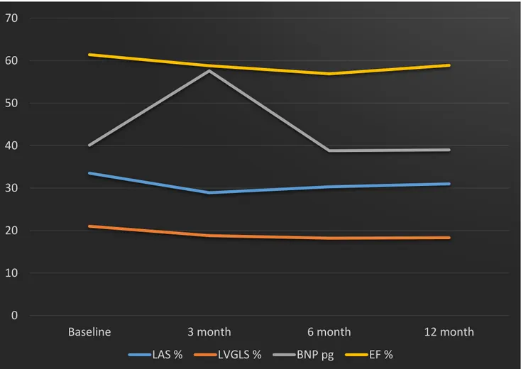
All patients remained in NYHA class I during follow-up; ECG did not show significant changes of repolarization phase nor QT corrected interval prolongation. Traditional echocardiographic parameters allowed to demonstrate: stable LV end-diastolic volumes during follow-up (79,6±22 vs 84,9±21,2 vs 81,7±20,2 vs 79,2±18,6 p n.s.); LV end-systolic volumes presented a transient increase at 3 and 6 months if compared to baseline values, with a trend to reduction at 12 months (31±11.4 vs 34.7±10.5 vs 35.7±10.8 vs 32.7±9.1; p 0,001 baseline vs 6 months). Table 3
LVEF always remained within normal range values, showing a slight reduction at 3 and 6 months after treatment initiation and a mild increase after discontinuation of chemotherapy: baseline 61,4±5,5%, 3 months 58,8±5,2% (p 0,012); 6 months 56,9±5,3% (p<0,001); 12 months 58,9±3,8%; (p 0,012).Table 3, Figure 3.
24
As far as diastolic function is concerned, E/A ratio was stable during follow-up, whereas E/e’ showed a trend to increase without reaching statistically significant values. Table 3
Speckle Tracking allowed to detect a stable reduction of LV GLS during follow-up: baseline GLS -21 ±2,5 %; 3 months GLS -18,8 ±2,2 % (p <0,001); 6 months -18,2 ±2,6 %(p<0,001); 12 months -18,3 ±2,9 % (p<0,001). Left atrial function evaluated through STE allowed to detect an early reduction of LAS , during reservoir phase at 3 and 6 moths and an improvement after discontinuation of chemotherapy: baseline 33,5±9,8%; 3 months 28,9±7 % (p 0,05); 6 months 30,3±8,5% (p 0,02) ; 12 moths 31±6 % (p ns). Table 4, Figures 3-4.
In agreement with LA reservoir reduction, LA stiffness showed a progressive increase at 3 e 6 months if compared to baseline, with a slight reduction at 12 months (0,30±0,29 vs 0,33±0,13 vs 0,37±0,19 vs 0,32±0,15; p n.s.). Table 4, Figure3.
25
Indexed LA volume showed a tendency to reduction during follow-up. This apparently unexpected finding can be interpreted as the consequence of an increased LA stiffness and a reduced LA compliance and compromised LA-LV coupling.
As Regards biomarkers, Troponin I did not show significant variations during follow-up, whereas BNP showed an early significant increase at 3 months if compared to baseline (40,1±26,4 vs 57,6±33,7 p 0.049), and a reduction at 6 (38.8±29.5) and at 12 months (39±18.5 p 0.047). Table 2, Figure 3.
9. Discussion
Early identification of LV subclinical dysfunction before deterioration of LVEF has been for many years the target of echocardiographic follow-up of patients who undergo potentially cardiotoxic treatment, however, as highlighted by most recent studies, LA may be an early target of
26
chemotherapy-induced cardiac damage and play a key-role in this particular setting.42-45
Our study confirms the presence of a relatively early cardiac damage induced by chemotherapy, with an involvement of both LA and LV. The main results of our study can be summarized as follows:
- In a selected and homogeneous population of women affected by breast cancer who underwent ANTs-based chemotherapy treatment for a period of 6 months, echocardiographic follow-up allowed to demonstrate an early and significant reduction of LA reservoir function at 3 and 6 months after treatment initiation, with a significant improvement at 12 months (six months after discontinuation of chemotherapy).
- LA stiffness progressively increases during follow-up, despite not reaching statistically significant values, while LA volumes shows a
27
slight reduction, probably due to an increased parietal stiffness and reduced LA compliance.
- LV longitudinal systolic function is compromised as represented by a significant but stable reduction of LVGLS during follow-up, despite normal LV ejection fraction.
- BNP increases after 3 months of chemotherapy (peak value) and reduces progressively at 6 and 12 month, as the consequence of an early activation of natriuretic peptides system, in response to an acute cardiac damage.
According to our findings, LA reservoir function is significantly and precociously compromised after 3 months of chemotherapy; these results are consistent with other evidences of literature that have recently demonstrated an impairment of LA longitudinal strain (reservoir and pump), even before a significant reduction of LVGLS, occurring after the first doses of chemotherapy, especially in patients with breast cancer.44-45
28
Assessment of the diastolic function has always played a key role in understanding the physiologic damage caused by ANTs¸ indeed several studies have reported LV diastolic abnormalities late after ANTs administration, especially in patients who survived from childhood malignancy; these abnormalities were assessed by isovolumic relaxation time or transmitral E/A ratio.46-47
In our study, despite a trend of E/e’ ratio to increase during follow-up, diastolic Doppler parameters were not significantly changed, which was discordant with previous studies; however the use of E/e’ ratio remains questionable in the oncological setting, as E and e’ velocities fluctuation in these patients could be the consequence of changes in loading conditions as a results of side effects associated with the chemotherapy (nausea, vomiting, and diarrhea) more than the results of a real change in LV diastolic performance.41
29
LA reservoir and stiffness evaluation may overcome these limitations, representing more stable parameters of LV end diastolic-pressures and more reliable indexes of subclinical cardiac diastolic dysfunction. In our study, a trend to increase of LA stiffness and an inverse relationship between LA volumes and stiffness was detected, most likely attributable to LA parietal fibrosis, structural marker of LA damage. The main consequence of this anatomical remodeling is represented by the loss of LA compliance and LA-LV coupling.
Cardiac troponin is the preferred biomarker for the measurement of myocardial injury and its low level elevations induced by doxorubicin are associated with histological evidence of myocardial injury. 48 However, the low prevalence of detection with standard assays would limit the utility of troponin measurement for clinical applications in patients after ANTs exposure. In our cohort of patients, Troponin I did not show significant variations, however BNP levels showed a rapid increase,
30
reaching a peak value after 3 months. A possible pathophysiological explanation for these findings is linked to the activation of a neuro-hormonal compensatory mechanism in response to an acute, inflammatory-like cardiac damage induced by the first dose of ANTs. Moreover LV GLS which was found to be impaired in our cohort of patients, despite a normal ejection fraction, confirms the presence of a subclinical LV systolic dysfunction, due to an early impairment of LV sub-endocardial fibers.
Although most of evidences were obtained in breast cancer patients, these data suggest that in patients treated with chemotherapy, the analysis of LA deformation in addition to LV systolic function evaluation, could be a sensitive and promising technique for detecting subclinical myocardial damage, even earlier than LV GLS.
31
Several limitations to this study warrant comment. The poor sample of
patients enrolled in the study and the presence of dropout at
echocardiographic follow-up represent both main limitations of our study
and may have affected the statistical significance of results. Secondly, we
used a software which was designed for LV analysis to obtain LA strain
curves as a results of lacking dedicated atrial software.
11. Conclusions:
Left atrial function estimated by 2DSTE longitudinal strain is significantly reduced in patients undergoing chemotherapy with ANTs but improves after discontinuation of therapy. Serial echocardiographic monitoring on these patients should include LA morphological and functional evaluation which may anticipate or accompany LV dysfunction and induce to start earlier cardio-protective treatment.
32
Table 1 Chemotherapy-related cardiac dysfunction (CRCD)
Cardioncology: State of the heart. M.C. Todaro et al. / International Journal of Cardiology 168 (2013)
Table 2 Clinical and laboratory data
baseline 3 months 6 months 12 months p
Patients, n 58 45 (13 drop-out) 44 (1 exitus) 35 (10 ongoing) SBP, mmHg 121±15.3 123±14.2 122±14.7 131±20.7*§# *0.001 vs base § 0.013 vs 3 #0.015 vs 6 DBP, mmHg 76.3±9.4 77.5±10.8 77.3±8.1 80±11.9 ns PP, mmHg 45.3±13.1 45.5±10.3 45.3±13.2 51.3±14*§ *0.005 vs 3 § 0.042 vs 6 HR, b/m 72.8±10.5 75.8±12.4*§ 67.9±16.1 70.1±9.6 *0.013 vs 6 §0.003 vs 12 Troponin, mg/dl 0.01±0.008 0.03±0.03 0.001±0.001 0.02±0.01 ns BNP, pg/ml 40.1±26.4 57.6±33.7 *§ 38.8±29.5 39±18.5 *0.049 vs base § 0.047 vs 6 SBP: Systolic blood pressure; DBP: Diastolic blood pressure; PP: Pulse pressure; HR: Heart rate; BNP: Brain Natriuretic Peptide
CRCD Drug prototype Findings on biopsy Mechanism Cumulative-dose relationship Reversibility Type 1 Doxorubicin (anthracyclines) Vacuoles, sarcomere disruption, necrosis Free oxygen radical formation yes no Type 2 Trastuzumab No ultrastructural changes Block of HER2 signaling No yes
33 Table 3 Echocardiographic parameters
baseline 3 months 6 months 12 months p
EDV,ml 79.6±22 84.9±21.2 81,7±20.2 79.2±18.6 ns ESV,ml 31±11.4 * 34.7±10.5 35.7±10.8 32.7±9.1 *0.001 vs 6 Svi, ml/mq 28.1±7.3 29.3±7.3 * 26.5±5.1 27±5.3 *0.051 vs 3 EF, % 61.4±5.5 *§# 58.8±5.2 56.9±5.3¥ 58.9±3.8 * 0.012 vs 3 § < 0.001 vs 6 # 0.012 vs 12 ¥ 0.041 vs 12 S', cm/sec 8±1.4 *§ 7.8±1.5 7.2±1.4 7.4±1.3 *0.019 vs 6 § 0.029 vs 12 GLS, % -21±2.5 *§ # -18.8±2.2 -18.2±2.6 -18.3±2.9 * < 0.001 vs 3 § < 0.001 vs 6 #< 0.001 vs 12 E/A 0.99±0.29 0.99±0.26 0.97±0.22 0.96±0.23 ns DT,msec 199±45.9 202.9±56.8 196.5±39.1 212±55 ns E/E' 8.3±4.1 8.6±2.1 9.1±2.7 9.1±2.6 ns
EDV: End-Diastolic Volume; ESV: End-Systolic Volume; SVi: Stroke Volume Index; EF: Ejection Fraction; GLS: Global Longitudinal Strain; DT: Deceleration Time
34 Table 4 Left atrial parameters
Baseline 3 months 6 months 12 months p
LAVi 32.6±9.1 * 30.1±6.8 29.4±7.6 28.5±7.9 * 0.007 vs 12
LALS 33.5±9.8 *§ 28.9±7.0 30.3±8.5 31±6 *0.005 vs 3
§ 0.026 vs 6
LA Stiffness 0.30±0.29 0.33±0.13 0.37±0.19 0.32±0.15 ns
35
Figure 1 Concentric representation of cardiotoxic effects of anthracyclines
Tocchetti CG et al. From molecular mechanisms to clinical management of antineoplastic drug-induced cardiovascular toxicity: A translational overview. Antioxid Redox Signal. 2017 May 15
36
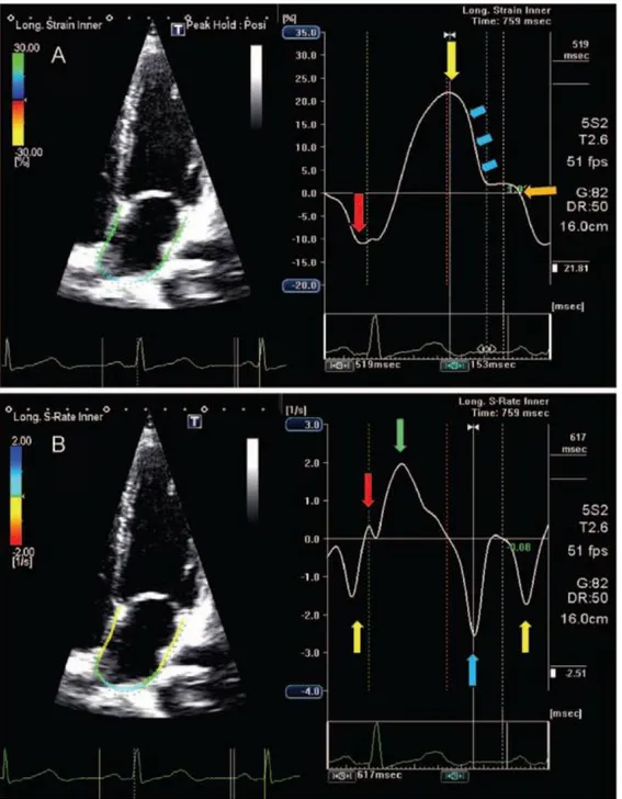
Figure 2 Left atrial strain and strain rate in a normal subject
Four-chamber view showing global left atrial (LA) longitudinal strain (A) and strain rate (B) in a normal subject. The trigger is before the P-wave on the electrocardiogram. (A) The negative peak of the contractile phase (red arrow), the positive peak of the reservoir phase (yellow arrows) and the conduit phase, which includes early diastole (blue arrows) and diastases (orange arrow). (B) The peak of the LA contractile phase in late diastole (yellow arrow), the early (red arrow) and late peak (green arrow) of the reservoir phase during ventricular systole, and the conduit phase in early diastole (blue arrow). Todaro MC et al. New echocardiographic techniques for evaluation of left atrial mechanics.. Eur Heart J Cardiovasc Imaging. 2012 Dec;13(12):973-84
37 Figure 3 Results
Graphic representation of main echocardiographic and laboratory parameters during follow-up. LAS (Left atrial Longitudinal Strain) is impaired at 3 (p 0.005) and 6 (p 0.026) months, recovering at 12 months; LVGLS (Left Ventricular Global Longitudinal Strain) is reduced at 3 months, remaining stable during follow-up ( p < 0.001); BNP (Brain Natriuretic Peptide) reaches its peak value at 3 months ( p 0.049) and decreases at 6 and 12 months; EF (Ejection Fraction) slightly reduces at 3 and 6 months, always remaining within normal range values.
0 10 20 30 40 50 60 70
Baseline 3 month 6 month 12 month
38 Figure 4
LA longitudinal strain (LAS) of a woman affected by breast cancer, before initiation of chemotherapy (panel A), at 3 months (panel B), at 6 months (panel C) and 12 months (panel D). A reduction of reservoir function is detected from baseline (40%) to 3 (33%) and 6 months (35%) follow-up, but an improvement of LAS is observed after discontinuation of chemotherapy at 12 months (38%).
40%
A
33%
B
D
38%
35%
C
39 References
1. Maria Chiara Todaro , Lilia Oreto , Rubina Qamar et al. Cardioncology: State of the
heart. International Journal of Cardiology 168 (2013) 680–687
2. Lenihan DJ, Cardinale D, Cipolla CM.The compelling need for a cardiology and
oncology partnership and the birth of the International CardiOncology Society. Prog
Cardiovasc Dis. 2010 Sep-Oct;53(2):88-93. doi: 10.1016/j.pcad.2010.06.002.
3. Lenihan DJ, Hartlage G, De Cara J et al. Cardio-Oncology Training: A Proposal From
the International Cardioncology Society and Canadian Cardiac Oncology Network for a
New Multidisciplinary Specialty. J Card Fail. 2016 Jun;22(6):465-71.
4. Jose Luis Zamorano, Patrizio Lancellotti, Daniel Rodriguez Muñoz et al. 2016 ESC
Position Paper on cancer treatments and cardiovascular toxicity developed under the
auspices of the ESC Committee for Practice Guidelines: The Task Force for cancer
treatments and cardiovascular toxicity of the European Society of Cardiology (ESC). Eur
40
5. Yeh ET, Bickford CL. Cardiovascular complications of cancer therapy: incidence,
pathogenesis, diagnosis, and management. J Am Coll Cardiol 2009;53:2231–47.
6. Ewer MS, Lippman SM. Type II chemotherapy-related cardiac dysfunction: time to
recognize a new entity. J Clin Oncol 2005;23:2900–2
7. Carlo Gabriele Tocchetti, Christian Cadeddu, Daniela Di Lisi et al. From molecular
mechanisms to clinical management of antineoplastic drug-induced cardiovascular
toxicity: A translational overview. Antioxid Redox Signal. 2017 May 15.
8. Cardinale D, Colombo A, Lamantia G et al. Anthracycline-induced cardiomyopathy:
clinical relevance and response to pharmacologic therapy. J Am Coll Cardiol
2010;55:213–220.
9. Lyu YL, Kerrigan JE, Lin CP, Azarova AM, Tsai YC, Ban Y, and Liu LF.
Topoisomerase II beta mediated DNA double-strand breaks: implications in doxorubicin
41
10. Steinherz LJ, Steinherz PG, Tan CT, Heller G, Murphy ML. Cardiac toxicity 4 to 20
years after completing anthracycline therapy. JAMA 1991;266:1672–7.
11. Von Hoff DD, Layard M, Rozencweig M, Muggia FM. Time relationship between last
dose of daunorubicin and congestive heart failure. Cancer Treat Rep 1977;61:1411–30.
12. Franco VI, Lipshultz SE. Cardiac complications in childhood cancer survivors treated
with anthracyclines. Cardiol Young 2015;25(Suppl 2):107–116.
13. Lipshultz SE, Colan SD, Gelber RD, Perez-Atayde AR, Sallan SE, Sanders SP. Late
cardiac effects of doxorubicin therapy for acute lymphoblastic leukemia in childhood. N
Engl J Med 1991;324:808–15.
14. Lipshultz SE, Lipsitz SR, Mone SM, et al. Female sex and drug dose as risk factors for
late cardiotoxic effects of doxorubucin therapy for childhood cancer. N Engl J Med
42
15. Khan AA, Ashraf A, Singh R, Rahim A et al Incidence, time of occurrence and response
to heart failure therapy in patients with anthracycline cardiotoxicity. Intern Med J. 2017
Jan;47(1):104-109. doi: 10.1111/imj.13305.
16. Dokmanovic M, King KE, Mohan N et al. Cardiotoxicity of ErbB2-targeted therapies and
its impact on drug development, a spotlight on trastuzumab. Expert Opin Drug Metab
Toxicol. 2017 Jul;13(7):755-766.
17. Martin M, Esteva FJ, Alba E, et al. Minimizing cardiotoxicity while optimizing treatment
efficacy with trastuzumab: review and expert recommendations. Oncologist 2009;14:1–
11.
18. Sengupta PP, Northfelt DW, Gentile F, et al. Trastuzumab-induced cardiotoxicity: heart
failure at the crossroads. Mayo Clin Proc 2008;83:197–203.
19. G. Curigliano, D. Cardinale, T. Suter et al Cardiovascular toxicity induced by
chemotherapy, targeted agents and radiotherapy: ESMO Clinical Practice Guidelines.
Ann Oncol (2012) 23 (suppl_7): vii155-vii166.
20. Eidem BW. Identification of anthracycline cardiotoxicity: left ventricular ejection
43
21. Marchandise B, Schroeder E, Bosly A, et al. Early detection of doxorubicin cardiotoxicity:
interest of Doppler echocardiographic analysis of left ventricular filling dynamics. Am
Heart J 1989;118:92–8.
22. Stoddard MF, Seeger J, Liddell NE, Hadley TJ, SullivanDM, Kupersmith J. Prolongation
of isovolumetric relaxation time as assessed by Doppler echocardiography predicts
doxorubicin-induced systolic dysfunction in humans. J Am Coll Cardiol 1992;20:62–9.
23. Tassan-Mangina S, Codorean D, Metivier M, et al. Tissue Doppler imaging and
conventional echocardiography after anthracycline treatment in adults: early and late
alterations of left ventricular function during a prospective study. Eur J Echocardiogr
2006;7:141–6.
24. Lang RM, Badano LP, Mor-Avi V et al. Recommendations for cardiac chamber
quantification by echocardiography in adults: an update from the American Society of
Echocardiography and the European Association of Cardiovascular Imaging. Eur Heart J
44
25. Geyer H, Caracciolo G, Abe H, Wilansky S et al.
Assessment of myocardial mechanics using speckle tracking echocardiography: fundame
ntals
and clinical applications. J Am Soc Echocardiogr. 2010 Apr; 23(4):351-69; quiz 453-5.
26. Stoodley PW, Richards DA, Hui R, et al. Two-dimensional myocardial strain imaging
detects changes in left ventricular systolic function immediately after anthracycline
chemotherapy. Eur J Echocardiogr 2011;12:945–52.
27. Vianna-Pinton R, Moreno CA, Baxter CM et al. Two-dimensional speckle-tracking
echocardiography of the left atrium: feasibility and regional contraction and relaxation
differences in normal subjects. J Am Soc Echocardiogr 2009;22:299-305.
28. Kim DG, Lee KJ, Lee S, Jeong SY, Lee YS, Choi YJ, et al. Feasibility of twodimensional
global longitudinal strain and strain rate imaging for the assessment of left atrial function:
a study in subjects with a low probability of cardiovascular disease and normal exercise
45
29. Todaro MC, Choudhuri I, Belohlavek M, et al. New echocardiographic techniques for
evaluation of left atrial mechanics.. Eur Heart J Cardiovasc Imaging. 2012
Dec;13(12):973-84. doi: 10.1093/ehjci/jes174. Epub 2012 Aug 21. Review.
30. Morris DA, Takeuchi M, Krisper M, et al. Normal values and clinical relevance of left
atrial myocardial function analysed by speckle-tracking echocardiography: multicenter
study. Eur Heart J Cardiovasc Imaging 2015;16:364-72.
31. Machino-Ohtsuka T, Seo Y, Tada H, Ishizu T, Machino T, Yamasaki H, Igarashi M, Xu
D, Sekiguchi Y, Aonuma K. Left atrial stiffness relates to left ventricular diastolic
dysfunction and recurrence after pulmonary vein isolation for atrial fibrillation. J
Cardiovasc Electrophysiol 2011;22:999–1006.
32. Cusmà Piccione M, Zito C, Khandheria B, Madaffari A, Oteri A, Falanga G, Donato
D, D'Angelo M, Carerj ML, Di Bella G, Imbalzano E, Pugliatti P, Carerj S. Cardiovascular
maladaptation to exercise in young hypertensive patients. Int J Cardiol. 2017 Apr
46
33. Ma XX, Zhang YL, Hu B, Zhu MR, Jiang WJ, Wang M, Zheng DY, Xue XP.
The usefulness of global left atrial strain for predicting atrial fibrillation recurrence
after catheter ablation in patients with persistent and paroxysmal atrial fibrillation. Arch
Cardiovasc Dis. 2017 May 18. pii: S1875-2136(17)30044-X
34. Longobardo L, Zito C, Carerj S, Khandheria BK Left atrium in heart failure with
preserved ejection fraction: the importance of function before anatomy. Eur Heart J
Cardiovasc Imaging. 2017 Mar 30
35. Todaro MC, Carerj S, Khandheria B, Cusmà-Piccione M, La Carrubba S,
Antonini-Canterin F, Pugliatti P1, Di Bello V, Oreto G, Di Bella G, Zito C.
Usefulness of atrial function for risk stratification in asymptomatic severe aortic stenosis.
J Cardiol. 2016 Jan;67(1):71-9. Epub 2015 May 21
36. Chunlai Shao, Jing Zhu, Jianchang Chen and Weiting Xu. Shao et al. Independent
prognostic value of left atrial function by two-dimensional speckle tracking imaging in
patients with non –ST segment- elevation acute myocardial infarction. BMC
47
37. Ernande L, Bergerot C, Girerd N3 et al. Longitudinal myocardial strain alteration is
associated with left ventricular remodeling in asymptomatic patients with type 2
diabetes mellitus. J Am Soc Echocardiogr. 2014 May;27(5):479-88
38. Cardinale D, Colombo A, Bacchiani G et al. Early detection of anthracycline cardiotoxicity
and improvement with heart failure therapy. Circulation 2015;131:1981–1988
39. Mor-Avi V, Lang RM, Badano LP, et al. Current and evolving echocardiographic techniques for the quantitative evaluation of cardiac mechanics: ASE/ EAE consensus statement on methodology and indications endorsed by the Japanese Society of Echocardiography. Eur J Echocardiogr 2011;12:167–205.
40. Cameli M, Caputo M, Mondillo S, Ballo P, et al, Feasibility and reference values of left atrial longitudinal strain imaging by two-dimensional speckle tracking. Cardiovasc Ultrasound 2009;7:6.
41. Plana JC, Galderisi M, Barac A, et al. Expert consensus for multimodality imaging evaluation of adult patients during and after cancer therapy. J Am Soc Echocardiogr
48
42. Shi J, Guo Y, Cheng L, Song F, Shu X. Early change in left atrial function in patients treated with anthracyclines assessed by real-time three-dimensional echocardiography. Sci
Rep. 2016 May 5;6:25512. doi: 10.1038/srep25512.
43. Sherif Moustafa, Katie Murphy, Bhargava K. Nelluri, et al. Temporal Trends of Cardiac Chambers Function with Trastuzumab in Human Epidermal Growth Factor Receptor II–
Positive Breast Cancer Patients. Echocardiography 2015. Oct 24
44. Maria Florescu, Lucia Stefania Magda, Dan Jinga, et al. Left Atrial Deformation is
Impaired After Epirubicin-based Chemotherapy in Patients with Breast Cancer.
Circulation. 2011;124:A13084
45. Moreno J, García-Sáez JA, Clavero M, Manganaro R, Moreno F, López J, Macaya C, Pérez
de Isla L. Effect of breast cancer cardiotoxic drugs on left atrial myocardium mechanics.
Searching for an early cardiotoxicity marker. Int J Cardiol. 2016 May 1;210:32-4. doi:
10.1016/j.ijcard.2016.02.093. Epub 2016 Feb 17
46. Rammeloo, L. A., Postma, A. & Sobotka-Plojhar, M. A. Low-dose daunorubicin in
induction treatment of childhood acute lymphoblastic leukemia: no long-term cardiac
damage in a randomized study of the Dutch childhood leukemia study group. Med Pediatr
49
47. Bu’Lock, F. A., Mott, M. G., Oakhill, A. & Martin, R. P. Left ventricular diastolic filling
patterns associated with progressive anthracycline-induced myocardial damage: a