Biomedicine & Pharmacotherapy 131 (2020) 110757
Available online 19 September 2020
0753-3322/© 2020 Published by Elsevier Masson SAS. This is an open access article under the CC BY-NC-ND license
(http://creativecommons.org/licenses/by-nc-nd/4.0/).
Immune dyscrasia in adult growth hormone deficiency: Evaluation of
hemolytic complement activity (CH50) and IgG subclasses
Edoardo Vergani
a,c, Carmine Bruno
a,c, Cecilia Napodano
b,c, Francesca Gulli
d,
Annunziata Stefanile
b,c, Gaia Piunno
a,c, Umberto Basile
b,c,*
, Antonio Mancini
a,c,*
aDipartimento di Medicina e Chirurgia Traslazionale, Universit`a Cattolica del Sacro Cuore, Rome, ItalybDipartimento di Scienze biotecnologiche di base, cliniche intensivologiche e peri-operatorie, Universit`a Cattolica del Sacro Cuore, Rome, Italy cFondazione Policlinico Universitario A. Gemelli IRCCS, Rome, Italy
dOspedale M. G. Vannini, Rome, Italy
A R T I C L E I N F O Keywords:
CH50 GHD
Immunoglobulins
Low grade chronic inflammation Free light chains
A B S T R A C T
CH50 is a screening assay for the activation of the classical complement pathway, the immunoglobulins- mediated one, activated in several inflammatory diseases. Adult growth hormone deficiency (aGHD) is recog-nized as a chronic inflammatory condition, although poorly evaluated under the profile of inflammatory bio-markers. The aim of this case-control observational study is to analyze CH50 and immunoglobulins G (IgG) subclasses production in aGHD, comparing this condition to healthy controls.
38 subjects were included and divided as follows: aGHD (n = 18, 6 females and 12 males); healthy controls (n = 20, 10 females and 10 males). GHD was diagnosed with dynamic test using Growth Hormone-Releasing Hormone (GHRH 50 μg i.v. + arginine 0,5 g/Kg), with a peak GH response < 9 μg/L when BMI was <30 kg/
m2 or < 4 μg/L when BMI was >30 kg/m2. The two groups were evaluated for hormonal and metabolic pa-rameters, CH50 and IgG subtypes.
IgG1 and IgG2 were significantly higher in controls than in aGHD, while IgG3 and IgG4 showed a trend to higher levels in controls, although not significant. Furthermore, CH50 levels were significantly higher in aGHD. These data substantiate the hypothesis of a dyscrasia in IgG subclasses production in aGHD. As IgG levels decrease, CH50 levels do not.
1. Introduction
Adult Growth Hormone Deficiency (aGHD), defined as a reduction in GH secretion caused by congenital or acquired disorders affecting the hypothalamus or the pituitary gland [1], is a pathological condition marked by low-grade chronic inflammation [2]. It shares some features with metabolic syndrome, including insulin resistance, altered lipidic metabolism, systemic inflammation [3–5] and oxidative stress (OS), even if a different pattern of antioxidant response has been shown [6,7]. The results of these phenomena are cardiovascular impairment and the subsequent complications [8]. Chronic low-grade inflammation (LGI) syndromes can be defined as systemic and chronic pathological
conditions characterized by a slight increase in inflammatory markers. Chronic LGI exhibits increased circulating cytokine levels and macro-phage infiltration in peripheral tissues, which do not cause, anyway, damage or loss of function to the involved tissues [9,10]. Different chronic inflammatory disorders and autoimmune diseases are accom-panied by elevated polyclonal free light chains of immunoglobulins (FLCs) levels in different body fluids and the increased FLCs concen-trations correlate with relapses of disease and enhanced activity of the immune system [11,12]; an augmented level of both FLCs types (kappa and lambda) has recently been described in aGHD [13]. Finally, it is known that GH, directly or via Insulin-like growth-factor-1 (IGF-1) ac-tivity, influences many aspects of immune response; both humoral and
Abbreviations: Adult, Growth Hormone Deficiency (aGHD); Oxidative, stress (OS); Low-grade, inflammation (LGI); Free, light chains of immunoglobulins (FLCs); Insulin-like, growth-factor-1 (IGF-1); Complement, total activity (CH50); Growth, Hormone-Releasing Hormone (GHRH); Growth, Hormone-Releasing Hor-mone + arginine (GHRH/ARG); Thyroid-stimulating, horHor-mone (TSH); Follicle-stimulating, horHor-mone (FSH); Luteinizing, horHor-mone (LH); Estradiol, (E2); Testosterone, (T); Glucose-6-phosphate, dehydrogenase (G6PDH); Standard, deviation (SD); Monocyte, chemoattractant protein 1 (MCP-1); Primary, empty sella (PES).
* Corresponding authors at: Largo A. Gemelli 8, 00168, Rome, Italy.
E-mail addresses: umberto.basile@policlinicogemelli.it (U. Basile), antonio.mancini@unicatt.it (A. Mancini).
Contents lists available at ScienceDirect
Biomedicine & Pharmacotherapy
journal homepage: www.elsevier.com/locate/biopha
https://doi.org/10.1016/j.biopha.2020.110757
cellular branches are modulated [14–18]. GHD has been described as a disorder characterized by altered IgG production, even though no clin-ical consequence of immunodeficiency is evident [19]. However, no data on IgG subclasses and complement function are available.
The complement cascade comprises over 20 serum proteins that form part of the innate immune system. Complement acts in a number of ways to clear invading organisms, with a major function of lysis of bacteria through formation of the membrane attack complex. Complement total activity (CH50) is a screening assay for the activation of the classical complement pathway, the immunoglobulins mediated-one. It is sensi-tive to the reduction, absence and/or inactivity of any component of the pathway. The CH50 tests the functional capability of serum complement components of the classical pathway to lyse sheep red blood cells pre- coated with rabbit anti-sheep red blood cell antibody (hemolysin). CH50 blood test is often ordered to evaluate complement component deficiency and evaluate complement activity in cases of immune com-plex diseases, such as glomerulonephritis and autoimmune diseases [20,
21].
The subclasses of IgG either may be useful to better understand a scenario of altered immune response [22]. The distribution of the IgG subclasses depends on the type of antigen and duration of antigen exposure, a concept known as “subclass restriction”. Thus, elevations of a given IgG subclass could reflect the nature of the underlying driving antigen. However, it should be noted that serum levels of IgG subclasses do not necessarily correlate with the amount of antibody deposition in the tissues, meaning that it is not possible to assume that the elevated IgG subclass seen in the serum is certainly involved in driving the disease process. In humans, IgG is the predominant antibody class (7− 15 g/L) and the four IgG subclasses, termed IgG1, IgG2, IgG3, and IgG4, func-tionally distinct because of different heavy chain gene usage, differ in their ability to fix complement (IgG3 > IgG1 > IgG2 > IgG4) and bind Fc receptors. The antigen driven subclass switching is sequential in the order IgG3, IgG1, IgG2 and IgG4 and is under T cell control [23,24]. Nevertheless, the four IgG subclasses are named in descending order of frequency: IgG1 is the most prevalent subclass, making up 60–70 % of the total IgG, followed by IgG2 (20–30 %), IgG3 (5–8 %) and lastly IgG4 that is the least frequent IgG subclass, comprising only up to the 5% of total IgG [22,25].
To the best of our knowledge no data are reported on CH50 and IgG subclasses beheaviour in aGHD. Therefore, we performed a case-control study in a group of adult GHD patients evaluating CH50 levels and IgG subclasses to further explore the pattern of inflammatory markers in this condition.
2. Materials and methods
2.1. Patients
Subjects involved in this study were admitted to the University Hospital “Policlinico Gemelli” and were enrolled after being given an explanation of purposes and nature of the study, conducted in accor-dance with the Declaration of Helsinki, as revised in 2013. The study was approved by the ethical committee of Catholic University of Rome. Upon assessment of the importance of the study and that the information resources were appropriately expressed, patients gave their informed consent.
We included 38 patients, divided as follows:
•aGHD (18 patients, 6 females and 12 males). GHD was diagnosed with dynamic test using Growth Hormone-Releasing Hormone (GHRH) 50 μg i.v. + arginine 0,5 g/Kg (GHRH/ARG), with a peak GH
response < 9 μg/L when BMI was < 30 kg/m2 or < 4 μg/L when BMI
was > 30 kg/m2. Etiologies of GHD were: 7 empty sella, 1 pineal cyst,
3 post-surgical hypopituitarism, 7 negative MRI pictures (idiopathic aGHD).
•healthy controls (20 patients, 10 females and 10 males).
Exclusion criteria were: age under 18 or over 75; history of cranial hypertension or active cranial hypertension, decompensated type 1 or 2 diabetes mellitus; autoimmune and infectious diseases; corticosteroid treatment (except for topic, inhalatory and oral hydrocortisone as replacement regimen); other diseases characterized with low insulin- like growth factor-1 (IGF-1) such as liver disease, malabsorption and malnutrition; renal failure; active malignancies. No patients received GH therapy at the moment of the study enrolment. The GHRH/ARG test was performed after an adequate replacement therapy of other pituitary- dependent axes; in particular, 1 patient was under levothyroxine treat-ment for secondary hypothyroidism, 3 patients under hydrocortisone treatment for secondary hypoadrenalism, 2 male patients under testos-terone undecanoate for secondary hypogonadism. All therapies were monitored by measuring peripheral hormones’ levels as appropriate.
2.2. Laboratory investigations
Both in patients and controls, a basal blood sample was collected at 08:00 a.m. after an overnight fast; it was centrifugated within 2 h and plasma aliquots stored at − 80 ◦C until assayed. The following
parame-ters were determined in both groups: glucose, insulin, total and HDL- cholesterol, triglycerides, IGF-1. The method used for hormone assays was chemo-luminescent immunoassay for insulin (reference range 3–20
μUI/mL) and IGF-1 (reference range 80− 330 ng/mL). Serum concen-trations of IGF-1 were measured by using immunochemiluminometric assays on a Roche Modular E170 analyzer according to the manufac-turer’s recommendations. (Roche Diagnostics, Indianapolis, IN, USA) using the consensus statement on the standardization and evaluation of IGF-1 [26]. The intra- and inter-assay coefficients of variation (CV) for all hormones were, respectively, < 5.0 % and < 7.0 %. Plasma con-centrations of glucose, total cholesterol, HDL-cholesterol and tri-glycerides were measured by using enzymatic assays and on Olympus AU2700 chemistry analyzer (Olympus America Inc., Center Valley, PA, USA) according to manufacter’s instruction. The intra-and inter-assay CV for total cholesterol and triglycerides were < 1.5 %, and < 2.5 %, respectively. The intra-and inter-assay CV for HDL-cholesterol were < 2.5 %, and < 3.0 %, respectively. LDL cholesterol was calculated by Friedewald’s equation: LDL = total cholesterol - (HDL + tri-glycerides/5). HOMA index was calculated according to the formula {[fasting insulin (μU/mL)] * [fasting glucose (mmol/L)]}/22.5 [27].
In aGHD group an endocrine evaluation including fT3, fT4, thyroid- stimulating hormone (TSH), follicle-stimulating hormone (FSH), lutei-nizing hormone (LH), estradiol (E2), testosterone (T) and cortisol levels was performed. The following methods were used for hormone assay: ChemoLuminescent Immunoassay for TSH (reference range 0.35–2.80
μUI/mL), fT3 (reference range 2.4–4.2 ng/ml), fT4 (reference range
8.5–16.5 pg/ml), FSH (reference range 2.5–11 mU/mL), LH (reference range 2.5–10 mU/mL), E2 (reference range <44 ng/mL), DHEAS (reference range 800− 3500 ng/mL), T (reference range. 0.20–2.00 ng/ ml), Cortisol (reference range 60− 220 ng/mL).
The Optilite CH50 Reagent is intended for the quantitative in vitro determination of CH50 in human serum and EDTA plasma using the Binding Site Optilite Turbidimetric assays analyser (The Binding Site Birmingham, UK) [28]. The test consists in liposomes encapsulating glucose-6-phosphate dehydrogenase (G6PDH) used to mimic an invading microorganism. On addition of sample, antibodies in the re-agent combine with dinitrophenyl groups on the surface of the lipo-somes. The resultant complex activates complement in the sample, which lyses the liposome, releasing G6PDH to react with glucose-6-phosphate and NAD in the reagent. The change in absorbance can be measured and is proportional to the complement activity in the sample. The comparison to a calibration curve gives a value for the unknown patient sample [29]. The four IgG subclasses concentrations were measured by turbidimetry through the employment of Human IgG and IgG subclass liquid reagent kits (The Binding Site Birmingham, UK) with Optilite instrument according to the manufacturer’s
recommendations. These kits are intended for quantifying human IgG and IgG subclasses. Concentrations are automatically calculated by reference to a standard curve stored within the instrument. Reference range for subclasses: 3.82–9.29 g/L for IgG1; 2.42–7.0 g/L for IgG2; 0.22–1.76 g/L for IgG3; 0.04–0.86 g/L for IgG4 [30]. Samples were tested according to the manufacturer’s instructions and serum dilutions, where necessary, were performed according to the manufacturer’s rec-ommendations. We selected 20 healthy blood donors in the local pop-ulation and they have fallen within the references interval indicated by manufacturers (data not shown) and then the ranges suggested were validated as indicated in CLSI EP 28A3C [31].
2.3. Statistical analysis
The statistical analysis was performed using Prism GraphPad 7, San Diego, CA, USA. The sample size was empirical for this observational study. The D’Agostino and Pearson test, using a p < 0.05, demonstrated a non-parametric distribution of all data analyzed. The differences be-tween CH50 and IgG subclasses in cases and controls were evaluated by Mann-Whitney test. The same test was used to compare hormonal and metabolic parameters between the two groups. The correlations be-tween IgG subclasses, CH50 and metabolic and hormonal parameters were evaluated through linear correlation analysis. When a correlation was found, Spearman test was used to find out its power. All reported P values were two-sided and a value of p < 0.05 was considered statisti-cally significant. There were no missing data.
3. Results
3.1. Patients characteristics
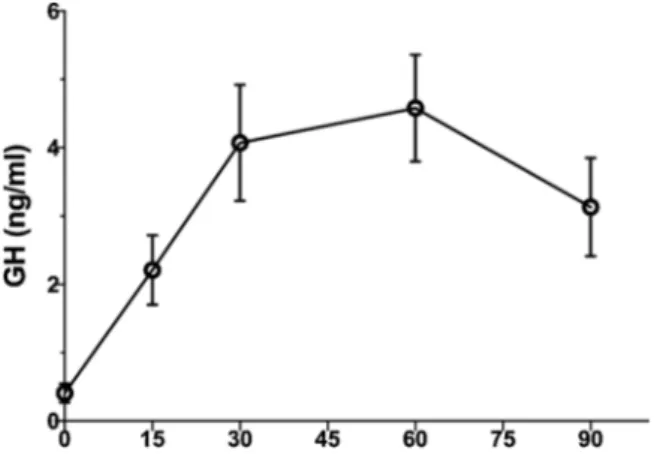
For the 18 partecipants affected by aGHD the mean ± standard de-viation (SD) age was 53.4 ± 12.1 years, while mean ± SD BMI was 29.2 ± 5.8 kg/m2. In aGHD group 33 % were females. Fig. 1 shows the
results of the GH response to maximal stimulation with GHRH + argi-nine in the tested patients. Mean peak levels were reached at 60 min during the test: 4.6 ± 2.9 μg/L. When considering separately obese vs
normal BMI subjects, we observed mean GH peak of 1.6 ± 1.4 μg/L in
patients with BMI equal or greater than 30 Kg/m2 and 5.4 ± 2.7 μg/L in
patients with BMI lower than 30 Kg/m2.
For the 20 volunterees of the control cohort 53.1 ± 9.9 years was the mean ± SD of age, while mean ± SD BMI was 23.97 ± 4.6 kg/m2. 50 % of
the control group were females.
3.2. Metabolic and hormonal data
Table 1 shows mean ± SD levels of metabolic and hormonal param-eters. As expected IGF-1 was significantly lower in aGHD (109.1 ± 38.8 ng/mL) than controls (194.7 ± 90.5 ng/mL). As metabolic parameters are concerned, a trend to increased basal insulin levels (14.3 ± 11.9 vs 9.8 ± 8.5) and consequently significant differences in HOMA-index were observed in aGHD (3.1 ± 2.5) in comparison to controls (1.2 ± 0.6). A trend to worse lipidic pattern, although not sig-nificant, was shown in aGHD cohort
3.3. Immunological parameters
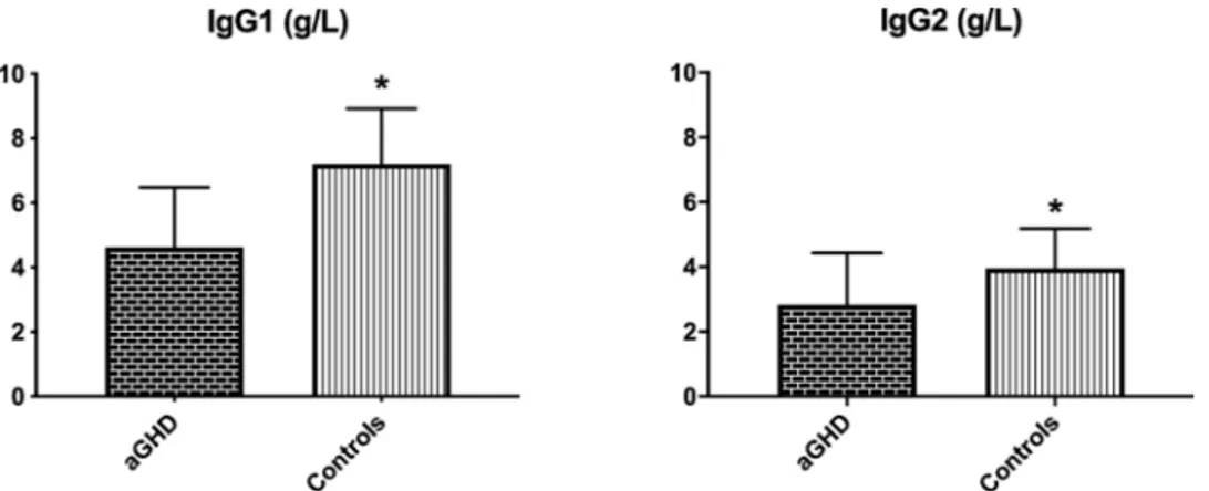
Fig. 2 and Fig. 3 show serum IgG levels in cases and controls. IgG1 and IgG2 were significantly higher in controls than in aGHD, whether IgG3 and IgG4 showed a trend, although not significant, to higher levels in controls than in aGHD. Furthermore, CH50 levels were significantly higher in aGHD (Fig. 4), although remaining within the reference range. At univariate analysis, in aGHD patients we found a significant inverse correlation between triglycerides and IgG1 (p = 0.03, Spearman corre-lation coefficient: − 0.50) and IgG2 (p = 0.04, Spearman correcorre-lation coefficient: − 0.03), while IgG2 also correlated positively with HOMA index (p = 0.02, Spearman correlation coefficient: 0.23). No correlation between IGF-1 or peak GH levels and immunological parameters was detected.
4. Discussion
Our data show that aGHD is characterized by a significant lower production of IgG1 and IgG2 and a trend toward lower production of IgG3 and IgG4 subclasses, consequently a less active complement classic pathway. This datum should be inserted in the complex picture of in-flammatory and/or oxidative alterations in patients affected by aGHD [32–34]. Moreover, correlation studies also suggested a relation with metabolic parameters in the same subjects.
GH and IGF-1 are involved in the regulation of the development and function of the immune system [35–37]. Indeed, they are produced and secreted by several immunocompetent cells and different cells in the immune system express their receptors [38]. Their action may be seen both on peripheral immune cells and on central organs of immunity, such as the bone marrow and the thymus [39]. GH drives chemotactic processes, stimulates monocytes phagocytic function, increases lymphocyte subpopulations such as CD3+, CD4+, CD8+, and improves the effectiveness of T lymphocyte response [40,41].
IGF-1 may exert an important role in linking OS and inflammation. A complex interplay does exist between IGF-1 and cytokines; IGF-1 stim-ulates IL-10 production [14,16], while inhibits the production of other proinflammatory cytokines [15,18]. On the contrary, lowering IGF-1
Fig. 1. Average of the results of the GHRH/ARG combined test response in
aGHD subjects enrolled in the study. Patients with total GHD (n = 18) have a peak of less than 9.0 μg/L when BMI was < 30 kg/m2 or < 4 μg/L when BMI
was > 30 kg/m2. Mean peak levels were reached at 60 min during the test:
4.6 ± 2.9 μg/L. When considering separately obese vs normal BMI subjects, we
observed mean GH peak of 1.6 ± 1.4 μg/L in patients with BMI equal or greater
than 30 Kg/m2 and 5.4 ± 2.7 μg/L in patients with BMI lower than 30 Kg/m2.
Table 1
Metabolic and hormonal parameters. Mean ± SD of metabolic and hormonal parameters in the two groups. As expected IGF-1 was significantly lower in aGHD than controls. A trend to increased basal insulin levels and consequently significant HOMA-index differences was observed in aGHD in comparison to controls. A trend to worse lipid pattern was shown in aGHD cohort.
aGHD Controls Total cholesterol (mg/dl) 179.5 ± 41.7 185.8 ± 42.1 HDL cholesterol (mg/dl) 50.6 ± 12.4 59.6 ± 19.5 LDL cholesterol (mg/dl) 112.8 ± 32.9 109.6 ± 38.1 Triglycerides (mg/dl) 102.4 ± 41.9 97.1 ± 38.1 Glucose (mg/dl) 87.2 ± 14.1 86.6 ± 19.7 Insulin (μUI/mL) 14.3 ± 11.9 9.8 ± 8.5 HOMA-IR 3.1 ± 2.5* 1.2 ± 0.6 IGF-1 (ng/mL) 109.1 ± 38.8* 194.7 ± 90.5 Notes: *p < 0.05 vs controls.
augments monocyte chemoattractant protein 1 (MCP-1) serum levels. MCP-1 in turn induces recruitment of monocytes and macrophages, as demonstrated again in type 2 diabetes and chronic hypertension, two conditions associated with OS [17]. Finally, IGF-1 favours the replica-tion and the survival of myeloid and lymphoid progenitors [42]. In GHD
patients, GH administration improves the function of the immune sys-tem [43]. As complement analysis is concerned, Cabellero-Villaraso et al. did not observe complement variations, evaluated as C3 and C4 serum levels, in GHD children before and after GH administration [43]. Differently from the previous cited study, we have analyzed classical complement pathway through CH50, thus finding higher levels in aGHD than healthy controls although not exceeding reference range. It seems presumable that classical complement pathway, the immunoglobulins mediated-one, is not activated in aGHD, probably for the lowering of IgG caused by lower GH secretion.
As previously stated, the lowering of IgG in aGHD is already known [19], even though no data on IgG subclasses are reported. Our datum of lower levels of IgG subtypes in aGHD is reasonable according to litera-ture, as a matter of fact the group of Yoshida [44] reported that GH enhance immunoglobulin synthesis (IgG1, IgG2, IgG3, IgG4, IgA1, IgA2, and IgM, for instance) in B lymphocytes. In particular, the same group supported the idea that GH-induced enhancement is not mediated by IGF-I as the enhancement is blocked by anti-GH antibody but not by anti-IGF-I antibody or anti-IGF-I receptor antibody [45]. Considering that GH receptor is predominantly expressed in B cells, they may be considered the most important site of action of GH in the immune system [46]. Furthermore, our group has recently described an enhanced secretion of FLCs in aGHD [13]; in our study aGHD patients presented higher k and λ levels than controls and higher k chains levels in com-parison to subjects affected by metabolic syndrome [13]. Based on the previous described data, it is possibile to assume the presence of a “dyscrasia” in the production of immunoglobulins in aGHD. Indeed, an increase in FLCs productions corresponds to a lower secretion of IgG with complete structure. We must emphasise that IgG subclasses in our “case” group, even if lower than healthy controls, were still in reference
Fig. 2. Mean ± SD IgG1 and IgG2 plasmatic levels in cases and controls. IgG1 and IgG2 were significantly higher in controls than in aGHD. * p < 0.05.
Fig. 3. Mean ± SD IgG3 and IgG4 plasmatic levels in cases and controls. IgG3 and IgG4 showed a trend, although not significant, to higher levels in controls than
in aGHD.
Fig. 4. Mean ± SD CH50 in cases and controls. CH50 levels were significantly
range, thus no worse immune response can be described in aGHD, as extensively reported in literature [19,38]. In addition, as stated previ-ously, none of the patients with aGHD reported infectious or autoim-mune processes at the moment of the evaluation.
As regard our aGHD cohort, the most frequent diagnoses were the idiopathic and primary empty sella (PES). Original suggestion of hypo-secretion of GH in primary PES was described by Brismar et al. [47,48]. The group of Gasperi showed a large proportion of PES patients with a reduced GH response to stimulation and low IGF-1 levels [49]. Furthermore, De Marinis et al. described isolated GH deficiency in a small group of patients, who presented benefits on quality of life after replacement therapy [50]. Moreover, it is known that GHD precedes other pituitary deficiencies in case of pituitary insult or damage, sug-gesting a greater sensitivity of somatotroph cells to different harmful stimuli [51]. Several aspects of pathophysiology of PES are unknown, as neurotransmitter control and autoimmune factors, other than the direct compression of the gland parenchyma. The prevalence of idiopathic GHD, 38 % in our cohort, is different among literature reports. In the observational HYPOCCS database, from 1996 to 2005, an increase from 13.9–19.3% was reported [52]. About 10 % patients were classified as idiopathic in the KIMS observational study [53]. The prevalence was also related to regional differences, from 2.6 % in the Netherlands to 65 % in UK; probably, a better knowledge about possible genetic or auto-immune aetiology of GHD, may narrow the field of “idiopathic” GHD. Nevertheless, there are some main potential restrictions to be considered about the present study. First, the number of subjects in the groups is slightly small, so the statistical power of the study is limited, even if aGHD is considered a rare condition; consequently, our findings will need to be confirmed in a larger population. Secondly, the design of the study and the power analysis cannot draw a cause-effect relation. Finally, more data and longitudinal studies will be needed to strentghten the hypotheses here proposed.
5. Conclusions
In conclusion, these data substantiate the hypothesis of a dyscrasia in IgG production in aGHD. An increase in FLCs production, previously reported by our group, could correspond to a lower secretion of IgG with complete structure. They should be inserted in the complex scenario of inflammation and oxidative stress markers underlying the greater morbidity and mortality of GHD, an underestimated disease with a physiopathology not still entirely understood. In the era of precision medicine, the ability to accurately measure specific predictors of patient outcomes may improve the quality of laboratory diagnosis in personal-ized clinical care. However, these data cannot be conclusive and further studies are needed to establish the role of such molecules as biological markers and their usefulness in prognostic and personalized treatment of such condition.
Funding
This research did not receive any specific grant from funding agencies in the public, commercial, or not-for-profit sectors.
Ethical approval
All procedures performed in studies involving human participants were in accordance with the ethical standards of the institutional and/or national research committee and with the Helsinki declaration and its later amendments or comparable ethical standards.
Informed consent
Informed consent was obtained from all individual partecipants included in the study.
CRediT authorship contribution statement
Edoardo Vergani: Conceptualization, Data curation, Formal
anal-ysis, Project administration, Resources, Software, Visualization.
Carmine Bruno: Conceptualization, Data curation, Formal analysis,
Project administration, Software, Visualization. Cecilia Napodano: Investigation, Methodology, Resources. Francesca Gulli: Investigation, Methodology, Resources, Visualization. Annunziata Stefanile: Inves-tigation, Methodology, Resources, Visualization. Gaia Piunno: Formal analysis, Resources, Visualization. Umberto Basile: Conceptualization, Investigation, Methodology, Project administration, Resources, Super-vision, Validation. Antonio Mancini: Conceptualization, Formal anal-ysis, Project administration, Supervision, Validation.
Declaration of Competing Interest
The authors report no declarations of interest.
References
[1] M.E. Molitch, D.R. Clemmons, S. Malozowski, G.R. Merriam, M.L. Vance, S. M. Shalet, Endocrine society: evaluation and treatment of adult growth hormone deficiency: an endocrine society clinical practice guideline, J. Clin. Endocrinol. Metab. 96 (2011) 1587–1609, https://doi.org/10.1210/jc.2005-2227. [2] A. Curr`o, D. Vergani, E. Bruno, C. Comi, S. D’Abate, C. Mancini, Plasmatic
lipocalin-2 levels in chronic low-grade inflammation syndromes: Comparison between metabolic syndrome, total and partial adult growth hormone deficiency, BioFactors. (2020), https://doi.org/10.1002/biof.1628.
[3] S.A. Beshyah, C. Freemantle, E. Thomas, O. Rutherford, B. Page, M. Murphy, D. G. Johnston, Abnormal body composition and reduced bone mass in growth hormone deficient hypopituitary adults, Clin. Endocrinol. (Oxf). 42 (1995) 179–189, https://doi.org/10.1111/j.1365-2265.1995.tb01860.x.
[4] J. Verhelst, R. Abs, Cardiovascular risk factors in hypopituitary GH-deficient adults, Eur. J. Endocrinol. 161 (Suppl 1) (2009) S41–49, https://doi.org/10.1530/ EJE-09-0291.
[5] J.O.L. Jorgensen, L. Thuesen, T. Ingemann-Hansen, S.A. Pedersen, I. Jorgensen, N. E. Skakkebaek, J.S. Christiansen, Beneficial effects of growth hormone treatment in GH-deficient adults, Lancet 333 (1989) 1221–1225, https://doi.org/10.1016/ S0140-6736(89)92328-3.
[6] A. Mancini, C. Di Segni, C. Bruno, G. Olivieri, F. Guidi, A. Silvestrini, E. Meucci, P. Orlando, S. Silvestri, L. Tiano, A. Pontecorvi, Oxidative stress in adult growth hormone deficiency: different plasma antioxidant patterns in comparison with metabolic syndrome, Endocrine 59 (2017) 130–136, https://doi.org/10.1007/ s12020-017-1468-1.
[7] A. Mancini, G.E. Martorana, M. Magini, R. Festa, S. Raimondo, A. Silvestrini, N. Nicolotti, A. Mordente, M.C. Mele, G.A.D. Miggiano, E. Meucci, Oxidative stress and metabolic syndrome: effects of a natural antioxidants enriched diet on insulin resistance, Clin. Nutr. ESPEN 10 (2015) e52–e60, https://doi.org/10.1016/j. clnesp.2014.11.002.
[8] A. Colao, The GH-IGF-I axis and the cardiovascular system: clinical implications, Clin. Endocrinol. (Oxf). 69 (2008) 347–358, https://doi.org/10.1111/j.1365- 2265.2008.03292.x.
[9] M.M.J. van Greevenbroek, C.G. Schalkwijk, C.D.A. Stehouwer, Obesity-associated low-grade inflammation in type 2 diabetes mellitus: causes and consequences, Neth. J. Med. 71 (2013) 174–187.
[10] S.P. Weisberg, D. McCann, M. Desai, M. Rosenbaum, R.L. Leibel, A.W. Ferrante, Obesity is associated with macrophage accumulation in adipose tissue, J. Clin. Invest. 112 (2003) 1796–1808, https://doi.org/10.1172/JCI200319246. [11] C.A. Hutchison, S. Harding, P. Hewins, G.P. Mead, J. Townsend, A.R. Bradwell,
P. Cockwell, Quantitative assessment of serum and urinary polyclonal free light chains in patients with chronic kidney disease, Clin. J. Am. Soc. Nephrol. 3 (2008) 1684–1690, https://doi.org/10.2215/CJN.02290508.
[12] U. Basile, F. Gulli, L. Gragnani, C. Napodano, K. Pocino, G.L. Rapaccini, M. Mussap, A.L. Zignego, Free light chains: eclectic multipurpose biomarker, J. Immunol. Methods 451 (2017) 11–19, https://doi.org/10.1016/j.jim.2017.09.005. [13] U. Basile, C. Bruno, C. Napodano, E. Vergani, K. Pocino, A. Brunetti, F. Gulli, S.
A. Santini, A. Mancini, Plasmatic free light chains as inflammatory marker in insulin resistance: comparison of metabolic syndrome with adult growth hormone deficiency, BioFactors 44 (2018), https://doi.org/10.1002/biof.1444.
[14] S. Teppala, A. Shankar, Association between serum IGF-1 and diabetes among U.S. Adults, Diabetes Care 40 (2010), https://doi.org/10.2337/dc10-0770. [15] E. Van Exel, J. Gussekloo, A.J.M. De Craen, M. Fr¨olich, A.B. Van Der Wiel, R.G.
J. Westendorp, Low production capacity of interleukin-10 associates with the metabolic syndrome and type 2 diabetes: the Leiden 85-plus study, Diabetes 51 (2002) 1088–1092, https://doi.org/10.2337/diabetes.51.4.1088.
[16] A.E. Schutte, M. Volpe, G. Tocci, E. Conti, Revisiting the relationship between blood pressure and insulin-like growth factor-1, Hypertension 63 (2014) 1070–1077, https://doi.org/10.1161/HYPERTENSIONAHA.113.03057.
[17] C. Pouvreau, A. Dayre, E.G. Butkowski, B. de Jong, H.F. Jelinek, Inflammation and oxidative stress markers in diabetes and hypertension, J. Inflamm. Res. 11 (2018) 61–68.
[18] V.V. Lima, S.M. Zemse, C.W. Chiao, G.F. Bomfim, R.C. Tostes, R. Clinton Webb, F. R. Giachini, Interleukin-10 limits increased blood pressure and vascular RhoA/ Rho-kinase signaling in angiotensin II-infused mice, Life Sci. 145 (2016) 137–143,
https://doi.org/10.1016/j.lfs.2015.12.009.
[19] V.C. Campos, M.R. Barrios, R. Salvatori, R.P. de Almeida, E.V. de Melo, A.C. S. Nascimento, A.R. de Jesus, M.H. Aguiar-Oliveira, Infectious diseases and immunological responses in adult subjects with lifetime untreated, congenital GH deficiency, Endocrine (2016), https://doi.org/10.1007/s12020-016-1061-z. [20] M. Costabile, Measuring the 50% haemolytic complement (CH50) activity of
serum, J. Vis. Exp. 19 (2010) 187–197, https://doi.org/10.3791/1923. [21] P. Vignesh, A. Rawat, M. Sharma, S. Singh, Complement in autoimmune diseases,
Clin. Chim. Acta (2017), https://doi.org/10.1016/j.cca.2016.12.017. [22] C. Napodano, M. Marino, A. Stefanile, K. Pocino, R. Scatena, F. Gulli, G.
L. Rapaccini, S. Delli Noci, G. Capozio, D. Rigante, U. Basile, Immunological role of IgG subclasses, Immunol. Invest. (2020) 1–18, https://doi.org/10.1080/ 08820139.2020.1775643.
[23] D. Lowe, R. Higgins, D. Zehnder, D.C. Briggs, Significant IgG subclass heterogeneity in HLA-specific antibodies: implications for pathogenicity, prognosis, and the rejection response, Hum. Immunol. 74 (2013) 666–672, https:// doi.org/10.1016/j.humimm.2013.01.008.
[24] J. Tan, X. Jin, R. Zhao, X. Wei, Y. Liu, X. Kong, Beneficial effect of T follicular helper cells on antibody class switching of B cells in prostate cancer, Oncol. Rep. 33 (2015) 1512–1518, https://doi.org/10.3892/or.2014.3684.
[25] P.H. Schur, IgG subclasses. A historical perspective, Monogr. Allergy 23 (1988) 1–11.
[26] D.R. Clemmons, Consensus statement on the standardization and evaluation of growth hormone and insulin-like growth factor assays, Clin. Chem. (2011), https:// doi.org/10.1373/clinchem.2010.150631.
[27] D.R. Matthews, J.P. Hosker, A.S. Rudenski, B.A. Naylor, D.F. Treacher, R.C. Turner, Homeostasis model assessment: insulin resistance and β-cell function from fasting plasma glucose and insulin concentrations in man, Diabetologia 28 (1985) 412–419, https://doi.org/10.1007/BF00280883.
[28] C. Tange, K. Townsend, S. Harding, Functional assessment of complement proteins C1q, C2, C3 and C5, Clin. Chim. Acta (2019), https://doi.org/10.1016/j. cca.2019.03.091.
[29] S. Yamamoto, K. Kubotsu, M. Kida, K. Kondo, S. Matsuura, S. Uchiyama, O. Yonekawa, T. Kanno, Automated homogeneous liposome-based assay system for total complement activity, Clin. Chem. 41 (1995) 586–590, https://doi.org/ 10.1093/clinchem/41.4.586.
[30] F. Klein, F. Skvaril, R. Vermeeren, A. Vlug, W.J. Duimel, The quantification of human IgG subclasses in reference preparations, Clin. Chim. Acta (1985), https:// doi.org/10.1016/0009-8981(85)90262-1.
[31] P. Wayne, Defining, Establishing, and Verifying Reference Intervals in the Clinical Laboratory ; Approved Guideline, third edition, 2008. C28-A3, CLSI.
[32] A. Mohn, D. Di Marzio, C. Giannini, R. Capanna, M. Marcovecchio, F. Chiarelli, Alterations in the oxidant-antioxidant status in prepubertal children with growth hormone deficiency: effect of growth hormone replacement therapy, Clin. Endocrinol. (Oxf). 63 (2005) 537–542, https://doi.org/10.1111/j.1365- 2265.2005.02378.x.
[33] M. Scacchi, E. Valassi, A.I. Pincelli, L.M. Fatti, F.P. Giraldi, P. Ascoli, R. Viarengo, B. Cestaro, F. Cavagnini, R. Cazzola, Increased lipid peroxidation in adult GH- deficient patients: effects of short-term GH administration, J. Endocrinol. Invest. 29 (2006) 899–904, https://doi.org/10.1007/BF03349194.
[34] D. Gonz´alez-Duarte, A. Madrazo-Atutxa, A. Soto-Moreno, A. Leal-Cerro, Measurement of oxidative stress and endothelial dysfunction in patients with hypopituitarism and severe deficiency adult growth hormone deficiency, Pituitary 15 (2012) 589–597, https://doi.org/10.1007/s11102-011-0374-4.
[35] J. Lebl, A. Sediva, M. Snajderova, S. Pruhova, V. Rakosnikova, Immune system in adults with childhood-onset growth hormone deficiency: effect of growth hormone therapy, Endocr. Regul. 34 (2000) 169–173.
[36] M.H. Aguiar-Oliveira, A.H.O. Souza, C.R.P. Oliveira, V.C. Campos, L.A. Oliveira- Neto, R. Salvatori, The multiple facets of GHRH/GH/IGF-I axis: lessons from lifetime, untreated, isolated GH deficiency due to a GHRH receptor gene mutation, Eur. J. Endocrinol. 177 (2017) R85–R97, https://doi.org/10.1530/EJE-16-1047. [37] G. Bodart, K. Farhat, C. Charlet-Renard, R. Salvatori, V. Geenen, H. Martens, The
somatotrope growth hormone-releasing Hormone/Growth Hormone/Insulin-Like growth Factor-1 Axis in Immunoregulation and immunosenescence, Front. Horm. Res. 48 (2017) 147–159, https://doi.org/10.1159/000452913.
[38] C.J. Auernhammer, C.J. Strasburger, Effects of growth hormone and insulin-like growth factor I on the immune system, Eur. J. Endocrinol. 133 (1995) 635–645,
https://doi.org/10.1530/eje.0.1330635.
[39] M. Geffner, Effects of growth hormone and insulin-like growth factor I on T- and B- lymphocytes and immune function, in: acta Paediatr, Int. J. Paediatr. Suppl. 423 (1997) 76–79, https://doi.org/10.1111/j.1651-2227.1997.tb18377.x. [40] S.B. Ramos, E.W. Brenu, R. Christy, B. Gray, L. McNaughton, L. Tajouri, M. Van
Driel, S.M. Marshall-Gradisnik, Assessment of immune function after short-term administration of recombinant human growth hormone in healthy young males, Eur. J. Appl. Physiol. (2011), https://doi.org/10.1007/s00421-010-1756-4. [41] M. Szalecki, A. Malinowska, M. Prokop-Piotrkowska, R. Janas, Interactions
between the growth hormone and cytokines – a review, Adv. Med. Sci. 111 (2018) 1307–1312, https://doi.org/10.1016/j.advms.2018.03.001.
[42] A.I. Esquifino, A. Arce, M.P. Alvarez, A. Szary, H. Brown-Borg, A. Bartke, Effects of overexpression of growth hormone on T cell activity in transgenic mice, J. Physiol. Biochem. 58 (2002) 161–168, https://doi.org/10.1007/BF03179853.
[43] J. Caballero-Villarraso, R. Aguado, M.D. Ca˜nete, L. Rold´an, R. Ca˜nete, M. Santamaría, Hormone replacement therapy in children with growth hormone deficiency: impact on immune profile, Arch. Physiol. Biochem. (2019), https://doi. org/10.1080/13813455.2019.1628070.
[44] A. Yoshida, C. Ishioka, H. Kimata, H. Mikawa, Recombinant human growth hormone stimulates B cell immunoglobulin synthesis and proliferation in serum- free medium, Acta Endocrinol. (Copenh) 126 (1992) 524–529, https://doi.org/ 10.1530/acta.0.1260524.
[45] H. Kimata, A. Yoshida, Effect of growth hormone and insulin-like growth factor-i on immunoglobulin production by and growth of human b cells, J. Clin. Endocrinol. Metab. 78 (1994) 635–641, https://doi.org/10.1210/ jcem.78.3.8126135.
[46] N. Hattori, Expression, regulation and biological actions of growth hormone (GH) and ghrelin in the immune system, Growth Horm, IGF Res. 19 (2009) 187–197,
https://doi.org/10.1016/j.ghir.2008.12.001.
[47] K. Brismar, S. Efendic, Pituitary function in the empty sella syndrome, Neuroendocrinology. 32 (1981) 70–77, https://doi.org/10.1159/000123133. [48] K. Brismar, Growth hormone secretion in empty sella syndrome, J. Endocrinol.
Investig. Off. J. Ital. Society Endocrinol. 5 (1982) 417–422, https://doi.org/ 10.1007/BF03350543.
[49] M. Gasperi, G. Aimaretti, E. Cecconi, A. Colao, C. Di Somma, S. Cannav`o, C. Baffoni, M. Cosottini, L. Curt`o, F. Trimarchi, G. Lombardi, L. Grasso, E. Ghigo, E. Martino, Impairment of GH secretion in adults with primary empty sella, J. Endocrinol. Invest. 25 (2002) 329–333, https://doi.org/10.1007/BF03344013. [50] L. De Marinis, S. Bonadonna, A. Bianchi, G. Maira, A. Giustina, Primary empty
sella, J. Clin. Endocrinol. Metab. 90 (2005) 5471–5477, https://doi.org/10.1210/ jc.2005-0288.
[51] S. Melmed, Idiopathic adult growth hormone deficiency GH secretion and action, J. Clin. Endocrinol. Metab. 98 (2013) 2187–2197, https://doi.org/10.1210/ jc.2012-4012.
[52] S.M. Webb, C.J. Strasburger, D. Mo, M.L. Hartman, S. Melmed, H. Jung, W.F. Blum, A.F. Attanasio, Changing patterns of the adult growth hormone deficiency diagnosis documented in a decade-long global surveillance database, J. Clin. Endocrinol. Metab. 94 (2009) 392–399, https://doi.org/10.1210/jc.2008-0713. [53] R. Abs, B.Å. Bengtsson, E. Hernberg-Ståhl, J.P. Monson, J.P. Tauber, P. Wilton, C. Wüster, GH replacement in 1034 growth hormone deficient hypopituitary adults: demographic and clinical characteristics, dosing and safety, Clin. Endocrinol. (Oxf). 50 (1999) 703–713, https://doi.org/10.1046/j.1365- 2265.1999.00695.x.