Abstract. – OBJECTIVE: Coronavirus dis-ease 2019 (COVID-19) is an emerging infec-tious disease that was first reported in Wuhan, China, and has subsequently spread world-wide. An association between increased ve-nous thromboembolism in patients with pneu-monia-related to COVID-19 has not yet been well described.
PATIENTS AND METHODS: We aimed to il-lustrate cases of pulmonary thromboembolism in patients with acute respiratory distress syn-drome related to COVID-19 treated in our inten-sive care unit. The medical records of patients affected by COVID-19 with acute respiratory dis-tress syndrome in our institute from 1/3/2020 to 31/3/2020 were retrospectively reviewed.
RESULTS: Our center registered a high prev-alence of thromboembolic events among 62 pa-tients affected by acute respiratory distress syndrome related to COVID-19 despite a regular antithrombotic prophylaxis. Out of these, 32 pa-tients were transferred to other hospitals, and 30 were treated in our center. Venous thrombo-embolism was registered in 12 (19.3%) cases. In particular, 11 diagnoses of pulmonary embolism and 1 diagnosis of deep vein thrombosis were formulated. We described a case series of ve-nous thromboembolism in nine patients treated in our Intensive Care Unit (ICU). Main pulmonary arteries were always involved in these patients. None of them died.
CONCLUSIONS: In conclusion, critically ill pa-tients with ARDS related to COVID-19 may have an increased risk of VTE that could be a leading cause of mortality. These patients require a high index of clinical suspicion and an accurate diag-nostic approach, in order to immediately start an appropriate anticoagulant treatment.
Key Words:
COVID-19, Acute Respiratory Distress Syndrome, Pulmonary embolism, Deep vein thrombosis.
Introduction
Coronavirus disease 2019 (COVID-19) was first described in Wuhan, China, and has subsequently spread worldwide1. Its outcome seems to be
deter-mined by the extent of the host immune system imbalance. The primary immune response usually leads to viral clearance. However, for unclear rea-sons, the secondary immune response may be ex-aggerated and, in some cases, may lead to multiple organ failure, acute respiratory distress syndrome (ARDS) and death2. This exaggerated response is
known as cytokine release syndrome (CRS), and it has an important role in the activation of co-agulation. Clinical observations have shown that patients with severe COVID-19 pneumonia have coagulation dysfunction3,4.
In recent studies, thromboembolic events were registered in patients with COVID-19 pneumonia5-7.
We retrospectively described cases of venous thromboembolism (VTE) in patients with ARDS related to COVID-19 that have been treated in our intensive care unit (ICU).
Patients and Methods
We retrospectively reviewed the medical re-cords of patients affected by ARDS related to
Y. LONGHITANO
1, F. RACCA
1, C. ZANZA
1,2,5, A. PICCIONI
2,
A. AUDO
3,
M. MUNCINELLI, R. SANTI
4, D. KOZEL
1, C. GERACI
1, M. TAVERNA
1,
V. BONATO
1, F. CASSINI
1, F. FRANCESCHI
21Department of Anesthesiology and Critical Care, Azienda Ospedaliera SS. Antonio e Biagio,
Alessandria, Italy
2Emergency Medicine, Fondazione Policlinico Gemelli IRCCS, Università Cattolica del Sacro Cuore,
Rome, Italy
3Department of Surgery, Azienda Ospedaliera SS. Antonio e Biagio, Alessandria, Italy
4Department of Internal Medicine, Azienda Ospedaliera SS. Antonio e Biagio, Alessandria, Italy 5Department of Emergency Medicine, Anesthesia and Critical Care, Michele and Pietro Ferrero
Hospital, Verduno (CN), Italy
Venous thromboembolism in critically
ill patients affected by ARDS related to
COVID-19 in Northern-West Italy
COVID-19 in our ICU from 1/3/2020 to 31/3/2020. COVID-19 diagnosis was made by clinical fea-tures and nose swab positivity. ARDS diagnosis followed the Berlin definition criteria8. All
pa-tients admitted to our ICU were intubated and mechanically ventilated. After the stabilization of clinical parameters, some patients were trans-ferred to other hospitals due to the overwhelming inflow of COVID-19 patients. Information about the outcome of transferred patients was obtained by interview over the phone. All patients treated in our center were handled in accordance with guidelines on the management of critically ill adults with COVID-199. All described patients
received antithrombotic prophylaxis with Enox-aparin 100 U/kg/24h. Patients more than 80 kg received Enoxaparin 5000 U twice a day. Specif-ic antiviral therapy includes Lopinavir/Ritonavir and hydroxychloroquine. In case of clinical signs of bacterial coinfection confirmed by procalci-tonin elevation and/or cultural positivity, antibi-otic therapy was introduced.
Results
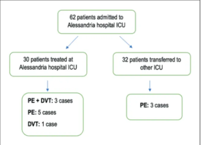
Alessandria hospital ICU admitted 62 patients with diagnosis of ARDS related to COVID-19. In this population, 12 VTE were registered (19.3%), in particular, 11 diagnoses of pulmonary embo-lism (PE) and one of deep vein thrombosis (DVT) were made (Figure 1). Only 25% of patients
com-plicated by thromboembolism were represented by females and patients’ median age was 53 (50-59) years old.
Among 62 patients, 32 patients were trans-ferred to other hospitals and 30 were treated in our center. Nine of reported diagnosis of VTE were made in our ICU; thus, the prevalence of VTE registered in our center is 30%. Thrombotic events took place on average after 64-9 days after
ICU admission and 1411-15 days after symptoms
onset. Demographic data and disease severity are shown in Table I. Principal clinical parameters registered the day when VTE was diagnosed are summarized in Table II and patients’ laboratory test results are described in Table III.
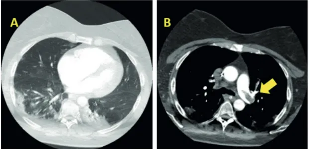
Only two patients (Patient 1 and Patient 3) with PE registered important hemodynamic in-stability which led to the diagnosis. In other cases, the clinical suspicion stemmed from alter-ation of gaseous exchanges and/or hemodynamic values not fully explained by the clinical picture. In the vast majority of patients, D-Dimer values were increased the day of diagnosis. However, only in five cases these values were markedly altered (Table III). In all patients with PE main pulmonary arteries were involved (Table II). In Figure 2, computed tomography scan imaging of ARDS and pulmonary emboli related to patient 1 are shown. DVT was associated to PE in three cases and in one case it is the only VTE finding. All of these patients were treated with the load and subsequent continuous infusion of Heparin. In addition, in patient 3, thromboendoarterecto-my was performed directly in ICU and in patient 1 i.v. thrombolytic therapy was administered. None of the described patients died.
Discussion
A correlation between increased VTE events and ARDS related to COVID-19 was observed. Out of these 62 patients 19.3% had a thrombo-embolic complication. Several factors contribute to the increase of VTE risk in ICU patients. Rec-ognized risk factors for DVT are related to one or more elements of Virchow’s triad: flow stasis, vessel injury and hypercoagulability. Flow sta-sis, due to prolonged immobility, mechanical ventilation, use of sedatives and neuromuscular block plays a major role in ICU patients10-12.
Moreover, in this population, vessel injury may be due to catheter insertion in central veins and hypercoagulability may be induced by sepsis or
Figure 1. Algorithm of thromboembolic complication distribution in intubated patients with moderate or severe ARDS related to Covid-19. All pulmonary embolism di-agnoses were confirmed by CT angiography except one, that was confirmed by transesophageal ultrasound due to patients’ very high hemodynamic instability. Legend: PE, pulmonary embolism; DVT, deep vein thrombosis.
dehydration10,11. Under such pathophysiological
conditions, the occurrence of additional DVT risk factors related to COVID-19 can precipitate thrombosis. The impact of COVID-19 on blood clotting has not yet been completely investi-gated. However, we know that CRS is thought to play an important role in disease severity13.
CRS is associated with increased levels of in-flammatory cytokines and activation of T lym-phocytes, macrophages, and endothelial cells. In particular, interleukin 6 and tumor necrosis factor seems to hold a key role leading to vas-cular leakage and to activation of complement, tissue factor and coagulation cascade14,15. Thus,
the high prevalence of the thrombotic events in patients with ARDS by COVID-19 may be induced by cross-talking between immune and coagulation systems.
PE is stratified into massive, sub-massive, and low-risk based upon the presence or absence of hypotension and right ventricular dysfunction or dilation. This stratification is associated with mortality risk16. However, many patients,
includ-ing those with large PE, have mild symptoms or are asymptomatic17. This nonspecific
presenta-tion may be magnified in patients mechanically ventilated with ARDS COVID-19 related. This is due to an overlap between PE manifestations and COVID-19 ARDS clinical signs. In fact,
alteration of arterial blood gases, D-dimer lev-els, and echocardiography are neither sensitive nor specific as diagnostic of suspected PE in this situation. All our patient at ICU admission had severe or moderate ARDS and increased of D-dimer values. Only in five cases, D-Dimer was markedly increased on the day of diagnosis. All patient on the day of diagnosis were deeply sedated and treated with high PEEP and aminic support before PE suspect. Only patient 1 and patient 3 registered severe hemodynamic insta-bility and deterioration of respiratory function due to PE (Table II). Instead, Patient 2 registered moderate hemodynamic instability with difficult control of blood pressure without impact on gas exchange. Those three cases of PE stemmed a clinical suspicion of VTE in other COVID-19 patients. Thus, even if other patients registered only minor alteration of gaseous exchanges and/ or hemodynamic alterations, PE diagnosis was made. Moreover, considering other VTE risk factors of our patients, Table I shows that only two subjects were obese and only one patient had a history of cancer.
Our experience emphasizes that identifying a PE in patients with COVID-19 ARDS is really challenging. On the other hand, a delay in diag-nosis of PE is associated with a poor outcome18.
As a consequence, these patients require a high
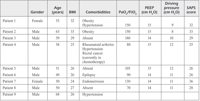
Table I. Demographic and clinical history of patients at ICU admission who subsequently developed thromboembolic events.
Driving
Age PEEP pressure SAPS
Gender (years) BMI Comorbidities PaO2/FiO2 (cm H2O) (cm H2O) score Patient 1 Female 53 32 Obesity
Hypertension 150 15 9 32
Patient 2 Male 63 33 Obesity 150 15 8 33
Patient 3 Male 59 29 Absent 180 14 10 29
Patient 4 Male 58 25 Rheumatoid arthritis 80 15 12 25 Hypertension
Rectal cancer (currently in chemotherapy)
Patient 5 Male 51 26 Absent 105 15 12 26
Patient 6 Male 49 26 Epilepsy 90 14 11 26
Patient 7 Female 50 24 Endometriosis 130 14 11 36
Patient 8 Male 50 27 Absent 70 14 11 28
Patient 9 Male 68 26 Hypertension
Legend: BMI, body mass index; DM, diabetes mellitus; PEEP, positive end-expiratory pressure; Driving Pressure = Plateau pressure – PEEP; SAPS, simplified acute physiology score.
index of clinical suspicion. So, an appropriate treatment with anticoagulation can be initiated in a timely fashion.
One of the hypotheses to explain a large dis-crepancy between low hemodynamic alterations of PE and the radiological picture found in many of our patients may be a significant vasodila-tion of pulmonary circulavasodila-tion. Indeed, Zhao et al19 showed that most patients with COVID-19
pneumonia have a vascular enlargement of the pulmonary lesion that might have been caused by an acute inflammatory response. This would al-low to not dramatically increase the resistance of the pulmonary circulation with consequent lower hemodynamic impact.
As shown in Table II, VTE diagnosis has been confirmed by CT scan with typical alterations for all patients except case 3 suffering sudden cardiac arrest. In this case diagnostic confirma-tion was obtained performing transesophageal cardiac ultrasound during cardiopulmonary re-suscitation. In the majority of patients, PaO2/FIO2 markedly improved 48 hours after diagnosis and treatment of PE.
Our data confirms previous studies that de-scribe thromboembolic events in patients with COVID-19 pneumonia5-7. Similar data can be
found in other viral pulmonary infections like ARDS related to H1N120,21. In particular, Obi et
al20 demonstrate a high incidence of VTE (44%)
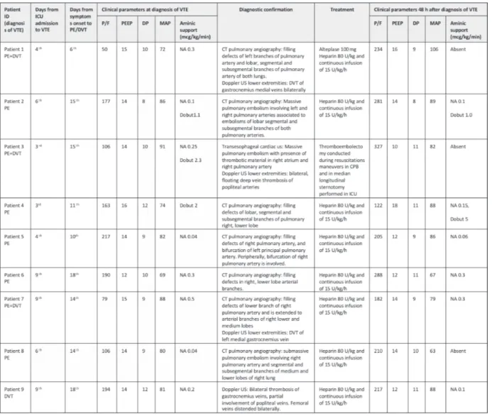
Table II. Clinical parameters of patients the day of diagnosis of thromboembolic event, diagnostic confirmation, treatment and outcome 48h after the diagnosis.
Legend: VTE, venous thromboembolism; PE, pulmonary embolism; DVT, deep vein thrombosis; ICU, intensive care unit; P/F, PaO2/FiO2; DP,
in observational cohort study of 36 critically ill patients with H1N1 ARDS. In this study PE was described in 10 patients and DVT in other 10 patients. Impressively this high rate of VTE was encountered despite antithrombotic prophylaxis.
Cui et al7 recently showed that 25% of patients
with severe COVID-19 pneumonia admitted to ICU developed lower extremity venous thrombo-sis. The DVT group had older age, lower lympho-cytes count, longer APTT and higher D-dimer.
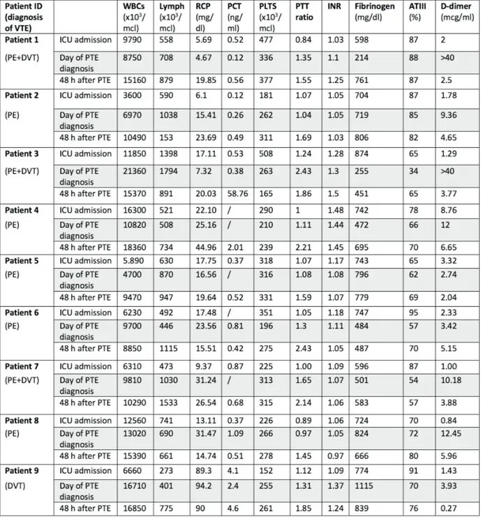
Table III. Lab test results related to the ICU patients’ admission, the day of diagnosis of thromboembolic event and 48 hours after the diagnosis.
Legend: VTE, venous thromboembolism; PE, pulmonary embolism; DVT, deep vein thrombosis; ICU, intensive care unit; WBCs, white blood cells; Lymph, Lymphocytes; RCP, reactive C protein; PCT, procalcitonin; PLTS, platelets; PTT ratio, partial thromboplastin time; INR, international normalized ratio; ATIII, antithrombin III.
Moreover, two recent case reports describe three patients with COVID-19 pneumonia complicated by acute pulmonary embolism confirmed by CT pulmonary angiography showing bilateral filling defects involving lobar, segmental, and subseg-mental branches of the pulmonary artery5,6.
Low-er-limb compression ultrasonography was nega-tive in one patient5 and was not reported in two
other patients6.
The concept of pulmonary thrombosis has been recently proposed as clinical pathological entity distinct from pulmonary embolism22,23.
Ac-cording to this hypothesis coagulative response induced by COVID-19 may promote a local dis-seminated intravascular coagulation and a pulmo-nary intravascular thrombosis. This pathophysio-logical mechanism may explain the presence of thrombotic material in pulmonary circulation in four of our patients, who did not show concomi-tant evidence of DVT. Thus, in our view, patients affected by ARDS related to COVID-19 may de-velop both pulmonary embolism and pulmonary thrombosis22, even though this hypothesis needs
further investigation.
Conclusions
Critically ill patients with ARDS related to COVID-19 may have increased risk of VTE, dis-seminated intravascular coagulation, pulmonary intravascular thrombosis and PE, which may
rep-resent a leading cause of mortality. Thus, these patients require a high index of clinical suspicion and an accurate diagnostic approach, in order to immediately start an appropriate anticoagulant treatment. Further studies are now needed to better understand the relationship between in-creased thrombotic events and ARDS related to COVID-19.
Conflict of Interest
The Authors declare that they have no conflict of interests.
References
1) World HealtH organization. Director-General’s
remarks at the media briefing on 2019-nCoV on 11 February 2020. https://www.who.int/dg/ speeches/detail/who-director-general-s-remarks- at-the-media-briefing-on-2019-ncov-on-11-febru-ary-2020 (Accessed on February 12, 2020) 2) Sarzi-Puttini P, giorgi V, Sirotti S, Marotto d, ardiz
-zone S, rizzardini g, antinori S, galli M. COVID-19,
cytokines and immunosuppression: what can we learn from severe acute respiratory syndrome? Clin Exp Rheumatol 2020; 38: 337-342.
3) Huang C, Wang Y, li X, ren l, zHao J, Hu Y, zHang
l, Fan g, Xu J, gu X, CHeng z, Yu t, Xia J, Wei Y, Wu
W, Xie X, Yin W, li H, liu M, Xiao Y, gao H, guo l,
Xie J, Wang g, Jiang r, gao z, Jin Q, Wang J, Cao
B. Clinical features of patients infected with 2019 novel coronavirus in Wuhan, China. Lancet 2020; 395: 497-506.
Figure 2. Computed tomography scan imaging of ARDS associated to COVID-19 and pulmonary emboli related to patient 1. A, Widespread bilateral consolidation and scattered patchy ground-glass opacities are concerning for ARDS. B, Pulmonary embolus across the bifurcation of pulmonary trunk is noted, as indicated by the arrow.
4) CHen n, zHou M, dong X, Qu J, gong F, Han Y,
Qiu Y, Wang J, liu Y, Wei Y, Xia J, Yu t, zHang X,
zHang l. Epidemiological and clinical
characteris-tics of 99 cases of 2019 novel corona virus pneu-monia in Wuhan, China: a descriptive study. Lan-cet 2020; 395: 507-513.
5) danzi gB, loFFi M, galeazzi g, gHerBeSi e. Acute
pul-monary embolism and COVID-19 pneumonia: a random association? Eur Heart J 2020; 41: 1858. 6) Xie Y, Wang X, Yang P, zHang S. COVID-19
com-plicated by acute pulmonary embolism radiology. Radiol Cardiothorac Imaging 2020; 2: e200067. 7) Cui S, CHen S, li X, liu S, Wang F. Prevalence of
venous thromboembolism in patients with severe novel coronavirus pneumonia. J Thromb Hae-most 2020; 18: 1421-1424.
8) ardS deFinition taSkForCe. Acute respiratory
dis-tress syndrome: the Berlin Definition. JAMA 2012; 307: 2526-2533.
9) alHazzani W Møller MH, araBi YM, loeB M, gong
Mn, Fan e, oCzkoWSki S, leVY MM, derde l, dzierBa
a, du B, aBoodi M, WunSCH H, CeCConi M, koH Y,
CHertoW dS, Maitland k, alSHaMSi F, BelleY-Cote e,
greCo M, laundY M, Morgan JS, keSeCioglu J, MC
-geer a, MerMel l, MaMMen MJ, aleXander Pe, ar -rington a, CentoFanti Je, Citerio g, BaW B, MeM -iSH za, HaMMond n, HaYden Fg, eVanS l, rHodeS
a. Surviving Sepsis Campaign: guidelines on the management of critically ill adults with Corona-virus Disease 2019 (COVID-19). Intensive Care Med 2020; 46: 854-887.
10) kaPlan d, CaSPer C, elliot g, Men S, Pendleton r,
kraiSS l, WeYriCH aS, griSSoM Ck, ziMMerMan ga,
rondina Mt. VTE incidence and risk factors in
pa-tients with severe sepsis and septic shock. Chest 2015; 148: 1224-1230.
11) geertS W, Cook d, SelBY r, etCHellS e. Venous
thromboembolism and its prevention in critical care. J Criti Care 2002; 17: 95-104.
12) CaPrini Ja. Thrombosis risk assessment as a
guide to quality patient care. Dis Mon 2005; 51: 70-78.
13) CHannaPPanaVar r, PerlMan S. Pathogenic human
corona virus infections: causes and consequenc-es of cytokine storm and immunopathology. Se-min Immunopathol 2017; 39: 529-539.
14) SHiMaBukuro-VornHagen a, gödel P, SuBkleWe M,
SteMMler H J, SCHlöSSer Ha, SCHlaak M, koCHanek M,
Böll B, Von BergWelt-Baildon MS. Cytokine release
syndrome. J Immunother Cancer 2018; 6: 56.
15) leVi M. Pathogenesis and diagnosis of
dissemi-nated intravascular coagulation. Int J Lab Hema-tol 2018; 40 Suppl 1: 15-20.
16) JaFF Mr, MCMurtrY MS, arCHer Sl, CuSHMan M,
goldenBerg n, goldHaBer Sz, JenkinS JS, kline Ja,
MiCHaelS ad, tHiStletHWaite P, VedantHaM S, WHite
rJ, zierler Bk; aMeriCan Heart aSSoCiation CounCil on CardioPulMonarY, CritiCal Care, PerioPeratiVeand
reSuSCitation; aMeriCan Heart aSSoCiation CounCilon
PeriPHeral VaSCular diSeaSe; aMeriCan Heart aSSoCia -tion CounCilon arterioSCleroSiS, tHroMBoSiSand VaS -Cular BiologY. Management of massive and
sub-massive pulmonary embolism, iliofemoral deep vein thrombosis, and chronic thromboembolic pulmonary hypertension: a scientific statement from the American Heart Association. Circulation 2011; 123: 1788-1830.
17) luCaSSen W, geerSing gJ, erkenS PM, reitSMa JB,
MoonS kg, Büller H, Van Weert HC. Clinical
deci-sion rules for excluding pulmonary embolism: a meta-analysis. Ann Intern Med 2011; 155: 448-460.
18) goldHaBer Sz, ViSani l, de roSa M. Acute
pul-monary embolism: clinical outcomes in the In-ternational Cooperative Pulmonary Embolism Registry (ICOPER). Lancet 1999; 353: 1386-1389.
19) zHao W, zHong z, Xie X, Yu Q, liu J. Relation
be-tween chest CT findings and clinical conditions of Coronavirus Disease (COVID-19) pneumonia: a multicenter study. AJR Am J Roentgenol 2020; 214(5): 1072-1077.
20) oBi at, tignanelli CJ, JaCoBS Bn, arYa S, Park Pk,
WakeField tW, Henke Pk, naPolitano lM.
Empiri-cal systemic anticoagulation is associated with decreased venous thromboembolism in critical-ly ill influenza A H1N1 acute respiratory distress syndrome patients. J Vasc Surg Venous Lymphat Disord 2019; 7: 317-324.
21) BunCe Pe, HigH SM, nadJaFi M, StanleY k, lileS WC,
CHriStian Md. Pandemic H1N1 influenza infection
and vascular thrombosis. Clin Infect Dis 2011; 52: e14-17.
22) Marongiu F, grandone e, BarCellona d. Pulmonary
thrombosis in 2019-nCoV pneumonia? J Thromb Haemost 2020; 18: 1511-1513.
23) Marongiu F, MaMeli a, grandone e, BarCellona d.
Pulmonary thrombosis: a clinical pathological en-tity distinct from pulmonary embolism? Semin Thromb Hemost 2019; 45: 778-783.