Alma Mater Studiorum – Università di Bologna
Alma Mater Studiorum – Università di Bologna
DOTTORATO DI RICERCA IN
Oncologia e Patologia Sperimentale
Ciclo XXVI
Settore Concorsuale di afferenza:
06/A2
06/A2
Settore Scientifico disciplinare:
MED04
MED04
TITOLO TESI
Genetic and environmental factors associated with
cardiovascular diseases and acute myocardial
infarction.
Presentata da: Dott.ssa Manuela Ianni
Coordinatore Dottorato
Relatore
Prof. Sandro Grilli
Prof. Federico Licastro
INDEX
-Introduction………pag. 4 Epidemiology………pag. 5 Classical risk factors………..pag. 6 New risk factors……… ...pag. 9 Inflammation……… ....pag. 11 Genetic risk factors………...pag. 14 Familiarity of CVD………...pag. 17 Environmental factors………...pag. 20
-Aim of the Study………pag. 24
-Materials and Methods………..pag. 26 Subjects and patients……….……....pag. 26 DNA estraction………...….………pag. 27 SNP detection………..………..pag. 27 Detection of EBV DNA………..……..pag. 28 Detection of HHV-6 DNA………..……..pag. 29 Statistical analysis………..……...pag. 29
-Results………..………pag. 30
Genotype and allele frequency………..……....pag. 30 Association between the triple genotype and cardiovascular risk………...pag. 33 Body mass index (BMI) and blood lipid profile………..…….pag. 34 Prevalence of CVE after 24 years of follow up………....pag. 37 Detection of EBV and HHV-6 DNA………...pag. 38
-Discussion………..pag. 44 Familiarity and AMI……….,..pag. 44 Genetic variations and AMI………,…pag. 45 Virus infections and AMI………pag. 47
-Conclusion……….pag. 50
INTRODUCTION
Cardiovascular Diseases (CVD) are the most frequent cause of morbidity and the leading cause of death in western societies and worldwide; furthermore, it remain today a great social and public sanity problem in Italy, due to the high number of people suffering these pathologies and the need for longterm care and rehabilitation trials (Braunwald E. 1997). CVD includes heart and blood vessel disease and their pathogenesis is related to atherosclerosis. Atherosclerosis is a condition that develops when a substance called plaque builds up in the walls of the arteries. This buildup narrows the arteries, making it harder for blood to flow through. If a blood clot forms, it can stop the blood flow and induce to heart attack or stroke.
The atherosclerosis leads to the development of atherosclerotic disease, the most frequent cardiovascular pathology, and its manifestations, in particular ischemic disease (acute myocardial infarction (AMI) and angina pectoris) and cerebral disease (ischemic ictus and hemorrhagic ictus).
A heart attack or AMI occurs when the blood flow to a part of the heart is blocked by a blood clot. If this clot cuts off the blood flow completely, the part of the heart muscle supplied by that artery begins to die. Usually, this is because one of the coronary arteries that supplies blood to the heart develops a blockage due to an unstable buildup of white blood cells, cholesterol and fat. The event is called "acute" when it is sudden and serious. The two main ways to determine if a person had AMI are electrocardiograms (ECGs) that trace the electrical signals in the heart and testing the blood for substances associated with damage to the heart muscle. Common blood tests are creatine kinase (CK-MB) and troponin. ECG testing is used to differentiate between two types of AMI based on the shape of the tracing. When the ST section of the tracing is higher than the baseline it is called an ST-elevation AMI (STEMI) which usually requires more aggressive treatment. Usually, when an event occurs in young age (35-45 years), the AMI is often fatal. In advanced age, often, the survivors to an AMI become a chronic patient that need continuous care all life long with high cost for society. Furthermore, the CVD are strongly associated to ageing and conduce to physically and cerebrally impairment.
An ischemic stroke happens when a blood vessel that feeds the brain gets blocked, usually, from a blood clot. When the blood supply to a part of the brain is shut off, brain cells will die. A hemorrhagic stroke occurs when a blood vessel within the brain bursts. The most likely cause is uncontrolled hypertension. The result will be the inability to carry out some of the previous functions as before like walking or talking. Some effects of stroke are permanent if too many brain cells die after a stroke due to lack of blood and oxygen to the brain. These cells are never replaced; others are only temporarily out of order.
Other types of CVD are: 1) Heart failure: means the heart is not pumping blood as well as it should, but the body's need for blood and oxygen is unchanged; 2) Arrhythmia: this is an abnormal rhythm of the heart that can beat too slow, too fast or irregularly, so the heart may not be able to pump enough blood to meet the body's needs; 3)Heart valve alterations: when heart valves do not open enough to allow the blood to flow through as it should two main alteration are observed valve stenosis and insufficiency.
Epidemiology
CVD kill an estimated 17 million people worldwide each year and over 4 million deaths in Europe. CVD weigh for 59% on global mortality versus 27% of tumor (World Health Organization. The top 10 causes of death, factsheet No. 310. Retrieved 14 August 2013 http://who.int/mediacentre/factsheets/fs310/en/index.html). CVD costs the Europe economy almost 196bn of euros per year (European Cardiovascular Disease Statistics 2012 Edition, p116. Retrieved 14 August 2013. http://www.escardio.org/about/Documents/EU-cardiovascular-disease-statistics-2012).In Italy, CVD are the most frequent cause of dead and AMI is the main clinical complication of CVD.
In our country, the last data regarding the epidemiology of CVD come from Istat and Istituto
Superiore di Sanità (ISS) in 2008. CVD cause 224.482 death per year (38,8% of total death). Of
these, 33% are cardiovascular event (CVE) (AMI, ischemic heart diseases, angina pectoris and other chronic form of ischemic heart diseases).
Classical risk factors
The knowledge of the etiology and pathogenetic mechanisms of CVD is still limited and incomplete. Well-known traditional risk factors include associated with CVD are:
Age: the risk of developing CVD increases between 40-50 years for men and 50-60 for women; Gender: the women are protected by sexual hormones up to the menopause and thereafter the percentage of CVE between women will increase. Therefore, gender is another important classical risk factor.
A family history of early heart disease: heart disease diagnosed before age 55 years in the father or a brother and before age 65 years in the mother or a sister increases the risk of CVD in offspring and sibling.
Race: African Americans are reported to be particularly at high risk to develop CVD than Asians, perhaps for the high levels of low-density lipoprotein cholesterol (LDL-C). The increased risk may be also associated with the different diets. Age, gender, race and familiarity are not-modifiable risk factors.
High cholesterol level: the Framingham Heart Study demonstrated that specially LDL-C, was the greater the risk of CVD. In 1984, the Lipid Research Clinics-Coronary Primary Prevention Trial revealed that lowering total and LDL (bad cholesterol levels) significantly reduced coronary heart disease (CAD). CAD was uncommon in people with cholesterol levels below 150 mg/dL. More recent series of clinical trials using statin drugs provided conclusive evidence that lowering LDL cholesterol reduces the rate of AMI, the need for percutaneous coronary intervention and the mortality associated with CAD-related causes (LaRosa JC et al., 2005)
Hypertension: in the Framingham Heart Study, even high-normal blood pressure (defined as a systolic blood pressure of 130-139 mm Hg, diastolic blood pressure of 85-89 mm Hg, or both) increased the risk of CVD 2-fold, as compared with healthy individuals (Vasan RS et al., 2001). A study by Allen et al. found that people who have increased or decreased in blood pressure during middle age had higher and lower remaining lifetime risk for CVD. This suggests that prevention
efforts should continue to emphasize the importance of lowering blood pressure in order to avoid hypertension (Allen N et al., 2012). Hypertension, along with other factors such as obesity contribute to the development of left ventricular hypertrophy (LVH). LVH founded to be an independent risk factor to CVD morbidity and mortality. It roughly doubles the risk of cardiovascular death in both men and women (Levy D et al., 1990).
Obesity: the Joint National Committee on Prevention, Detection, Evaluation, and Treatment of High Blood Pressure (JNC VII) emphasizes weight control, adoption of the Dietary Approaches to Stop Hypertension (DASH) diet, with sodium restriction and increased intake of potassium and calcium-rich food, moderation of alcohol consumption to less than 2 drinks daily and increased physical activity (Chobanian AV et al., 2003). Obesity was associated with elevated vascular risk in population studies. In addition, this condition was associated with glucose intolerance, insulin resistance, hypertension, physical inactivity, and dyslipidemia (Rexrode KM et al., 1998). A study by Das et al examined more than 50,000 patients from the National Cardiovascular Data Registry with STEMI. The results suggested that although patients who were extremely obese (body mass index [BMI] >40) present at a younger age with STEMI, they have less extensive CAD and better LV function. However, as expected, their in-hospital mortality following STEMI was increased (adjusted odds ratio, 1.64) (Das SR et al., 2011).
Diabetes Mellitus: a disorder of metabolism associated with either insulin deficiency or insulin resistance. Glucose builds up in the blood stream, overflows through the kidneys into the urine, and results in the body losing its main source of energy, even though the blood contains large amounts of glucose. Patients with diabetes are 2-8 times more likely to experience future CVE than age-matched and ethnically matched individuals without diabetes (Howard BV et al., 2002). A recent study suggested a potential reduction of all-cause and CVD mortality in women with diabetes mellitus who consumed whole-grain and bran (He M et al., 2010). Another study suggested that meat consumption was associated with a higher incidence of coronary heart disease and diabetes mellitus (Micha R et al., 2010). A meta-analysis performed by Nordmann et al founded that the Mediterranean diet had more favorable changes in weighted mean differences
of body weight, body mass index, systolic blood pressure, diastolic blood pressure, fasting plasma glucose, total cholesterol, and high-sensitivity C-reactive protein than low-fat diets (Nordmann AJ et al., 2011).
Sigarettes smoking: Cessation of cigarette smoking constitutes the single most important preventive measure for CAD. As early as the 1950s, studies reported a strong association between cigarette smoke exposure and heart disease. Persons who consume more than 20 cigarettes daily have a 2- to 3-fold increase in total heart disease. Continued smoking is a major risk factor for recurrent heart attacks (Rea TD et al., 2002). Smoking is a risk factor for CVD in women and men; however, a systematic review and meta-analysis by Huxley and Woodward suggested that in some countries, smoking by women is on the rise; the study suggested that proper counseling and nicotine addiction programs should focus on young women (Huxley RR et al., 2011).
Physically exercise: The cardioprotective benefits of exercise include reducing adipose tissue, which decreases obesity; lowering blood pressure, lipids, and vascular inflammation; improving endothelial dysfunction, improving insulin sensitivity, and improving endogenous fibrinolysis (Thompson PD et al., 2003). In addition, regular exercise reduces myocardial oxygen demand and increases exercise capacity, translating into reduced coronary risk. Studies shown that even 15 minutes a day or 90 minutes a week of moderate-intensity exercise may be beneficial (Greenland P et al., 2010).
Modifiable risk factors are cigarettes smoke, diabetes, hypertension, hypercholesterolemia, high Body Mass Index (BMI), obesity, sedentary style changing the diet and follow a good life-style habit. Many sanitary preventative interventions of ISS to promote the good life life-style, to stop smoking, improve physically activities, and eat better (few salt, fat, or sweet) have decreased the incidence of CVD, in fact, in the last 40 years the mortality for CVD it’s the half.
It was observed that more than half of patients with atherosclerotic complications, such as AMI, does not demonstrate classical risk factors.
New risk factors
Others factors are now considered as new risk factors (Ridker PM, 1999, Rifai et al, 2001). C-reactive protein (CRP): is a protein in the blood that demonstrates the presence of inflammation, which is the body's response to injury or infection; CRP levels rise if inflammation is present. The inflammation process appears to contribute to the growth of arterial plaque, and in fact, inflammation characterizes all phases of atherothrombosis and is actively involved in plaque formation and rupture. According to some research results, high blood levels of CRP may be associated with an increased risk of developing CAD, CVE and having a heart attack (Arroyo-Espliguero R et al., 2004; Rifai N et al., 2001). In the Jupiter trial, in healthy persons without hyperlipidemia but with elevated high-sensitivity CRP levels, the statin drug rosuvastatin significantly reduced the incidence of major CVE (Ridker PM et al., 2008). The 2010 ACCF/AHA guideline for assessment of cardiovascular risk in asymptomatic adults states that measurement of CRP can be useful in selecting patients for statin therapy and may be reasonable for cardiovascular risk assessment, depending on the patient’s age and risk level. CRP measurement is not recommended for cardiovascular risk assessment in asymptomatic high-risk adults, low-risk men 50 years or younger, or low-risk women 60 years or younger (Wang TJ et al., 2006).
An elevated lipoprotein(a) [Lp(a)] level is an independent risk factor of premature CAD (Hjemdahl P, 2002; Braunwald E, 1997) and is a significant risk factor for premature atherothrombosis and CVE. Measurement of Lp(a) is more useful for young individuals with a personal or family history of premature vascular disease and repeat coronary interventions. The 2010 ACCF/AHA guideline for assessment of cardiovascular risk in asymptomatic adults states that, in asymptomatic intermediate-risk adults, lipoprotein-associated phospholipase A2 might be reasonable for cardiovascular risk assessment (TJ et al., 2006). Lp(a) may be used to identify people at increased cardiovascular risk, but as of yet, there have been no studies on Lp(a) lowering because of the lack of available agents that are effective in reducing this value. Therefore, LDL lowering is probably the best strategy in people with elevated Lp(a) levels.
(Arrojo et al., 2004).
Homocysteine: a natural by-product of the dietary breakdown of protein methionine. In the general population, mild to moderate elevations are due to insufficient dietary intake of folic acid. Homocysteine levels may identify people at increased risk of heart disease, but again, due to the lack of agents that effectively alter the homocysteine levels, studies have not shown any benefit from lowering the homocysteine level (Miller, A et al., 1997). Levels of fibrinogen: an acute-phase reactant, increase during an inflammatory response. This soluble protein is involved in platelet aggregation and blood viscosity, and it mediates the final step in clot formation. Significant associations were found between fibrinogen level and risk of CVE in the Gothenburg, Northwick Park, and Framingham heart studies (Wilhelmsen L et al., 1984; Levenson, J., et al., 1995).
Low serum testosterone levels have a significant negative impact on patients with CAD. More studies are needed to assess better treatment (Malkin CJ et al., 2010). One meta-analysis suggested that the presence of erectile dysfunction increases the risk of CVD, coronary heart disease, stroke, and all-cause mortality. This additional risk may be independent of conventional cardiovascular risk factors (Dong JYet al., 2011).
One study suggested that women aged 50 years or younger who undergo a hysterectomy were at an increased risk for CVD later in life (Ingelsson E et al., 2011). Oopherectomy also increases the risk for both coronary heart disease and stroke.
A prospective cohort study (n=2312) by Kestenbaum et al evaluated older patients without CAD over 14 years. Vitamin D and parathyroid hormone were measured and the outcomes included AMI, heart failure, cardiovascular death, and all-cause mortality. Vitamin D deficiency was associated with increased mortality and AMI (each 10 ng/mL drop in vitamin D was associated with 9% greater increase in death and 25% increase in AMI). Parathyroid hormone excess was associated with a 30% increased risk of heart failure. Further randomized controlled trials are required (Kestenbaum B et al., 2011).
Inflammation
Inflammation appears to be important in the pathogenesis of atherosclerotic disease, since atherosclerotic plaques and lesions are associated with infiltration of activated immune cells and increased expression and synthesis of inflammatory markers (Hansson GK. 2001).
Cytokines are produced in a variety of tissues and regulate the expression of a number of inflammatory molecules, leading to destabilization and finally rupture of vulnerable atheromatic plaques. They also participate in the pathophysiology of acute coronary syndromes by direct effects on myocardial contractility and apoptosis. Several lines of evidence indicate that increased inflammatory cytokine levels and inflammatory cytokine activity detectable in peripheral blood have a prognostic role since they are useful markers predicting future CVE in patients with advanced atherosclerosis and in patients after coronary syndromes and may be useful in identifying subjects with a worse clinical course (Tousoulis D et al., 2006; De Gennaro L et al., 2012).
However, the existing data are limited and controversial. Potentially thousands of molecules are relevant to this apparently complex disease. Research paid much attention to sensitive specific serum biomarkers for vulnerable plaques as diagnostic tools for the identification of patients with acute coronary syndrome, but also to help us to identify high-risk patients. Furthermore, AMI is a myocardial necrosis occurring due to persistent coronary ischemia, in which inflammation plays an important role and heart failure is a common complication.
Elevated plasma levels of interleukine (IL-) 6 associated with increased risk of future AMI in a prospective study that involving 14916 apparently healthy men were found. These data thus support a role for cytokine-mediated inflammation in the early stages of atherogenesis (Ridker PM et al., 2000). IL-6 plasma levels was also associated with increased atherosclerosis when the control group was compared with the group free of subclinical CVD (Jenny, N.S et al., 2002). IL-6 levels differentiated those with subclinical CVD from those without. Although the -174C allele was not associated with incident events, associations of the genotype with inflammation and AMI, combined with the plasma IL-6 results, suggest that IL-6 may chronically predispose an
individual to develop atherosclerosis.
Chiappelli et al. suggested that the C allele of the promoter polymorphism in the IL-6 gene is a risk factor for AMI in the elderly and the production of the IL-6 differentially affected different genotypes of the IL-6 −174 promoter polymorphism (Chiappelli M et al., 2005).
Elevated admission level of IL-6, but not of soluble CD40 ligand (sCD40L), metalloproteinase- 9 (MMP-9), or tissue inhibitor of metalloproteinase-1 (TIMP-1), might indicate the onset of STEMI, and could provide prognostic value for future cardiac mortality within 2 y in patients with STEMI (Tan J et al., 2008).
A significant increase in IL-6 serum level as well as a significant decrease in IL-1 receptor alpha (Ra) for patients with a history of AMI was found. A trend toward higher concentration of inflammatory mediators was noticed in relation to the increase in severity of the aortic valve disease. An "inflammatory pattern" was associated with aortic sclerosis pathology and these results suggested the persistence of a chronic inflammation in patients who experienced acute coronary events (Rugina M et al., 2007).
The frequencies of genetic apo E (APOE) isoforms E2, E3 and E4 were determined in 523 patients with AMI. Significant difference in the frequency of APOE4 but not in the frequencies of isoforms E3 and E2 between patients and controls were observed (Utermann G et al., 1984). Several studies confirmed these results (Bennet AM et al., 2007).
Admission levels of serum MMP-9 and TIMP-1 closely correlated with left ventricular structure and function, which may be involved in the process of post-infarction remodeling of myocardium in AMI (Tan J et al., 2012).
Tumor Necrosis Factor alpha (TNF-α) may be an early marker of myocardial damage because of the early increase of its level after ischemic injury instead of being late consequence of extensive tissue necrosis. A significant increase was still seen at 48 hours post admission in patients with signs of heart failure but not in those without signs of heart failure. A significant positive correlation was found between plasma TNF-alpha level and calcium-dependent protein kinases level at admission. TNF-α level may be also an important indicator of the severity of AMI and the
occurrence of heart failure (Fahim MR et al., 2004). Plasma concentrations of TNF-α are persistently elevated among post-AMI patients at increased risk for recurrent coronary events. These data support the hypothesis that a persistent inflammatory instability is present among stable patients at increased vascular risk (Ridker PM et al., 2000a).
Monocyte chemotactic protein-1 (MCP-1) appears to play a critical role at multiple stages in atherosclerosis, including the initiation of the fatty streak, promotion of plaque instability, and remodeling after myocardial infarction. In a large cohort of patients with acute coronary syndromes, an elevated baseline level of MCP-1 was associated both with traditional risk factors for atherosclerosis as well as an increased risk for death or AMI. For this reason, it is appears to play a crucial role at multiple stages of atherosclerosis (De Lemos JA et al., 2003 ).
Between the pro- and anti-inflammatory components of the immune system there is a dynamic balance, but it is know that violation of which is an important mechanism of development of many pathological states, in this case, cardiac insufficiency. In the acute phase of AMI, inflammatory activation is enhanced with predominant proinflammatory response. In the course of the healing process within 6 months inflammation is suppressed and the balance between pro- and anti-inflammatory activation is restored. The size of AMI, BMI, lipid levels and the baseline levels of inflammatory markers influence the levels of inflammatory factors (Karpiński Ł et al., 2009).
Balance between the pro- and antiinflammatory cytokines reflects the index of inflammatory activity. A study demostrated that uncomplicated AMI patients, there is initially a high level of both proinflammatory and antiinflammatory cytokines. After 10 days, were observed a decline in the IL-6 level and an increase in the TNF-α and IL-10 levels. In complicated AMI, as compared to uncomplicated AMI patients were observed initially high levels of TNF-α and IL-6 and reduced levels of IL-8 and 1L-10, patients. In dynamics of supervision of this group, further increase of all proinflammatory cytokines and a decline of IL-10 was registered (Havrylenko TI et al., 2012).
coronary syndrome and systolic dysfunction or acute heart failure (De Gennaro L et al., 2012). Some findings suggested that IL-8 and IL-12 was involved in the process of ischemic heart disease, and serum IL-12 may be a marker for differentiating AMI from unstable angina pectoris (Zhou RH et al., 2001). A number of studies associated blood levels of individual proinflammatory plasma biomarkers and gene variants that favor inflammation or cholesterol synthesis with AMI, but until now, none of these factors alone are sufficient to define risk for individuals. Furthermore, phenotype markers, blood levels of a given marker, may considerably vary with gender, age, concomitant diseases, metabolic disorders and diet, be sensitive to other environmental variables and significantly change with time in the same subject.
Genetic risk factors
Plasma levels of lipids and proinflammatory molecules fluctuate over time and in relation to age, sex, diet, lifestyle, medication, and environmental factors, limiting their usefulness in etiologic and predictive contexts. Inherited gene variants are, in contrast, constant and might provide a better indication of high intrinsic risk. Genetic markers may be used to overcome these limitations. For instance, gene polymorphisms (SNPs), known to influence the phenotypic expression of a given inflammatory molecule, may become informative in assessing individual risk of CVE and recent studies have focused on the genetic background of inflammatory cytokines as possible intrinsic risk factors associated with CVD. These investigations assessed gene variations largely distribute in human population and showed that some alleles of cytokine genes were more frequently distributed in patients with CVD than in controls (Humphries SE et al., 2001; Andreotti F et al., 2002).
Cytokines as regulators of activity of inflammation play significant role in mechanisms of formation of atherosclerotic plaques and in processes of their destabilization. One of leading genetic factors determining level of their production appears to be polymorphism of cytokine genes structure at their promoter loci (Konenkov VI et al., 2012).
of new SNPs associated with CHD and AMI (Patel RS et al., 2011), but their clinical relevance is still unclear because a single gene variant can make a limited contribution to the total genetic load of AMI, and both common and rare gene polymorphisms may differentially affect susceptibility to the disease.
Data from a cross-sectional study suggested that the functional interaction of three genes the allele C of IL-1b, the allele C of IL-6 and e4 allele of APOE was strongly associated with AMI, affects pathogenetic mechanisms and an impaired regulation of immune responses plays a pivotal role in the disease (Licastro F et al., 2004). In the setting of non-ST-elevation acute coronary syndromes, genetic variation at the IL-1 gene locus contributes to the changes in soluble markers of endothelial inflammation (Ray KK et al., 2002).
A number of studies explored the association of ischaemic heart disease with SNPs of the inflammatory molecules TNF-α and β, transforming growth factors (TGF) beta1 and 2, IL-1 and its receptor antagonist (IL-1ra), the receptor for lipopolysaccharide (CD14), P and E selectins, and platelet endothelial cell adhesion molecule (PECAM) 1. The data provided some evidence that alterations in the genetics of the inflammatory system may modify the risk of ischaemic heart disease (Andreotti F et al., 2002).
Several gene variants that promote inflammation and cholesterol metabolism were associated with AMI. Promoter SNPs with functional relevance in the expression of the cognate inflammatory gene are often found at elevated frequency among patients with AMI. AMI across a broad age range’ carried multiple proinflammatory alleles (IL6, TNF, IL10, Alpha 1-antichymotrypsin (ACT)). ‘A subset of AMI in middle age’ had numerous proinflammatory alleles (IL6, TNF, ACT, interferon gamma IFN-γ, 3-hydroxy-3-methyl-glutaryl-CoA reductase (HMGCR)). ‘AMI after age 80’ had a reduced risk set (IL6, IL10, IFN-γ) (Licastro F et al., 2011; Licastro F et al., 2007).
IL-10 reflects a proinflammatory state in patients with acute coronary syndrome and was suggested that IL-10 is as effective a biomarker for the risk prediction of future CVE as other markers of systemic inflammation (Mälarstig A et al., 2008). Also IL-8 -251 A/T polymorphism is
associated with acute coronary syndrome risk in the Chinese Han population and the A allele of IL-8 -251 A/T may be an independent predictive factor for acute coronary syndrome (Zhang X et al., 2011). IL-18 gene is a susceptibility locus for AMI, a finding of potential interest in the clinical practice (Koch W et al., 2011a).
Six different and functionally relevant SNPs of the genes coding for IL-10, TNF-α, and TNF-β are neither separately nor in cooperation associated with the risk of CAD or AMI in angiographically examined study patients (Koch W et al., 2001b). Also a relationship among genetically determined TNF-α and pro-thrombin (FII) production and increased levels of tissue damage markers of AMI, suggest that a complex genetic background, might be involved in susceptibility to AMI in young men influencing the extension and severity of the disease (Vaccarino L et al., 2013).
Although many risk factors are intercorrelated, raising the possibility of a higher level of genetic control by a small number of master genes that control fundamental physiological systems. Such genes are likely to be relevant to the combined processes of plaque instability and coronary thrombosis.
The complex pathogenesis of AMI implicates phenotypic and genetic heterogeneity. A new mathematical algorithm named Auto Contractive Map (AutoCM), was applied in AMI patients to detect and evaluate the relationships among genetic factors, clinical variables and classical risk factors. Genes were selected because their strong regulatory effect on inflammation and SNP in these gene were located in the promoter region. In the connectivity map generated by AutoCM a group of variables was directly linked with the AMI status: gender (male), early age at onset (50-65 years), HMGCR gene (CC wild type genotype), IL-1β, ACT, IL-6 GG and vascular endothelial growth factor (VEGF) CC genotypes. This direct link suggested a possible pathogenetic association with AMI. Other genetic, clinical and phenotypic variables were associated to the disease under a statistically defined hierarchy showed in the new connectivity map generated by AutoCM. These analyses suggested also that genotypes of few inflammatory genes, a SNP in HMGCR gene, middle age, gender, low HDL and diabetes were very informative variables to
predict the risk of AMI (Licastro F et al., 2010).
Familiarity of CVD
Parental history is a widely accepted risk factor for offspring CVE, although the mechanisms remain unclear. Many studies examined the contribution of conventional and genetic risk factors in explaining the excess risk of CVE in offspring with positive parental history. Whether parental CVD confers increased risk independent of other risk factors remains controversial. Prior studies relied on offspring report, without complete validation of parental events.
Despite some study underline an association of positive parental history with blood pressure, total and high-density lipoprotein cholesterol, CRP, and physical activity, less than 15% of the risk was explained through conventional and novel risk factors. The greatest risk of CVD was observed in positive parental history participants with elevated CRP or hypertension and substantially increases the risk of CVD (Hamer M et al., 2009).
Genotypes of inflammatory molecules seem to play an opposite role in atherosclerosis and longevity: pro-inflammatory genotype seems to contribute significantly to the risk of coronary heart disease, but some study supported the hypothesis that the genetic background favoring CVD. So several study in centenarian verify the role of proinflammatory alleles, such as pyrin and C-C chemokine receptor type 5 (CCR5), in AMI and longevity. The results suggested the genetic background favoring CVD and that the centenarian genetic background may be useful for investigating genetic key components of age-associated diseases as atherosclerosis (Candore G, et al., 2006).
A family history of CVD and AMI is frequently encountered in clinical practice, and a premature heart attack in parents is associated with a high risk of the disease in their offspring. A small subgroup of children, whose parents suffered a heart attack in their late thirties and early forties, may be at particularly high cardiovascular risk was evaluated about weight, blood pressure, plasma cholesterol levels, obesity and smoking and it is concluded that cardiovascular risk factor screening among children with a positive family history of premature atherosclerotic
complications is appropriate and cost-effective (Rumboldt M et al., 2003).
To assess the risk of CVE, the diagnostic effectiveness of molecular-genetic markers (SNPs in APOE, ACE (I/D) and MTHFR (C677T) genes) in combination with conventional risk factors was studied. The combination of results of molecular-genetic testing with conventional risk factors allows to increase the predictability of cardiovascular risks (Nazarenko GI et al., 2009). Some study examined the association of parental CVD with 8-year risk of offspring CVD, using pooled logistic regression. Parental CVD independently can predict future offspring events in middle-aged adults. Addition of parental information may help clinicians and patients with primary prevention of CVD, when treatment decisions may be difficult in patients at intermediate risk based on levels of single or multiple risk factors (Lloyd-Jones DM et al., 2004).
The relevance of familial factors to CVD is further supported by the finding that siblings with CVD are at increased risk of future CVE in middle-aged adults regardless of the presence of established risk factors (Murabito JM et al., 2005).
The associations of parental longevity with carotid intima-media thickness, carotid plaques, and aortic arterial stiffness in adult offspring was also examined and the results indicate that there are modifications of structure and function of large arteries according to paternal longevity (Zureik M et al., 2006).
Data from the longitudinal Framingham Heart Study confirmed that subjects with long-lived parents have a better cardiovascular risk profile in middle age than those whose parents died younger and centenarians have better cardiovascular risk profiles compared to younger old people (Terry DF et al., 2007).
Some reports revealed that CVD (i.e. hypertension, diabetes, angina and/or myocardial infarction) are less common in centenarians respect to 70 and 80 years old persons. A combination of genetic factors and lifestyle aspects appears to contribute to the longevity of centenarians. A role on this better cardiovascular risk profile may be played by the increasing use of pharmacologic treatments in the elderly population (especially for hypertension and dyslipidemia), but the contribution of drug treatments to promote extreme longevity is not confirmed. In fact,
centenarians in general needed fewer drugs at younger ages due to a healthy lifestyle. The importance of the genetic contribution was demonstrated by the inheritance of low-risk cardiovascular profiles in centenarian offspring and lower prevalence of cardiovascular CVD in this population as compared with their spouses or with age-matched subjects without centenarian parents (Galioto A et al., 2008).
Furthermore, the Offs of centenarians have a better cardiovascular risk profile and retain some important cardiovascular advantages over time than those of parents not enjoying a long life (Adams ER et al., 2008). It is interesting to note that structural vascular changes can be observed in Offs with a parental history of AMI at a young age, regardless of the presence of a number of the classical cardiovascular risk factors. Increased carotid intima-media thickness is an early manifestation of atherosclerosis. A parental history of premature AMI was associated with an increase in carotid carotid intima-media thickness in children-adolescents and young adults. Offspring of coronary patients showed an unfavourable lipid profile compared to controls; however, the association between a premature AMI and carotid intima-media thickness was independent of lipids, apolipoproteins and other traditional risk factors. So, vascular structural changes are detectable in Offs of parents with premature AMI at a young age and occur independently of several traditional cardiovascular risk factors (Barra S et al., 2009).
Once again, the Framingham Heart Study shown that parental stroke before the age of 65 years is associated with a 3-fold increase in the risk of stroke in their Offs, and that parental history can be used as clinical risk marker of an individual’s propensity to stroke (Seshadri S et al., 2010). The distribution of some SNPs influencing a inflammation and found an higher prevalence of pro-inflammatory SNPs (SNP A2080G of pyrin gene, SNP Gly670Arg of PECAM gene, C1019T of Cx 37 gene, SNP G1059C of PCR gene) and a lower prevalence of anti-inflammatory SNPs (Asp299Gly of TLR4 gene, SNP -1082 G/A of IL10 gene, CCR5Δ32) in young patients with AMI was found. Results of these studies shown that early AMI could be associated with a genetic predisposition to an intense inflammatory response, also to an hyperviscosity syndrome (Incalcaterra E et al., 2010).
All these findings suggest that genetic studies of offspring may be useful to identifying transmissible genetic traits in CVD.
Environmental factors
Studies about associations of infections with viruses and other pathogens, with CVD, frailty and/or mortality are conflicting. Since high levels of antibodies against these pathogens occur in the elderly, the role of pathogens in morbidity and mortality of vulnerable elderly was explored. Increasing evidence supports a link between serological evidence of prior exposure to infectious pathogens, pathogen burden, and the risk for future AMI and death in patients with coronary artery disease. The role of viral infections in the pathogenesis of atherosclerosis remains controversial largely due to inconsistent detection of the virus in atherosclerotic lesions, but the identification of specific viral signatures can help to understand pathogenetic mechanisms in AMI.
In the past, evidence for enterovirus (EV), adenovirus, and cytomegalovirus (CMV) was in the focus of attention; in the meantime, "new" cardiotropic pathogens such as parvovirus B19, Epstein-Barr virus (EBV), and human herpesvirus (HHV-) 6 in patients with dilated cardiomyopathy with and without inflammation have been detected. Their persistence in the myocardium correlates with a decline in pumping capability within 6 months. While the virus is still being eliminated, the second phase of the disease begins, which is characterized by autoimmune phenomena and often a cardiac inflammatory response which likewise correlates with a worsening prognosis. The transition to the third and final phase with development of dilated cardiomyopathy occurs gradually and can take years. The goal of every diagnostic and therapeutic intervention must be to eradicate the virus and eliminate the inflammatory response to prevent the disease from progressing to terminal cardiac insufficiency (Pankuweit S et al., 2010). Some study evaluated the intimal presence of four pathogens in human coronary atheroma, clinically associated with acute coronary syndromes and stable angina and the effect of pathogen burden on the expression of human heatshock protein 60 (hHSP60), a key protein in
(auto-)immune pathogenesis of atherosclerosis. The data demonstrate that there exist an impact of intimal pathogen burden in plaque instability, and suggest the presence of (auto-)immunoreactions against upregulated hHSP60 is an important pathomechanism that may contribute to acute coronary syndromes (Andrié R et al., 2003).
A study assess the detection of human HHV-6 and HHV-8, two DNA viruses with a distinct tropism for endothelium and lymphocytes, with the aim to know if these virus may be associated with coronary instability and to found a correlation between infection-driven inflammatory burden and acute manifestation of coronary artery disease.
HHV-8 viremia was undetectable in all three groups of study: patients with AMI, patients with stable coronary artery disease and patients without coronary and carotid artery atherosclerosis subjected to cardiac valve replacement. HHV-6 viremia was detected in a substantial fraction of the samples examined without differences between groups (Magnoni M et al., 2012).
To evaluate the association of ischemic heart disease with the number of pathogens among individuals with infection serum samples of patients with ischemic heart disease as the AMI or unstable angina and healthy subjects for the presence of antibodies to Helicobacter pylori (H. pylori), CMV, HSV-1 and HSV-2 were tested. The rate of subjects with high infection burden (3 seropositivities) was significantly higher in ischemic heart disease group than to control group. The seroprevalence of anti-H. pylori, anti-CMV and anti-HSV-1 antibodies in AMI and unstable angina groups was significantly higher than control group. The infection burden was significantly increased in patients with ischemic heart disease and this parameter should be considered as an independent risk factor for development of ischemic heart disease (Jafarzadeh A et al., 2011). Some study described also a dual role of infections as risk factors for coronary heart disease and showed that EV, HSV and Chlamydia pneumoniae (C. pneumoniae) HSP60 IgG antibody titers were associated with increased risk of coronary heart disease and protection from infections, usually suffered during the childhood before the era of MMR vaccination, may predispose the individual to coronary heart disease (Pesonen E et al., 2007).
atherosclerotic vascular diseases were assessed to elucidate whether infectious agents such as herpes viruses could be implicated in the inflammatory atherosclerotic process and to examine the relation between the levels of these antibodies lipid profile and high-sensitive C-reactive protein (hsCRP) in these patients. The level of CMV- and EBV- specific antibodies were elevated among vascular disease patients and the presence of CMV-specific IgG was associated with the development of the disease. Serum lipids and hsCRP were increased among the patients; however, no significant correlation was detected between antiviral IgG levels and lipid profile or hsCRP (Al-Ghamdi A, 2012).
The C. pneumoniae, HSV-1, CMV and EBV DNA in atherosclerotic plaques from carotid arteries obtained from 17 patients were investigated. Genomic sequences of C. pneumoniae, HSV-1, CMV were not found in any atherosclerotic lesion. Therefore, these results did not support the hypothesis of an association between these infectious agents and atherosclerosis. Conversely, three patients were found to be positive for EBV DNA, thus indicating that, at least in a limited number of patients, EBV could play a role in atherogenesis (Tremolada S et al., 2011).
The intimal presence of 4 pathogens in atheroma, clinically associated with acute coronary syndromes and stable angina and the effect on the expression of intimal CRP, tissue factor and hHSP60 to demonstrate the impact of pathogen burden in plaque instability and suggest local proinflammatory, prothrombotic, and proimmunogenic effects was investigated. Analysis revealed C. pneumoniae was present in 73%, H. pylori in 31%, CMV in 16%, and EBV in 40%. Also the expressions of CRP, tissue factor, and hHSP60 were significantly higher in acute coronary syndromes lesions and the number of infectious pathogens correlated significant with the expressions of CRP, tissue factor and hHSP60 (Andrié RP et al., 2010).
In a cross-sectional study, the presence of C Pneumoniae, Mycoplasma pneumoniae (M. pneumoniae), CMV and EBV in atherosclerotic and non-atherosclerotic vascular samples was investigated. The results suggest that C. pneumoniae, M. pneumoniae and CMV were present with similar frequency both in atherosclerotic and non-atherosclerotic vessels (Bayram A et al., 2011).
The presence of HHV-6, HHV-7 and HHV-8 DNA in carotid, iliac and coronary artery specimens obtained from a group of subjects with atherosclerosis and a control group were investigated. HHV-6 but not HHV-7 and HHV-8 DNA were found in atherosclerotic vascular tissues. However, further research on broader study groups and with different protocols is needed to determine whether these viruses play a role in the formation of atherosclerosis (Kaklikkaya I et al., 2010). Explanted heart of 28 patients, who underwent heart transplantation were screened for EV, Adenovirus Type 3 (AdV3) and HHV6 sequences in order to assess the incidence of cardiac viral infection that may be implicated in the pathogenesis of cardiomyopathy, and estimate viral distribution in the myocardium. AdV3 and HHV6 sequences coexisted in one case with inflammatory cardiomyopathy (Tátrai E et al., 2011).
The presence of CMV immediate early genomic region of DNA in 40 arterial specimens from coronary plaques and 27 samples from normal vessels were evaluated. CMV DNA was detected in 9 out of 26 patients (34.6%) in both non-atherosclerotic tissues and atherosclerotic plaques. No statistically significant differences were observed between normal and diseased vessels. These findings do not support a direct causative role of CMV in the development of atherosclerotic plaques (Xenaki E et al., 2009).
Some publications shed light to the role of AdV3, HHV- 6 and EV viruses in the development of dilated cardiomyopathy. AdV3, HHV- 6 and EV genomes in DNA and RNA were isolated from five regions of the heart muscle. In 2 patients AdV3 and in 1 patient both AdV3 and HHV-6 were detected. The AdV3 genome was the most frequent virus genome in explanted heart tissues. The identified viral sequences suggested previous viral infection, which could have played a role in the development of dilated cardiomyopathy (Tátrai E et al., 2007).
AIM OF STUDY
CVD are rising as a cause of death and disability worldwide and also a great social and medical problem and AMI is the main clinical complication. The knowledge about the aetiology and pathogenetic mechanisms of coronary vessels disease is limited and incomplete. AMI is a multifactorial disease with a complex pathogenesis in which lifestyles, individual genetic backgrounds and environmental risk factors contribute to the pathogenetic mechanisms and clinical manifestations.
More than half of patients with atherosclerosis do not show classical risk factors. New risk factors for CVD are emerging as the results of a growing understanding of the process of atherogenesis. Inflammation appears to be important in the pathogenesis of atherosclerotic disease, since atherosclerotic plaques and lesions are associated with infiltration of activated immune cells and increased expression and synthesis of inflammatory markers. Elevated blood levels of cytokines and interleukins in patients with CVD have been also found. However, phenotype markers, may considerably vary in several physiological condition or be sensitive to other environmental variables and significantly change with time in the same subject. Genetic markers may be used to overcome these limitations. For instance, gene polymorphism, known to influence the phenotypic expression of a given inflammatory molecule, may become informative in assessing individual risk of CVE.
Furthermore, it is known that a family history and a still largely undefined genetic background greatly influence the early clinical manifestation of AMI and CVD.
We investigated genetic variations represented by SNPs in the promoter region of a number of genes regulating metabolic and immune functions that have previously been found to be associated with an increased risk of AMI in case/control studies as possible genetic markers of an increased risk of CVD in children of parents with a positive history of AMI (Licastro F et al., 2010).
familiarity for AMI and a control group of patients without history of CVD to improve our understanding about genetic background involved in the development of atherosclerosis and its complications.
A second control group of comparable age from the WHO-MONICA Brianza study to compare CVE prevalence during the 24 years follow-up of our Offs was also used.
In second instance we investigated the same SNPs in genes in another group of AMI to confirm the obtained results.
Finally, DNA samples from peripheral blood leukocytes (PBL) of all patients were analyzed for the presence of EBV and HHV-6 to assess the involving of a viral etiology as risk factors of CVD and AMI.
MATERIALS AND METHODS
Subjects and patients
The study involved 154 Offs from Northern Italy, each of whom had one parent who had experienced an AMI before the age of 65 years. An evaluation was made of the classic AMI risk factors (metabolic parameters, smoking, diabetes, obesity, a sedentary lifestyle) together with ECG records, anthropometric indices, arterial pressure and medical history. The Offs were re-screened after a follow-up of 24 years in order to assess any changes in behavioral and biological risk factors and collect blood samples for laboratory and genetic evaluation. The CVD risk factors and drug treatments recorded at baseline and 24 years later were compared with those of the age-matched population of the WHO-MONICA Project, which were collected in Brianza, Northern Italy, during independent surveys carried out in 1984, 1991 and 2004 (Ferrario M et al., 2001). A further group of 269 consecutive patients with a clinical diagnosis of AMI based on electrocardiographic changes and standard laboratory findings, and confirmed by echocardiography and coronary angiography (Licastro F et al., 2010), who were admitted to the Cardiology Unit of Ferrara University Hospital during 2006–2007, was investigated.
Another group of patients with cardiomyophaty (CMP) belonged from Cardiology Unit of Ferrara Hospital and included 177 AMI (patients that at the moment of hospitalization had enzimatic dismission index tipical of myocardial necrosis) with two types of myocardial infarctions based on the shape of the tracing. An ST section of the tracing higher than the baseline is called an ST elevation AMI (STEMI) which usually requires more aggressive treatment and another one without ST elevation (NSTEMI: Non ST Elevation Myocardial Infarction) (Moe KT et al., 2010). The healthy CTR were 315 subjects without a family history of CVD participating in the “Conselice study of brain aging” conducted in Northern Italy in 1999–2005 (Ravaglia G et al., 2001), none of whom showed any signs of CVD or inflammatory diseases before or during the study. The plasma cholesterol and lipid profiles of the Offs, AMI patients and CTR were determined on the basis of standard laboratory procedures. The research protocol was approved by our Institutional Review Boards, and all of the participants gave their written informed
consent.
DNA estraction
5 ml of blood diluted in a Falcon with ¾ volumes of Phosphate Buffer Saline 1X were extracted from the patients and were centrifuged for 20 minutes at a temperature of 4 ° C at 4000 rpm . 20 ml of saline solution Nonidet P40/NaCl (Nonidet P40 0,1% e NaCl 0,9%) were added to the pellet and it is centrifuged again for 15 minutes at 4 º C at 4000 rpm . 5 ml of lyses buffer (Urea 7 M; NaCl 0,3 M;EDTA 10 mM; Tris-HCl 10 mM a pH 7,5) and then 1 ml of 10 % SDS were added to the pellet and incubated for the lysis in thermostat bath at 37 º C for 10 minutes. 7 ml of phenol / chloroform / isoamyl alcohol 25:24:1 were added and centrifuged for 15 minutes at 4 º C to 3900 rpm. the supernatant was collected it was measured the volume. 50 ml Sodium acetate is added to the supernatant to obtain a final concentration of 0.2 M. Then 2-3 volumes of 95% ethanol were added to the solution; shaking slightly, you can see a suspension of filamentous DNA ( jellyfish ). The suspension was centrifuged at 4 º C at 3500 rpm for 20 minutes. To collect the DNA, the pellet coated into the tube was resuspended in 1 ml of 70% ethanol. The pellet thus collected was centrifuged at 13000 rpm for 10 minutes. Removed the ethanol, the pellet was dried and finally was resuspended in water by controlling the viscosity to add a quantity of water adequate. The concentration of DNA was read in a spectrophotometer at 260 nm . The samples obtained are maintained at -20 º C (Grimaldi LM et al., 2000).
SNP detection
The presence of SNPs in the promoter regions of the VEGF (−2578 C/A, rs699947), ACT (−51 G/T, rs1884082), HMG-CR genes (−911 C/A, rs3761740), IL-1β (−511 C/T, rs16944) and IL-10 (−1082 G/A, rs1800896) were detected by real-time PCR. The SNP-specific primers and probes were designed using the TaqMan genotyping assay (ABI, Foster City, CA) in a 25 μl total volume of BIORAD CFX 96 in accordance with the manufacturer’s instructions (Licastro F et al., 2010). IFN-γ genes (+874 T/A, rs2430561) genotype was assayed by RT-PCR using allele specific
modified LNA primers (TTTATTCTTACAACACAAAATCAAATC+T, TTTTATTCTTACAACACAAAATCAAATC+A, TGTGCCTTCCTGTAGGGTATTATTA). The polymorphic SNP was located at the 3’-position of the forward primers and a single-LNA base was incorporated at this position (+) (Proligo, Italy).
RT-PCR was perfprmed in 96 well plates using a BIORAD CFX 96 platform. Reaction volume (25 µl), included a SYBR Green PCR Master Mix with the enzyme, Mg2+ and dNTP (ABI, Foster
City, CA, USA; 200 nmol/L) PCR primers and genomic DNA (0.5 ng/µl). A start of 10 min at 95°C was followed by 40 cycles at 95°C for 15 s and 60°C for 60 s. (Licastro F et al., 2010).
Detection of EBV DNA
DNA samples were analyzed by qPCR. For the standard curve, we performed a nested PCR with an EBV-positive sample under the same conditions as described above. The PCR products were loaded onto a 2% agarose gel and purified with a QIA quick gel extraction kit (Qiagen). Concentration of DNA was determined using a Beckman DU 460 spectrophotometer in which the purified PCR product was placed onto the apparatus and the optical density (OD) measured (λ = 260 nm). OD values were converted to the appropriate concentrations (ng/μL). The following equation was used to calculate the copy numbers from a known PCR product concentration: weight of PCR fragment (g/μL)/(660 g per mol × number of base pairs of the PCR fragment) × (6.023 × 10²³) = number of genomic copies/μL (Malorny B et al., 2003). Tenfold dilutions were made of this cleaned-up DNA. In each run we added virus-specific standards (102, 103, 104, 105 copies/5 μL), which were used to generate the reference curve to quantify viral DNA in individual samples. Primer sequences and PCR cycling conditions are listed: GCCAGAGGTAAGTGGACTTTAAT, GAGGGGACCCTGAGACGGGT
(Amplicon size/protein: 96 bp), (PCR cycle conditions: 50 °C: 2 min, 95 °C: 15 min, 45 cycles of 94 °C: 2 min, 60 °C: 30 s, 72 °C: 30 s, melt curve 65 °C to 95 °C, 4 °C.), (Limited optical Detection: 33 copies/reaction).
Detection of HHV-6 DNA
DNA samples from peripheral leukocytes were analyzed by qPCR. Standard curve for HHV-6 was prepared as described above for the EBV standard curve. For the external PCR, 250 ng/5 μL of DNA template was added to a 50-μL mixture containing 1 μmol/L of each external primer, 200 μmol/L of dNTP (Fermentas), reaction Buffer 1X (Euroclone), MgCl2 2 mmol/L (Euroclone), and 2 U of Thermus aquaticus polymerase (EuroTaq, Euroclone). For the internal PCR, 10 μL of the previous PCR reaction was added to a 50-μL mixture under the same conditions as the external PCR. Primer sequences and PCR cycling conditions are listed: TCCATTATTTTGGCCGCATTCGT,
TGTTAGGATATACCGATGTGCGT (Amplicon size/protein: 173 bp), (PCR cycle conditionsand Limited optical Detection as described above for EBV).
Statistical analysis
The different genotypes were statistically analyzed using contingency tables and the chi-square (χ2) test, and the odds ratios (OR) and confidence intervals (CI) and their statistical significance were also calculated. The level of statistical significance was set at 5%.The mean values of the various quantitative variables were compared by means of one-way analysis of variance (ANOVA) followed by appropriate posthoc comparisons and Bonferroni’s correction. Statistical tests were two-sided, and significance was set at p<0.05.
The Hardy-Weinberg equilibrium was verified for the two control groups. A logistic regression model was used to evaluate the effect of several clinical variables and the SNPs on the risk of CVD and AMI.
RESULTS
Genotype and allele frequency
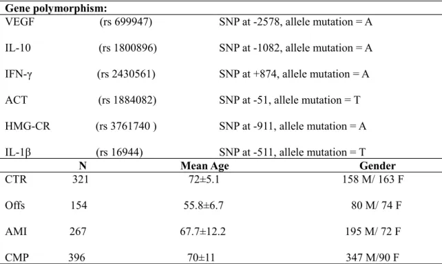
Table 1shows the SNP numbers, gene positions and mutated alleles of the investigated VEGF, ACT, HMGCR, IL-1β, IL-10 and IFN-γ genes, along with the number, mean age and gender of the subjects in the different groups.
Table 1. SNPs list and the number, age and gender distribution of CTR, Offs, AMI and CMP Gene polymorphism: VEGF (rs 699947) IL-10 (rs 1800896) IFN-γ (rs 2430561) ACT (rs 1884082) HMG-CR (rs 3761740 ) IL-1β (rs 16944) SNP at -2578, allele mutation = A SNP at -1082, allele mutation = A SNP at +874, allele mutation = A SNP at -51, allele mutation = T SNP at -911, allele mutation = A SNP at -511, allele mutation = T
N Mean Age Gender
CTR 321 72±5.1 158 M/ 163 F Offs 154 55.8±6.7 80 M/ 74 F AMI 267 67.7±12.2 195 M/ 72 F CMP 396 70±11 347 M/90 F
The genotype distribution and allele frequency of the VEGF gene between Offs, AMI and CTR groups are shown in Table 2.
The CC genotype was more frequent in the Offs than in CTR (63% vs 40.9%, p = 0.0001), and also more frequent in the AMI group (65% vs 40.9%, p = 0.0001; OR= 2.689). The percentage of VEGF C carriers was significantly higher in the AMI group than in the CTR (95.3% vs 84.5%, p = 0.0001; OR = 3.735), whereas the percentage of A carriers was significantly lower in the Offs (37.7%, p = 0.0001) and the AMI group (35%, p = 0.0001; OR = 0.373) than the CTR (59.1%). The AA genotype was more frequent in the CTR than in the AMI group (15.5% vs 4.7%, p = 0.0001; OR = 0.269).
The genotype distribution and allele frequency of the IL-10 gene between Offs, AMI and CTR groups are shown in Table 3.
Table 3 shows the IL-10 genotype distribution and allele frequency in the three groups. The GG genotype was more frequent in the CTR than in the Offs (30.5% vs 13%, p = 0.0001) or the AMI group (21.1%, p = 0.016; OR = 0.609). The A allele was significantly less frequent in the CTR than in the Offs (69% vs 87.7%, p = 0.0001) or the AMI group (78.9%, p = 0.012; OR = 1.674). There were also some differences between the Offs and the AMI group: the GG genotype was more frequent in the latter (21.1% vs 13%, p = 0.037; OR= 1.795); there was a higher percentage of A carriers among the Offs (87.7% vs 78.9%, p = 0.02), in whom the frequency of the AA genotype was also higher (35.1% vs AMI =25.7%, p = 0.043).
The TT genotype was more frequent in the CTR than in the Offs (30.6% vs 15.7%, p = 0.001), whereas the percentage of A carriers was higher among the Offs than the CTR (84.3% vs 69.4%, p = 0.001). No difference in the distribution of the IFN-γ polymorphism between the AMI group and the CTR was detected, but the frequency of the TT genotype was slightly higher in the AMI group than in Offs (24.2% vs 15.7%, p = 0.036; OR= 1.761) and the A allele was less frequent in the AMI group than Offs (75.8% vs 84.3%, p = 0.044; OR= 0.584). The SNPs in the promoter region of the ACT, HMGCR and IL-1β genes were also investigated, but not significant difference in allele and genotype frequencies between the groups was found (data not shown).
Association between the triple genotype and cardiovascular risk.
The concomitant presence of the CC genotype of VEGF, the A allele of IL-10 and the A allele of IFN-γ between Offs, AMI and CTR groups (that we called “triple genotype”) was also determined and resulted to be associated with an increased risk of AMI, as shown in Table 5.
This “triple genotype” was more frequent in the Offs (46.4%) and the AMI (31.8%) than in CTR (17.3%), and the differences were highly statistically significant (Offs vs CTR: p = 0.0001, OR = 4.129; AMI vs CTR: p = 0.0001, OR = 2.224).
Body mass index (BMI) and blood lipid profile.
Data regarding BMI and serum lipid profile from Offs, CTR and IMA have been reported in Table 6.
BMI values of Offs and IMA were slightly increased (27 ± 4) as compared with ideal age matched reference value. Moreover, BMI from our CTR group was higher than Offs and IMA, however this difference may be mainly ascribed to the older age of CTR. Lipid profile from the Offs population was substantially in the normal range for their age cohort. Once again CTR
population showed slightly increased blood levels of total cholesterol, LDL and triglycerides as expected according their age range.
Comparison for BMI, total cholesterol, HDL, LDL, triglycerides and VDL values between Offs carriers and non carriers for the triple genotype was performed. The results were display in Table 7.
As shown in Table 7, no statistical difference for BMI, total cholesterol, HDL, LDL, triglycerides and VDL values between Offs carriers and non carriers for the triple genotype was detected.
Prevalence of CVE after 24 years of follow up.
The prevalence rates of a history of ischemic heart disease (AMI or angina pectoris), stroke, ischemic heart disease plus stroke, hypertension, diabetes and smoking in 154 Offs at the beginning (age 23–35 years; Table 8, panel A) and at the end of the follow up period (age 50– 60 years; Table 8, panel B) were compared with those of gender and age matched subjects from the MONICA Brianza population.
No difference in the event rates was present between the two populations at the beginning of the follow up period. On the contrary, at the end of the follow up the prevalence of ischemic heart disease among the male Offs was three times higher, that of stroke was eight times higher, that of stroke and ischemic heart disease was three times higher, and that of diabetes was twice as high. However, there was no increased prevalence of CVE among female Offs during the same period.
Detection of EBV and HHV-6 DNA
Data regarding primers, PCR conditions and assay sensitivity for the detection of CMV, EBV, and HHV-6 nucleic acid positivity are reported in Table 9.
DNA from CMP, Offs and CTR PBL samples were analyzed by qRT-PCR for the presence of HHV6 and EBV.
In all samples, 63.1% of CMP patients, 58.1% of CTR and 47.7% of Offs were positive for EBV DNA. No differences between CMP and CTR and between Offs and CTR were found (CMP vs CTR χ2=1.091; p=0.296; Offs vs CTR χ2=2.621; p=0.105). Instead, statistically significant differences between CMP and Offs were found (CMP vs Offs χ2=8.460; p=0.004; OR=1.877; CI (1.224-2.879)).
A correction for age by a logistic regression analysis between CMP, CTR and Offs was applied but no statistically significant differences were found (CMP vs CTR p=0.952; CMP vs Offs p=0.432; Offs vs CTR p=0.483).
CMP patients were stratified in patients with AMI (CMP AMI) or Angina (aCMP). Data regarding EBV positivity in CMP AMI and aCMP, Offs and CTR are shown in Table 11.
Among CMP patients, 59.9% of CMP AMI and 65.9% of aCMP were positive for EBV DNA and no differences regarding the presence of EBV in CMP group were present. Statistically significant differences were observed between aCMP and Offs and CMP AMI and Offs (aCMP vs Offs: χ2=9.927; p=0.002; OR=2.117; CI=1.323-3.388. CMP AMI vs Offs: χ2=3.936; p=0.047; OR=1.622; CI=1.004-2.618).
EBV positive Offs and CTR subjects were stratified according to the presence of triple genotype carriers or not carriers. Data regarding EBV positivity in triple genotype carriers or not carriers Offs and CTR are shown in Table 12.
As shown in Table 12, 42.3% of Offs versus 16% of CTR were triple genotype carriers and EBV positive and the differences are statistically different (Offs vs CTR: χ2=10.841; p=0.001; OR=3.850; CI (1.684-8.802). The presence/absence of the triple genotype in Offs and CTR
subjects appeared to influence EBV positivity.
In Table 13, data regarding HHV-6 positivity in PBL samples are shown.
In all samples, 42..4% of CMP patients, 63.6% of CTR and 56% of Offs were positive for HVV-6 DNA. At first glance, the HHV-6 positivity seemed to have a correlation with age. A logistic regression was applied to correct analysis for age between CMP, CTR and Offs but no statistically significant differences were found (CMP vs CTR: Log Reg for Age p=0.312; CMP vs Offs: Log Reg for Age p=0.466; Offs vs CTR: Log Reg for Age p=0.558).
CMP patients were stratified in AMI patients (CMP AMI) and angina (aCMP). Data regarding HHV-6 positivity in CMP AMI and aCMP, Offs and CTR are shown in Table 14.
Among investigated subjects, 41.2% of CMP AMI patients, 43.3% of aCMP, 63.6% of CTR and 34.8% of Offs were positive for HVV-6 DNA. No differences regarding the presence of HHV-6 between the two groups of CMP or between CMP AMI, aCMP, Offs and CTR are present. (CMP AMI vs aCMP: χ2=0.172; p=0.678; CMP AMI vs CTR: χ2=10.534; p=0.001; OR=0.405; CI= (0.233-0.704); aCMP vs Offs: χ2=2.462; p=0.117; CMP AMI vs Offs: χ2=1.431; p=0.232; aCMP vs CTR: χ2=9.194; p=0.002; OR=0.437; CI=0.255-0.751).
HHV-6 positive Offs and CTR were stratified according to the presence of triple genotype carriers or not carriers. Data regarding HHV-6 positivity in triple genotype carriers or not carriers Offs and CTR are shown in Table 15.
In the analyzed groups, 42.6% of Offs and 16.7% of CTR were triple genotype carrier .The presence/absence of the triple genotype in Offs and CTR subjects appeared to affect HHV-6 positivity and the differences were statistically different (Offs vs CTR: χ2=7.656; p=0.006 ; OR=3.704 CI (1.426-9.617)).
DISCUSSION
The multiple pathogenetic pathways leading to AMI include classical and new risk factors and multiple genetic traits and environmental factors such as infections microorganisms might be associated with the disease. Recent genome-wide association (GWA) studies have contributed substantially to the discovery of new SNPs associated with CHD and AMI (Patel RS et al., 2011) but their clinical relevance is still unclear. In fact, a single gene variant can make a limited contribution to the total genetic load of AMI, and both common and rare gene polymorphisms may differentially affect susceptibility to the disease. These factors may also partially explain the contradictory results of genetic association studies using the candidate gene approach in AMI case/control studies (Kullo IJ et al., 2007; Hamsten A et al., 2008; Chiappelli M et al., 2005).
Familiarity and AMI
Parental history is a widely accepted risk factor for offspring of parents with CVE and AMI, although the mechanisms remain unclear. For instance, Offs with positive parental history for CVE (<65 years) resulted at higher risk of incident CVE compared with Offs with negative parental history. Despite Offs with positive parental history showed classical risk factors such as increased blood pressure, total and high-density lipoprotein cholesterol, CRP levels and decreased physical activity, less than 15% of the excess risk was explained through these risk factors. (Hamer M et al., 2009).
Family history of AMI, stroke, and related risk factors in first-degree relatives was studied. By looking at the extent to which parental history was associated with affected siblings within disease category. 15.7% cases of acute coronary syndromes occurred in families with ≥2 affected first-degree relatives compared with 5.1% strokes. Therefore, heritability of coronary events appeared to be greater than that of cerebral events and AMI was more likely to cluster in families than stroke (Banerjee A et al., 2011).
It was recently shown the presence of a relation between a family history of heart attack and the occurrence of early AMI in young women. In fact, the rate of AMI among women first-degree