Synthesis and Biological Activity of Peptide
α‑Ketoamide
Derivatives as Proteasome Inhibitors
Salvatore Pacifico,
†Valeria Ferretti,
†Valentina Albanese,
†Anna Fantinati,
†Eleonora Gallerani,
†Francesco Nicoli,
†Riccardo Gavioli,
†Francesco Zamberlan,
‡Delia Preti,
*
,†and Mauro Marastoni
††Department of Chemical and Pharmaceutical Sciences, University of Ferrara, Via Luigi Borsari 46, 44121 Ferrara, Italy ‡School of Chemistry, University of Nottingham, University Park, NG7 2RD, United Kingdom
*
S Supporting InformationABSTRACT: Proteasome activity affects cell cycle progres-sion as well as the immune response, and it is largely recognized as an attractive pharmacological target for potential therapies against several diseases. Herein we present the synthesis of a series of pseudodi/tripeptides bearing at the C-terminal position different α-ketoamide moieties as pharmaco-phoric units for the interaction with the catalytic threonine residue that sustains the proteolytic action of the proteasome.
Among these, we identified the 1-naphthyl derivative 13c as a potent and selective inhibitor of the β5 subunit of the 20S proteasome, exhibiting nanomolar potency in vitro (β5 IC50= 7 nM,β1 IC50= 60μM, β2 IC50> 100 μM). Furthermore, it significantly inhibited proliferation and induced apoptosis of the human colorectal carcinoma cell line HCT116.
KEYWORDS: Proteasome, pseudopeptides,α-ketoamides, β subunits inhibition
T
he 26S proteasome is a sophisticated multicatalytic enzymatic complex of key importance for intracellular protein degradation and homeostasis in eukaryotic organ-isms.1,2It appears as a hollow cylinder consisting of a central 20S proteolytic core (CP) capped by two 19S regulatory particles, which are responsible for the recognition of polyubiquitinated substrates and their guiding and transport within the CP.3The 20S proteasome is formed by four stacked heptameric rings of α- and β-type subunits with the typical stoichiometryα7β7β7α7. In eukaryotic cells, eachβ ring hosts three catalytic active sites with distinct proteolytic specificity: the β1 subunit promotes the so-called caspase-like (C-L) or post acidic (PGPH) activity responsible for the processing of the substrates after acid residues; theβ2 subunit displays the trypsin-like (T-L) activity with hydrolysis after basic residues; the β5 subunit undertakes the chymotrypsin-like activity (ChT-L), cutting substrates after hydrophobic and aromatic residues. Despite the different specificity, the three active sites share a common catalytic mechanism that involves a key N-terminal threonine residue (Thr1) responsible for the nucleophilic attack at the specific peptide bond of the substrate.4,5Proteasome activity affects cell cycle progression as well as the immune response, and it is largely recognized as a very attractive pharmacological target for potential therapies against a series of diseases.6,7 In particular, several classes of both specific and nonspecific inhibitors of proteasome activity have been developed.7−9 The translational potential of this system has been well established in cancer therapy with the clinical success of the three proteasome inhibitors bortezomib, carfilzomib, and ixazomib approved for the treatment of
hematological malignancies such as multiple myeloma.10−14 Nonetheless, despite the efficacy of these drugs, several limitations in their clinical employment have emerged.15 In particular, a percentage of patients does not respond to the treatment and a high relapsing frequency has been observed, probably due to the development of resistances over time. In addition, dose-limiting toxicity has been witnessed after administration of proteasome inhibitors currently in use. Peripheral neuropathy is one of the most recurrent side effects which has been attributed to off-target interactions.15Thus, the design of a new generation of inhibitors with high selectivity for the active site of the target enzyme continues to be an active field of research since it could overcome some of the typical side effects described for the first generation of drugs.8,15,16
In this context, our research efforts have been aimed at the design, synthesis, and biological characterization of new classes of peptides able to inhibit the proteasome activity.17−21Each of these is characterized by a distinct pharmacophoric unit, consisting of different electrophilic groups potentially able to interact with the hydroxyl group of the side chain of the Thr1 residue of the enzyme responsible for the proteolytic action. Herein, we present a new contribution in thisfield consisting of the development of a series of peptide-based derivatives bearing at the C-terminal residue anα-ketoamide pharmaco-phoric unit as the electrophilic substrate. This moiety has
Received: May 24, 2019
Accepted: June 6, 2019
Published: June 6, 2019
pubs.acs.org/acsmedchemlett Cite This:ACS Med. Chem. Lett. 2019, 10, 1086−1092
1086
Downloaded via UNIV DEGLI STUDI DI FERRARA on July 22, 2019 at 13:03:21 (UTC).
already been successfully introduced into the structure of various pseudopeptidic enzyme inhibitors.22,23Of note, some examples of proteasome inhibitors bearing an α-ketoamide function have also been reported in the literature, such as the cyclic polypeptide 95A and its diastereoisomers TMC-95B−D.24,25 Moreover, this electrophilic function has been inserted in the backbone of linear peptides targeting the proteasome.26According to these and more recent studies, the α-ketoamide motif is emerging as the most promising group with a possible therapeutic application against the proteasome because of its ability to induce a potent but reversible inhibition of the enzyme’s activity if compared to other investigated C-terminus warheads (i.e., α-ketoaldehyde, α,β-epoxy ketone, boronic acid, vinyl sulfone).27,28 Thus, we focused our attention on the trileucine derivative 1a (shown in
Figure 1) that has been previously reported to inhibit the
proteasome catalytic subunits at nanomolar concentrations,29 and with the above evidence in mind, we designed two series of derivatives modeled on its structure, as depicted inFigure 1. The first series (6a−6f) was conceived with the aim of simplifying the tripeptide structure of 1a to shorter dipeptide analogues in which the α-keto benzylamide pharmacophoric unit of the parent compound was maintained but combined with different C-terminal residues. In the second series (13a− 13e), the trileucine sequence of 1a was functionalized with different α-keto (cyclo)alkyl/(hetero)arylamides. A novel convergent synthetic approach has been designed and applied for the obtainment of the latter derivatives. The available strategies for the synthesis of α-ketoamide derivatives were recently described in an exhaustive review.25The synthesis of thefirst series of target dipeptides (6a−6f) was performed as depicted inScheme 1and in analogy to procedures previously reported by Stein et al.27Briefly, different Fmoc protectedL- α-amino acids (2a−2f) were initially converted into the respective aldehydes (3a−3f) by a known two-step method reported by Fehrentz and Castro.30 This was based on the initial conversion of 2a−2f into the corresponding N,O-dimethyl hydroxamates via activation of the carboxyl group with 1-ethyl-3-(3-(dimethylamino)propyl)-carbodiimmide (WSC) and N-hydroxybenzotriazole (HOBt) and subsequent treatment with N,O-dimethylhydroxylamine. In the next step, the hydroxamate derivatives were efficiently reduced to the corresponding aldehydes 3a−3f with LiAlH4. These were then treated with benzyl isocyanide in the presence of acetic acid according to the multicomponent Passerini reaction. The resulting N-Fmoc-O-acetyl hydroxyamide intermediates were
fully deprotected with a mild basic treatment to furnish derivatives 4a−4f. The desired α-hydroxyamides 5a−5f were then obtained as inseparable diastereomeric mixtures by standard coupling with Z-Leu-OH followed by side chain deprotection with TFA when necessary (5b and 5d). Finally, the oxidation with 2-iodoxybenzoic acid gave the ketoamides 6a−6f.
For the synthesis of the second series of tripeptide analogues 13a−13e, in which the nitrogen atom of the α-ketoamide group was substituted with different (cyclo)alkyl/(hetero)aryl moieties, we designed the alternative synthetic pathway reported inScheme 2. This novel approach has the advantage of not requiring the use of isocyanides that are in some cases toxic and mostly characterized by a well-known aggressive and extremely unpleasant smell. First, Boc-protected leucinal 7 was reacted with vinylmagnesium bromide to give the vinyl derivative 8 as a diastereomeric mixture.31 The following protection with 2,2-dimethoxypropane led to a mixture of cis− trans oxazolidine 9, whose double bond was oxidized with RuO2and NaIO4.
The resulting carboxylic intermediate 10 was coupled with different amines in the presence of HATU and DIPEA. The subsequent deprotection with trifluoroacetic acid gave 11a− 11e as diastereomeric mixtures. These were then coupled under standard conditions with the N-protected dipeptide Z-Leu-Leu-OH to give theα-hydroxyamides 12a−12e that were successively oxidized to the correspondingα-ketoamides 13a− 13ewith 2-iodoxybenzoic acid. Theα-hydroxyamides 5a−5f/ 12a−12e and the α-ketoamides 6a−6f/13a−13e were purified via preparative HPLC and characterized by ESI-MS and 1H NMR spectroscopy.
The capability of the synthesized compounds of inhibiting each of the three proteasome activities was measured through an in vitro assay which is based on the employment of Suc-Figure 1. New α-ketoamide peptide derivatives as potential
proteasome inhibitors.
Scheme 1. Synthesis of the First Series of Ketoamide Dipeptide Derivativesa
aReagents and conditions: (a) N,O-Dimethylhydroxylamine
hydro-chloride, WSC, HOBt, Et3N, DMF; (b) LiAlH4, THF; (c) benzyl
Isocyanide, CH3COOH; (d) LiOH, THF/MeOH; (e) Z-Leu-OH,
WSC, HOBt, DMF; (f) TFA (for 5b and 5d); and (g) 2-iodoxybenzoic acid, DMSO.
LLVY-AMC (for the ChT-L), Boc-LRR-AMC (for the T-L), and Z-LLE-AMC (for the PGPH) as specific fluorogenic substrates.32Briefly, semipurified proteasomes were pretreated with increasing concentrations (0.01−100 μM) of the new pseudopeptides (5a−5f, 6a−6f, 12a−12e, and 13a−13e) in an activity buffer. The trileucine derivatives 1a and 1b (Table 1, synthesized according to reported procedures28,29) have been evaluated under the same conditions for comparative reasons along with the known proteasome inhibitor MG132 (Z-LLL-CHO)33as an internal standard. The inhibitory activity of all compounds is reported inTable 1and expressed as IC50values in micromolar concentrations.
The effects of 5a−5f and 12a−12e against the three catalytic activities of proteasomes were evaluated in order to establish the actual importance of the ketone’s carbonyl of the α-ketoamide pharmacophore in the interaction with Thr1. It has been reported that such interaction leads to a reversible hemiacetal adduct resulting from the nucleophilic attack of the Thr1 hydroxyl group on the ketone carbonyl of the α-ketoamide portion.28Interestingly, although devoid of electro-philic properties, the α-hydroxyamide moiety of 5a−5f and 12a−12e mimics the tetrahedral intermediate which is supposed to follow the nucleophilic addition of Thr1 to the α-ketoamide. However, our SAR study strongly confirms that the presence of the electrophilic α-ketoamide moiety is mandatory for the inhibitory activity toward all three catalytic sites as its conversion to the corresponding α-hydroxyamide led to a marked loss of potency (compare 1a with 1b, 5a−5f with 6a−6f, and 12a−12e with 13a−13e). Likewise, short-ening of the trileucine motif of 1a to the dileucine analogue 6f resulted in a general decrease of potency, particularly evident against the chymotryptic-like activity (β5 IC50 = 0.014 and 0.92 nM for 1a and 6f, respectively). Nonetheless, we explored the possibility of restoring the activity with the introduction of C-terminal amino acids other than the leucine residue of 6f (i.e., Phe, Tyr, 1-Nal, Asn, and Val for compounds 6a−6e, respectively). This led to a further reduction of potency with IC50values that reached the high micromolar range against the
three investigated activities. Thus, compound 6f (with the sequence Z-Leu-Leu-CONHBn) was confirmed as the most active of thisfirst series as able to inhibit the three activities of the enzyme complex with IC50values of about 1μM, without significant selectivity in the biological response. Most of the analyzed dipeptide derivatives had a mild selectivity for the postacidic and/or chymotryptic activities, with the exception of 6e, characterized by a C-terminal valine, that inhibited theβ2 tryptic subunit (IC50 = 2.11 μM) with significant selectivity over the β1 (7-fold) and β5 (30-fold) subunits. These preliminary results suggested that both the length of the peptide sequence and the C-terminal leucine residue would contribute in eliciting a better inhibition. Thus, we designed the second series of tripeptide α-ketoamides (compounds 13a−13e inTable 1) in which the Z-LLL template of 1a was maintained but combined with different substituents on the nitrogen of theα-ketoamide moiety. The resulting compounds exhibited from submicromolar to low nanomolar potency in inhibiting theβ5 chymotryptic activity (β5 IC50ranging from 7 to 770 nM), with various levels of selectivity over theβ1 and β2 subunits. Among these, derivative 13c, in which the α-keto benzylamide of 1a was replaced by the 1-naphthylamide moiety, displayed the highest potency with an IC50value of 7 nM against the chymotryptic activity and very low or no activity against theβ1 and β2 subunits (IC50values of 57 and >100μM, respectively). Thus, the introduction of a bulky and lipophilic aromatic bicycle at the C-terminal portion of the linear peptide structure led to a slight improvement of theβ5 activity inhibition (2-fold) associated with a remarkable increase of selectivity (selectivity ratios: β1/β5 = 8.200, β2/ β5 > 14.200) in comparison with the parent compound 1a. In this series of molecules, the investigated ketoamide sub-stitutions determined the following order of potency: 1-naphthyl-NH > isobutyl-NH ≈ Bn-NH > 4-F-benzyl-NH > morpholin-4-yl > tetrahydroisoquinolin-2-yl. These data would suggest that the presence of a more flexible primary amide, compared to a secondary one, would enhance the inhibition of the ChT-L activity. This is particularly evident when comparing the activities of 1a and 13e, since the latter’s isoquinoline derivative can be considered a constrained analogue of the parent benzylamide compound.
The antiproliferative activity of selected compounds (6b, 6f, 1a, 13a, 13c, and 13d) was evaluated at concentrations ranging from 0.1 to 100μM against the human colorectal carcinoma cell line HCT116 and compared to that of the reference inhibitor MG132. The effect of the compounds on cell viability was measured at 72 h and reported in Table 1 expressed as IC50. Interestingly, a significant antiproliferative activity was observed for all the examined molecules with variable degrees of potencies, basically reflecting their capability to inhibit the β5 subunit of the proteasome, mediating the chymotryptic activity (ChT-L). Indeed, the Leu-Leu derivative 6f (IC50 ChT-L = 920 nM and IC50proliferation = 8.6μM,Table 1) seems to be more potent than the Leu-Tyr analogue 6b (IC50 ChT-L= 12.43 μM and IC50 proliferation >100 μM), while both compounds were shown to have lower activity than the reference pseudotripeptide 1a (IC50ChT-L= 14 nM and IC50 proliferation= 0.78μM) and MG132 (IC50ChT-L = 7 nM and IC50 proliferation = 0.82 μM). Notably, the tripeptide analogues 13a, 13c, and 13d, which all showed an IC50for ChT-L < 100 nM, induced a strong antiproliferative effect, comparable or even superior to that of the reference pseudotripeptide 1a and of MG132, as all the three Scheme 2. Synthesis of the Second Series of Ketoamide
Tripeptide Derivativesa
aReagents and conditions: (a) vinylmagnesium bromide, CH
2Cl2; (b)
p-toluensulfonic acid, 2,2-dimethoxypropane, CH2Cl2; (c) NaIO4,
ruthenium(IV) oxide hydrate, acetone; (d) appropriate amines a−e, HATU, DIPEA, DMF; (e) TFA; (f) Z-Leu-Leu-OH, HATU, DIPEA, DMF; (g) 2-iodoxybenzoic acid, DMSO; 11−13a R = 4-fluorobenzyl-NH; 11−13b R = morpholin-4-yl; 11−13c R = 1-naphthyl-NH; 11− 13dR = isobutyl-NH; 11−13e R = tetrahydroisoquinolin-2-yl.
compounds displayed an IC50 proliferation <1 μM. In particular, the compound 13c, which showed the highest activity inhibiting the β5 subunit of the proteasome, also exerted the strongest antiproliferative effect.
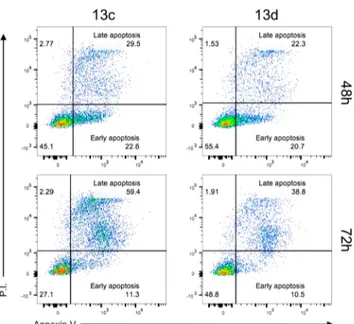
Finally, to determine if this effect was associated with cell death induction, apoptosis levels were measured in HCT116 cells treated for 48 and 72 h with two selected compounds, i.e., 13c and 13d. The positivity to Annexin V and negativity to propidium iodide (P.I.) identifies early apoptotic cells while double positivity to both Annexin V and P.I., late apoptotic cells. As shown inFigure 2, 13c exhibited increased levels of late apoptotic cells already at 48 h, and this effect was even higher at 72 h, consistently with the lower IC50proliferation shown in comparison to 13d.
Docking studies were performed for a subset of compounds (6a, 6b, 5f, 6f, and 13c) selected in respect to their biological profile, and the best binding poses of each molecule in the β1, β2, and β5 binding sites were compared to those of the known α-ketoamide tripeptide 1a.29
Figure 3A shows compound 13c, the most potent and selective β5 inhibitor identified in this work, docked in theβ5 binding pocket alongside the reference compound 1a (Figure 3B). Moreover, the best binding poses for compounds 1a and 13c in theβ1 and β2 catalytic subunits
have been illustrated in Figures S1 and S2of the Supporting Information. For each proposed binding pose, a schematic diagram of the inhibitor−protein interactions has been also supplied. For both molecules, the α-ketoamide group was found to interact in all binding sites with the active Thr1 residue via hydrogen bonding interactions, with donor− acceptor distances in the range of 2.6−3.3 Å, typical of medium/strong H-bonds. Furthermore, 1a and 13c were found to be surrounded by (or interacting with) several residues which the structural analyses indicated as involved in the formation of the receptor pockets.5For molecule 13c, the important residues Thr21, Gly47, and His108 were directly interacting with the ligand (Figure 3A). As a reference, for the ChT-L active center, residues Thr21, Gly47, Ala49, and Ala50 were found to be involved in the binding of known inhibitors such as Bortezomib.5 Moreover, docking results at the β5 subunit show that the distal aromatic regions of 13c, unlike 1a, were able to establish a significant number of π/π stacking and CH/π interactions with the residues His108, Trp,25
Tyr170, and Gly47. Of note, these interactions involve both the benzyl and naphthyl terminal groups, favoring the anchorage of the molecule to the proteasome active site. In our models, a lower number of residues of theβ1 (Gly47) and β2 (Gly47, Gln22) Table 1. Inhibition of the Proteasome Subunits byα-Hydroxy/Ketoamide Peptides and Their Effect on Cell Proliferation (The Values Reported Are the Mean± SEM of Three Independent Experiments)
cmpd R IC50(μM) PGPH (β1) IC50(μM) T-L (β2) IC50(μM) ChT-L (β5) IC50(μM)aprolif. MG132 1.45± 0.31 4.59± 0.06 0.007± 0.001 0.82± 0.03 1a 0.73± 0.01 0.43± 0.01 0.014± 0.001 0.78± 0.01 1b 17.82± 0.90 >100 0.53± 0.04 ND Dipeptide Analogues 5a Bn >100 57.98± 4.38 >100 ND 6a Bn 82.36± 6.72 17.61± 1.18 51.09± 3.34 ND 5b 4-OH-Bn >100 51.63± 5.06 70.45± 5.93 ND 6b 4-OH-Bn 50.11± 3.84 21.54± 2.05 12.43± 0.92 >100 5c 1-naphthyl >100 >100 >100 ND 6c 1-naphthyl 73.22± 5.91 >100 >100 ND 5d CH2CONH2 53.15± 4.27 >100 >100 ND 6d CH2CONH2 89.47± 8.17 >100 >100 ND 5e isopropyl 30.57± 2.76 >100 82.07± 7.55 ND 6e isopropyl 15.45± 1.21 2.11± 0.24 63.34± 6.13 ND 5f isobutyl 20.44± 1.76 >100 50.21± 4.33 ND 6f isobutyl 1.09± 0.12 1.32± 0.11 0.92± 0.08 8.6± 0.34 Tripeptide Analogues 12a 4-F-Bn-NH- >100 >100 >100 ND 13a 4-F-Bn-NH- 11.5± 0.81 0.59± 0.01 0.056± 0.003 0.93± 0.13 12b morpholin-4-yl >100 >100 >100 ND 13b morpholin-4-yl 0.56± 0.02 85.4± 3.12 0.48± 0.02 ND 12c 1-naphthyl-NH >100 >100 8.1 ND 13c 1-naphthyl-NH 57.5± 4.06 >100 0.007± 0.001 <0.10 12d isobutyl-NH >100 >100 >100 ND 13d isobutyl-NH 5.9± 0.32 0.43± 0.03 0.012± 0.002 0.81± 0.12 12e tetrahydroisoquinolin-2-yl >100 >100 >100 ND 13e tetrahydroisoquinolin-2-yl >100 38.9± 0.98 0.77± 0.04 ND aIC
50proliferation values against HCT116 cells for the most potent compounds in the enzyme inhibition assays.
subunits would make contact with the distal aromatic portions of 13c, and this could account for the overall selectivity profile of the molecule.
For what is concerning the docking results obtained for the dipeptide derivative 6f (Figure S3) in comparison with the tripeptide analogue 1a, both molecules show their capability to fit well all of the three investigated binding sites making contact with key residues of the binding pockets. In β1, for instance, the conserved residues are Lys,33Thr21, Ser118, Arg45. Inβ2, it is worth mentioning the presence of the residue Cys118 of subunitβ3, which is responsible for the character of the S3 specificity pocket for the selective β2 inhibitor Mal-βAla-Val-Arg-al.5 It is also worth noting that the tripeptide 1a, with a longer sequence by one nonpolar alkyl amino acid, is able to establish van der Waals interactions with further residues that insist around the binding pocket, something that the shorter dipeptide 6f presented in this work is not able to do.
The docking simulation of the other 6a, 6b, and 5f molecules did not give good results in comparison with those presented above, as far as the mutual Thr1-ketoamide group position is concerned. The five best poses for each molecule in the three binding pockets are reported in the Supporting Information (Figures S4−S6).
In summary, this paper describes the synthesis, biological evaluation, and docking analysis of two series of pseudodi/ Figure 2.Effect of compounds 13c and 13d on apoptosis induction.
HCT116 tumor cells cultured for 48 or 72 h in the presence or absence of the indicated compounds at a concentration of 1 μM. Numbers in the dot plots represent percentages of cells. One representative experiment out of three is shown.
Figure 3.Molecules 13c (A) and 1a (B) in theβ5 binding pocket with a schematic diagram of the inhibitor−protein interactions.
tripeptides as proteasome inhibitors. The entire investigated molecules feature at the C-terminal portion anα-ketoamide as the pharmacophoric unit able to interact with and block the catalytic threonine of the active subunits of the 26S proteasome. Our stepwise SAR optimization work led to the identification of 13c as a potent and selective inhibitor of the β5 subunit of the 20S proteasome with nanomolar potency in vitro. The compound significantly inhibited proliferation and induced apoptosis of the human colorectal carcinoma cell line HCT116, confirming the potential of β5-selective proteasome inhibitors in cancer therapy. Recent findings would also suggest that the inhibition of theβ5 activity by selective ligands could have some therapeutic perspectives in the cardiovascular area since promoting beneficial effects in rat models of ischemia reperfusion injury.34
■
ASSOCIATED CONTENT*
S Supporting InformationThe Supporting Information is available free of charge on the
ACS Publications website at DOI: 10.1021/acsmedchem-lett.9b00233.
Detailed synthetic procedures, spectroscopic data and full characterizations of the described compounds, procedures for biological experiments, computational procedures, and Figures S1−S6 from the docking studies (PDF)
■
AUTHOR INFORMATIONCorresponding Author
*Phone: +39-532-455501. E-mail:[email protected].
ORCID
Delia Preti:0000-0002-1075-3781
Author Contributions
S.P., V.A., and A.F. performed the chemical synthesis. V.F. performed and interpreted the docking study. E.G., F.N., and R.G. performed the in vitro molecular pharmacology studies. F.Z. drafted the manuscript. M.M. and D.P. oversaw and developed the project. All authors have given approval to the final version of the manuscript.
Funding
D.P. is supported by the funds FAR 2017 (Fondo di Ateneo per la Ricerca Scientifica) and FFABR 2017 (Finanziamento delle attività base di ricerca) of the University of Ferrara. M.M. is supported by the funds FAR 2017 of the University of Ferrara. The FACSCanto II was funded by Ferrara University Grant“Bando per l’acquisizione di strumenti per la ricerca di ateneo-2015”.
Notes
The authors declare no competingfinancial interest.
■
REFERENCES(1) Glickman, M. H.; Ciechanover, A. The ubiquitin-proteasome proteolytic pathway: destruction for the sake of construction. Physiol. Rev. 2002, 82, 373−428.
(2) Ciechanover, A. Proteolysis: from the lysosome to ubiquitin and the proteasome. Nat. Rev. Mol. Cell Biol. 2005, 6, 79−87.
(3) Finley, D. Recognition and processing of ubiquitin-protein conjugates by the proteasome. Annu. Rev. Biochem. 2009, 78, 477− 513.
(4) Groll, M.; Heinemeyer, W.; Jager, S.; Ullrich, T.; Bochtler, M.; Wolf, D. H.; Huber, R. The catalytic sites of 20S proteasomes and
their role in subunit maturation: a mutational and crystallographic study. Proc. Natl. Acad. Sci. U. S. A. 1999, 96, 10976−10983.
(5) Borissenko, L.; Groll, M. 20S proteasome and its inhibitors: crystallographic knowledge for drug development. Chem. Rev. 2007, 107, 687−717.
(6) Sijts, E. J.; Kloetzel, P. M. The role of the proteasome in the generation of MHC class I ligands and immune responses. Cell. Mol. Life Sci. 2011, 68, 1491−1502.
(7) Kisselev, A. F.; Van der Linden, W. A.; Overkleeft, H. S. Proteasome inhibitors: an expanding army attacking a unique target. Chem. Biol. 2012, 19, 99−115.
(8) Groll, M.; Huber, R.; Moroder, L. The persisting challenge of selective and specific proteasome inhibition. J. Pept. Sci. 2009, 15, 58− 66.
(9) Micale, N.; Scarbaci, K.; Troiano, V.; Ettari, R.; Grasso, S.; Zappalà, M. Peptide-based proteasome inhibitors in anticancer drug design. Med. Res. Rev. 2014, 34, 1001−1069.
(10) Moreau, P.; Richardson, P. G.; Cavo, M.; Orlowski, R. Z.; San Miguel, J. F.; Palumbo, A.; Harousseau, J. L. Proteasome inhibitors in multiple myeloma: 10 years later. Blood 2012, 120, 947−959.
(11) Adams, J. The development of proteasome inhibitors as anticancer drugs. Cancer Cell 2004, 5, 417−421.
(12) Moreau, P. The emerging role of carfilzomib combination therapy in the management of multiple myeloma. Expert Rev. Hematol. 2014, 7, 265−290.
(13) Shirley, M. Ixazomib: first global approval. Drugs 2016, 76, 405−411.
(14) Allegra, A.; Alonci, A.; Gerace, D.; Russo, S.; Innao, V.; Calabrò, L.; Musolino, C. New orally active proteasome inhibitors in multiple myeloma. Leuk. Res. 2014, 38, 1−9.
(15) Park, J. E.; Miller, Z.; Jun, Y.; Lee, W.; Kim, K. B. Next-generation proteasome inhibitors for cancer therapy. Transl Res. 2018, 198, 1−16.
(16) Gozzetti, A.; Papini, G.; Candi, V.; Brambilla, C. Z.; Sirianni, S.; Bocchia, M. Second generation proteasome inhibitors in multiple myeloma. Anti-Cancer Agents Med. Chem. 2017, 17, 920−926.
(17) Marastoni, M.; Baldisserotto, A.; Canella, A.; Gavioli, R.; De Risi, C.; Pollini, G. P.; Tomatis, R. Arecoline tripeptide inhibitors of proteasome. J. Med. Chem. 2004, 47, 1587−1590.
(18) Baldisserotto, A.; Ferretti, V.; Destro, F.; Franceschini, C.; Marastoni, M.; Gavioli, R.; Tomatis, R. Alpha,beta-unsaturated N-acylpyrrole peptidyl derivatives: new proteasome inhibitors. J. Med. Chem. 2010, 53, 6511−6515.
(19) Franceschini, C.; Trapella, C.; Calia, R.; Scotti, A.; Sforza, F.; Gavioli, R.; Marastoni, M. C-terminal trans,trans-muconic acid ethyl ester partial retro-inversopseudopeptides as proteasome inhibitors. J. Enzyme Inhib. Med. Chem. 2013, 28, 1034−1039.
(20) Scotti, A.; Trapella, C.; Ferretti, V.; Gallerani, E.; Gavioli, R.; Marastoni, M. Studies of C-terminal naphthoquinone dipeptides as 20S proteasome inhibitors. J. Enzyme Inhib. Med. Chem. 2016, 3, 456−463.
(21) Marastoni, M.; Trapella, C.; Scotti, A.; Fantinati, A.; Ferretti, V.; Marzola, E.; Eleonora, G.; Gavioli, R.; Preti, D. Naphthoquinone aminoacid derivatives, synthesis and biological activity as proteasome inhibitors. J. Enzyme Inhib. Med. Chem. 2017, 32, 865−877.
(22) Chang, K. O.; Kim, Y.; Groutas, W. C.; Hua, D.; Saif, L. J. Broad-spectrum antivirals against 3C or 3C-like proteases of picornavirus-like supercluster: picornaviruses, caliciviruses and coronaviruses. u,S, Patent Application 20140243341, August 28, 2014 .
(23) Dondapati, J. S.; Godi, S.; Babu, A. P. QSAR studies on peptide alpha-ketoamides and alpha-ketohydroxamate derivatives as calpain I inhibitors. J. Enzyme Inhib. Med. Chem. 2008, 23, 757−762.
(24) Koguchi, Y.; Kohno, J.; Nishio, M.; Takahashi, K.; Okuda, T.; Ohnuki, T.; Komatsubara, S. TMC-95A, B, C, and D, novel proteasome inhibitors produced by Apiospora montagnei Sacc. TC 1093. Taxonomy, production, isolation, and biological activities. J. Antibiot. 2000, 53 (53), 105−109.
(25) De Risi, C.; Pollini, G. P.; Zanirato, V. Recent developments in general methodologies for the synthesis ofα-ketoamides. Chem. Rev. 2016, 116, 3241−3305.
(26) Lynch, S. M.; Narayanan, A.; Steiner, S. Ketoamide immunoproteasome inhibitors. World Patent WO 2014056748, April 17, 2014.
(27) Stein, M. L.; Cui, H.; Beck, P.; Dubiella, C.; Voss, C.; Krüger, A.; Schmidt, B.; Groll, M. Systematic comparison of peptidic proteasome inhibitors highlights theα-ketoamide electrophile as an auspicious reversible lead motif. Angew. Chem., Int. Ed. 2014, 53, 1679−1683.
(28) Voss, C.; Scholz, C.; Knorr, S.; Beck, P.; Stein, M. L.; Zall, A.; Kuckelkorn, U.; Kloetzel, P. M.; Groll, M.; Hamacher, K.; Schmidt, B. α-Keto phenylamides as P1’-extended proteasome inhibitors. ChemMedChem 2014, 9, 2557−2564.
(29) Braun, H. A.; Umbreen, S.; Groll, M.; Kuckelkorn, U.; Mlynarczuk, I.; Wigand, M. E.; Drung, I.; Kloetzel, P. M.; Schmidt, B. Tripeptide mimetics inhibit the 20 S proteasome by covalent bonding to the active threonines. J. Biol. Chem. 2005, 280, 28394−23401.
(30) Fehrentz, J. A.; Castro, B. An efficient synthesis of optically active α-(t-butoxycarbonylamino)-aldehydes from α-amino acids. Synthesis 1983, 1983 (8), 676−678.
(31) DeMattei, J. A.; Leanna, M. R.; Li, W.; Nichols, P. J.; Rasmussen, M. W.; Morton, H. E. An Efficient Synthesis of the Taxane-Derived Anticancer Agent ABT-271. J. Org. Chem. 2001, 66, 3330−3337.
(32) Hansen, M. B.; Nielsen, S. E.; Berg, K. Re-examination and further development of a precise and rapid dye method for measuring cell growth/cell kill. J. Immunol. Methods 1989, 119, 203−210.
(33) Lee, D. H.; Goldberg, A. L. Proteasome inhibitors: valuable new tools for cell biologists. Trends Cell Biol. 1998, 8, 397−403.
(34) Sanchez, G.; Berrios, D.; Olmedo, I.; Pezoa, J.; Riquelme, J. A.; Montecinos, L.; Pedrozo, Z.; Donoso, P. Activation of chymotrypsin-like activity of the proteasome during ischemia induces myocardial dysfunction and death. PLoS One 2016, 11 (8), e0161068.