Università degli Studi di Ferrara
DOTTORATO DI RICERCA IN
"BIOCHIMICA, BIOLOGIA MOLECOLARE E
BIOTECNOLOGIE"
CICLO XXVI
COORDINATORE Prof. Bernardi Francesco
The Endothelial Protein C Receptor enhances
FVIIa mediated hemostasis
Settore Scientifico Disciplinare BIO/10
Dottorando Tutore
Dott. Pavani Giulia Prof. Bernardi Francesco Prof. Margaritis Paris
INDEX
ABSTRACT ... 3
SOMMARIO ... 4
INTRODUCTION ... 5
The blood coagulation system ... 5
The clotting cascade ... 6
Coagulation Factor VII ... 8
FVIIa in Hemophilia ... 11
FVIIa mechanism of action in hemophilia ... 12
FVIIa molecular engineering ... 13
Protein C anticoagulant pathway ... 14
The Endothelial Protein C Receptor ... 14
FVII/a is a ligand for EPCR ... 17
AIM OF THE STUDY ... 22
METHODS ... 23
Construction of expression vectors ... 23
Expression and purification of recombinant proteins ... 23
Analytical Ultracentrifugation ... 24
Generation of CHO-‐K1 stable cell lines expressing full-‐length murine EPCR and tissue factor ... 24
Phospholipid Binding assay ... 25
Isothermal titration calorimetry (ITC) ... 25
Cell-‐surface binding assay ... 26
Factor Xa generation assay ... 27
Thrombin generation, coagulation assays and determination of mFVIIa antigen .. 27
Animal experiments and procedures ... 28
Murine carotid artery thrombosis and murine tail clip hemostasis models ... 28
Statistical analysis ... 29
RESULTS ... 30
Generation of mFVIIa constructs with affinity to mEPCR ... 30
Expression of mFVIIa chimeras ... 31
Generation of CHO-‐K1 stable cell lines expressing full-‐length murine EPCR and/or TF ... 32
mFVIIa chimeras bind to mEPCR when expressed on the cell surface or in solution ... 33
Partial substitution of the Gla domain of mFVIIa does not affect its in vitro activity ... 36
The mFVIIa-‐FMR molecule binds mEPCR in vivo ... 42
EPCR binding enhances FVIIa hemostatic activity in vivo ... 43
DISCUSSION ... 48
CONCLUDING REMARKS ... 53
ACKNOWLEDGMENTS ... 54
ABSTRACT
Recombinant activated human Factor VII (rFVIIa) is an established hemostatic agent in hemophilia but its mechanism of action remains unclear. Although tissue factor (TF) is its natural receptor, rFVIIa also interacts with the endothelial protein C receptor (EPCR) through its γ-carboxyglutamic acid (Gla) domain with unknown hemostatic consequences in vivo. Here, we study whether EPCR facilitates rFVIIa hemostasis in hemophilia using a mouse model system. Murine activated FVII (mFVIIa) is functionally homologous to rFVIIa, but binds poorly to murine EPCR (mEPCR). We modified mFVIIa to gain mEPCR binding using 3 amino acid changes in its Gla-domain. The resulting molecule mFVIIa-FMR specifically bound mEPCR in vitro and in vivo and was identical to mFVIIa with respect to TF affinity and procoagulant functions. Using two macrovascular injury models in hemophilic mice, administered mFVIIa-FMR exhibited superior hemostatic properties compared to mFVIIa. These effects were specific to the mFVIIa-FMR and mEPCR interaction since antibody blocking of mEPCR abolished them. Since mFVIIa-FMR models both the TF-dependent as well as EPCR binding properties of rFVIIa, our data unmask a novel contribution of EPCR on the action of rFVIIa administration in hemophilia. This may prompt the rational design of improved and safer rFVIIa therapeutics
.
SOMMARIO
Il Fattore VII attivato ricombinante (rFVIIa) è un agente emostatico utilizzato per il trattamento dei pazienti emofilici con inibitori, ma il suo meccanismo d’azione rimane ad oggi non completamente elucidato. Nonostante il Fattore Tissutale (TF) sia il suo cofattore naturale più importante, il rFVIIa interagisce attraverso il dominio γ-carbossiglutammico (Gla) anche con il recettore endoteliale della Proteina C (PC) con conseguenze funzionali ancora sconosciute.
In questo studio abbiamo caratterizzato l’effetto del legame ad EPCR sull’attività emostatica del FVIIa, utilizzando un modello murino di emofilia. Nel topo il FVIIa murino (mFVIIa), nonostante l’alta omologia funzionale e strutturale con l’omologo umano, non interagisce con EPCR. Per trasformare il mFVIIa in un ligando di mEPCR senza intaccare le sue proprietà coagulative abbiamo generato una variante di mFVIIa (mFVIIa-FMR) modificando tramite mutagenesi 3 residui del dominio Gla. Questa variante è in grado di legare in modo specifico mEPCR e mantenere le stesse caratteristiche funzionali di mFVIIa in vitro. Per queste sue proprietà mFVIIa-FMR rappresenta la molecola ideale per caratterizzare in vivo l’effetto di EPCR sull’attività del FVIIa. In topi emofilici sottoposti a danno vascolare. mFVIIa-FMR ha dimostrato maggiori proprietà emostatiche rispetto al mFVIIa. Questo effetto procoagulante dipende dal legame specifico di mFVIIa-FMR con EPCR.
Questo lavoro identifica per la prima volta EPCR come un nuovo modulatore dell’attività del FVIIa in vivo. La comprensione più dettagliata del meccanismo con cui EPCR aumenta l’attività coagulante del FVIIa potrà promuovere lo sviluppo di nuove molecole terapeutiche con maggiore attività emostatica per il trattamento dell’emofilia.
INTRODUCTION
The blood coagulation system
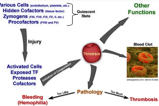
Coagulation involves a multitude of proteins and cellular partners that act in concert in response to a vascular injury to generate thrombin, which is responsible for the generation of the fibrin plug (Figure 1). However, while fibrin generation is required for the arrest of excessive bleeding, unregulated clotting may lead to vascular occlusion and result in myocardial infarction, stroke, pulmonary embolism, or venous thrombosis. Thus, the regulation of the delicate balance between the procoagulant and anticoagulant mechanisms is of fundamental for survival(Dahlback, 2005)
The dynamics of the blood coagulation system are dominated by three membrane-bound procoagulant complexes which assemble at the site of vascular injury on platelets, the damaged endothelium and subvascular tissue (Figure 2)(Mann et al., 2003). The assembly and regulation of these complexes and their opposing anticoagulant systems dictate the coagulant response.
Fig. 1 The blood coagulation response, image courtesy of Dr. Rodney M. Camire, The
The clotting cascade
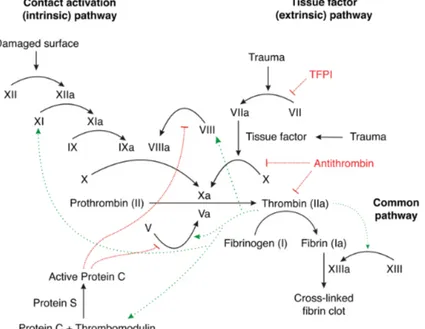
The clotting cascade is shown in Figure 3. Initiation of coagulation begins by exposure of Tissue Factor (TF) to the bloodstream, through physical damage to the lining of the blood vessel or increased expression in endothelial and monocytes in response to certain inflammatory signals. About 1-2 % of Factor VII (FVII) circulates in its activated form (FVIIa) and binds rapidly to TF after exposure (Vadivel and Bajaj, 2012). The FVIIa-TF complex efficiently converts factor IX (FIX) and factor X (FX) to active enzymes FIXa and FXa, which together with factor VIIIa (FVIIIa) and factor Va (FVa), respectively, propagate the coagulation process.(Dahlback, 2005)
With sufficient stimulus, the FXa produced activates some prothrombin. This initial thrombin activates the procofactors and platelets required for presentation of the intrinsic FXase (FVIIIa-FIXa) and prothrombinase (FVa-FXa) complexes that drive the subsequent propagation phase. During this phase the intrinsic FXase drives most of the FXa generated, and the reaction becomes TF independent. (Mann et al., 2003).
When FXa combines with FVa on the activated platelet surface or other activated membranes (e.g. endothelium), the complex FXa/FVa (prothrombinase) efficiently converts prothrombin to thrombin, resulting in fibrin generation and, ultimately, clot formation.
Fig. 2 The 3 vitamin K–dependent procoagulant complexes. The thicker arrow
associated with intrinsic factor Xase generation of factor Xa reflects the improved catalytic efficiency of this complex. Adapted from (Mann et al., 2003)
The assembly of the prothrombinase complex is crucial since it is 300,000-fold more active than factor Xa alone in catalyzing prothrombin activation.(Mann, 2003)
Fig 3. General overview of the clotting cascade. Green arrows indicate positive
feedback reactions, in red inhibitory pathway. (Wikipedia Commons)
It is critical that once a procoagulant response to vascular injury has been initiated that an appropriate anticoagulant response is concurrently mounted. As such, the components of the prothrombinase complex are targets of multiple regulatory mechanisms to arrest the production of thrombin. First, TF dependent activation of coagulation is regulated by tissue factor pathway inhibitor (TFPI), a FXa-dependent inhibitor of the FVIIa/TF complex (Shirk and Vlasuk, 2007). The mechanism of inhibition by TFPI involves its binding first to FXa through domain 2 and then to the TF/FVIIa complex through domain 1(Rao and Mackman, 2010). FXa is also inhibited by serpins such as antithrombin (AT), α1-antitrypsin. The cofactor, FVa, is a target for degradation by activated protein C (aPC). aPC, a serine protease derived from its plasma precursor protein C (PC) in a thrombin dependent activation process (Bravo et al., 2012; McVey et al., 1998), plays a critical role in inactivating the non-enzymatic cofactor components of both the prothrombinase and the intrinsic FXase complexes, factors Va and VIIIa, respectively. Additional key components of the PC pathway include: thrombomodulin and endothelial
protein C receptor (EPCR) which regulate PC activation; protein S which acts as a cofactor for aPC; and protein C inhibitor, a suppressor of aPC formation (McVey et al., 2001; Wildhagen et al., 2011).
Coagulation Factor VII
Factor VII is a vitamin K-dependent glycoprotein that participates in the initiation of the extrinsic pathway of blood coagulation. It is expressed and synthesized in the liver where is secreted as single-chain zymogen composed of 406 amino acid residues (Mr 50,000 kD). The factor VII gene (F7) consists in 9 exons and it is located on chromosome 13q34, 2.8 kb upstream of the FX gene. F7 shares significant similarity with other genes coding for vitamin K-dependent proteins(Greenberg et al., 1995). It is believed that these proteins have been assembled by exon-shuffling events via intronic recombination (Patthy, 1985). Inherited FVII deficiency is an autosomal recessive disease with variable clinical symptoms (Mariani and Bernardi, 2009). FVII deficiency is not associated with complete absence of functional FVII, suggesting that a complete lack of FVII is incompatible with life (McVey et al., 1998). As described in clinical reports, homozygous carriers die shortly after birth due to severe hemorrhage (McVey et al., 2001). The same phenotype was observed in F7 knockout mice models (Rosen et al., 2005).
The genetic similarity with other clotting factors is also reflected by high structural homology and domain organization at a protein level (Figure 4). FVII consist of a γ-carboxyglutamic acid (Gla) domain which enables reversible binding to phospholipid membranes, a hydrophobic stack, which is involved in formation of the ternary complex, two epidermal growth factor-like domains (EGF 1-2), and a protease domain (Bajaj et al., 2006). The Gla domain is a hallmark of all vitamin K-dependent coagulation proteins; FVII has 10 glutamic acid residues that undergo a characteristic post-translational carboxylation at the γ-carbon. Gla residues are necessary for calcium binding which induces a conformational change in the Gla domain required for phospholipid/receptor binding and therefore clotting activity (Larson et al., 1998). FVII Gla domain coordinates 7 divalent cations: crystal structures obtained in presence of 2.5 mM Mg2+/5 mM Ca2+ indicate that metal positions 1, 4, and 7 are occupied by Mg2+ while the remaining 4 sites
coordinate Ca2+. Other post-translational modifications in FVII include N-glycosylations (Asn145, Asn322) and O-glycosylations (Ser52, Ser60). There are also two other calcium binding sites located in EGF1 and in the serine protease domain(Hansson and Stenflo, 2005).
Approximately 99% of FVII circulates as inactive zymogen. The initial event, cleavage of the peptide bond between Arg152/Ile153, converts the zymogen FVII into a two-chain serine protease linked by a disulfide bond that exists in equilibrium between a predominant zymogen-like form and an active FVIIa. In the zymogen-like conformation, the new N-terminus of Ile153 is solvent-exposed and accessible to modification, whereas in the active FVIIa, a salt bridge forms between the α-amino group of Ile 153 and the β-carboxyl group of Asp343, stabilizing the insertion of Ile153 into the protein (Toso et al.,
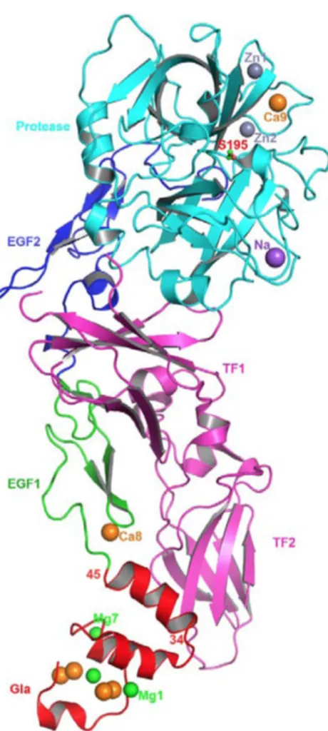
Fig 4 Structure of FVIIa/sTF complexes.
The FVIIa/sTF structure of Bajaj et al., at 1.8 angstrom resolution (PDB id 2A2Q) in the presence of Ca2+ and Mg2+ is shown. FVIIa consists of Gla (red), EGF1 (green), EGF2 (blue) and protease (cyan) domains, and the sTF (magenta) contains two fibronection type III domains. The different metal ions Ca2+(orange), Mg2+(green), Zn2+ (light blue) and Na+ (purple), bound to different domains of FVIIa are shown as spheres. Adapted from (Vadivel and Bajaj, 2012)
2003). TF binding facilitates the stabilizing insertion of the N-terminal tail of the protease domain of FVII into the FVII activation pocket and the formation of a salt bridge with an Asp343 side chain (Higashi et al., 1994; Petersen et al., 1999). Therefore, although activated, FVIIa does not express its full procoagulant functionality until bound to TF.(Monroe and Key, 2007). This extreme cofactor dependence of FVIIa ensures that the blood clotting cascade is triggered at the appropriate place and time (Persson, 2006). The complex FVIIa/TF catalyzes the activation of both factor IX and factor X, the latter initially being the more efficient substrate. FVIIa/TF/FX complex constitutes the extrinsic FXase, which produces the initial amount of FXa necessary to trigger the coagulant cascade. As the extrinsic tenase complex is rapidly shut down by TFPI, generation of thrombin is indirectly maintained by the FXa generation through the intrinsic tenase complex (where FIXa participates). (Mann et al., 2003) (Figure 5).
Fig.5 A computational estimation of the temporal dependence of the relative FXa
production by the extrinsic FXase (◊) and the intrinsic FXase (■) complexes. The clot time (CT, arrow) represents the time at which free thrombin levels are predicted to reach 2nM. (from (Hockin et al., 2002))
In addition to the TFPI inhibition of the TF-FVIIa-FXa complex (see above), antithrombin III (AT III), the major plasma inhibitor of coagulation proteases, also
inhibits free FVIIa at a very slow rate(Rao et al., 1995), but rapidly in complex with TF in the presence of heparin (Rao LV, Blood 1995). This has important implications for the clinical use of recombinant FVIIa (rFVIIa) for management of bleeding. As a matter of fact, a pharmacokinetic study in hemophilic patients suggests that complex formation between rFVIIa and AT is the main clearance pathway for exogenously administered rFVIIa, accounting for 65% of a therapeutic dose (90 µg/kg) of rFVIIa (AGERSØ et al., 2011).
FVIIa in Hemophilia
Genetic or acquired deficiencies in Factor VIII and Factor IX (precursors of the intrinsic FXase) lead to the most common bleeding disorders, hemophilia A and hemophilia B, respectively. One in 5000 male newborns is a carrier of a defective gene leading to factor VIII deficiency, and 1 in 30 000 male newborns has factor IX deficiency (Stonebraker et al., 2012; 2010). The most effective treatment of congenital hemophilia A and B patients consists of regular prophylactic infusions of plasma-derived or recombinant FVIII or FIX, respectively. However, a major impediment for replacement therapy is the development of inhibitory antibodies (inhibitors) against the treatment(Walter et al., 2013). The incidence of inhibitor formation is approximately 30% for hemophilia A and 3% for hemophilia B. As eradication of the inhibitor therapy is not feasible in a portion of patients, hemostasis can only be achieved with “inhibitor bypassing agents”, such as recombinant activated Factor VII (rFVIIa).
Recombinant FVIIa (rFVIIa, NovoSeven® ) was first approved in Europe in 1996 for the treatment of bleeding episodes in patients with congenital hemophilia and inhibitors to FVIII or FIX or acquired hemophilia(Abshire and Kenet, 2004).
Due to the short half-life rFVIIa is administered repeatedly, starting with a bolus injection of 90–120 µg/kg, given every second hour, until hemostasis is achieved. This regimen was shown to be effective for both treatment of acute bleeding episodes (on-demand treatment) and prevention of surgery-associated bleeding The incidence of thrombotic events in hemophilic patients with the use of rFVIIa is extremely low. It appears to be lower than the thrombotic risk seen with other clotting factor concentrates with known
thrombogenic potential(Kenet and Martinowitz, 2008). In addition to congenital hemophilia with inhibitors, rFVIIa is used in patients with acquired haemophilia, congenital FVII deficiency or Glanzmann’s thrombasthenia. rFVIIa is also prescribed for off-label purposes, including reversal of anticoagulant therapy, neonatal coagulopathies; severe hepatic disease; high-risk surgical procedures (Ratko et al., 2004). In the past years the off-label use of rFVIIa for hospitalized patients has increased, despite concerns about efficacy and safety, including evidence suggesting an increased rate of thromboembolic events(Yank et al., 2011).
FVIIa mechanism of action in hemophilia
Despite its widespread use, the mechanism of action of high dose administration of rFVIIa in hemophilia remains controversial and two possible models have been proposed. The first postulates that rFVIIa generates hemostatic amounts of Factor Xa via a TF-rFVIIa complex on the surface of TF-expressing cells at the site of injury. As a result, the therapeutic rFVIIa doses are large since rFVIIa must compete with endogenous Factor VII (FVII) for binding to TF (Butenas et al., 2003; Veer et al., 2000). The second proposal centers on a TF-independent mode of action and the poor affinity of FVII for anionic phospholipid membranes (Kjalke et al., 2007), a necessary scaffold for coagulation reactions to occur. Consequently, large doses of rFVIIa are required for treatment in order to overcome this deficiency and generate hemostatic amounts of FXa in a TF-independent fashion on platelets(Hoffman et al., 2011). Mathematical modeling and in vitro experiments have attempted to unify these two proposals, implicating a predominantly TF-dependent and a minor, TF-independent component in rFVIIa's action (Shibeko et al., 2012). However, the functional separation of the TF and phospholipid binding properties in FVII and activated FVII (FVIIa) is challenging since both reside in the relatively short Gla domain of the molecule. As a result, the apparent controversy in the contribution of TF in rFVIIa’s mode of action has not been sufficiently addressed, especially in vivo. A better understanding of rFVIIa’s mechanism of action could suggest ways to improve its therapeutic and safety profile.
FVIIa molecular engineering
One of the limitations of rFVIIa therapy is the high dose required to provide effective hemostasis (Abshire and Kenet, 2004). Different efforts to enhance the procoagulant activity of rFVIIa have focused on chemical modification/formulation or the rational design of novel, high-specific activity variants of rFVIIa. Despite the activation, FVIIa has a zymogen like conformation and needs TF in order to transition in the active enzyme form.
Without structural information about the latent, zymogen-like conformation of free FVIIa, altering the properties of rFVIIa by mutagenesis was dictated by rational design. Three substitutions were introduced in the serine protease domain of FVIIa (V158D/E296V/ M298Q-FVIIa; NN1731) based on the structure of other members of the trypsin family (Persson et al., 2001). The NN1731 molecule adopts a conformation intermediate between that of the free (zymogen-like) and the TF-bound (active) forms of FVIIa. Thus, it acquires its enhanced activity by partial mimicry of TF-induced activation even in absence of cofactor(Rand et al., 2008). NN1731 showed enhanced procoagulant and antifibrinolytic activities in an in vitro model of hemophilia, as well as higher hemostatic efficacy in a mouse model of hemophilia relative to wild-type (Bouwens et al., 2013; TRANHOLM et al., 2003).
Novo Nordisk A/S started a clinical trial with the NN1731 analogue (Vatreptacog alfa) with the aim of providing an improved bypassing agent offering more rapid, reliable and sustained resolution of acute bleeds in patients with hemophilia and inhibitors (Hoffman et al., 2011; Regan et al., 1996). Vatreptacog alfa has successfully passed through clinical trials and demonstrated a superior efficacy of compared to rFVIIa at lower doses in hemophilia patients with inhibitors (DE PAULA et al., 2012; Sturn, 2003). However, data from longer use of this variant uncovered a few patients developed anti-drug antibodies, including one patient with a potentially neutralizing effect in one sample. Some of these patients also developed cross-binding antibodies to rFVIIa and therefore the study has been halted. Similar impediments were also encountered in a human clinical trial using an enhanced activity rFVIIa molecule from Bayer Pharmaceuticals. As a
result, new approaches have to be exploited in order to improve rFVIIa-based therapeutics.
Protein C anticoagulant pathway
Thrombin generation is the key event during coagulation. Ultimately, thrombin production is directly or indirectly regulated by anticoagulant systems, such as serpin inhibition, TFPI and protein C (PC) anticoagulant pathway. When thrombin binds thrombomodulin (TM) on the endothelial surface, its specificity switches to anticoagulant substrates resulting in activation of protein C and thrombin activatable fibrinolysis inhibitor (TAFI, a fibrinolysis inhibitor present in plasma). The procoagulant properties of thrombin are lost on binding to TM because TM occupies the functionally important exosite I in thrombin and thereby blocks interactions with other thrombin-binding proteins. TM is expressed ubiquitously on the vascular endothelium, predominately in the capillaries where the ratio between the endothelial cell surface and blood volume reaches its peak. The high concentration of TM in the capillary circulation ensures that thrombin binds to TM (Kd ≈ 0.5 nmol) and activates PC bound to the endothelial protein C receptor (EPCR,(Dahlback, 2005; Thiyagarajan et al., 2007)). Activated PC (aPC) inactivates FVa and FVIIIa by proteolysis thereby down-regulating FXa and thrombin production. APC also exerts a number of cytoprotective and anti-inflammatory activities (Balazs, 2006; Valle et al., 2012) although such molecular mechanisms remains not fully elucidated (Bouwens et al., 2013; Xu et al., 2000).
The Endothelial Protein C Receptor
The endothelial protein C receptor (EPCR) is a type 1 transmembrane glycoprotein that shares sequence homology with the major histocompatibility complex class 1 (MHC I) and CD1 family of molecules (Regan et al., 1996; Taylor et al., 2001). It is primarily expressed on the endothelium of large blood vessels, while it is almost absent on the microcirculation.
EPCR is also present on the surface of monocytes, CD56+ natural killer cells, neutrophils (Fukudome et al., 1998; Sturn, 2003) eosinophils and brain capillary endothelial
cells(Centelles et al., 2010; Thiyagarajan et al., 2007).MoreoverEPCR expression can be detected in embryonic giant trophoblast cells, and also identifies hematopoietic stem cells within murine bone marrow (Balazs, 2006; van Hylckama Vlieg et al., 2007). A soluble form of EPCR (sEPCR) circulates in plasma, which is generated in vitro through proteolytic cleavage by metalloprotease activity inducible by thrombin and other inflam- matory mediators (LI, 2005; Xu et al., 2000), but its physiological relevance in vivo is not known.
Protein C binding to EPCR increases the rate of protein C activation by the thrombin-thrombomodulin complex on the endothelial surface of a factor of 20 in vivo (LI et al., 2005; Taylor et al., 2001)). The influence of EPCR on protein C activation kinetics is primarily on the Km for the reaction. In fact, the Km of the thrombin-TM complex for protein C in the absence of EPCR is far below the plasma concentration of protein C. EPCR binding appears to concentrate protein C on the endothelial cell surface of large vessels, favoring its activation (Fukudome et al., 1998; Medina et al., 2007). Accordingly to these observations, it has been shown that blocking EPCR in mice promotes thrombosis development(Centelles et al., 2010; Stearns-Kurosawa et al., 1996), and epidemiological studies in humans indicated that the presence of anti-EPCR autoantibodies in plasma resulted as a risk factor for deep venous thrombosis (DVT) in the general population(Oganesyan et al., 2002; van Hylckama Vlieg et al., 2007).
EPCR null mice are not viable, and EPCR expression on the feto-maternal surface is critical for embryo development while is not essential in the embryo (Hansson and Stenflo, 2005; LI, 2005). On the other hand, Tie2 (endothelial)-directed EPCR expressing transgenic mice did not exhibit any gross hemorrhagic or thrombotic abnormalities and had normal growth and viability despite of markedly lower levels of plasma protein C. Those mice, however, exhibit an eightfold increase in aPC generation in response to infusion of thrombin and were partially resistant to a lethal dose of bacterial lipopolysaccharide(LI et al., 2005; Reuning et al., 1993).
In addition to controlling the coagulation by modulating protein C-mediated anticoagulant pathway, EPCR has been shown to play an important role in many pathophysiologic processes, such as inflammation responses to infection and trauma, hematopoiesis, and autoimmunity. Many of these effects are mediated through
EPCR-dependent cleavage of protease-activated receptor 1 (PAR-1) and the downstream signaling cascade (GHOSH et al., 2006; Medina et al., 2007; Preston, 2006).
In addition to binding protein C, EPCR binds proteinase 3, the autoantigen in Wegener’s granulomatosis. This complex then binds to Mac-1 on activated neutrophils where it apparently inhibits tight leukocyte adhesion to activated endothelium (Oganesyan et al., 2002; Stearns-Kurosawa et al., 1996). The proteinase 3 and protein C binding sites appear to be distinct.
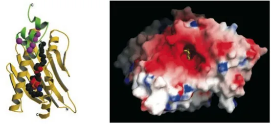
Structural information derived from X-ray crystallography revealed a high similarity with CD1d, a protein involved in the immune response against glycolipid antigens. One molecule of phosphatydilethanolamine was found embedded in a hydrophobic pocket of EPCR in a groove typically involved in antigen presentation (Figure 6).
Fig. 6 Left The recombinant sEPCR (rsEPCR) molecule with a portion of the protein C Gla domain and a lipid molecule. In EPCR (yellow ribbon), two α-helices
and an eight-stranded β-sheet create a groove that is filled with phospholipid (the space-filling balls in the center). Binding of Ca2+ ions (magenta spheres) to the protein C Gla domain (green ribbon) exposes the N-terminal ω loop, which in the absence of EPCR interacts with the phospholipid surfaces on the membrane. The model of the complex consists of residues 7–177 of rsEPCR and the first 33 residues of the protein C Gla domain. Right, Surface representation of the rsEPCR molecule. Electrostatic potentials are mapped on the surface (red negative, blue positive). The head group of the lipid is solvent-exposed (yellow stick model in the center), whereas the fatty acid chains are buried deep in the hydrophobic groove
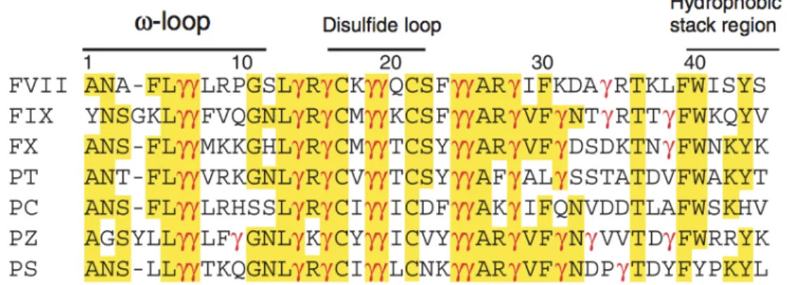
A crystal structure of part of the protein C Gla domain (residues 1–33) bound to soluble EPCR indicated that hydrophobic residues Phe-4 and Leu-8 contained within the ω-loop of the protein C Gla domain and Gla residues 7, 25 and 29, were directly involved in the interaction (Figure 6)(Oganesyan et al., 2002; Preston, 2006). Some of these residues are conserved in other vitamin K dependent enzyme such as FVII, FX and Prothrombin (PT), as indicated in Figure 7.
Fig. 7 Amino acid sequences in the Gla domains of human vitamin K-dependent plasma proteins. Numbers are based on human factor VII. Amino acid residues that are
identical in at least four of the proteins are marked in yellow. γ indicates the γ-carboxyglutamic acid residues. Residues that form the ω-loop, the disulfide loop, and the hydrophobic stack region are marked. (adapted from (GHOSH et al., 2006; Hansson and Stenflo, 2005; López-Sagaseta et al., 2007))
FVII/a is a ligand for EPCR
The idea of an additional binding site for FVII/FVIIa other than TF on the cell surface was first proposed in 1993 by Reuning et Al(Hedner, 2008; Reuning et al., 1993). Binding experiments on LPS-stimulated or non-stimulated endothelial cell (HUVEC) revealed the presence of at least two independent binding sites for FVII/FVIIa of which about 10% are TF specific. This report also suggested that the FVIIa binding site on non-stimulated HUVEC appeared to be a common binding site for vitamin K-dependent proteins. The FVII/a receptor was identified as the endothelial protein C receptor only 13 years later (GHOSH et al., 2006; Pendurthi and Rao, 2010; Preston, 2006).
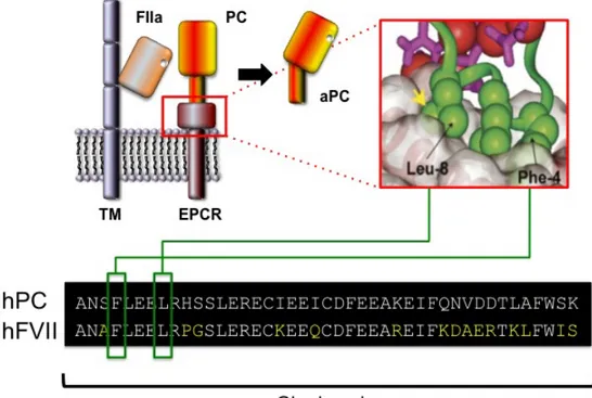
FVII Gla domain shares high homology with PC, in particular the PC residues directly involved in EPCR binding (Phe 4 and Leu 8) are conserved in FVII. In addition, crystal structures data of EPCR bound aPC and FVII indicate that the spatial disposition of the residues required for interaction is conserved among the two proteins (Figure 8) (López-Sagaseta et al., 2007; Oganesyan et al., 2002).
Fig 8 Protein C activation on the endothelial surface by the trombin-thrombomodulin complex. Thrombin (FIIa) bound to trombin-thrombomodulin cleaves
EPCR-bound protein C (PC) to generate activated PC (aPC). A magnification of the crystal structure shows the residues of PC directly involved in the interaction with the receptor. On the bottom, the same residues are indicated on the protein alignment of human PC (hPC) and FVII Gla domains. Adapted from (Preston, 2006; Puy et al., 2010)
The interaction between FVII/a and EPCR is dependent on Ca2+ and Mg2+, as observed for PC/APC. The affinity of both molecules for the receptor is also comparable (Kd of 40
nM and 30 nM for FVII and PC/APC, respectively), as assessed by diverse approaches including surface plasmon resonance or binding on endothelial cells with fluorescently or radioactively labeled FVII/FVIIa (Disse et al., 2011; GHOSH et al., 2006; López-Sagaseta et al., 2007). Despite the similar affinity, PC concentration in plasma is 6 fold higher compared to FVII, implicating that PC would occupy most of the receptor at
physiological concentration. Nevertheless under certain conditions (e.g. therapeutic administration of rFVIIa in hemophiliacs), plasmatic concentrations of the two proteins become comparable (Hedner, 2008; NAYAK et al., 2009). Hence, under these circumstances, FVIIa interaction with EPCR can have relevant implication on the hemostatic response, either modulating protease activity or down-regulating PC activation by competition for receptor occupancy. In order to determine the effect and magnitude of FVIIa-EPCR interaction on coagulation under such therapeutic settings, in vivo models are required.
Up to this date, conflicting data were reported on the effect of EPCR on FVIIa function in vitro. It has been shown that EPCR binding was not affecting FVIIa-mediated FX activation on Human Umbilical Vein Endothelial Cells (HUVECs)(GHOSH et al., 2006; Pendurthi and Rao, 2010). In contrast, the micromolar addition of sEPCR to plasma was able to prolong clotting time in a PT assay and significantly reduce FXa generation on neoplastic cells (H727) (López-Sagaseta et al., 2007). According to Puy(Puy et al., 2010), this effect was caused by an EPCR dependent sequestration of FVIIa from phosphatidylserine-rich regions, a necessary scaffold for coagulation. More recently, Disse et al (Disse et al., 2011) showed that EPCR specifically inhibits activation of FX by soluble TF-FVIIa only in presence of Mg2+. In this model, sEPCR did not primarily target the Gla domain of FVIIa but rather the Gla domain of FX to attenuate FX activation. A possible explanation for these controversial findings on FVIIa activity may be due to different factors, including the cell system used, the source of TF and the relative abundance/localization of TF and EPCR on the surface. Most important, these studies lack in vivo evidences to support the magnitude of this interaction in a flow system. Such evidence will provide essential mechanistic data in the action of pharmacologic FVIIa administration in hemophilia as well as potential approaches that exploit such interaction for improved FVIIa therapeutics (protein or gene-based).
There is accumulating evidence suggesting that clotting factors in the perivascular space, and not only circulating plasma factors, have the potential to contribute to hemostatic protection. The EPCR-PC/APC complexes have been shown to internalize into the cytoplasm of the endothelial cells. The receptor-ligand complexes accumulate in a recycling compartment before being targeted back to the cell surface (NAYAK et al.,
2009). However, it may facilitate the transcytosis of PC/aPC from the apical to the basal side of the cell thus helping to clear it from the circulation and reach the extravascular space. Also EPCR-hFVII/VIIa complexes are internalized by endothelial cells and undergo transcytosis from the apical to the basolateral surface (NAYAK et al., 2009). In vivo data have shown that EPCR appears to be involved in human FVIIa (hFVIIa) uptake in mice (GOPALAKRISHNAN et al., 2010b) and in other murine models hFVIIa has been found in perivascular tissues where it associates with TF after bolus administration (Hoffman et al., 2007) . Interestingly, hFVIIa found in the perivascular space retained coagulant activity for extended period of time (up to 7 days) (GOPALAKRISHNAN et al., 2010a).
It has been observed that prophylactic treatment of hemophilic patients with rFVIIa was effective in reducing bleeding episodes up to 3 moths after suspension of treatment(Konkle et al., 2007). The mechanism that underlies the extended protection observed in these patients during the follow up is still unknown, considering the short half life of rFVIIa in plasma (Erhardtsen, 2000). The study of EPCR-FVIIa interaction in vivo might also be important to understand potential mechanisms involved in rFVIIa-induced secondary prophylaxis.
In order to determine the effect and magnitude of FVIIa-EPCR interaction on coagulation under such therapeutic settings, in vivo models are required. However, such in vivo models must take into account any species-specific incompatibilities. This is certainly the case when one looks at the interaction of FVIIa-EPCR in a mouse system Human and murine EPCR (hEPCR and mEPCR, respectively) share high homology: 21 out of 23 residues of hEPCR directly involved in ligand binding are conserved in mEPCR (LIAW et al., 2001), and the other two are conservatively substituted (Ile-51 by Leu and Arg-81 by Lys). Two residues in the human PC Gla domain appear to influence the PC-EPCR interaction, namely Phe4 and Leu8 (Preston RJ, JBC 2006). Mouse PC, the natural ligand for mouse EPCR, in contrast to human PC, has Phe4 and Met8. However, mouse FVII has Leu4 and Leu8 and has been shown to be a poor ligand for mEPCR (µM affinity).(Margaritis et al., 2004; Puy et al., 2011) One could utilize human rFVIIa but it has a poor affinity for mouse TF (Petersen 2005), hence limited hemostatic effects. Therefore, although the mouse has been indispensable in hemostasis research, the
peculiarity of the mouse EPCR and FVIIa introduces additional complications in efforts in vivo hemostatic effect of a FVIIa-EPCR interaction under conditions of therapeutic FVIIa administration.
AIM OF THE STUDY
Understanding the role of an EPCR-FVIIa interaction in vivo in hemophilia treatment with rFVIIa will have mechanistic and translational ramifications. This thesis aims to define the effects of the EPCR-FVIIa interaction in a setting of FVIIa administration in hemophilia, in vivo. For this, we utilize rational design and functional characterization of a mouse FVIIa molecule that has mouse EPCR binding capacity as a true gain-of-function, without altering other properties of mouse FVIIa. These unique set of characteristics essentially mimic those of human rFVIIa. This allowed us to utilize this novel mouse FVIIa molecule in in vivo hemostatic assays and discern the role of the FVIIa-EPCR interaction in the hemostatic function of therapeutic rFVIIa administration.
METHODS
Construction of expression vectors
Construction of a pcDNA3.1-based (Invitrogen, Carlsbad, CA) expression plasmid vector for murine FVIIa wildtype (mFVIIa) has been previously described (Larson et al., 1998; Margaritis et al., 2004). Chimeras of mFVIIa harboring parts of the murine protein C Gla domain were generated by exchange cloning of gene-synthesized appropriate fragments (Genscript, Piscataway, NJ). All mFVIIa constructs contained the Arg-Lys-Arg-Arg-Lys-Arg (RKRRKR [2RKR]) sequence inserted at Arg-Lys-Arg-Arg-Lys-Arg152- Ile153 of the mature protein and a C-terminal HPC4 epitope tag (EDQVDPRLIDGK) for purification (Margaritis et al., 2004; Philo, 2006). Murine soluble EPCR (amino acids 1-214, msEPCR, NCBI accession L39017) was generated by gene synthesis (Genscript) and cloned into pcDNA3.1 vector. A C-terminal Factor Xa cleavage site (IEGR) followed by an HPC4 epitope tag was also included. Full-length murine EPCR (mEPCR) was cloned into pcDNA3.1 (pcDNA3-mEPCR) following reverse transctiption of total RNA from a murine hemangioendothelioma cell line (Babul and Stellwagen, 1969; Fukudome et al., 1998), a kind gift from Dr. Charles Esmon (University of Oklahoma, OK). The murine tissue factor (mTF) cDNA (NCBI accession NM_010171) was generated by gene synthesis (Genscript) cloned into pcDNA3.1 with a Zeocin resistance selectable marker (pcDNA3-mTF). The identity of all cloned fragments and resulting vectors was verified by DNA sequencing.
Expression and purification of recombinant proteins
Murine FVIIa, mFVIIa chimeras and msEPCR were expressed in human embryonic kidney (HEK 293) cells, following stable DNA transfection as previously described (Camire et al., 2000; Margaritis et al., 2004). Purification of recombinant proteins from conditioned medium was performed using a C-terminal epitope tag (HPC4), as previously reported (Higgins and Mann, 1983; Margaritis et al., 2004). Murine FVIIa and chimeras were further purified on a CHT5-I hydroxyapatite column (BioRad Laboratories,
Hercules, CA), to separate the γ- carboxylated from the uncarboxylated material (Larson et al., 1998). Protein purity was assessed by SDS-PAGE using 4–12% polyacrylamide gels (Invitrogen). Protein concentration for mFVIIa or its chimeras were determined using an average molecular weight and extinction coefficient of 50,000 and 1.39, respectively.
Analytical Ultracentrifugation
To calculate the protein concentration of recombinant msEPCR, analytical ultracentrifugation was used to determine the average molecular weight and protein extinction coefficient. Sedimentation velocity was performed at 45K rpm in (20 mM HEPES, 150 mM NaCl, pH 7.4) and 20°C using an XL-I instrument using interference optics in a AN60-Ti rotor and two sector cells with sapphire windows. Molecular weight and sedimentation coefficient were obtained by fitting g(s*) distributions using DCDT software, as previously described (Philo, 2006). The extinction coefficient was determined by differential refractometry (Babul and Stellwagen, 1969). We determined that the molecular weight of the recombinant msEPCR preparation was 39,700 ± 2,800 Da, the sedimentation coefficient (s20,w) was 2.72 ± 0.05 S. Gamma-carboxyglutamic acid analysis on all recombinant proteins was performed as previously described (Camire et al., 2000).
Generation of CHO-‐K1 stable cell lines expressing full-‐length murine EPCR and tissue factor
To generate a stable cell line expressing full-length mEPCR or mTF, CHO-K1 cells (ATCC® Catalog No. CCL-61) were transfected using Lipofectamine 2000 (Invitrogen, for mEPCR) and selected in 1 mg/ml G418-sulfate or 400 µg/ml zeocin growth medium (Invitrogen, for mTF). Resistant colonies were screened by flow cytometry using an anti-mEPCR antibody (anti-mouse CD201-APC 0.5 µg/ml, eBioscience, San Diego, CA) or an anti-mTF antibody (goat anti-mTF at 4 µg/ml, Cat. AF3178, R&D Systems, Minneapolis, MN) followed by a Alexa-Fluor 488 labeled donkey anti-goat IgG (2 µg/ml, Cat. A-11055, Invitrogen). Geneticin- or zeocin-resistant CHO-K1 cells (negative controls for cell binding experiments) were generated by stable transfection of an empty
pcDNA3 vector harboring a neomycin or zeocin resistance marker (respectively) and selection in appropriate medium, as described above.
Phospholipid Binding assay
Phospholipid vesicles containing phosphatidylcholine and phosphatidylserine (PC:PS, 75:25 % [w/w]) were prepared as described previously(Higgins and Mann, 1983). Binding assays were performed as previously described (Dahlback, 2005; Saller et al., 2006; Vadivel and Bajaj, 2012). Recombinant mFVIIa or mFVIIa-FMR was incubated for 2 h on plates coated with 50 µl of 80 µM PC:PS vesicles in TBS (50 mM Tris, 150 mM NaCl, 5 mM CaCl2 pH 7.4)-0.2% BSA (TBS-BSA). Plates were washed 3 times with TBS-0.5% Tween-20, to remove non-specifically bound material. Bound protein was visualized colorimetrically (at 492nm) with mouse monoclonal anti-HPC4 antibody (1 µg/ml in TBS-BSA) and a secondary rabbit anti-mouse HRP conjugated antibody (1:1000 in TBS-BSA, Dako) Following subtraction of non-specific signal (phospholipids alone or protein alone), calculation of Kdapp calculation and non-linear regression analysis
was performed using the GraphPad Prism v5.0b Macintosh software package.
Isothermal titration calorimetry (ITC)
All proteins were dialyzed overnight versus 20 L of 20 mM HEPES, 150 mM NaCl, 1.6 mM Ca2+, 0.6 mM Mg2+ and pH 7.4 (“Reaction Buffer”). Proteins were concentrated after dialysis by centrifugal ultrafiltration using an Amicon Ultracel 30K device (Millipore, Billerica, MA) and centrifuged to remove particulate material. Buffers and proteins were degassed before use. ITC experiments were performed at 25°C using a MicroCal ITC200 microcalorimeter (GE Healthcare, Piscataway, NJ). A reaction mixture for ITC (204 µl) of mFVIIa-WT (25 µM) or its chimeras was loaded into the sample cell. Titration curves were initiated by injection of 0.8 µl msEPCR (250 µM) followed by successive 2 µl injections of msEPCR every 180 s, under continuous stirring. Injections of ligand into reaction buffer were performed to determine baseline corrections. The integrated heats from each injection, normalized to the moles of ligand per injection,
were fit to a single-site binding isotherm using ORIGIN v7 (OriginLab Corp., Northampton, MA). The integrated peak of the first injection was excluded from the fit. Lack of proteolysis during the experiment was verified by SDS PAGE analysis of samples prior to and after ITC. The heat of dilution of msEPCR into buffer under the same experimental conditions (background) was subtracted from the fitted data.
Cell-‐surface binding assay
Determination of ligand binding to cell-surface exposed receptor (mEPCR or mTF) was performed as previously described(GHOSH et al., 2007)with some modifications. Specifically, confluent monolayers in a 24-well plate of mEPCR-expressing (CHO-K1-mEPCR), or G418 resistant CHO-K1 cells were incubated with increasing concentration of ligand in Buffer “B” (10 mM HEPES, 150 mM NaCl, 4 mM KCl, 11 mM Glucose, 1mg/ml bovine serum albumin [BSA], 1.6 mM Ca2+, 0.6 mM Mg2+) at 4 °C on ice for 1 h. After 4 washes with ice-cold Buffer “B”, the surface-bound ligand was eluted with Buffer “C” (10 mM HEPES, 150 mM NaCl, 4 mM KCl, 11 mM Glucose, 1 mg/ml BSA, 5 mM ethylenediaminetetraacetic acid [EDTA]) and subjected to polyacrylamide gel electrophoresis and detection by western blotting. Protein presence in the eluates was detected with a mouse monoclonal anti-HPC4 antibody (5µg/ml, (Margaritis et al., 2004)) and a secondary goat anti-mouse IgG antibody (IR-Dye CW800, 1:10000 dilution, Li-Cor Biosciences, Lincoln, NE). Blots were imaged at 169 µm with Odyssey imager (Li-Cor Biosciences). After image background subtraction, band intensities were quantified and corrected by nonspecific binding using the ImageJ software (Schneider et al., 2012). Only the heavy chain intensity of each recombinant protein was used for quantification due to the non-specific signal from BSA in the reaction buffer overlapping with the trace amounts on zymogen in each blot. Integrated densities were plotted and affinity constants (Kd) were calculated using a one-site specific binding curve fitting with GraphPad Prism v5.0b (GraphPad Software, Inc., La Jolla, CA). Conversion to nM was done using the integrated densities of known amounts of ligand in Buffer “C”, electrophoresed and blotted as described above.
Factor Xa generation assay
Mouse FX (mFX) and activated mFX (mFXa) were kind gifts from Dr R. M. Camire (The Children’s Hospital of Philadelphia, PA). CHO-K1 expressing mTF were seeded on a 96 well plate (1.3E4 cells/well, Cat. 353872, BD PrimariaTM Clear MicrotestTM Plate) 48 h before the experiment. Confluent monolayers were washed twice with Reaction Buffer (20 mM HEPES, 150 mM NaCl, 0.1% (w/v) PEG 8000 pH 7.45, 1.6 mM Ca2+, 0.6 mM Mg2+) and placed on an Eppendorf MTP Thermoblock at 37°C. One hundred pM of mFVIIa or mFVIIa-FMR in reaction buffer were added. The reaction was initiated after 3 minutes by adding murine FX (mFX, 160 nM final concentration) in a 100 µl total reaction volume. At selected time points (0-30 min) an aliquot of the reaction mixture was diluted in Quench Buffer (20 mM HEPES, 150 mM NaCl, 0.1% (w/v) PEG 8000 pH 7.45, 50 mM EDTA). Chromogenic substrate S-2222 (DiaPharma, West Chester, OH) was added at a final concentration of 400 µM and activated mFX (mFXa) generation was monitored by measuring A405 at 22°C using a VMax plate reader (Molecular Devices, Menlo Park, CA). The concentration of mFXa generated over time was calculated by interpolation of the initial rate of hydrolysis of S-2222 by known amounts of mFXa (0-11 nM) in Quench Buffer.
Thrombin generation, coagulation assays and determination of mFVIIa antigen Thrombin generation was determined according to methods previously described ((Ivanciu et al., 2011), (Bunce et al., 2011)). Prothrombin time (PT) assays on plasma, cell culture supernatants, and purified proteins were determined as previously described (Margaritis et al., 2011) using human Factor VII deficient plasmas (George King Bio-Medical Inc, Overland Park, KS) and Innovin (Siemens Healthcare, Malvern, PA). Quantification was performed using standard curves of increasing concentration of each recombinant protein. Determination of uninhibited (free) mFVIIa or mFVIIa-FMR in mouse plasma was performed by an ELISA, as previously described (Margaritis et al., 2011) and/or a clotting-based assay with standard curves of increasing concentration of each recombinant protein.
Animal experiments and procedures
All experimental procedures were reviewed and approved by the Institutional Animal Care and Use Committee (IACUC) at The Children's Hospital of Philadelphia. Weight and age (8-12 weeks) matched male Hemophilia B (Lin et al., 1997), Hemophilia A (Bi et al., 1995) mice backcrossed on C57BL/6 background for at least 6 generations and hemostatically normal male mice (C57BL/6 background, The Jackson Laboratory, Bar Harbor, ME) were used in all experiments. Blood was collected via the retro-orbital plexus into 3.8% citrate. For EPCR blocking experiments, a rat monoclonal anti-EPCR antibody (RCR-252, Cat. E6280, Sigma) or isotype rat IgG (Cat. 0108- 01, Southern Biotech, Birmingham, AL), both in PBS, were infused via tail vein within 1 hour prior to the administration of mFVIIa or variant. Quantification of recovery and antigen levels in Figure 6 was determined by a clotting-based as well as an antigen-based (see above) assay, reported as an average of both.
Murine carotid artery thrombosis and murine tail clip hemostasis models
Ferric chloride–induced injury was performed according to our previously published procedures (Ivanciu et al., 2011) using 7.5% FeCl3. In some experiments, antibody (RCR-252 or isotype IgG [see section above]; 50 µg in 100µl of PBS) was infused 30 min prior to the initiation of the FeCl3 carotid artery injury model. The tail clip assay was performed according to our published procedures(Ivanciu et al., 2011; Margaritis et al., 2004; 2011; 2009) with some modifications. Mice were anesthetized with isoflurane, the tail was pre-warmed at 37 °C and the distal portion transected at a diameter of 3 mm. The tail was then placed in a conical tube containing 14 ml of saline at 37 °C and blood was collected for 2 min. Procoagulant or PBS was then infused via the jugular vein, the injured tail was immediately moved to a fresh tube of saline (14 ml at 37 °C), and blood was collected for an additional 8 min. Quantitative assessment of blood loss was determined by measuring total hemoglobin by absorbance at 575 nm following red cell lysis and converted to total blood loss (µl) using an appropriately established standard curve.
Statistical analysis
Two-tailed Mann-Whitney U and Kruskal-Wallis tests were performed using the GraphPad Prism v5.0b Macintosh software package (GraphPad Software). Statistical differences were considered significant when P < 0.05.
RESULTS
Generation of mFVIIa constructs with affinity to mEPCR
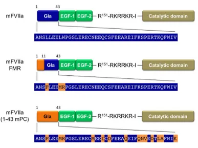
Since protein C (PC) interacts with EPCR through the Gla domain (Oganesyan et al., 2002), we generated two chimeric mFVIIa molecules with modified Gla domains using either part or the entire Gla domain of murine protein C (mPC) (Figure 1). Both chimeric molecules, as well as mFVIIa, contain a PACE/furin cleavage site enabling their secretion in the two-chain, activated form, as we have previously shown (Margaritis et al., 2004; 2009; 2011). The chimeric mFVIIa-(1-43 mPC) molecule contains the full mPC Gla domain and therefore should exhibit maximal affinity of the chimeric molecule to mEPCR. However, such modification of the Gla domain could perturb the coagulant activity of the resulting molecule. For this reason, we generated a minimally-mutated chimeric molecule mFVIIa-FMR that contains the first 11 amino acids of secreted mPC, resulting in just three amino acid changes relative to mFVIIa (L4F, L8M and W9R, Figure 1). We utilized this part of the mPC Gla domain because it defines the specificity of the PC-EPCR interaction (Preston, 2006).
Fig. 1 Structure of the recombinant proteins used in this study. All proteins have a
γ-carboxyglutamic acid (Gla) domain, two epidermal growth factor (EGF) domains (EGF-1 and EGF-2) and a catalytic domain. A recognition site (Arg-Lys-Arg-Arg-Lys-Arg [RKRRKR]) for a paired amino acid cleaving enzyme (PACE)/furin type protease is inserted after the Arg at position 151. The sequence of the Gla domain is shown and highlighted in blue for each protein. Changes in the Gla domain compared to mFVIIa taken from the murine PC Gla domain are highlighted in orange. Numbering refers to the mature form of each protein.
Expression of mFVIIa chimeras
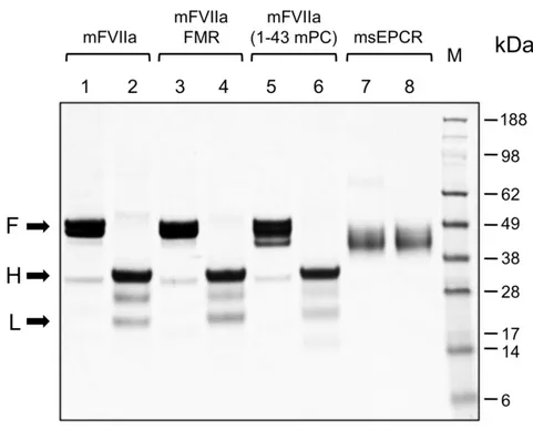
We generated stable clones of HEK293 cells expressing either mFVIIa, mFVIIa-FMR, mFVIIa-(1-43 mPC) or murine soluble EPCR (msEPCR) and purified recombinant proteins. For mFVIIa and its chimeras, the inclusion of a hydroxyapatite column purification step ensured that purified material had a full complement of Gla domain modifications, confirmed by γ-carboxyglutamic acid analysis (data not shown). SDS-PAGE of purified proteins under reducing or non-reducing conditions revealed that mFVIIa and its chimeras were secreted and purified in the activated, two-chain form (Figure 2, lanes 2, 4 and 6). In order to accurately determine the msEPCR protein concentration, the E280 0.1% extinction coefficient of the msEPCR protein preparation (0.66) was determined by differential refractomery in the analytical centrifuge (Babul and Stellwagen, 1969).
Fig. 2 Coomassie brilliant blue staining of SDS PAGE gel of purified proteins. Five
µg of mFVIIa, mFVIIa chimeras or msEPCR were electrophoresed under reducing or non-reducing conditions. The apparent molecular weights of the marker standard (M) are indicated in kDa. Lanes 1,3,5 and 7: non-reducing conditions; lanes 2,4,6,8: reducing conditions. Arrows point to the full-length (F) (under non-reducing conditions) and the heavy (H) and light (L) chains (under reducing conditions) of each protease.
Generation of CHO-‐K1 stable cell lines expressing full-‐length murine EPCR and/or TF
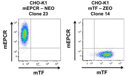
In order to assess the ability of the mFVIIa chimeras to bind to murine EPCR as a gain-of-function when the receptor is presented in a phospholipid membrane context, we generated a stable cell line expressing full-length mEPCR (CHO-K1-mEPCR) in a homogeneous fashion, verified by flow cytometry (Fig 3). We also generated a CHO-K1-based mTF expressing cell line (CHO-K1-mTF) for cofactor binding experiments and functional assays. To control for non-specific binding, CHO-K1 cells stably transfected with empty expression vector were also generated.
Fig. 3 Scatter plots of CHO-K1 clones expressing mEPCR or mTF on the cell surface. CHO-K1 cells were dissociated in non-enzymatic dissociation buffer (Gibco)
and stained for mEPCR and mTF. Receptor expression was evaluated by flow cytometry, quadrants were defined by negative controls (CHO-K1 naïve cells stained for both receptors).
mFVIIa chimeras bind to mEPCR when expressed on the cell surface or in solution
CHO-K1-mEPCR (or control CHO-K1) cells were incubated with increasing concentration of ligand (mFVIIa or its chimeras) at 4°C for 1 h in presence of physiological concentrations of Ca2+ and Mg2+ ions to ensure maximal binding, as has been previously shown for human PC and EPCR (Kurosawa, 1996). Analysis of bound ligand showed that mFVIIa lacked appreciable affinity to mEPCR (Figure 4, left panel) even at high concentration (1 µM), as expected (Puy et al., 2011; Sen et al., 2012). In contrast, both mFVIIa-FMR and mFVIIa-(1-43 mPC) showed specific and dose-dependent binding to mEPCR (Figure 4, middle and right panel, respectively). Fluorescent densitometric analysis allowed us to estimate a Kd of interaction of 0.34 ± 0.07 µM and 0.19 ± 0.06 µM for mFVIIa-FMR and mFVIIa-(1-43 mPC), respectively.
Fig. 4 Recombinant mFVIIa chimeras bind to mEPCR on CHO-K1 cells in contrast to mFVIIa. Increasing concentration of ligand (10-1000 nM) was incubated on CHO-K1
or CHO-K1-mEPCR cells in the presence of 1.6 mM Ca2+ and 0.6 mM Mg2+ for 1h at 4°C. Bound fraction was eluted and visualized by fluorescence-based western blotting (top part, images shown in greyscale) and band intensities were quantified and corrected for nonspecific binding (on CHO-K1 cells). Integrated band intensities were plotted (bottom part) and Kd constants were calculated. Conversion to nM was done using the
integrated densities of known amounts of each ligand and values were normalized for cell numbers. Depicted curves for each protein are a representative of three experiments.
To further validate the interaction between mFVIIa chimeras and mEPCR, isothermal titration calorimetry (ITC) was used. This solution-based assay can determine the magnitude of the two thermodynamic terms that define the binding affinity between two proteins ΔH and ΔS (Leavitt and Freire, 2001), as well as the Kd and stoichiometry of interaction (n). In ITC, titration of msEPCR into mFVIIa, resulted in weak heat measurements that did not allow for the accurate determination of thermodynamic terms (Figure 5, left panel). This is indicative of a weak interaction between the two proteins (Kd > 8 µM), confirming our results from cell binding experiments (see Figure 4). In contrast, mFVIIa-FMR and mFVIIa-(1-43mPC) exhibited significant heat flow as a result of specific interaction with msEPCR, with a stoichiometry of ~1 for both chimeras, as
shown by the resulting isotherms (Figure 5, middle and right panels, respectively). Moreover, the equilibrium dissociation constants obtained from data fitting (0.48 ± 0.02 µM for mFVIIa-FMR and 0.11 ± 0.01 µM mFVIIa-[1-43mPC], Table 1) were in good agreement with our findings on CHO-K1-mEPCR cells.
Fig 5. Recombinant mFVIIa chimeras bind to murine soluble EPCR (msEPCR) in contrast to mFVIIa. Upper portion of each panel: heat flow tracings were obtained upon
successive injections (2 µl) of msEPCR into the sample cell containing 25 µM mFVIIa or mFVIIa chimera in dialysate at 25°C. The first injection (0.5 µl) was eliminated from the analysis. The heat flow profile from an identical injection of msEPCR into dialysate was used as a reference and is shown offset (top of upper panel) for clarity. Bottom portion of each panel: corrected integrated heats (Kcal/mole of injectant) were analyzed. The line is drawn according to parameters listed in Table 1.
Table 1. Thermodynamic constants of interaction of msEPCR with mFVIIa or its chimeras using isothermal titration calorimetry.
Kd: dissociation constant; n: stoichiometry of interaction; ΔG: change in Gibbs free
energy (G); ΔH: change in enthalpy (H); ΔS: change in entropy (S); ND: not determined. All parameters are listed as ± 67% confidence intervals.
Collectively, our data confirm that binding to mEPCR, either when it is presented on the cell surface or in solution, can be engineered as a gain-of-function to mFVIIa by modification of its Gla domain. This can be achieved by a small change in the ω-loop of the protease (mFVIIa-FMR), thus suggesting that the mEPCR minimal binding region is located in the first 11 residues of mPC Gla domain.
Partial substitution of the Gla domain of mFVIIa does not affect its in vitro activity
The Gla domain of human FVII contributes to the interaction with tissue factor (Banner et al., 1996) thus affecting function within the initiation of coagulation. Therefore, prior to further in vivo experimentation, we tested whether Gla domain modification in the mFVIIa chimeras perturbs coagulant function. Using a clotting based assay, we found that the partial Gla domain modification in mFVIIa-FMR did not affect activity (Figure 6 A, P > 0.05 vs. mFVIIa). In contrast, full transplant of the mPC Gla domain to mFVIIa in the mFVIIa-(1-43 mPC) chimeric molecule abolished activity (Figure 6A, P < 0.05 vs. mFVIIa or FMR). Therefore, we decided to further characterize the mFVIIa-FMR molecule only, relative to mFVIIa.
To investigate the mFVIIa-FMR affinity for the natural FVII/FVIIa cofactor (TF), we used CHO-K1 cells stably expressing full-length murine TF (mTF, Figure 3, right) or CHO-K1 controls. We performed binding experiments with increasing ligand (mFVIIa or
Sample Kd (µM) n ΔG (Kcal mol-1) ΔH (Kcal mol-1) ΔS (cal mol-1K-1) mFVIIa >8 ND ND ND ND mFVIIa-FMR 0.48 ± 0.02 0.89 -8.6 - 4.7 ± 0.03 13.3 mFVIIa-(1-43 mPC) 0.11 ± 0.01 0.85 -9.5 -4.6 ± 0.03 16.4
mFVIIa-FMR) concentration added, as we described for CHO-K1-mEPCR cells. Both Ca2+ and Mg2+ cations were present at their physiological concentration. We found that mFVIIa-FMR and mFVIIa exhibited similar affinity for mTF (Figure 6B), as demonstrated by the estimated Kd of 14.9 ± 11.1 nM (mFVIIa-FMR) and 16.9 ± 8.9 nM (mFVIIa). The affinity of mFVIIa to mTF is lower than previously reported (Petersen et al., 2005) but could be due to the assay system employed or the source of TF (soluble of mTF vs full length), as observed with human FVIIa and human TF (see (Sen et al., 2010) for references). Our data suggest that the modifications in the Gla domain present in mFVIIa-FMR do not affect its interaction with mouse TF or the function of the molecule, compared to mFVIIa. Therefore, mFVIIa-FMR is functionally equivalent to mFVIIa but with the added ability to bind mEPCR.
Fig. 6 Comparison of murine TF-dependent binding and in vitro activity of mFVIIa and mFVIIa chimeras. (A) Using a prothrombin time clotting assay, mFVIIa and
FMR exhibit similar activity. Asterisk indicates P < 0.05 vs. mFVIIa or mFVIIa-FMR. Data are expressed as mean ± 1 SD. NS: not statistically significant difference. (B) Binding of mFVIIa (!) or mFVIIa-FMR (") on CHO-K1 cells expressing mTF. Increasing concentration of ligand (10-250 nM) was incubated on CHO-K1 or CHO-K1-mTF cells and bound fraction analyzed as described in Figure 3. Both ligands exhibit similar mTF binding.
The Gla domain is also involved in the interaction with phospholipid membranes and therefore modulates FVIIa activity. We tested the ability of mFVIIa and mFVIIa-FMR to bind phosphatidylcholine:phosphatidylserine (75/25) vesicles to simulate an activated phospholipid surface, as described by (Saller, 2005). As shown in Figure 8, both mFVIIa
molecules bound liposomes with similar affinity, mFVIIa-FMR showing a slightly reduced Kd compared to mFVIIa (730 ± 55 nM vs 420 ± 24 nM, respecitvely).
Figure 7. Phospholipid binding affinity of mFVIIa and mFVIIa-FMR. Increasing
concentration (0-4µM) of each mFVIIa or mFVIIa-FMR was incubated on a 96-well plate precoated with a mixture of 75:25 of phosphatidylcholine:phosphatidyl serine phopsholipids. Following washing of unbound material, the bound protein was detected by western blot. Quantification of bound material allowed for determination of calculated affinity of each variant for such procoagulant phospholipids.
In order to verify that mFVIIa-FMR and mFVIIa can form functional complexes with mTF in the context of a cell membrane, we determined the rate of generation of activated murine FX (mFXa) following incubation of CHO-K1-mTF cells with either mFVIIa or mFVIIa-FMR. As shown in Figure 8, both mFVIIa and mFVIIa-FMR exhibited similar rates of mFXa generation (2.20 ± 0.07 nM/min for mFVIIa and 2.00 ± 0.03 nM/min for mFVIIa-FMR). 0.0 0.5 1.0 1.5 2.0 2.5 3.0 3.5 4.0 0.0 0.2 0.4 0.6 0.8 1.0 [Protein] (µM)
OD (492 nm)
mFVIIa mFVIIa-FMRFig. 8 Generation of mFXa over time on CHO-K1 cells expressing mTF following
incubation of mFVIIa (!) or mFVIIa-FMR ("). Both ligands exhibit similar rates of mFXa generation.
In order to assess if mEPCR binding could influence mFXa generation, we co-transfected and subcloned clone 14 of CHO-K1-mTF (previously used for mFXa generation assays), generating a CHO-K1-mTF/mEPCR expressing cell line. mTF expression between CHO-K1-mTF and CHO-K1-mTF/mEPCR was comparable, as confirmed by flow cytometry (Figure 9) analysis and mFVIIa titration assays (Figure 10).
Fig. 9 Scatter plots and histogram of CHO-K1 clones expressing mEPCR and mTF on the cell surface. Left, CHO-K1 cells were dissociated in non-enzymatic dissociation
buffer (Gibco) and stained for mEPCR and mTF. Receptor expression was evaluated by flow cytometry, quadrants were defined by negative controls (CHO-K1 naïve cells stained for both receptors). Right, histogram of CHO-K1 naïve (blue line), CHO-K1-mTF (red line) and CHO-K1-mTF/mEPCR (light blue line), stained for mTF.
Fig. 10 Functional TF determination on CHO-K1 expressing mTF or mTF and mEPCR mFX activation was measured at 10 min in HBS PEG 0.1% pH 7.45, 5 mM
Ca2+, 37 C° in presence of increasing concentration of mFVIIa or FMR (0.005 – 5 nM), 160 nM mX and 400 µM S-2222.
In the presence of mTF and mEPCR, mFXa generation rates were 1.71 ± 0.10 nM/min for mFVIIa and 1.87 ± 0.08 nM/min for mFVIIa-FMR (Table 2). There is a trend for a slightly higher rate of mFXa generation vs. mTF alone (see Table 2). However, under the same experimental conditions, mFX activation on CHO-K1-mEPCR cells (no mTF) was undetectable (data not shown), collectively suggestive of small (if at all) synergistic mTF-mEPCR synergistic effect on mFX activation in vitro. Unfortunately, further analysis on such effect is difficult given that the local concentration of the receptor on the cell surface in relation to mTF might be suboptimal (we did not quantify receptor density).
Sample nM mXa / min
mTF mTF mEPCR
mFVIIa 2.20 ± 0.07 1.71 ± 0.10
mFVIIa-FMR 2.00 ± 0.03 1.87 ± 0.08
Table 2 Comparison of activation rates of mFX by mFVIIa or mFVIIa-FMR on
CHO-K1 expressing mTF alone or mTF and mEPCR on the cell surface. Rates are listed as ± 67% confidence intervals.