UNIVERSITÀ DEGLI STUDI DI FOGGIA
DOTTORATO DI RICERCA
IN
“INNOVAZIONE E MANAGEMENT DI ALIMENTI AD ELEVATA VALENZA SALUTISTICA”
(XXX CICLO)
TESI DI DOTTORATO
Oral Cancer: from the research of biomarkers for diagnosis and
prognosis to the treatment with nutraceuticals
Dottorando: Dott. Giuseppe Troiano
Tutor: Prof. Lorenzo Lo Muzio
2
Abstract
Oral Squamous Cell Carcinoma (OSCC) accounts for more than 500.000 new cancer cases every year globally, and in high-risk countries is one of the most common cancer in men. When diagnosed at a late stage, treatment of OSCC is highly debilitating and the disease if often fatal. Patients diagnosed in early stage OSCC has a favorable prognosis and requires less aggressive treatment. The overall 5-year survival rate for OSCC is below 50% and has only improved modestly over the past two decades despite improvements in treatment modalities.
The diagnostic delay seems to be one of the main obstacles to overcome in order to diagnose tumors in early stage. The research of clinical biomarker aims to identify molecules capable of predicting the risk of the disease' presence and help clinicians in the early diagnosis. Furthermore, another field of research is related to the research of prognostic biomarker, in order to address patients towards personalized treatment modalities on the basis of the molecular aggressiveness of the neoplasia. Moreover, there is the need to find new therapeutic drugs aiming to improve the survival rate of OSCC patients. In this field, nutraceuticals, any substance considered to be a food or a food ingredient that provides medical and health benefits, showed promising effects for the treatment of some types of cancer. However, further studies are useful before to include them in the current therapeutic protocols.
The aim of this thesis was twofold, first the research of possible clinical biomarkers of OSCC diagnosis and prognosis; and secondly addressed to assess the effects of treatment with Curcumin and Genistein on OSCC cell lines.
Results of several studies included in this thesis, performed for the research of clinical biomarker, showed the identification of promising proteins for the diagnosis of OSCC. Among these, neurotrophin-3 (NT-3) revealed the better diagnostic prediction values when quantified in plasma of patients with OSCC.
3 Related to the prognostic prediction we performed meta-analysis of the literature for the long non-coding RNA (lncRNA) HOTAIR and demonstrated as this marker seems to be able to predict words prognosis when expressed at higher levels in the tumor bulk.
Finally, we assessed the treatment effects with curcumin and genistein on OSCC cancer cell lines. Results showed a reduction in: viability, migration, proliferation of cancer cells and increasement in the rate of apoptosis after treatment with different dosages of this nutraceuticals.
In conclusions, we identified promising biomarker for the prediction of diagnosis and prognosis of OSCC patients. In addition, results of our studies showed that treatment with curcumin and genistein lead to inhibition of: viability, migration, proliferation, and increase in the rate of apoptosis of OSCC cell lines.
4 SUMMARY
Introduction ... 6
1.1. Introduction and thesis motivation ... 6
Research of diagnostic and prognostic biomarkers in OSCC... 8
- 2.1. Background ... 8
- 2.2. Literature Review of circulating miRNA for the detection of OSCC ... 9
- 2.3 Analysis of circulating miRNAs and proteins for the detection of patients with tongue squamous cell carcinoma ... 12
2.3.2 Materials and methods ... 13
2.3.3 Results ... 15
- 2.4 Meta-analysis of HOTAIR as prognostic factor in patients with squamous cells carcinoma of the head and neck ... 20
Effects of treatment with Curcumin and Genistein on OSCC cell lines ... 24
- 3.1. Background ... 24
- 3.2. Materials and Methods ... 25
- 3.3. Results... 28
Discussion and Conclusions... 34
References ... 37
Appendix 1: Journal publications ... 42
Appendix 2: Abstracts and Conference Proceedings... 49
Appendix 3: Oral Presentations... 53
Appendix 4: Honors and Awards ... 54
5 Dedicated to the memory of my mother
6
Introduction
1.1. Introduction and thesis motivation
The most recent statistics from the US estimated the incidence for cancer of the oral cavity at 2.7 per 100,000 (3.7 in men and 1.8 in women) in 2012, with substantial differences by sex, age, and region (1). Furthermore, highest incident for oral cavity cancer was reported in south-central Asia (40.9% of all incident cases) (2). The incidence of oral cavity cancer was consistently greater among men than women, with an M/F rate ratio of 2.1, ranging from 5.2 for Central and Eastern Europe to 1.4 for Northern Africa, Western Asia and Oceania (2). Risk factors involved in the development of oral squamous cell carcinoma (OSCC) are considered: tobacco smoke, alcohol consumption, reverse smoke and betel chewing. Reaching an early diagnosis of symptomatic OSCC is crucial for a better prognosis in terms of disease stage, survival and quality of life (3). In fact, patients diagnosed in early stages have a longer life expectancy compared to patients who get the diagnosis in advanced stages (4). The overall 5-year survival rate for OSCC is below 50% and has only improved modestly over the past two decades despite considerable improvement of treatment modalities (5).
Recently the 8th edition of the American Joint Committee on Cancer (AJCC) introduced in the staging system of the oral cavity squamous cell carcinoma (OSCC) the depth of invasion (DOI) as a variable for the T category (6). The new staging manual highlights the importance to distinguish between tumors with different DOI and the need to use the latter for staging purposes. However, although tumor-node-metastasis (TNM) staging is routinely used to predict tumor behavior and, hence, to inform the choice of treatment strategies for OSCC, patients with same TNM stages may result in dramatically different survival time (7).
Due to these reasons, the scientific community put its attention to the research of biomarkers for the early diagnosis and prognosis prediction. The prognosis prediction aims to develop personalized treatment planned on the basis of the molecular profiling of the tumor. In this way, clinicians could
7 modulate the aggressiveness of the treatment on the basis of a molecular risk profile. From the point of view of diagnosis, a biomarker of early diagnosis could decrease the rate of patients treated in advanced stages and strongly increase the overall survival rate of patients with OSCC.
In fact, as previously mentioned, the advent of new treatment modalities who demonstrated benefits for other types of malignancies, failed to improve survival for OSCC patients. With regards to the advanced OSCC, guidelines from the National Comprehensive Cancer Network still today recommend surgical excision followed by concurrent single– agent cisplatin chemo-radiotherapy in T3 or T4 lesions demonstrating adverse features of extracapsular spread and/or positive margins (8). For such reasons, there is the need to figure out the molecular basis of this pathology in order to provide clinicians new treatment options they can use in the clinical practice.
‘Nutraceutical’’ term (a combination of the words ‘‘nutrition’’ and ‘‘pharmaceutical’’) refers to any substance considered to be a food or a food ingredient that provides medical and health benefits (9). Recently, some nutraceutical agents showed anti-inflammatory, antioxidant and anticancer properties in several studies (10, 11). Among these, curcumin and genistein are among the nutraceuticals the most promising (12-14).
Therefore, the aim of this thesis was twofold, first, the research of possible clinical biomarkers of OSCC diagnosis and prognosis; and secondly to assess the effects of treatment with Curcumin and Genistein on OSCC cell lines.
8
Research of diagnostic and prognostic biomarkers in OSCC
- 2.1. Background
The World Health Organization (WHO) defines a biomarker as any substance, structure or process that can be measured in the body or its products and influences to predict the incidence of outcome or disease (Biomarkers in Risk Assessment: Validity and Validation, Environmental Health Criteria Series, No222, WHO). Biomarkers can be used for patient assessment in multiple clinical settings, including estimating the risk of disease and distinguishing benign from malignant tissues.
Cancer biomarkers can be classified according to the disease state, such as predictive, diagnosis and prognosis biomarkers (15). A diagnostic biomarker aims to detect or confirm the presence of a disease or condition of interest or to identify individuals with a subtype of the disease. Instead, a prognostic biomarker informs about a likely cancer outcome (e.g., overall survival, disease-free survival, and cause-specific survival) independent of the received treatment. The identification of prognosis biomarkers could guide clinicians in modulating the best therapeutic approach based on the molecular aggressiveness of the pathology. Predictive biomarkers, sometimes referred to biomarkers used to identify individuals who were more likely than similar individuals without the biomarker to experience a favorable or unfavorable effect from exposure to a medical product or an environmental agent (16).
These biomarkers allow clinicians to select a set of chemotherapeutic agents which will work best for an individual patient. For example, Herceptin is useful in breast cancer lesions showing only Her2/Neu overexpression, whereas tamoxifen is the preferred treatment for other breast cancer lesions. Thus Her-2/Neu is a predictive cancer biomarker for a subset of breast cancer therapies (17). Likewise, drugs such as erlotinib or gefitinib work only in lung cancer patients with specific mutations in the epidermal growth factor receptor (EGFR) gene (18). Although, several studies have been performed aiming to find out biomarkers of diagnosis and prognosis for OSCC, no molecule
9 has still been introduced in the clinical practice. For such reason, there is the need to go deep in the study related to biomarkers of diagnosis and prognosis in OSCC.
- 2.2. Literature Review of circulating miRNA for the detection of OSCC
Although, proteins still represent the final effectors of the cell processes, more recently the discovery of coding RNA added a new layer to our understanding of biological processes. The term non-coding RNA (ncRNAs) encompasses microRNA (miRNAs), long nonnon-coding RNAs (lncRNAs), circular RNAs (circRNAs), and others (19). Although ncRNAs do not encode proteins, they are master regulators of gene expression through various mechanisms (20, 21). In the field of cancer research, it has become apparent that aberrations within the noncoding genome drive fundamental cancer phenotypes in addition to the best-known protein coding mutations (22). MiRNAs are the most studied group of ncRNAs. They are long in between 19 and 23 nucleotides. They are able to negatively regulate target messenger RNAs (mRNAs) by partially binding to their 3’ untranslated region (23). In addition, miRNAs seem to be generated from intergenic genomic sequences or intronic regions of protein-coding genes (23).
The majority of mammalian miRNAs are transcribed by the RNA polymerase II into a long precursor containing a typical stem-loop structure (pri-miRNAs) (Fig. 1) [11].
10 Figure 1: Mechanisms involved in synthesis of miRNAs. It starts in the nucleus and ends with secretion once the miRNa has reached its final stage.
These initial transcripts undergo cleavage by the ribonuclease enzyme III Drosha to produce the pre-miRNAs of around 70 nucleotides and with hairpin structure. Pre-pre-miRNAs are then transported into the cytoplasm by a nuclear export factor (Exportin-5), where they are cleaved by an enzyme called Dicer into duplexes of 21–23 nucleotides (24). Afterwards, one strand of the duplex, the passenger strand (miRNA⁄), is degraded and the other incorporated together with Argonaute proteins or lipoproteins to form the RNA-induced silencing complexes (RISC) leading to the formation of mature miRNA (25). The presence of miRNAs was also discovered in blood and several other body fluids including: urine, tears, pleural effusions and saliva, exhibiting different expression patterns in cancer patients compared to healthy individuals. In order to clarify the role of miRNAs as possible biomarkers of diagnosis in OSCC we performed a systematic review of the literature including studies
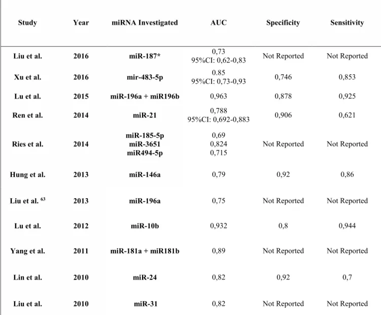
11 performed on blood, serum or plasma who investigated the expression of specific miRNAs. We reported their potential use as cancer biomarkers. This systematic review was performed according to the Preferred Reporting Items for Systematic reviews and meta- Analysis (PRISMA). The criteria for inclusion in this systematic review were: studies on human blood, plasma or serum evaluating miRNA expression in patients with oral cancer compared to healthy controls. Presence of at least one miRNA dysregulated in the test group confirmed by either qRT-PCR or ddPCR. At the end of the selection process 16 studies [56–71] were assessed as adequate for inclusion in the study. Of the studies included, only 11 reported Receiver Operating Characteristics (ROCs) of circulating miRNAs in order to discriminate between cancer and non-cancer patients (Table 1).
Table 1: Data for Receiver Operating Characteristic (ROC) as: Area Under the Curve (AUC), specificity and sensitivity from 11 studies.
Study Year miRNA Investigated AUC Specificity Sensitivity
Liu et al. 2016 miR-187* 95%CI: 0,62-0,83 0,73 Not Reported Not Reported
Xu et al. 2016 mir-483-5p 95%CI: 0,73-0,93 0.85 0,746 0,853
Lu et al. 2015 miR-196a + miR196b 0,963 0,878 0,925
Ren et al. 2014 miR-21 95%CI: 0,692-0,883 0,788 0,906 0,621
Ries et al. 2014 miR-185-5p miR-3651 miR494-5p 0,69 0,824
0,715 Not Reported Not Reported
Hung et al. 2013 miR-146a 0,79 0,92 0,86
Liu et al. 63 2013 miR-196a 0,75 Not Reported Not Reported
Lu et al. 2012 miR-10b 0,932 0,8 0,944
Yang et al. 2011 miR-181a + miR181b 0,89 Not Reported Not Reported
Lin et al. 2010 miR-24 0,82 0,92 0,7
12 These studies revealed very promising results of Area Under Curve (AUC), showing promising diagnostic ability in the detection of OSCC through the quantification of specific miRNAs in blood and derivatives. However, most of the included studies suffer from the presence of different source of bias bound to the heterogeneous sampling and to the use of not fully reliable housekeeping genes. Such shortcomings should be overcome in future studies focusing on single subsites of tumors arising in the oral cavity and using a panel of reliable reference genes.
- 2.3 Analysis of circulating miRNAs and proteins for the detection of patients with
tongue squamous cell carcinoma
2.3.1 Introduction
As above mentioned, in the last years the research of biomarkers has been enriched by the possibility of studying a multitude of molecules besides the well-known proteins. Among these cell-free miRNAs have a great potential because they are stable, even when subjected to extreme conditions such as high temperature, wide variation in pH or multiple freeze-thaw cycles (26). The use of amplification reactions, such as qRT-PCR, for the study of circulating miRNAs allows the detection of molecules also at a low concentration. Unlike detection of circulating nucleic acids, two potential problems with serum protein biomarkers were their low concentrations due to the dilution in the circulation and the limited ability to assay multiple proteins from a small sample volume. Recently, the proximity extension assay (PEA) has been developed. It is a highly sensitive method based on pairs of antibodies linked to oligonucleotides with partial complementariness for each other (PEA probes) (27). When the two different antibodies bind to their target protein, probes are brought into proximity and the two oligonucleotides accordingly bind (28).
The new sequence achieved can be extended, amplified and measured by quantitative real-time PCR, where the number of PCR templates is proportional to the initial concentration of antigen in the sample (29). In addition for a high sensitivity, specificity is also improved due to the requirement for simultaneous binding of two different antibodies to epitopes on the same antigen (30). The purpose
13 of this study was to analyze the expression of both circulating protein and miRNAs in the plasma of patients with squamous cell carcinoma of the tongue.
2.3.2 Materials and methods - Patients Material
Whole blood was collected from a total of 19 patients and 23 healthy volunteers, mean age 58 and 50 years respectively. Tumor biopsies (T) and biopsies from corresponding clinically normal tumor free tissue (TF) adjacent to the tumor was collected from 15 patients, with a mean age of 59 years. Three milliliters of peripheral blood were collected into vacutainers containing EDTA using standardized venipuncture procedures. Handling and processing was the same for all samples. Tubes were left standing for at least 30 minutes at room temperature after collection, centrifuged at 1300 g for 10 min at room temperature and the top layer, the plasma, was immediately aliquoted.
- Extraction of miRNAs from plasma and tissues
Total RNA including miRNA was extracted from frozen plasma samples using miRCURY RNA Isolation kit –Biofluids (Exiqon, Vedbaek, Denmark). A standard protocol for RNA isolation was used according to the manufacturer, with 250 μl of plasma as starting material and stored at -80 °C until further use. Total RNA including miRNA was extracted from frozen plasma samples using miRCURY RNA Isolation kit –Biofluids (Exiqon, Vedbaek, Denmark). A standard protocol for RNA isolation was used according to the manufacturer, with 250 μl of plasma as starting material.
After centrifugation to remove debris, 200 μl supernatant was extracted and RNA eluted in 50 μl RNase free water. AllPrep DNA/RNA/miRNA Universal Kit (Qiagen, Hilden, Germany) was used for miRNA extraction from tumor and tumor free tissue. The fresh frozen biopsies (less than 20 mg) were homogenized in 600 μl Buffer RLT Plus containing β-mercaptoethanol using a Precellys Tissue homogenizer (Bertin Technologies, Artigus Pres Boreaux, France). After DNase treatment and washing, total RNA including miRNA was eluted twice in 30 μl and the eluates pooled. Purified RNA
14 was stored at -80 °C until cDNA preparation. Quantity and purity of RNA was measured using a NanoDrop ND-1000 spectrophotometer (Thermo Scientific, Wilmington, DE, USA).
- Discovery and validation of miRNAs in plasma
Twenty microliters of RNA extracted from plasma from 13 patients and 13 controls was sent on dry ice to Exiqon Services (Vedbæk Denmark) for miRNA analysis with real-time PCR panel. The panel used was miRCURY LNA Universal RT microRNA PCR Human Panel I. Briefly 10 μl RNA was reversed transcribed in 50 μl using the miRCURY LNA Universal RT microRNA PCR, polyadenylation and cDNA synthesis kit (Exiqon, Vedbæk, Denmark). cDNA was diluted 50x and assayed in 10 μl PCR reactions according to the kit protocol. Amplification was performed in a LightCycler® 480 Real-time PCR system (Roche) in 382 well plates, and amplification curves analyzed using Roche LC software, both for determination of Cq (by the 2nd derivative method) and for melting curve analysis. All samples went through a quality control of RNA isolation and cDNA synthesis using Exiqon´s RNA spike-in kit. All data were therefore normalized by the formula; normalized Cq = global mean Cq – assay Cq (sample). For validation of findings from the panel in plasma, miR-150 and three miRNAs used for normalization, miR-30e, miR-93 and miR-222, were quantified by individual qRT-PCR assays. RNU6 and SNORD 44 were used for normalization of miR-150 tissue data, obtained from 8 of the 13 SCCT samples included in the plasma analysis plus an additional 7 samples. Real time RT-PCR was performed using an IQ5 multicolor real-time PCR detection system with IQ SYBR Green Supermix (Bio-Rad Laboratories, Hercules, CA, USA). For each assay, 4 μl cDNA, diluted 40X, was used in 10 μl reactions. Cycling conditions were enzyme activation at 95°C for 10 min followed by 45 cycles of denaturation at 95°C for 10 sec and annealing/extension at 60°C for 60 sec.
15 - Proximity Extension Assay
Plasma from 19 controls (9 of the samples included in the miRNA analysis and an additional 10) and 19 SCCT patients (12 of the 13 analyzed for miRNA in plasma and an additional 7 patients with SCCT) were sent to Clinical biomarkers facility, Science for Life Laboratory (Uppsala, Sweden) for analysis with proximity extension assay. One μl EDTA-plasma was used in each of two different panels comprising 92 and 71 proteins respectively, Proseek multiplex Oncology I, v2 and Proseek multiplex Inflammation I (Olink Bioscience, Uppsala, Sweden).
2.3.3 Results
- miRNA expression in TSCC patients
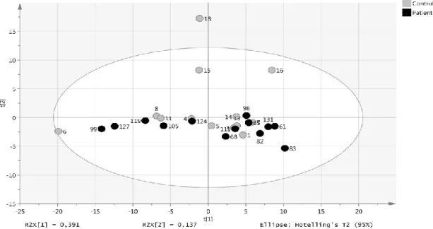
A panel of circulating miRNAs was analyzed in plasma from healthy individuals (13 controls) and patients with SCCT (13 patients). Of the 372 miRNAs analyzed, 152 were detected in all samples. None of the samples showed signs of hemolysis, evaluated by analyzing the ratio between miR-451 and miR-23a-3p. All samples had values below 7.0, where a ratio above 7.0 is considered as risk of being affected by hemolysis. Principal component analysis (PCA) showed that the levels of these miRNAs did not discriminate healthy individuals from SCCT patients (Figure 2).
Figure 2: miRNA levels in patients with tongue tumor compared with healthy controls. Score plot (t1/t2) from PCA modelling based on 152 miRNAs detected in all samples. Each dot represents one patient indicated by the patient ID.
16 Statistical evaluation of individual miRNA levels showed miR- 150 to be significantly decreased in patients with SCCT compared to controls, Benjamini-Hochberg FDR adjusted p-value 0.007 (Figure 3A). Single qRT-PCR assay was then applied to validate miR-150 results using a combination of three miRNAs for normalization (miR-93, miR-222 and miR-30e). The normalized Cq value from single qRT-PCR assays of plasma samples showed high consensus with the normalized Cq value from the panel (p < 0.001, Rho = 0.913).
miR-150 was further analyzed in tissue samples using paired tumor and tumor free samples from 15 SCCT patients; 8 of the patients included in the plasma analysis and an additional 7 patients. In contrast to the decreased plasma levels in tumor patients, there was significant up-regulation (p-value 0.02) of miR-150 in tumor tissue compared to tumor free tissue adjacent to the tumor (Figure 3B).
17 Figure 3: miR-150 levels in plasma and tissue. (A) Individual miR-150 levels in plasma from controls and tumors based on Cq values from the miRNA-panel. Normalized Cq = global mean Cq – assay Cq (sample). A higher value thus indicate that miR-150 is more abundant in that particular sample. (B) Fold change in miR-150 level in tongue tumor compared to tumor free tongue tissue from the same patient.
- Protein expression in TSCC patients
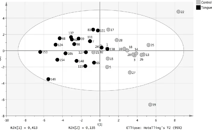
A total of 146 proteins involved in oncogenesis and inflammation were analyzed by proximity extension assay of plasma from 19 SCCT patients (the same 13 as included in the miRNA analysis and an additional 6 patients) and 19 healthy controls (9 that were included in the miRNA analysis and 10 additional controls). Using a cut-off of p < 0.05, 23 markers were differentially expressed between SCCT patients and healthy individuals. Principal component analysis of these 23 proteins separated SCCT patients and healthy individuals from each other, although not completely (Figure 4).
Figure 4: Levels of plasma proteins with tongue tumor compared with healthy controls. Score plot (t1/t2) from PCA modelling based on 23 plasma proteins with significantly different levels in patient and control samples.
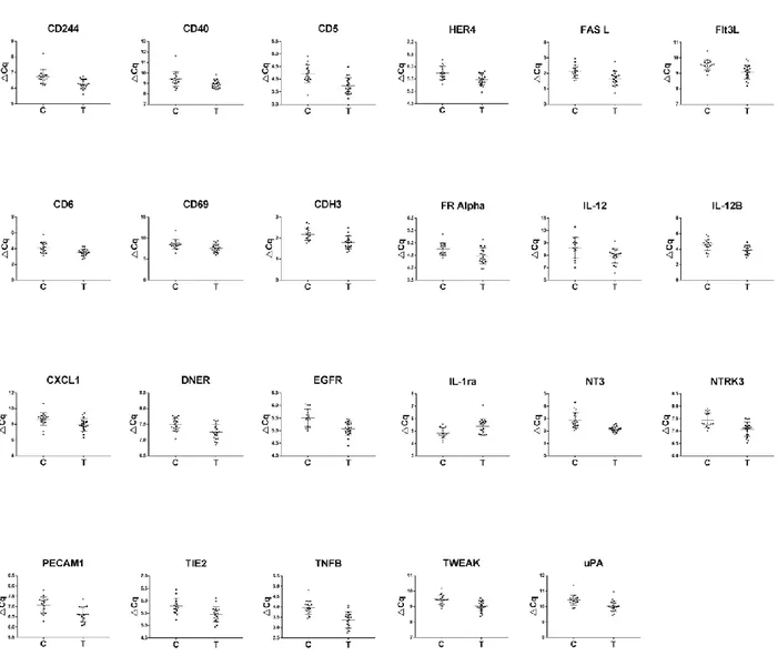
18 All but one of these proteins showed down-regulation in SCCT patients compared to healthy controls, the exception being IL1-ra, and there was a large overlap in protein levels between the groups for all proteins (Figure 5).
Figure 5: Differentially expressed proteins. Scatter plots of significantly differentially expressed proteins in plasma from patients with squamous cell carcinoma of the tongue compared to healthy controls
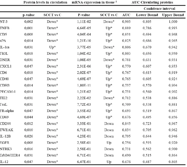
19 Calculating a receiver operating curve (ROC) showed the AUC for individual proteins to range from 0.676 to 0.903, indicating fair to good discrimination between those with and without disease (Table 2).
20 The protein with highest AUC value was NT-3 (neurotrophin-3; NTF3), which is also the only candidate with both high sensitivity and high specificity. For the 23 differentially expressed proteins, corresponding gene expression was available in a recently published study of RNA profiling in nine of the patients. Sixteen mRNAs showed significantly different expression when comparing tumor tissue to control, whilst mRNA levels for 7 of the proteins was not significantly different between control and tumor tissues. Consistency in the direction of change for circulating protein and tissue mRNA levels were identified in only three of the 16 differentially expressed genes (DNER, NT3 and TIE2), where protein levels were lower in plasma from patients than controls and mRNA levels were lower in tumor tissue compared to tumor-free tissue. Consistency in the direction of change for circulating protein and tissue mRNA levels were identified in only three of the 16 differentially expressed genes (DNER, NT3 and TIE2), where protein levels were lower in plasma from patients than controls and mRNA levels were lower in tumor tissue compared to tumor-free tissue.
- 2.4 Meta-analysis of HOTAIR as prognostic factor in patients with squamous cells
carcinoma of the head and neck
Squamous cell carcinoma of the head and neck (SCCHN) encompasses a wide and frequent range of neoplasms arising from the epithelium of different sites such as: oral cavity, nasal cavity, oropharynx, hypopharynx and larynx. It has been reported that around 90% of all tumors in this region are squamous cell carcinomas. SCCHNs are among the ten most frequent malignant neoplasms in humans; and due to the high rate of early metastasis to regional cervical lymph-nodes treatment of these patients is a challenge for clinicians. OSCC belongs to the wide group of SCCHN, being the most present subtype with about 24% of all the SCCHN cases. Long non-coding RNAs (lncRNAs) are transcripts longer than 200 nucleotides previously defined as “junk DNA” (31). They are usually transcribed by RNA polymerase II and can undergo splicing and polyadenylation processes. Although they are not translated into proteins, many of them are linked to some complexes involved in chromatin modification causing an overexpression or overexpression or silencing of target genes of
21 which most are in cancer pathogenesis silencing of target genes of which most are in cancer pathogenesis. The HOX transcript antisense intergenic RNA (HOTAIR) is one of the most studied lncRNAs (32). Different studies have focused on the capacity of HOTAIR to cooperate with different chromatin modifying complexes, above all the Polycomb Repressive Complex 2 (PRC2), through its 5′-terminal binding domain (33).
HOTAIR lets PRC2 recognize the target gene, leading to Histone H3 lysine-27 trimethylation, causing a silencing effect. HOTAIR is also able to cooperate with the LSD1 complex and can be regulated by different factors such as miR141, Ago2, c-Myc, TGF-β and small interfering RNAs (siRNAs). The goal of this systematic review and meta-analysis was to assess the link between HOTAIR expression and patient prognosis in SCCHN in order to highlight its potential role as prognostic biomarker. Inclusions criteria were: (i) studies focusing on the expression of HOTAIR in SCCHN including more than 50 patients in total, (ii) studies showing analysis of correlation between different levels of HOTAIR expression and overall survival, (iii) studies reporting a Hazard Ratio (HR) and 95% confidence interval (CI) or Kaplan-Meier curve for HR estimation and (iv) studies including quantitative analysis performed with qRT-PCR, in situ hybridization (ISH), fluorescence in situ hybridization (FISH), droplet digital PCR (ddPCR) or RNA sequencing data. Accordingly, studies performed on cell lines or animal models, reviews, case reports, overlapping publications and all studies reporting insufficient data for estimation of HR and 95% CI, were excluded. No restriction was applied concerning year of publication. The bibliographic research was performed on the databases: PUBMED, SCOPUS, Web of Knowledge and EMBASE. In addition, a direct search on the bibliographies of previously published systematic reviews on the topic was also performed. Overall effects were investigated with a fixed effect model where I2 was less than 50%, if higher, a random effects model was used. Data for overall survival were synthetized as HR and standard error (SE), while for lymph-node metastasis, tumor stage and histological grade the odds ratio (OR) was evaluated. In cases in which HR and its 95% CI was not reported in the article, it was extracted from Kaplan- Meier curves using the method of Tierney et al (34).
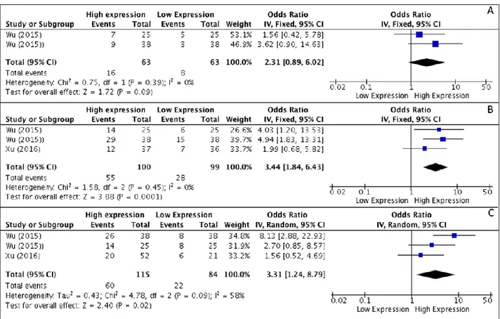
22 At the end of the selection process, 4 studies met the inclusion criteria and were included in the analysis. All studies were performed in China and published between 2012–2016. Results of meta-analysis, on the basis of two studies, revealed that higher levels of HOTAIR expression were not associated with higher degree of differentiation (OR, 2.31; 95% CI: [0.89, 6.02]; p = 0,09) (Figure 6A), whereas a correlation to advanced tumor stages was seen (OR, 3.44; 95% CI: [1.84, 6.43]; p < 0,001) (Figure 6B). As heterogeneity was not detected (I2 = 0%) a fixed effects model was used. Analysis of correlations performed with a random effect model (I2 = 58%) revealed a correlation between HOTAIR expression and the rate of LNM (OR, 3.31; 95% CI: [1.24, 8.79]; p = 0,02) (Figure 6C).
Figure 6: (A–C) Forest plot showing different expression of lncRNA HOTAIR and histological grade (A), tumor stage (B) and lymph-node metastasis (C); the frequency of HOTAIR expression was considered in patients with: Advance Stage (III–IV), positive LNM and Poor/Low Grade of differentiation
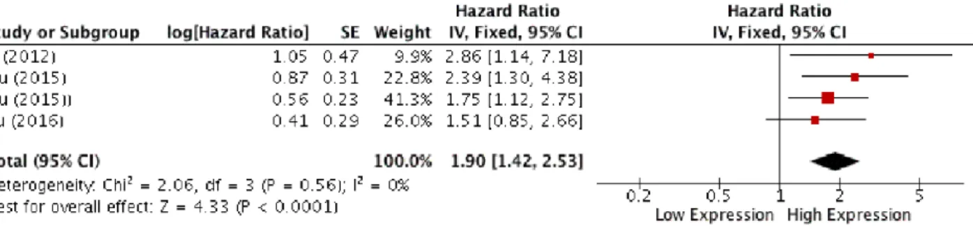
Analysis of overall survival pooling HRs showed high expression of HOTAIR to be associated with poor OS in patients with SCCHN (HR, 1.90; 95% CI: [1.42, 2.53]; p < 0,0001), (Figure 7).
23 Figure 7: Forest plot for the association between HOTAIR expression and overall survival.
Summary of data extracted from the included studies used for the pooled analysis are shown in Table 3.
Table 3: Synthesis of data extracted from the included studies related to outcomes pooled in the meta-analysis
24
Effects of treatment with Curcumin and Genistein on OSCC cell lines
- 3.1. Background
‘‘Nutraceutical’’ (a combination of the words ‘‘nutrition’’ and ‘‘pharmaceutical’’) refers to any substance considered to be a food or a food ingredient that provides medical and health benefits. The use of natural agents is promising because not only they have minimal toxicity to humans compared to conventional chemotherapies, but also, they could target numerous signaling pathways. This is beneficial as malignant transformation and progression are multistage processes caused by gene alterations in more than one signaling pathway. This is one of the most plausible explanations why monomodal therapy typically fails in cancer treatments as the specific inhibitors often target only a single gene in a signaling pathway. Therefore, the impact of natural agents on cancer treatment could be more efficacious, as they can be used alone or as adjuvant in combination chemotherapy to improve therapeutic efficacy by overcoming drug resistance and/or reducing drug-induced toxicities. Hence, many of the anticancer agents currently used in cancer therapies have been developed from natural products such as plants (vincristine, vinblastine, etoposide, paclitaxel, camptothecin, topotecan, and irinotecan), marine organisms (cytarabine), and microorganisms (dactinomycin, bleomycin, and doxorubicin). Besides these, there are also plant-derived dietary polyphenols such as curcumin, genistein, resveratrol, epigallocatechin-3-gallate, indole-3-carbinole and others. A number of studies involving cultured cancer cells and animal models have illustrated the protective role of these dietary polyphenols, and mechanistic studies have demonstrated that they exert their antiproliferative and/or proapoptotic effects to prevent the occurrence and/or spread of various cancers by targeting numerous key elements in intracellular signaling network involved in carcinogenesis.
Curcumin is one of the components of curry and a popular dietary spice worldwide. It is the primary active constituent of turmeric, a botanical agent derived from the rhizome (root) of the Curcuma longa, a perennial herb belonging to the ginger family that is broadly cultivated in south and
south-25 east Asia. Turmeric is comprised of a group of three curcuminoids, i.e., curcumin (diferuloylmethane), demethoxycurcumin, and bisdemethoxycurcumin, as well as volatile oils, sugars, proteins and resins. Curcumin is a hydrophobic polyphenol that is nearly insoluble in water. Importantly, it has limited systemic bioavailability due to its rapid metabolism, largely through conjugation to sulfates and glucuronides. In humans, this metabolism presumably occurs in the gastrointestinal tract rather than in the liver. Curcumin has been considered pharmacologically safe, based on the fact that it has been consumed for centuries as a dietary spice at doses up to 100 mg/day. Moreover, its safety and tolerability became evident in phase I studies when it was administered at doses as high as 8 g per day.
Genistein (4′,5,7-trihydroxyisoflavone) is an isoflavone, found in soybeans and in all its derivatives such as flour, sauces, oil, milk and cheese. It is also found in other legumes such as lentils, beans, peas, chickpeas and whole grains such as wheat, rice, barley, rye and oats. It has highanti-cancer properties, including the inhibition of tyrosine kinase proteins and inhibition of cell cycle at G2/M phase; it is also able to promote apoptosis by activation of caspase-9 and -3.
The aim of this study was to assess the effects of treatment with different dosages of curcumin and genistein on oral cancer cell lines.
- 3.2. Materials and Methods
-Cell cultures and treatments
PE/CA-PJ15, PE/CA-PJ49, HSC-3 are cell lines of tongue squamous cells carcinoma (European Collection of Cell Cultures, ECACC). Cells were maintained at standard conditions of temperature and atmosphere (37°C and 5% CO2, respectively) for all tests used. DMEM culture medium with 4,500 mg/L glucose was used for PE/CA-PJ15 and PE/CA-PJ49 cells and Roswell Park Memorial Institute 1640 medium (RPMI 1640) for HSC-3 cells (Life Technologies, Gibco, Grand Island, NY, USA). Both culture media were supplemented with 10% fetal bovine serum, L-glutamine (2 mM) and penicillin–streptomycin (100 U/mL) (Sigma Aldrich, Saint Louis, MO, USA). Genistein (Abcam,
26 Cambridge, UK) and curcumin were dissolved in dimethylsulfoxide (DMSO) at a stock concentration of 5 mM. Serial dilutions of Genistein at 20, 50 and 100 μM and of Curcumin at 1, 5, 10, 20 and 50μM were prepared. The cell lines were treated for 24, 48 and 72 hours at the concentrations previously mentioned.
- xCELLigence System
The xCELLigence DP Real-Time Cell Analyzer (RTCA) is used for real-time monitoring of cell proliferation, cytotoxicity, and migration. This system is based on recording the electronic impedance with a unitless parameter called Cell Index (CI). The CI calculation is based on the following formula: CI = (Zi - Z0)/15çϛ where Zi is the impedance at the start of the experiment. Hence, CI is a self-calibrated value derived from the ratio of measured impedances. When many cells are attached on the surface of a particular E-Plate the CI gets high. For this reason, we have used it to determine the variation of cell adhesion after curcumin treatments. The RTCA System was used accordingly to the manufacturer instructions.
- MTT assay and Trypan blue exclusion test
Vybrant® MTT Cell Proliferation Assay Kit (Thermo Fisher Scientific, Waltham, MA, USA) was used to evaluate cell viability of PE/CA-PJ15, PE/CA-PJ49, and HSC-3 cells after treatments with genistein. We seeded 5×10⁴ cells in a total volume of 250 μL/well in a 96-well plate with 20, 50 and 100 μM of genistein for 24, 48 and 72 hours. Then, 100 μL of fresh culture medium and 10 μL of 12 mM MTT stock solution were added to each well after the cells were incubated for 4 hours at 37°C. To each well, SDS-HCl solution (10 mL of 0.01 M HCl to 1 gm of SDS) was added for an incubation period of 12 hours in a humidified chamber at 37°C. The absorbance was read at 570 nm using the Multiskan™ GO Microplate Spectrophotometer (Thermo Fisher Scientific). While PE/CA-PJ15, PE/CA-PJ49, HSC-3 cells were treated with genistein and curcumin at the same concentrations and time points, Trypan blue exclusion method was used to validate the cell viability results obtained with
27 MTT assay. A suspension of each cell line was mixed with 0.4% Trypan blue solution and, after 10 minutes, cells were counted automatically with JuLI™ FL (NanoEntek, Pleasanton, CA, USA). - Scratch assay
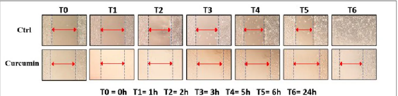
A monolayer of each cell line was scraped with a p200 pipette tip and then washed twice with Dulbecco’s phosphate buffered saline 1X (Life Technologies, Gibco) in order to remove debris. Then, cells were treated with the IC50 dose of genistein at 24 hours. Untreated cells were used as control. Initially, we acquired the first image (T0) and then the subsequent ones after 1 hour (T1), 2 hours (T2), 3 hours (T3), 5 hours (T4), 6 hours (T5) and 24 hours (T6) after treatment. ImageJ software (National Institutes of Health, Bethesda, MD, USA) was used to calculate the size of wound and analyze all the acquired images. GraphPad Prism 7 (GraphPad Software, Inc., CA, USA) software was used for statistical evaluation.
- Western blotting analysis
After treatments cells were lysated to obtain proteins. They were measured and, then, separated by 15% SDS polyacrylamide gel electrophoresis and transferred to a nitrocellulose membrane (Bio-Rad, Hercules, CA, USA). Bovine serum albumin (BSA) 5% was used for 1 hour as blocking solution. Membranes were incubated with vitronectin (1:150; BD Biosciences), OCT4 (1:700; Novus Biologicals, Littleton, CO, USA), survivin (1 : 1,000; Cell Signaling Technology Inc., Danvers, MA, USA), and β-actin (1:5,000; Sigma Aldrich), overnight at 4°C. Then, peroxidase-conjugated secondary antibody was used (1 : 2,500; Santa Cruz Biotechnology, Dallas, TX, USA). Signals were acquired with enhanced chemiluminescence kit (ClarityTM Western ECL Substrate, Bio-Rad). UVP ChemiDoc-It®TS2 Imaging System (Analytik Jena AG, Jena, Germany) was used.
- Statistical analysis
GraphPad Prism 7 software was used for Student’s t-test and one-way analysis of variance (ANOVA) and all data were expressed as the M ± SEM;17 a P-values ,0.05, P,0.01, P,0.001 were accepted as statistically significant.
28
- 3.3. Results
- Effects on cell adhesion
Through xCELLigence system, we monitored adhesion kinetics in real time of the treated cells, using untreated cells as control. After 24, 48 and 72 hours, the CI values were taken and adhesion curves were analyzed. Data obtained show clear effects of genistein and curcumin on all cells used even at 24 hours after the treatment. The resulting curves for Curcumin showed a difference of adhesion between control and treated cells. The reduction of adhesion was proportionally increased respect to the concentration of curcumin used for all cell lines. Even after long times of treatment, the effects of curcumin were significant.
CI values to 72 hours were considerably reduced in all conditions analyzed, even considering the limitations of xCELLigence system, such as the normal reduction in cell growth space for long analysis times. However, the results obtained confirmed that the increase in curcumin concentrations leaded to a proportional reduction of cell adhesion, even for very long treatments. In the evaluation of adherence rate, we converted the CI values in percent values in order to obtain a better indication of the reduction of cell adhesion, considering the control value of CI, at each time point, as the maximum value of adhesion at that period. In order to better evaluate the effect of curcumin on cell adhesion, we evaluated the expression of integrin β1, an integral membrane glycoprotein that binds the extracellular matrix proteins playing a key role in cell adhesion. Figure 8 a, b showed the reduction of integrin expression by 50% in cells treated with 10 μM of curcumin after 24 hours compared to control.
29
Figure 8 (A/B): Expression of integrin β1. Downregulation of integrin β1 expression by 50% in cells treated with 10 μM of curcumin after 24 hours compared to control (a), (b).
30 While, after 48 hours the same reduction was present for cells drawn with about 5μM of curcumin. Instead, at 72 hours after treatment the values were significantly reduced compared to the treatments after 24 and 48 hours. In particular, a reduction of 50% adhesion relative to the control was evident in cells treated with 5μM of nutraceutical. In addition, we found a great reduction of integrin expression in all the cells treated with 20 and 50μM of curcumin to 48 hours and 72 hours after treatment.
- Effects on cell viability
Through the MTT assay, we verified a decrease in cell viability proportional to increasing concentrations of curcumin and genistein for all treated cell lines. We observed a 50% reduction of viability in HSC3 cells treated with 5-10 μM of curcumin at 24 hours after treatment and 1-5 μM at 48 and 72 hours. PE/CA-PJ15 cells showed the same reduction for the concentrations of 10 μM of curcumin at 24 and 48 hours and about 10-20 μM at 72 hours. A reduction of about 50% was observed in PE/CA-PJ49 cells treated with 5-10 μM of curcumin at all time points. Through trypan blue test, the cell viability was reduced for HSC-3 and PE/CA-PJ49 cells in the same way as MTT assay. While for the PE/CA-PJ15 cells a 50% reduction in cell viability was observed for cells treated with 10-20 μM of curcumin at all time points unlike MTT test. For genistein, we detected a reduction of about 50% of cell viability in each treated cell line with concentrations between 20 and 50 μM at 24, 48 and 72 hours. Trypan blue assay confirmed almost all the data obtained with the MTT assay.
- Effects on cell migration
A monolayer of each cell line (PE/CA-PJ15, PE/CA-PJ49, HSC-3) was scraped with a p200 pipette
tip and then washed twice with Dulbecco’s phosphate buffered saline 1X (Life Technologies, Gibco) in order to remove debris. Then, cells were treated with the IC50 dose of genistein at 24 hours. Untreated cells were used as control. Initially, we acquired the first image (T0) and then the subsequent ones after 1 hour (T1), 2 hours (T2), 3 hours (T3), 5 hours (T4), 6 hours (T5) and 24 hours (T6) after treatment. The treated cells migrated very little in the cut area. The most significant effects were evident starting from T4 and, specifically, the wound of treated cells does not show any healing
31 after 24 hours. The untreated control showed an increasing level of healing of the wound during the considered times total healing after 24 hours.
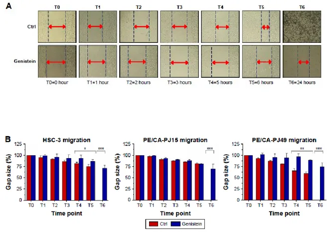
Evaluations of the timing at T4, T5 and T6 were most significant with P-value ,0.05, P,0.01, P,0.001 (Figure 9 a,b).
Figure 9: Effects of genistein on cell migration(A) and Significant increase of the gap size is more evident from T4 in tongue carcinoma cells compared to untreated control (Ctrl) (A). All experiments were performed at least 3 times and results are presented as the mean ± SD. *P,0.05, **P,0.01, ***P,0.001 (B).
32 ImageJ software (National Institutes of Health, Bethesda, MD, USA) was used to calculate the size of wound and analyze all the acquired images. GraphPad Prism 7 (GraphPad Software, Inc., CA, USA) software was used for statistical evaluation. The same protocol was adopted using curcumin standing for the genistein. To evaluate cell migration, we captured images at the beginning of the treatment with 10 μM of curcumin (T0) and at intervals (T1, T2, T3, T4, T5 and T6) during the migration of the cells to close the scratch, and we compared the images to determine the rate of cell migration (Figure 10a).
Figure 10: Effects of curcumin on cell migration(a); All experiments were performed at least three times and results are presented as the means ± SD. *p < 0.05, **p < 0.01, ***p < 0.001 (b).
33 The concentration of curcumin used was the average IC50 value at 24 hours, previously estimated. All experiments were performed at least three times and the results were presented as means ± SD. We noticed a significant increase of the gap size directly proportional to the increase of the treatment time with curcumin, while it decreased in the untreated control. The increase was most evident from T3-T4 onwards and it was present in all of TSCC cell lines. For T4 and T5 times we had a very high significance compared to control (p <0.05 and p <0.01) for all cell lines. Instead, the wound treatment with curcumin after 24 hours (T6) prevented the complete healing of the wound, while it disappeared completely in the untreated control (p <0.001).
- Effects on cell apoptosis
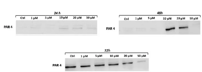
After treating tongue cancer cells with different curcumin concentrations, we evaluated the expression of PAR4. PAR4 is a pro-apoptotic and tumor suppressor protein that selectively induces apoptosis in cancer cells by activating the extrinsic mechanisms. We noticed an increased expression of PAR4, pro-apoptotic protein, for concentration higher than 10 μM at 24 and 48 hours and for all treatments at 72 hours. This demonstrated, probably, that curcumin induces apoptosis in cells treated with 10 μM already after 24 hours and that possible apoptosis is more evident after 72 hours of treatment (Figure 11).
Figure 11: Curcumin reduces promotes apoptosis. Variation in level expression of PAR4 showed at 24 hours (a), 48 hours (b) and 72 hours (c). Curcumin upregulates PAR4. The results shown in Figure b belong to two gels.
34
Discussion and Conclusions
The tumor markers are playing an increasingly important role in cancer detection and management. (35). A cancer biomarker may be a molecule secreted by a tumor cell or a specific response of the body to the presence of cancer (36). Cancer biomarkers can be classified based on the disease state, including predictive, diagnosis and prognosis biomarkers (16). In our research activities, we focused mainly on the research of diagnostic and prognostic biomarkers. The first ones are used to detect or confirm presence of a disease or condition of interest or to identify individuals with a subtype of the disease. In OSCC such kind of molecules have been studied mainly in blood and saliva, aiming to find a marker that can help clinicians in the early diagnosis of the disease, decrease the diagnostic delay and improve the survival rate of OSCC patients (37-39). In the last years, the research of circulating biomarkers has been enriched by the study of new molecules in addition to the "classic" proteins. Among these, great attention has been paid on the study of miRNAs. Such molecules are a class of small non-coding RNAs long 19–23 nucleotides, able to negatively regulate target messenger RNAs (mRNAs) by partially binding to their 30 untranslated region (40). In cancer miRNAs are often dysregulated and implicated in different cellular process such as differentiation, proliferation, apoptosis and metastasis (41, 42). The presence of miRNAs was also discovered in blood and several other body fluids including: urine, tears, pleural effusions and saliva, exhibiting different expression patterns in cancer patients compared to healthy individuals (43). Although, the exact mechanism with which miRNAs are released into the body fluids is still unclear a lot of studies have been performed in order to assess whether circulating miRNAs could be used as diagnostic biomarker in OSCC. In order to synthesize results of literature, we performed a systematic review about the evaluation of circulating miRNAs in blood and derivatives as diagnostic biomarkers in OSCC (44). Results of the review revealed that 11 studies reported very good values of Area Under the Curve (AUC), sensitivity and specificity. However, issues related to methods and sampling have to be overcome before to use circulating miRNAs in the clinical practice.
35 Unlike detection of circulating nucleic acids that can be amplified prior to detection using array technologies, two potential problems with serum protein biomarkers are their low concentrations due to dilution in the circulation and the limited ability to assay multiple proteins from a small sample volume by means of immunoenzymatic assays (45). Recently, the development of proximity extension assay (PEA) has overcome such limitations allowing the simultaneous quantification of proteins in body fluids (46).
On the basis of such evidences, we performed a study aiming to assess both circulating proteins (with PEA) and miRNA (by qPCR-panel) in patients with squamous cells carcinoma of the tongue (47). We identified 23 proteins that seems to be able to detect the disease, among these neurotrophin-3 (NT-3) showed the better predictive values. While, no one of the 372 circulating miRNAs analyzed showed good values for use as diagnostic biomarker. To note, miR-150 was downregulated in blood but upregulated in tissues of patients, suggesting a possible role in the pathogenesis of OSCC. Focusing on the prognosis, the research aims to find a biomarker used to identify likelihood of a clinical event, disease recurrence or progression in patients who have the disease or medical condition of interest (16). On the prognostic side, focused on the study of a new molecules called long non-coding RNAs. tools for treatment of the disease. Long non-non-coding RNAs (lncRNAs) are transcripts longer than 200 nucleotides that do not code for protein production (31).
Their action seems to be linked to chromatin modification causing an overexpression or silencing of target genes of which most are in cancer pathogenesis (48). In particular, we performed a meta-analysis of studies that quantified the expression of the lncRNA HOTAIR in patients with squamous cells carcinoma of the head and neck (SCCHN) (49). HOTAIR is a well-known oncogene, 2.2 kb long and transcribed from the antisense strand of the HOXC gene located on chromosome 2, and consisting of 6 exons (50). Although the exact mechanism of action is not well known, HOTAIR seems to be able to regulate the expression of very important genes, such as: p53, PTEN, E-cadherin and HER-2 (32, 33, 51). Results of our meta-analysis based on the inclusion of 4 studies revealed that HOTAIR is a promising prognostic biomarker in SCCHN patients. HOTAIR expression correlated
36 with: advanced tumor stages (OR, 3.44; 95% CI: [1.84, 6.43]; p < 0,001), development of lymphnode metastasis (OR, 3.31; 95% CI: [1.24, 8.79]; p = 0,02) and poor overall survival (HR, 1.90; 95% CI: [1.42, 2.53]; p < 0,0001). However, such preliminary results need to be further expanded increasing the power of evidences and focusing on the single subsite of SCCHN.
Although treatment options for OSCC patients have improved in the last decades, the overall survival rate is still low, underlining the importance of expanding research in new therapeutic options. We addressed our attention to the study of the effects of two nutraceuticals (Curcumin and Genistein) on OSCC cell lines. Results of our studies revealed that both molecules exert dose dependent inhibition of viability, adhesion proliferation, migration of OSCC cell lines. In addition, curcumin seems to be able to induce apoptosis through downregulation of survivin, that is a well-known inhibitor of apoptosis (52).
Although such effects are very promising, it is to underline that one of the major barriers to the use of nutraceuticals in clinical practice is related to their low systemic bioavailability (53). In order, to increase the amount of the products absorbed systemically further studies on new pharmaceutical formulations of nutraceuticals should be addressed, aiming to overcome such limitations and allow the use of nutraceuticals in the clinical practice.
37
References
1. Malvezzi M, Arfe A, Bertuccio P, Levi F, La Vecchia C, Negri E. European cancer mortality predictions for the year 2011. Ann Oncol. 2011;22(4):947-56.
2. Shield KD, Ferlay J, Jemal A, Sankaranarayanan R, Chaturvedi AK, Bray F, et al. The global incidence of lip, oral cavity, and pharyngeal cancers by subsite in 2012. CA Cancer J Clin. 2017;67(1):51-64.
3. van Harten MC, Hoebers FJ, Kross KW, van Werkhoven ED, van den Brekel MW, van Dijk BA. Determinants of treatment waiting times for head and neck cancer in the Netherlands and their relation to survival. Oral Oncol. 2015;51(3):272-8.
4. Kerdpon D, Jantharapattana K, Sriplung H. Factors related to diagnostic delay of oral squamous cell carcinoma in southern Thailand: Revisited. Oral Dis. 2017.
5. Le Campion A, Ribeiro CMB, Luiz RR, da Silva Junior FF, Barros HCS, Dos Santos KCB, et al. Low Survival Rates of Oral and Oropharyngeal Squamous Cell Carcinoma. Int J Dent. 2017;2017:5815493.
6. Dirven R, Ebrahimi A, Moeckelmann N, Palme CE, Gupta R, Clark J. Tumor thickness versus depth of invasion - Analysis of the 8th edition American Joint Committee on Cancer Staging for oral cancer. Oral Oncol. 2017;74:30-3.
7. Bitu CC, Destro MF, Carrera M, da Silva SD, Graner E, Kowalski LP, et al. HOXA1 is overexpressed in oral squamous cell carcinomas and its expression is correlated with poor prognosis. BMC Cancer. 2012;12:146.
8. Lau A, Li KY, Yang WF, Su YX. Induction chemotherapy for squamous cell carcinomas of the oral cavity: A cumulative meta-analysis. Oral Oncol. 2016;61:104-14.
9. Wu X, Cheng J, Wang X. Dietary Antioxidants: Potential Anticancer Agents. Nutr Cancer. 2017;69(4):521-33.
38 10. Pandey MK, Gupta SC, Nabavizadeh A, Aggarwal BB. Regulation of cell signaling pathways by dietary agents for cancer prevention and treatment. Semin Cancer Biol. 2017;46:158-81.
11. Correia-da-Silva M, Sousa E, Pinto MMM, Kijjoa A. Anticancer and cancer preventive compounds from edible marine organisms. Semin Cancer Biol. 2017;46:55-64.
12. Pandima Devi K, Rajavel T, Daglia M, Nabavi SF, Bishayee A, Nabavi SM. Targeting miRNAs by polyphenols: Novel therapeutic strategy for cancer. Semin Cancer Biol. 2017;46:146-57. 13. Dandawate PR, Subramaniam D, Jensen RA, Anant S. Targeting cancer stem cells and signaling pathways by phytochemicals: Novel approach for breast cancer therapy. Semin Cancer Biol. 2016;40-41:192-208.
14. Scarpa ES, Ninfali P. Phytochemicals as Innovative Therapeutic Tools against Cancer Stem Cells. Int J Mol Sci. 2015;16(7):15727-42.
15. Mishra A, Verma M. Cancer biomarkers: are we ready for the prime time? Cancers (Basel). 2010;2(1):190-208.
16. BEST (Biomarkers, EndpointS, and other Tools) Resource. Silver Spring (MD)2016.
17. Roses RE, Paulson EC, Sharma A, Schueller JE, Nisenbaum H, Weinstein S, et al. HER-2/neu overexpression as a predictor for the transition from in situ to invasive breast cancer. Cancer Epidemiol Biomarkers Prev. 2009;18(5):1386-9.
18. Sharma SV, Bell DW, Settleman J, Haber DA. Epidermal growth factor receptor mutations in lung cancer. Nat Rev Cancer. 2007;7(3):169-81.
19. Momen-Heravi F, Bala S. Emerging role of non-coding RNA in oral cancer. Cell Signal. 2017;42:134-43.
20. Majem B, Rigau M, Reventos J, Wong DT. Non-coding RNAs in saliva: emerging biomarkers for molecular diagnostics. Int J Mol Sci. 2015;16(4):8676-98.
21. Bisogno LS, Keene JD. RNA regulons in cancer and inflammation. Curr Opin Genet Dev. 2017;48:97-103.
39 22. Iyer MK, Niknafs YS, Malik R, Singhal U, Sahu A, Hosono Y, et al. The landscape of long noncoding RNAs in the human transcriptome. Nat Genet. 2015;47(3):199-208.
23. Guay C, Regazzi R. Circulating microRNAs as novel biomarkers for diabetes mellitus. Nat Rev Endocrinol. 2013;9(9):513-21.
24. Bartel DP. MicroRNAs: genomics, biogenesis, mechanism, and function. Cell. 2004;116(2):281-97.
25. Seok H, Ham J, Jang ES, Chi SW. MicroRNA Target Recognition: Insights from Transcriptome-Wide Non-Canonical Interactions. Mol Cells. 2016;39(5):375-81.
26. Schwarzenbach H, Nishida N, Calin GA, Pantel K. Clinical relevance of circulating cell-free microRNAs in cancer. Nat Rev Clin Oncol. 2014;11(3):145-56.
27. Blokzijl A, Nong R, Darmanis S, Hertz E, Landegren U, Kamali-Moghaddam M. Protein biomarker validation via proximity ligation assays. Biochim Biophys Acta. 2014;1844(5):933-9. 28. Greenwood C, Ruff D, Kirvell S, Johnson G, Dhillon HS, Bustin SA. Proximity assays for sensitive quantification of proteins. Biomol Detect Quantif. 2015;4:10-6.
29. Fredriksson S, Gullberg M, Jarvius J, Olsson C, Pietras K, Gustafsdottir SM, et al. Protein detection using proximity-dependent DNA ligation assays. Nat Biotechnol. 2002;20(5):473-7. 30. Gullberg M, Gustafsdottir SM, Schallmeiner E, Jarvius J, Bjarnegard M, Betsholtz C, et al. Cytokine detection by antibody-based proximity ligation. Proc Natl Acad Sci U S A. 2004;101(22):8420-4.
31. Evans JR, Feng FY, Chinnaiyan AM. The bright side of dark matter: lncRNAs in cancer. J Clin Invest. 2016;126(8):2775-82.
32. Yu X, Li Z. Long non-coding RNA HOTAIR: A novel oncogene (Review). Mol Med Rep. 2015;12(4):5611-8.
33. Wu Y, Zhang L, Zhang L, Wang Y, Li H, Ren X, et al. Long non-coding RNA HOTAIR promotes tumor cell invasion and metastasis by recruiting EZH2 and repressing E-cadherin in oral squamous cell carcinoma. Int J Oncol. 2015;46(6):2586-94.
40 34. Tierney JF, Stewart LA, Ghersi D, Burdett S, Sydes MR. Practical methods for incorporating summary time-to-event data into meta-analysis. Trials. 2007;8:16.
35. Saxena S, Sankhla B, Sundaragiri KS, Bhargava A. A Review of Salivary Biomarker: A Tool for Early Oral Cancer Diagnosis. Adv Biomed Res. 2017;6:90.
36. Rivera C, Oliveira AK, Costa RAP, De Rossi T, Paes Leme AF. Prognostic biomarkers in oral squamous cell carcinoma: A systematic review. Oral Oncol. 2017;72:38-47.
37. Lee LT, Wong YK, Hsiao HY, Wang YW, Chan MY, Chang KW. Evaluation of saliva and plasma cytokine biomarkers in patients with oral squamous cell carcinoma. Int J Oral Maxillofac Surg. 2017.
38. Blatt S, Kruger M, Ziebart T, Sagheb K, Schiegnitz E, Goetze E, et al. Biomarkers in diagnosis and therapy of oral squamous cell carcinoma: A review of the literature. J Craniomaxillofac Surg. 2017;45(5):722-30.
39. Gao W, Guo CB. Factors related to delay in diagnosis of oral squamous cell carcinoma. J Oral Maxillofac Surg. 2009;67(5):1015-20.
40. Friedman RC, Farh KK, Burge CB, Bartel DP. Most mammalian mRNAs are conserved targets of microRNAs. Genome Res. 2009;19(1):92-105.
41. Scapoli L, Palmieri A, Lo Muzio L, Pezzetti F, Rubini C, Girardi A, et al. MicroRNA expression profiling of oral carcinoma identifies new markers of tumor progression. Int J Immunopathol Pharmacol. 2010;23(4):1229-34.
42. Liang Z, Bian X, Shim H. Downregulation of microRNA-206 promotes invasion and angiogenesis of triple negative breast cancer. Biochem Biophys Res Commun. 2016;477(3):461-6. 43. Ortiz-Quintero B. Cell-free microRNAs in blood and other body fluids, as cancer biomarkers. Cell Prolif. 2016;49(3):281-303.
44. Troiano G, Boldrup L, Ardito F, Gu X, Lo Muzio L, Nylander K. Circulating miRNAs from blood, plasma or serum as promising clinical biomarkers in oral squamous cell carcinoma: A systematic review of current findings. Oral Oncol. 2016;63:30-7.
41 45. Karakus R, Buyrukcu BA, Aybay C. A new efficient method for eliminating the interference effect of human serum and increasing the sensitivity and recovery rate of enzyme immunoassay. J Immunoassay Immunochem. 2005;26(2):109-24.
46. Assarsson E, Lundberg M, Holmquist G, Bjorkesten J, Thorsen SB, Ekman D, et al. Homogenous 96-plex PEA immunoassay exhibiting high sensitivity, specificity, and excellent scalability. PLoS One. 2014;9(4):e95192.
47. Boldrup L, Troiano G, Gu X, Coates P, Fåhraeus R, Wilms T, et al. Evidence that circulating proteins are more promising than miRNAs for identification of patients with squamous cell carcinoma of the tongue. Oncotarget. 2017;8(61):103437-48.
48. Di Gesualdo F, Capaccioli S, Lulli M. A pathophysiological view of the long non-coding RNA world. Oncotarget. 2014;5(22):10976-96.
49. Troiano G, Alberto VC, Boldrup L, Gu X, Muzio LL, Sgaramella N, et al. Expression of the long non-coding RNA HOTAIR as a prognostic factor in squamous cell carcinoma of the head and neck: A systematic review and meta-analysis. Oncotarget. 2017;8(42):73029-36.
50. Tani H, Mizutani R, Salam KA, Tano K, Ijiri K, Wakamatsu A, et al. Genome-wide determination of RNA stability reveals hundreds of short-lived noncoding transcripts in mammals. Genome Res. 2012;22(5):947-56.
51. Li D, Feng J, Wu T, Wang Y, Sun Y, Ren J, et al. Long intergenic noncoding RNA HOTAIR is overexpressed and regulates PTEN methylation in laryngeal squamous cell carcinoma. Am J Pathol. 2013;182(1):64-70.
52. Xie S, Xu H, Shan X, Liu B, Wang K, Cai Z. Clinicopathological and prognostic significance of survivin expression in patients with oral squamous cell carcinoma: evidence from a meta-analysis. PLoS One. 2015;10(2):e0116517.
53. Saranya TS, Rajan VK, Biswas R, Jayakumar R, Sathianarayanan S. Synthesis, characterisation and biomedical applications of curcumin conjugated chitosan microspheres. Int J Biol Macromol. 2017.
42
Appendix 1: Journal publications
1) Linda Boldrup, Giuseppe Troiano, Xiaolian Gu, Philip Coates, Robin Fåhraeus, Torben Wilms, Lena
Norberg-Spaak, Lixiao Wang, Karin Nylander. Evidence that circulating proteins are more promising than miRNAs for identification of patients with squamous cell carcinoma of the tongue. Oncotarget. 2017 Sep 30; doi: /10.18632/oncotarget.21402.
2) Giuseppe Troiano, Vito Carlo Alberto Caponio, Linda Boldrup, Xaolian Gu, Lorenzo Lo Muzio,
Nicola Sgaramella, Lixiao Wang, Karin Nylander. Expression of the long non-coding RNA HOTAIR as a prognostic factor in squamous cell carcinoma of the head and neck: a systematic review and meta-analysis. Oncotarget. 2017 Aug 21;8(42):73029-73036. doi: 10.18632/oncotarget.20373.
3) Ardito F, Pellegrino MR, Perrone D, Troiano G, Cocco A, Lo Muzio L. In vitro study on anti-cancer
properties of genistein in tongue cancer. Onco Targets Ther. 2017 Nov 13;10:5405-5415. doi: 10.2147/OTT.S133632
4) Giuseppe Troiano, Khrystyna Zhurakivska, Lorenzo Lo Muzio, Luigi Laino, Marco Cicciù, Lucio Lo
Russo. Combination of Bone Graft and Resorbable Membrane for Alveolar Ridge Preservation: a Systematic Review, Meta-analysis and Trial Sequential Analysis. J Periodontol. 2017 Sep 12:1-17. doi: 10.1902/jop.2017.170241.
5) Domenico Ciavarella, Michele Tepedino, Crescenzio Gallo, Graziano Montaruli, Khrystyna
Zhurakivska, Ludovica Coppola, Giuseppe Troiano, Claudio Chimenti, Michele Laurenziello, Lucio Lo Russo. Post-orthodontic position of lower incisors and gingival recession: A retrospective study. Journal of Clinical and Experimental Dentistry 9(12), pp. e1425-e1430
43
6) Giuseppe Troiano, Mario Dioguardi, Armando Cocco, Luigi Laino, Gabriele Cervino, Marco Cicciu,
Domenico Ciavarella, Lorenzo Lo Muzio. Conservative vs Radical Approach for the Treatment of Solid/Multicystic Ameloblastoma: A Systematic Review and Meta-analysis of the Last Decade. Oral Health Prev Dent. 2017;15(5):421-426. doi: 10.3290/j.ohpd.a38732.
7) Alessio Buonavoglia, Dorina Lauritano, Donatella Perrone, Fatima Ardito, Giuseppe Troiano, Mario
Dioguardi, V Candotto, FJ Silvestre, Lorenzo Lo Muzio. Evaluation of chemical-physical properties and cytocompatibility of TheraCal LC. J Biol Regul Homeost Agents. 2017 Apr-Jun;31(2 Suppl 1):1-9
8) Alan Scott Herford, Marco Cicciù, Livium Florian Eftimie, Megan Miller, Fabrizio Signorino, Fausto
Famà, Gabriele Cervino, Giuseppe Lo Giudice, Ennio Bramanti, Floriana Lauritano, Giuseppe
Troiano, Lorenzo Lo Muzio, Luigi Laino. rhBMP-2 applied as support of distraction osteogenesis: a
split-mouth histological study over nonhuman primates’ mandibles. Int J Clin Exp Med 2016;9(9):17187-17194.
9) Luigi Laino, Giuseppe Troiano, Dardo Menditti, Alan Scott Herford, Alberta Lucchese, Gabriele
Cervino, Floriana Lauritano, Rosario Serpico, Marco Cicciù. Use of collagen matrix to improve wound repair after mucosal biopsy: a multicenter case series. Int J Clin Exp Med 2017;10(5):8363-8368 / ISSN:1940-5901/IJCEM0047628
10) Fatima Ardito, Michele Giuliani, Donatella Perrone, Giuseppe Troiano, Lorenzo Lo Muzio. The
crucial role of protein phosphorylation in cell signaling and its use as targeted therapy (Review). Int J Mol Med. 2017 Jun 22. doi: 10.3892/ijmm.2017.3036.
44
11) Michele Laurenziello, Graziano Montaruli, Crescenzio Gallo, Michele Tepedino, Laura Guida, Letizia
Perillo, Giuseppe Troiano, Lorenzo Lo Muzio, Domenico Ciavarella. Determinants of maxillary canine impaction: Retrospective clinical and radiographic study. J Clin Exp Dent. 2017;9(11):e1304-9. doi:10.4317/jced.54095
12) Giuseppe Troiano, Mario Dioguardi, Luisa Limongelli, Angela Tempesta, Gianfranco Favia, Michele
Giuliani, Francesca Sanguedolce, Lorenzo Lo Muzio. Can Inspection of the Mouth Help Clinicians Diagnose Crohn's Disease? A Review. Oral Health Prev Dent. 2017;15(3):223-227. doi: 10.3290/j.ohpd.a38158.
13) Angela Pia Cazzolla, Giuseppe Troiano, Khrystyna Zhurakivska, Eugenio Maiorano, Gianfranco
Favia, Maria Grazia Lacaita, Giuseppe Marzo, Franca Dicuonzo, Stefano Andresciani, Lorenzo Lo Muzio. Langerhans cell histiocytosis of the maxillae in a child treated only with chemotherapy: a case report. J Med Case Rep. 2017 May 9;11(1):130. doi: 10.1186/s13256-017-1286-3
14) Giuseppe Troiano, Luigi Laino, Khrystyna Zhurakivska, Marco Cicciù, Lorenzo Lo Muzio, Lucio Lo
Russo. Addition of enamel matrix derivatives to bone substitutes for the treatment of intrabony defects: A systematic review, meta-analysis and trial sequential analysis. J Clin Periodontol. 2017 May 6. doi: 10.1111/jcpe.12742.
15) Klas Strindlund, Giuseppe Troiano, Nicola Sgaramella, Philip J. Coates, Xiaolian Gu, Linda Boldrup,
Luigi Califano, Robin Fahraeus, Lorenzo Lo Muzio, Fatima Ardito, Giuseppe Colella, Gianpaolo Tartaro, Renato Franco, Lena Norberg-Spaak, Mohammad Saadat, Karin Nylander. Patients with high c-MYC-expressing squamous cell carcinomas of the tongue show better survival than those with low- and medium-expressing tumours. J Oral Pathol Med. 2017 Apr 10. doi: 10.1111/jop.12579.
45
16) Alan Scott Herford, Marco Cicci, Livium Florian Eftimie, Megan Miller, Fabrizio Signorino, Fausto
Famà, Gabriele Cervino, Giuseppe Lo Giudice, Ennio Bramanti, Floriana Lauritano, Giuseppe
Troiano, Lorenzo Lo Muzio, Luigi Laino. rhBMP-2 applied as support of distraction osteogenesis: a
split-mouth histological study over nonhuman primates’ mandibles. Int J Clin Exp Med 2016;9(9):17187-17194 / ISSN:1940-5901/IJCEM0025809.
17) Giuseppe Troiano, Mario Dioguardi, Armando Cocco, Michele Giuliani, Cristiano Fabiani, Alfonso
D'Alessandro, Domenico Ciavarella, Lorenzo Lo Muzio. Centering Ability of ProTaper Next and WaveOne Classic in J-Shape Simulated Root Canals. ScientificWorldJournal. 2016;2016:1606013. doi: 10.1155/2016/1606013
18) Marco Cicciù, Alan Scott Herford, Gabriele Cervino, Giuseppe Troiano, Floriana Lauritano, Luigi
Laino. Tissue Fluorescence Imaging (VELscope) for Quick Non-Invasive Diagnosis in Oral Pathology. J Craniofac Surg. 2017 Mar;28(2):e112-e115. doi: 10.1097/SCS.0000000000003210.
19) Giuseppe Troiano, Linda Boldrup, Fatima Ardito, Xaolian Gu, Lorenzo Lo Muzio, Karin Nylander.
Circulating miRNAs from blood, plasma or serum as promising clinical biomarkers in oral squamous cell carcinoma: A systematic review of current findings. Oral Oncol. 2016 Dec;63: 30-37. doi: 10.1016/j.oraloncology.2016.11.001.
20) Giuseppe Troiano, Mario Dioguardi, Armando Cocco, Giovanni Giannatempo, Luigi Laino,
Domenico Ciavarella, Elio Berutti, Lorenzo Lo Muzio. Influence of Operator's Experience on the Shaping Ability of Protaper Universal and Waveone Systems: A Comparative Study on Simulated Root Canals. Open Dent J. 2016 Oct 7; 10:546-552.