Department of Pharmacy
INTERNATIONAL RESEARCH DOCTORATE PROGRAM IN MOLECULAR PHYSIOPATHOLOGY, DIAGNOSIS AND THERAPY
OF METABOLIC DISEASES Coordinator: Prof. Maurizio Bifulco
XII cycle NS 2010–2013
NOVEL INSIGHTS INTO THE BIOLOGICAL EFFECTS
OF THE ISOPRENOID DERIVATIVE
N6-ISOPENTENYLADENOSINE:
INVOLVEMENT OF THE METABOLIC SENSOR AMPK
IN ANGIOGENESIS INHIBITION
Tutor PhD Student Prof. Patrizia Gazzerro Paola Picardi
This dissertation is based upon the following articles:
Picardi P, Pisanti S, Ciaglia E, Margarucci L, Ronca R, Giacomini A, Malfitano
AM, Casapullo A, Laezza C, Gazzerro P, Bifulco M. Antiangiogenic effects of N6-isopentenyladenosine, an endogenous isoprenoid end product, mediated by AMPK activation. FASEB J. 2013. [Epub ahead of print] PubMed PMID: 24265487.
Ciaglia E, Pisanti S, Picardi P, Laezza C, Malfitano AM, D’Alessandro A, Gazzerro P, Vitale M, Carbone E, Bifulco M. N6-isopentenyladenosine, an endogenous isoprenoid end product, directly affects cytotoxic and regulatory functions of human NK cells through FDPS modulation. J Leukoc Biol. 2013. 94:1207-19
Kai# mikroi# kairoi# mega@lwn pragma@twn aiòtioi gi@gnontai
Dhmosqe@nhv, Peri# sunta@xewvTABLE OF CONTENTS
LIST OF PUBLICATIONS LIST OF ABBREVIATIONS ABSTRACT
1 BACKGROUND 1
1.1 N6-isopentenyladenosine, an endogenous isoprenoid 1 end-product, from plants..
1.2 ..to mammals 2
1.3 Biological effects of N6-isopentenyladenosine 5 1.4 Basic aspects of angiogenesis 9 1.5 Angiogenesis in cancer 10
1.6 A snapshot of melanoma 13
1.7 AMP-activated protein kinase, sensor of cellular energy 16
1.8 Double face of autophagy: promoter of cell survival 18
or cell death? 1.9 Immune regulation in cancer: focus on melanoma 23
2 AIMS OF THE STUDY 29
3 MATERIALS AND METHODS 31
4 RESULTS 45
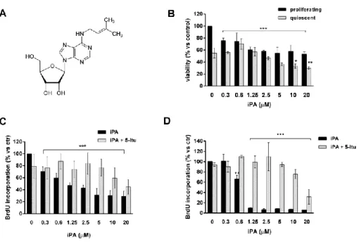
4.1 iPA inhibits endothelial cell proliferation 45
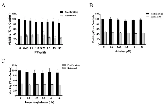
4.2 The peculiar structure of iPA is responsible for the 46
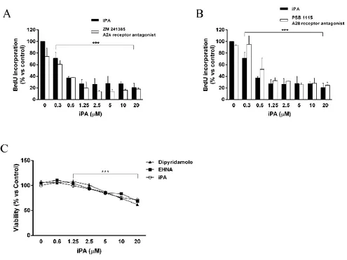
antiproliferative effects on endothelial cells 4.3 The classical adenosine pathways are not involved in 49
the biological effects exerted by iPA 4.4 iPA must be phosphorylated into iPAMP to exert 51
its biological effects through AMPK activation 4.5 iPA induces S-phase stasis and apoptosis, activating the 55
intrinsic caspase cascade and DNA damage machinery 4.6 iPA inhibits endothelial cell migration, invasion, and 58
4.7 iPA inhibits angiogenesis in vivo 61 4.8 iPA inhibits angiogenic phenotype of melanoma cells 63 4.9 iPA inhibits melanoma cells proliferation and colony formation 63 4.10 iPA induces a G1-phase stasis and apoptosis in melanoma cells 65 4.11 iPA induces AMPK-dependent activation of autophagy 67
in melanoma cells
4.12 iPA activates autophagy and apoptosis pathways to induce 70 cell death in a coordinated and cooperative manner
4.13 iPA selectively expands NK cells in human primary PBMCs culture72 4.14 iPA directly stimulates human purified NK cells 73 4.15 iPA synergizes with IL-2 for the proliferation and activation 76
status of human purified NK cells
4.16 Stimulatory effect of iPA on NK cell-mediated antitumor 78 cytotoxicity on the surface expression of NKp30
4.17 Cytokines and chemokines secretion profile of iPA 80 treated-NK cells upon interaction with K562 cells
4.18 MAPK signal transduction is uniquely modulated by iPA 82 plus IL-2 stimulation
4.19 iPA-mediated effects on IL2- stimulated NK cells are 84 MAPK dependent
4.20 Analysis of the mechanism underlying the stimulatory effect 85 of iPA: a role for the farnesyl diphosphate synthase
5 DISCUSSION 89
5.1 N6-isopentenyladenosine inhibits angiogenesis in vitro 89 and in vivo through AMPK activation
5.2 N6-isopentenyladenosine, activating AMPK, induces 96 autophagy and apoptosis in a cooperative manner in melanoma cells
5.3 N6-isopentenyladenosine directly affects cytotoxic and 101 regulatory functions of human NK cells
6 CONCLUSIONS 109
7 REFERENCES 115
LIST OF PUBLICATIONS
1) Picardi P, Pisanti S, Ciaglia E, Margarucci L, Ronca R, Giacomini A, Malfitano AM, Casapullo A, Laezza C, Gazzerro P, Bifulco M. Antiangiogenic effects of N6-isopentenyladenosine, an endogenous isoprenoid end product, mediated by AMPK activation. FASEB J. 2013. [Epub ahead of print] PubMed PMID: 24265487.
2) Ciaglia E, Pisanti S, Picardi P, Laezza C, Malfitano AM, D’Alessandro A, Gazzerro P, Vitale M, Carbone E, Bifulco M. N6-isopentenyladenosine, an endogenous isoprenoid end product, directly affects cytotoxic and regulatory functions of human NK cells through FDPS modulation. J Leukoc Biol. 2013; 94:1207-19.
3) Pisanti S, Picardi P, D'Alessandro A, Laezza C, Bifulco M. The endocannabinoid signaling system in cancer. Trends Pharmacol
Sci. 2013; 34(5):273-82.
4) Pisanti S, Picardi P, Prota L, Proto MC, Laezza C, McGuire PG, Morbidelli L, Gazzerro P, Ziche M, Das A, Bifulco M. Genetic and pharmacological inactivation of cannabinoid CB1 receptor inhibits angiogenesis. Blood. 2011; 117(20):5541-50.
LIST OF ABBREVIATIONS
5-Itu = 5-iodotubercidin ADK = adenosine kinase AICAR =
5-aminoimidazole-4-carboxamide riboside
AMP = adenosine monophosphate AMPK = AMP-activated protein
kinase
AO = acridin orange
APC = antigen presenting cell Atg = autophagy-related gene ATP = adenosine triphosphate CCL = chemokine (C-C motif)
ligand
CFSE = carboxyfluorescein
diacetate succinimidyl ester
CTL = cytotoxic T lymphocyte DC = dendritic cell
DMAPP = dimethylallyl
pyrophosphate
EHNA = erythro-9
(2-hydroxy-3-nonyl)-adenine
eNOS = endothelial nitric oxide
synthase
FDPS = farnesyl diphosphate
synthase
Hif = hypoxia-inducible factor HLA = human leukocyte antigen HO-1 = heme oxygenase 1 HUVEC = human umbilical vein
endothelial cell IFN-γ = interferon-γ IL-2 = interleukin-2 iPA = N6-isopentenyladenosine iPAMP = N6-isopentenyladenosine 5'-monophosphate
IPP = isopentenyl pyrophosphate IPT = isopentenyl transferase LC3 = microtubule-associated
protein light chain 3
LC-MS/MS = liquid
chromatography–
coupled tandem mass spectrometry
LKB1 = liver kinase B1
MAPK = mitogen-activated protein
kinases
MMP = matrix metalloproteinase MFI = mean fluorescence intensity mTOR = mammalian target of
rapamycin NK = natural killer PARP = poly(ADP-ribose) polymerase pATM = phospho-ataxia telangiectasia mutated
pATR = phospho-ATM and
Rad3-related PBMC = peripheral blood mononucleated cell pChk1 = phospho-checkpoint kinase 1 PI = propidium iodide SRM = selected-reaction monitoring TNF-α = tumor necrosis factor-α VEGF = vascular endothelial
growth factor
ZMP =
ABSTRACT
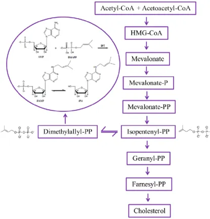
N6-isopentenyladenosine (iPA) is a modified adenosine characterized by an isopentenyl chain derived by dimethylallyl pyrophosphate (DMAPP), an intermediate of the metabolic pathway of mevalonate, that is known to be deregulated in cancer.
iPA is an endogenous isoprenoid-derived product present in mammalian cells as a free nucleoside in the cytoplasm, or in a tRNA-bound form, displaying well established pleiotropic biological effects, including a direct anti-tumor activity against several cancers. However, the precise mechanism of action of iPA in inhibiting cancer cell proliferation remains to be clarified.
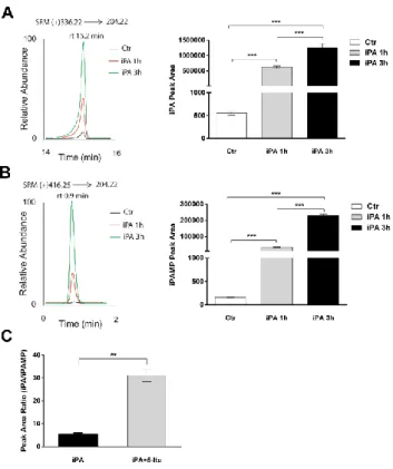
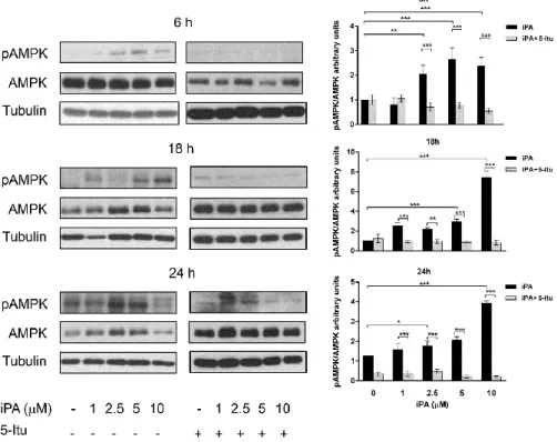
In this work, we investigated whether iPA could directly interfere with the angiogenic process, fundamental to cancer growth and progression, and if the growth and proliferation of human melanoma cells, known for their highly angiogenic phenotype, could be affected by the treatment with iPA. Finally, we investigated if iPA could have an immunomodulatory role targeting directly human natural killer (NK) cells, components of innate immunity that participate in immunity against neoplastic cells, in order to provide a cooperative and multifactorial mode of action of iPA to arrest cancer growth. To evaluate the potential involvement of iPA in angiogenesis, we employed human umbilical vein endothelial cells (HUVECs) as a suitable in vitro model of angiogenesis, by evaluating the viability, proliferation, migration, invasion, tube formation, and molecular mechanisms involved. Data were corroborated in mice by using a gel plug assay. iPA dose- and time-dependently inhibited all the neoangiogenesis stages, with an IC50 of 0.98 µM. We demonstrated for the first time that iPA was monophosphorylated into iPA 5'-monophosphate (iPAMP) by adenosine kinase (ADK) inside the cells. iPAMP is the active form that
inhibits angiogenesis through the direct activation of AMP-kinase (AMPK). Indeed, all effects were completely reversed by pre-treatment with 5-iodotubercidin (5-Itu), an ADK inhibitor. The isoprenoid intermediate isopentenyl pyrophosphate (IPP), which shares the isopentenyl moiety with iPA, was ineffective in the inhibition of angiogenesis, thus showing that the iPA structure is specific for the observed effects. Thus, iPA is a novel AMPK activator and could represent a useful tool for the treatment of diseases where excessive neoangiogenesis is the underlying pathology.
The activation of AMPK seems to be the mechanism by which iPA exerts also its anticancer effects in A375 human melanoma cells. Indeed, in order to evaluate if iPA could be a useful agent able to inhibit tumor angiogenesis, we tested in vitro the ability of iPA to arrest the proliferation and the growth of melanoma, a tumor known for its highly angiogenic phenotype. In particular, we performed co-cultures of HUVEC and A375 cells, and we evaluated melanoma cells vitality to delucidate their cell fate following iPA-treatment. Moreover, the molecular mechanism elicited by iPA in this cancer model was analyzed. According with endothelial cells, iPA (from 2.5 to 10 µM) was able to inhibit melanoma cell growth, inducing autophagy and subsequently apoptosis, through AMPK activation, acting asan AMP mimetic.
In the last part of this work, we analyzed a possible role of iPA in immune regulation. In analogy to the unique specificity for phosphoantigens like IPP, shown by human Vγ9Vδ2 T cells, we report for the first time the ability of iPA to selectively expand and directly target human NK cells. Interestingly, at submicromolar concentrations, iPA stimulated resting human NK cells and synergized with IL-2 to induce a robust ex vivo activation with significant secretion of CCL5 and CCL3 and a large increase in TNF-α and IFN-γ production when compared with IL-2 single
cytokine treatment. Moreover, iPA was able to promote NK cell proliferation, upregulating the expression of specific NK cell activating receptors, as well as CD69 and CD107a expression. Cytotoxic activity of NK cells against tumor targets was due to a selective potent activation of MAPK signaling intermediaries downstream IL-2 receptor. The effect resulted at least in part from the fine modulation of the farnesyl diphosphate synthase (FDPS) activity, the same enzyme implicated in the stimulation of the human γδ T cells. The iPA-driven modulation of FDPS can cause an enhancement of post-translational prenylation essential for the biological activity of key proteins in NK signaling and effector functions, such as Ras. These unanticipated properties of iPA provide additional piece of evidence of the immunoregulatory role of the intermediates of the mevalonate pathway and open novel therapeutic perspectives for this molecule as an immune-modulatory drug.
Taken together, these results highlight a sinergic mode of action of iPA as antitumor agent, interfering with neovascularization and hence blood supply to nourish cancer cells, inducing autophagy and apoptosis directly in tumor cells and stimulating the immune response to attack neoplastic cells.
1. BACKGROUND
1.1 N6-isopentenyladenosine, an endogenous isoprenoid end-product, from plants..
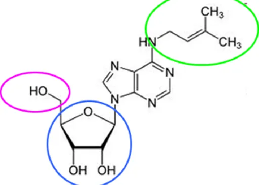
N6-isopentenyladenosine (iPA) is a modified adenosine characterized by an isopentenyl chain linked to the nitrogen at position 6 of the purine base (Fig. 1).
Figure 1. Chemical structure of iPA
iPA belongs to a class of substances known as cytokinins, which are important purine derivatives that serve as hormones that control many processes in plants (Bifulco et al., 2008). Cytokinin literally means stimulus of cell movement (cytokinesis) but it is also used in terms of cell division. They were discovered as factors promoting cell division in tobacco tissue cultures (Skoog et al., 1965) and have been shown to regulate several other developmental events, such as de novo bud formation, release of buds from apical dominance, leaf expansion, delay of senescence, promotion of seed germination, and chloroplast formation (Mok and Mok, 2001). Thus, cytokinins are plant growth regulators defined by their ability to promote cell division in tissue cultured in presence of auxin (Astot et al., 2000). The discovery of auxin, the first
organic substance defined as hormone of the plants, led to the discovery of other chemical substances regulating the plant growth. As in the animal world, it was unlikely that plant growth and development depended on the control of a unique hormone. Indeed, it was found a growth factor derived from DNA degradation that stimulated the growth of plant tissues in vitro (Skoog et al., 1965). This substance was named kinetin and the class of regulators was named cytokinins. The general term “kinin” was created to describe chemicals that promote cell division, but it was subsequently replaced by “cytokinin” for confusion with the term kinin from the realm of zoology (Oka, 2003). In 1963 the first cytokinin in plants was discovered from the immature cariosside of corn (Zea mais): the substance was named zeatin (Oka, 2003). iPA is one of the cytokinins isolated from plants and its ribosides, ribotide and 2- methyltioderivate are known (Bifulco et al., 2008). Many studies suggest that cytokinins are mainly generated in the radical system and transported to the aerial part through the xilematic flux. The metabolism of cytokinins seems to occur in various tissues of the plant, either by elimination of the side chain through cytokinins oxidases or by ribosylation. Glucosylation seems to occur in the leaves: glucosides could store hormones which, when required, are transported in specific organs. All the known cytokinins present adenine substitution on position N6, with an isoprenoid or an aromatic group (Bifulco et al., 2008).
1.2 ..to mammals
iPA was found in transfer RNA (tRNA) of many eukaryotic and prokaryotic cells (Burns et al., 1976) and it is the only known cytokinin existing in animal tRNAs, specifically at position 37 of the tRNA molecules that bind codons starting with uridine (Persson et al., 1994). It is
a rare component of some ribosomal RNA and was isolated from yeast through the hydrolysis of the serine of tRNA. Free iPA, not linked to tRNA, was found in cell extracts of Saccharomyces cerevisiae and
Schizosaccharomyces pombe (Laten and Zahareas-Doktor, 1985).
However, the cell levels of free iPA are not decreased in yeast strains having mutations that result in reduced amounts of isopentenylated tRNA (Faust and Dice, 1991). This result demonstrates that free iPA is derived from a biosynthetic pathway independent from isopentenyl tRNA degradation and probably related to modifications of monophosphate adenosine.
The adenosine is generated through the classic biosynthetic pathway of nucleosides, while the isoprenoid chain derives from a product of the mevalonate pathway and, more specifically, from dimethylallyl pyrophosphate (DMAPP), which is in equilibrium with its isomer isopentenyl pyrophosphate (IPP) (Faust and Dice, 1991). iPA is typically found next to the 3' of the tRNA anticodon that binds to codons containing uracile as first base (tRNALeu, tRNAPhe, tRNASer, tRNATrp, tRNATyr): this site suggests a role in the process of transduction. The tRNA containing iPA binds more efficiently the ribosome than the unmodified analogous nucleoside, thus demonstrating that the absence of isopentenyladenosine decreases the efficacy of transduction in vitro and in vivo (Warner et al., 2000; Moustafa et al., 2001; Bifulco et al., 2008).
It was previously believed that the RNA degradation was the main source of free cytokinins, including iPA. Nevertheless, calculation on the rate of RNA turnover demonstrated that a de novo biosynthetic pathway independent from tRNA should be present in plants; a further step was the discovery of an enzyme for cytokinin biosynthesis in Dictiostelum
discoideum. Cell extracts from this organism can convert adenosine
N6-isopentenyladenosine-5'-monophosphate (iPAMP) and the correspondent nucleoside iPA (Fig. 2). Moreover, in 1984 it was shown that the product of the T-DNA gene4 (ipt) of the crown gall-forming bacterium,
Agrobacterium tumefacien, was an isopentenyl transferase, IPT (Bifulco et
al., 2008). IPTs are a family of enzymes, conserved from microorganisms to mammals, that catalyze the addition of iPA on residue 37 of tRNA molecules using DMAPP as donor of the isopentenyl group. IPT is also involved in the synthetic pathway independent of isopentenylated tRNA degradation (Golovko et al., 2000; Spinola et al., 2005).
Figure 2. Proposed biosynthesis of iPA in plants
Astot and coworkers provided then evidence for an alternative iPAMP independent cytokinin biosynthetic pathway both in Arabidopsis thaliana expressing the Arabidopsis tumefaciens ipt gene and as part of normal
wild-type plant metabolism (Astot et al., 2000). Sequence analysis shows the homology between the human and Saccharomyces cerevisiae and
Escherichia coli IPT aminoacid sequence. All well-conserved motifs
present in tRNA-IPT can be found in the deduced human protein sequence (Golovko et al., 2000). The full-length TRIT1 transcript is able to complement the yeast tRNA-IPT biochemical activity, producing amounts of iPA-tRNA similar to those produced by the yeast gene. In mammalian cells, iPA is found in selenocysteine (Sec) tRNA that decodes UGA stop codons and inserts Sec residues into nascent peptides. The ability to decode UGA as a Sec codon is affected when the tRNASec lacks iPA at position 37 as demonstrated in Xenopus (Spinola et al., 2005). iPA is therefore the only cytokinin modified tRNA species found in animals. However, biosynthesis of iPA in mammals has not been fully disclosed.
1.3 Biological effects of N6-isopentenyladenosine
It has been proved that in plants cytokinins are able to induce callus (clusters of dedifferentiated plant cells that proliferate indefinitely in a disorganized manner) to re-differentiate into adventitious buds, just like human cancer cells. Because of these similarities, cytokinins may also affect the differentiation of human cancer cells through mechanisms of action that could involve common signal events of the signal transduction (Bifulco et al., 2008). Indeed, whilst iPA biosynthesis in mammals has not been fully disclosed, some of its biological effects are known, including anti-tumour action on human and murine cells.
More than forty years ago, it has been shown by Gallo and colleagues that iPA can exert a promoting or inhibitory effect on human cell growth, on the bases of used concentration and the cell cycle phase. They reported a biphasic effect of iPA: an inhibitory action at high μM concentrations and
a stimulatory effect at lower concentrations (0.1-1.0 μM). Moreover, they demonstrated that the addition of iPA 12 hours after phytohemagglutinin stimulation determined a decrease of mitotic figures, suggesting that iPA effects depend on the phase of cell cycle. It was also demonstrated that the effects on DNA synthesis are preceded by the inhibition of RNA and protein synthesis. The mechanism of RNA inhibition could be the result of the competition of iPA with the adenosine-3'-phosphate, adenosine or 2'- deoxyadenosine 5'-triphosphate during RNA synthesis. However, the addition of such substances, even at higher concentrations of iPA, did not decrease its inhibitory effect (Gallo et al., 1969). Inhibitory effect of iPA has been demonstrated also on the growth of sarcoma cells at very high concentration (22 and 100 μM). Studies from these cell extracts demonstrated in silico that iPA may be a potential substrate for adenosine kinase, and also a weak inhibitor of adenosine deaminase, glucose-6-phosphate-dehydrogenase and methylase of mammalian tRNAs. iPA acts also as potent inhibitor of the uptake of purine and pirimidine nucleosides. The authors suggest that iPA cytotoxicity for these cells might be due to its conversion in 5'-monophosphate even if they don’t definitively prove it. This derivative molecule from iPA, at high intracellular levels, could be cytotoxic since it affects the enzymes involved in purine metabolism (Divekar et al., 1973).
More recent studies demonstrated that iPA exerts a significant apoptotic effect on viable human lymphocytes. The capacity of the modified ribonucleosides to induce apoptosis was studied also in several human cell lines and it was observed that iPA was the most active cytokinin. The cancer cell lines Caco-2 and HL-60 were more reactive to an apoptotic stimulation than non-malignant human peripheral blood lymphocytes (Meisel et al., 1998). In a thyroid cell system iPA was able to influence cAMP dependent organization of the microfilaments. The effects of iPA
were exerted both on the synthesis of cAMP and on its activity, probably due to the substitution on the nitrogen 6 of adenosine (Laezza et al., 1997). Of note, iPA is the only adenosine substituted on nitrogen 6 with physiologic relevance. Then, the same research group investigated the anti-proliferative effect of iPA, highlighting a dose-dependent arrest of the G1 cell phase transition associated with a reduction of cells in the S phase through the inhibition of farnesyl diphosphate synthase (FDPS) and protein prenylation. Interestingly, iPA effect was not mediated by the adenosine receptors but was due to a direct modulation of FDPS enzyme activity as a result of its uptake inside the cells (Laezza et al., 2006). The antiproliferative activity of iPA was confirmed also in 9 human epithelial cancer cell lines derived from different types of malignant tissues (Spinola et al., 2007): it was observed a complete suppression of clonogenic activity in 8 of the cell lines after exposure to iPA at a concentration of 10 μM. The observed effects appeared to reflect a block in DNA synthesis, not involving a FDPS down-regulation in lung cells model. These findings are consistent with the reported inhibition of proliferation of rat thyroid tumor cells induced by iPA. Although iPA has been reported to induce apoptosis as well as differentiation in a human myeloid leukemia cell line (Ishii et al., 2002), any pro-apoptotic effect has been reported in thyroid and epithelial cancer cells (Laezza et al., 2006; Spinola et al., 2007). Moreover, iPA was able to cause a pronounced change in cell morphology, associated to a disorganization of actin fibers in the cytoplasm, suggestive of a cell stress condition. Antiproliferative effects of iPA was confirmed also on human colon cancer cells, through inhibition of DNA synthesis and induction of apoptosis (Laezza et al., 2009).
There are some evidence of the activity of iPA also in patients. A far away work described a clinical study on the therapeutic use of iPA on twenty leukaemia patients who were treated daily with intravenous infusion of
iPA. Patients received the substance drug orally or intravenous, showing transient and short lasting effects of the therapy probably due to iPA instability (Mittelman et al., 1975). Other researchers had already reported that iPA inhibited the growth of various cancers, but iPA LD50 was different and dependent on the way of administration and species (from ca. 200 to 800 mg/Kg). However, they focused particularly on toxic effects (hepatotoxicity, pancreatic atrophy and antiproliferative effects in lymphoid tissue) of iPA in rats and dogs and did not gain further insights into the observed antitumour effects (Suk et al., 1970).
No additional example of in vivo anticancer activity has been reported for humans or, in general, mammalians up to the present. Only in 2006, the successful inhibition of a rat tumour by iPA injected in a nude mouse directly at the tumour site has been reported, without detectable toxic or hypolocomotor effects on the treated animals (Laezza et al., 2006). Later, Colombo and colleagues have shown no significant in vivo activity of iPA injected intraperitoneally into nude mice inoculated with ovarian-carcinoma cells, suggesting that the pharmacokinetics of the compound do not allow to reach efficacious concentrations. The lack of activity might rest in the rapid clearance and short half-life in vivo of circulating nucleosides and, most likely, of circulating iPA (Colombo et al., 2009). Taken together, these data suggest that this isoprenoid end product might be used for antineoplastic therapy, representing a potential new anticancer drug. However, it would be necessary to increase knowledge about the exact biological role of iPA inside mammalian cells even in the sense of its ability to control cancer cell growth (Bifulco et al., 2008).
1.4 Basic aspects of angiogenesis
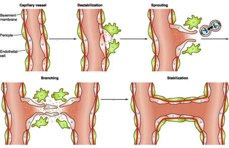
The term ‘‘angiogenesis’’ is commonly used to reference the development of new blood vessels growth from preexisting vasculature. It plays an important role in physiologic conditions, including development, reproduction, wound healing and bone morphogenesis, and in pathologic ones as cancer, intraocular neovascular disorders, rheumatoid arthritis, psoriasis and others. The entire process, that consists in localized enzymatic degradation of basal membrane, migration and proliferation of endothelial cells, and lumen formation of the new capillary (Fig. 3), is finely controlled by a tuned balance between positive and negative effectors (Carmeliet, 2003). In the embryo, new vessels form de novo via the assembly of mesoderm-derived endothelial precursors (angioblasts) that differentiate into a primitive vascular labyrinth (vasculogenesis) (Swift and Weinstein, 2009). Subsequent vessel sprouting (angiogenesis) creates a network that finally remodels into arteries and veins (Adams and Alitalo, 2007). Recruitment of pericytes and vascular smooth muscle cells that enwrap nascent endothelial cell tubules provides stability and regulates perfusion (arteriogenesis) (Jain, 2003). In the adult, vessels are quiescent and rarely form new branches.
Vessels can also grow through other mechanisms, such as the splitting of pre-existing vessels through intussusception or the stimulation of vessel expansion by circulating precursor cells (Fang and Salven, 2011; Makanya et al., 2009).
Figure 3. The cellular steps involved in angiogenesis
Blood vessels supply oxygen and nutrients and provide gateways for immune surveillance. As this network nourishes all tissues, it is not surprising that structural or functional vessel abnormalities contribute to many diseases. Inadequate vessel maintenance or growth causes ischemia in diseases such as myocardial infarction, stroke, and neurodegenerative or obesity-associated disorders, whereas excessive vascular growth or abnormal remodeling promotes many ailments including cancer as routes for tumor cells to metastasize, inflammatory disorders, and eye diseases (Carmeliet, 2003; Folkman, 2007).
1.5 Angiogenesis in cancer
Angiogenesis is widely considered as an essential process to nourish the growing tumor and to remove metabolic waste products (Folkman, 1971). Intratumoral hypoxia leads to the stabilization and expression of the pro-tumorigenic hypoxia-inducible factor (Hif)α transcription factors. Under
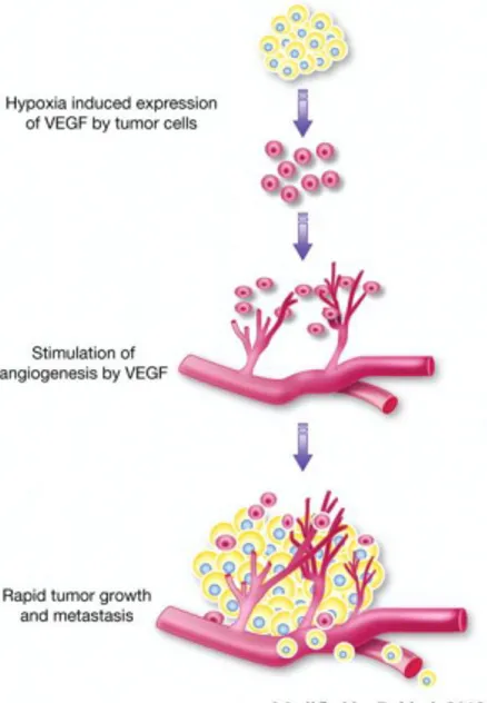
normal oxygen levels, Hif-1α and Hif-2α are short-lived because prolyl hydroxylase (PHD) proteins mediate their degradation (Fong and Takeda, 2008). During hypoxia, PHD proteins show reduced activity, which results in the formation of heterodimerization by Hif-1α and Hif-2α with Hif-1β. This complex is able to trigger the expression of genes containing hypoxia-responsive elements, including the gene encoding vascular endothelial growth factor (VEGF) (Qing and Simon, 2009). Under hypoxic conditions, pericytes are able to secrete VEGF which can act in a paracrine manner on endothelial cell proliferation (Reynolds et al., 2000). In consequence, the quiescent vascular network is getting disturbed and activated (Fig. 4).
Figure 4. Tumor associated-angiogenesis
Whereas blood vessel growth is tightly controlled under physiological conditions, tumor progression is highly associated with the acquisition of an angiogenic phenotype based on a transition from the avascular to the vascular phase (Folkman et al., 1989). This process is called the “angiogenic switch”, in which the balance of pro- and anti-angiogenic
molecules leads toward angiogenic inducers (Folkman and Hanahan, 1991). Tumor vasculature is often characterized by the presence of poorly developed so called “immature” vessels with multiple branch-points and deregulated vessel sprouting which contributes to tumor hypoxia. In particular, tumor vessels display abnormal structure and function with seemingly chaotic organization. Endothelial cells lack a cobblestone appearance, are poorly interconnected and occasionally multilayered. Also, arterio-venous identity is ill defined and shunting compromises flow. The basement membrane is irregular in thickness and composition, and fewer, more loosely attached hypocontractile mural cells cover tumor vessels, though tumor-type-specific differences exist. The resulting irregular perfusion impairs oxygen, nutrient and drug delivery (Goel et al., 2011). Neovascularization enhances tumor cell proliferation, local invasion and hematogenous metastasis. However, the abnormalities of tumor vessels provide the potential for targeting these vessels without destroying the normal vasculature (Arap et al., 2002).
Vessel leakiness, together with growing tumor mass, increases the interstitial pressure and thereby impedes nutrient and drug distribution. The loosely assembled vessel wall also facilitates tumor cell intravasation and dissemination. As a consequence of poor oxygen, nutrient and growth factor supply, tumor cells further stimulate angiogenesis in an effort to compensate for the poor functioning of the existing ones. However, this excess of proangiogenic molecules only leads to additional disorganization as the angiogenic burst is nonproductive, further aggravating tumor hypoperfusion in a vicious cycle.
Nowadays anti-angiogenetic pharmacological approaches are approved for several advanced metastatic cancers, in combination with validated chemotherapy or cytokine therapy, aimed at blocking vessel growth targeting VEGF (Potente at al., 2011). The hypoxic and acidic tumor
milieu constitutes a hostile microenvironment that is believed to drive selection of more malignant tumor cell clones and further promotes tumor cell dissemination. The uneven delivery of chemotherapeutics, together with a reduced efficacy of radiotherapy owing to the lower intratumoral oxygen levels, limit the success of conventional anticancer treatments. Moreover, tumors would be able to switch mechanisms of vascular growth and some of these mechanisms rely less on VEGF, so they would possess the means to escape from treatment with VEGF receptor inhibitors. Identifying the molecular basis of these alternative modes of vessel growth will thus be critical to improve the efficacy of antiangiogenic treatment (Potente et al., 2011).
Despite efforts to stimulate angiogenesis therapeutically by proangiogenic factors, most trials failed to meet these expectations. Alternative strategies, based on pro-angiogenic cell therapies or targeting of microRNAs, offer new opportunities but are in (pre)clinical development (Bonauer et al., 2010). Nonetheless, only a fraction of cancer patients show benefit as tumors evolve mechanisms of resistance or are refractory toward VEGF receptor inhibitors. Conflicting results about the benefit of VEGF blockade have kick-started a debate on whether antiangiogenic treatment may trigger more invasive and metastatic tumors. On the upside, ‘‘sustained normalization’’ of abnormal tumor vessels may offer benefit for fighting metastasis (Goel et al., 2011).
1.6 A snapshot of melanoma
Melanoma is a cutaneous tumor characterized by abnormal proliferation of melanocytes that invade the basement membrane. Recent epidemiologic evidence has documented both an increase in incidence of and in mortality from malignant melanoma. The importance of this relatively uncommon
malignant neoplasm (incidence less than 5/100,000 population) is attested to the fact that despite accounting for only 3% of all cutaneous malignant neoplasms, malignant melanoma causes 67% of the deaths attributable to skin cancer (Cummins et al., 2006). Malignant melanoma has a poor prognosis and limited therapeutic options, particularly in advanced tumors. The 5-year-survival rate of advanced tumors with distant metastases is still as low as 5-10% (Balch et al., 2009). Surgical resection of the primary tumour offers a significant chance of cure and remains the mainstay of treatment where possible. However, melanoma, which can progress to a stage of spreading with low invasive potential (radial growth phase) to the next stage (vertical growth phase), disseminates early, both intradermally and through lymphatic and haematogenous routes and therefore despite resection, once there is invasion into the dermis of the skin, the outlook changes dramatically.
Chemotherapy with the alkylating agent dacarbazine as monotherapy was established as treatment for metastatic melanoma thirty years ago and remains the standard of care outside of clinical trials. However the response rate of melanoma to dacarbazine is only in the region of 15 to 20% and it has not been shown to improve overall survival. Therefore, there is an urgent need to find more effective treatments for this condition (Zaki et al., 2012). An area of significant interest for modern cancer therapeutic strategies is targeting angiogenesis, as this is one of the cardinal patho-physiological features of invasive cancer, essential for tumour growth and metastasis (Hanahan and Weinberg, 2000). Indeed, melanoma is well-recognised to be a highly vascular tumour. However the impact on tumor-induced angiogenesis for disease progression and metastasis remains controversial (Helfrich and Schadendorf, 2011). Anyway, it is well known that the overproduction of VEGF165 and its association with VEGF receptor (VEGFR) expression promotes melanoma
cell growth and survival through MAP kinase and phosphatidyl inositol-3-kinase (PI3K) signaling pathways (Graells et al., 2004). Moreover, immunohistochemical studies suggest that VEGF is expressed by 20–77% of human primary melanomas. VEGF also increased during follow-up (Zaki et al., 2012). Expression of VEGF, R1, R2 and VEGF-R3 was observed to be significantly higher in malignant melanocytes when compared with samples from their benign counterparts, supporting the hypothesis that in particular the upregulation of VEGF-R2 may select for an angiogenic phenotype. Clinically, the expression and activation of MMPs is related to the degree of invasiveness and metastasis of melanoma and also correlates with low survival rates in melanoma patients (Hofmann et al., 2000). In summary, it is clear that angiogenesis represents a relevant process to target in melanoma. There is therefore a strong biological rationale to incorporate the anti-angiogenic strategy into therapeutic plans. Whereas cytotoxic chemotherapy targets DNA repair and the cell cycle to induce cell killing through apoptosis, antiangiogenic therapy is more cytostatic. The main targets are: pro-angiogenic ligands; kinases that are intrinsic to extracellular endothelial cell receptors or growth factor pathways essential for survival and proliferation within endothelial cells; MMPs and integrins. Agents used in these anti-angiogenic strategies may be further sub-divided according to their chemical entity in monoclonal antibodies (mAbs), small molecule kinase inhibitors that abrogate receptor or cytoplasmic kinases and VEGF-trap, that is a composite decoy receptor based onVEGFRs, fused to an Fc segment of IgG1 (Holash et al., 2002). Early phase trials suggest clinical activity using a variety of anti-angiogenic drugs, but the data are as yet too immature to provide conclusive evidence of clinical benefit in melanoma patients. Given the existence of redundant pathways that may compensate for inhibition of a single angiogenic axis, it is highly unlikely that targeting a single pathway
will be sufficient to achieve optimal tumor control in melanoma. Therefore there are ongoing questions over whether targeting multiple ligands, receptors or kinases within several signaling pathways may be advantageous over specific inhibition of single targets.
1.7 AMP-activated protein kinase, sensor of cellular energy
AMP-activated protein kinase (AMPK) is a highly conserved sensor of cellular energy status that exists in the form of heterotrimeric complexes containing a catalytic α-subunit combined with regulatory β- and γ- subunits (Hardie et al., 2012). Metabolic stress caused by nutrient deprivation, prolonged exercise, ischemia or hypoxia mediates the activation of AMPK, initiating a cellular program to conserve energy by turning on generating catabolic pathways and switching off ATP-consuming anabolic pathways through direct phosphorylation of target proteins and modulation of gene expression (Hardie et al., 2006). Changes in the AMP- or ADP/ATP ratio, alterations in intracellular calcium concentrations as well as AMP mimetics cause AMPK activation. Recently, in addition to AMPK role in energy homeostasis, emerging evidence indicates that AMPK regulates endothelial cell function, modulating vascular tone by activation of endothelial nitric oxide synthase (eNOS) (Morrow et al., 2003). Moreover, AMPK exerts important anti-inflammatory effects by blocking the expression of adhesion molecules and chemokines by endothelial cells (Ewart et al., 2008). Significantly, many of the beneficial actions of AMPK on endothelial cell function are related to the activation of eNOS or the induction of the vasoprotective protein heme oxygenase HO-1 (Liu et al., 2011). Although the ability of AMPK to preserve endothelial cell viability is well appreciated, the role of AMPK in regulating endothelial cell proliferation is limited and not well
understood (Reihill et al., 2011). AMPK activation is related with inhibition of endothelial cells proliferation and migration (Peyton et al., 2012) or with decreased angiogenesis and inhibition of macrophage infiltration associated to the inhibition of retinoblastoma proliferation in
vivo (Theodoropoulou et al., 2013).
Although AMPK has not yet been formally demonstrated to be a tumour suppressor, there are many indications that this is the case: AMPK activation inhibits cell growth and proliferation; AMPK exerts many effects of LKB1, a known tumour suppressor; AMPK up-regulates oxidative metabolism and would therefore be expected to mediate an ‘anti- Warburg’ effect; and similar to other known tumour suppressors, AMPK activation is down-regulated in many tumour cells, by loss of LKB1 and other mechanisms (Dandapani and Hardie, 2013). These considerations led to studies investigating whether the use of AMPK-activating drugs in humans might affect the incidence of cancer. Indeed, Type 2 diabetics taking the AMPK-activating drug metformin have significantly reduced the incidence of cancer compared with those on other medications (Decensi et al., 2010). The nucleotide ZMP, also referred as AICAR (5-amino-4-imidazolecarboxamide 1-β-Dribofuranoside) monophosphate, is an intermediate in purine nucleotide synthesis, but also acts as an AMP mimetic that mimics all of the effects of AMP on the AMPK system (Corton et al., 1995). Incubation of most cells with the equivalent riboside (AICAR) causes accumulation of ZMP, because its uptake and phosphorylation to ZMP is rapid compared with subsequent metabolism; it has therefore been widely used to activate AMPK in intact cells and in
vivo. Recently, some reports have implicated AMPK in the regulation of
autophagy. The activation of AMPK leads to the suppression of mammalian target of rapamycin (mTOR), thereby activating autophagy (see below) (Wang et al., 2011).
1.8 Double face of autophagy: promoter of cell survival or cell death?
The word autophagy is derived from the Greek roots “auto” (self) and “phagy” (eating) and broadly refers to the cellular catabolic processes in which cytoplasmic materials are transported to lysosomes for degradation. Christian de Duve, who was awarded the Nobel Prize for his work on lysosomes, first used the term autophagy in 1963 (Wang and Qin, 2013). There are several different types of autophagy: macroautophagy, microautophagy and chaperone-mediated autophagy (Fig. 5). Macroautophagy (hereafter referred to as autophagy) is the main route for the sequestration of cytoplasm into the lytic compartment.
Figure 5. Three main forms of autophagy
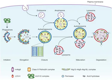
Autophagy is a lysosomal pathway involved in the turnover of cellular macromolecules and organelles. The first step of autophagy is the envelopment of cytosol and/or organelles in the isolating membrane, which wraps around the cargo forming an autophagosome, a vesicle surrounded by a double membrane. Autophagosome then undergoes a progressive maturation by fusion with endolysosomal vesicles creating an
autolysosome, where the cargo is degraded. Autophagy is controlled by a set of evolutionarily conserved autophagy-related proteins (Atg proteins). The initial nucleation and assembly of the primary autophagosomal membrane requires a kinase complex that consists of class III phosphatidylinositol 3-kinase (PI3K), p150 myristylated protein kinase and beclin 1 (also known as Atg6). The further elongation of the isolation membrane is mediated by two ubiquitin-like conjugation systems one of them resulting in the conversion of microtubule-associated protein 1 light chain 3 (LC3; also know as Atg8) from free form (LC3-I) to a lipid-conjugated membrane-bound form (LC3-II) (Fig. 6). The accumulation of LC3-II and its localization to vesicular structures are commonly used as markers of autophagy (Høyer-Hansen and Jäättelä, 2007). Autophagy in mammalian systems occurs under basal condition and can be stimulated by stress, including starvation, oxidative stress, or by treatment with the pharmacological agent rapamycin. In addition to its roles in maintaining normal cellular homeostasis by liberating nutrients from macromolecules and by assisting the clearance of misfolded proteins and damaged organelles, autophagy plays a role in the destruction of some bacteria within the cells.
Autophagy is also involved in removing damaged or excess organelles. For example, mitochondria that have lost their membrane potential and peroxisomes in response to changing environmental cues can be selectively removed by autophagy (Rodriguez-Enriquez et al., 2004). Endoplasmic reticulum (ER) stress caused by the accumulation of unfolded proteins threatens cell survival and triggers the evolutionarily conserved ER to nucleus signaling pathway called the unfolded protein response (UPR), which reduces global protein synthesis and induces the synthesis of chaperones and other proteins that increase the capacity of the ER to fold its client proteins. When the ER stress is extensive or sustained, and the
function of ER cannot be restored, it leads to the removal of the affected cells by apoptosis. Accumulating data now indicate that ER stress is also a potent trigger of autophagy (Høyer-Hansen and Jäättelä, 2007). Whereas the induction of autophagy by ER stress is conserved from yeast to mammals, the signaling pathways responsible for autophagy induction and its cellular consequences appear to vary according to the cell type and the stimulus. A better understanding of the signaling pathways controlling autophagy and the cellular fate in response to ER stress will hopefully open new possibilities for the treatment of the numerous diseases related to ER stress. In addition to activating the UPR by the pathways discussed above, ER-stress leads to a release of calcium from the ER into the cytosol, which, in turn, can activate various kinases and proteases potentially involved in autophagy signaling, as AMPK. It is not clear whether activation of AMPK is enough to trigger autophagy or whether other independent signals are required in parallel. The data presented above encourage the development of autophagy promoting therapies for diseases associated with protein aggregates either in the ER or the cytosol. If this proves to be the case, combination therapies with ER stressors and autophagy inhibitors may prove useful in cancer therapy. The direct link between ER stress and autophagy was reported recently. Thus, it is natural that many burning questions concerning the signaling pathways linking ER stress to autophagy, the mechanisms by which ER is selected as autophagic cargo, the crosstalk between ER stress-induced autophagy and cell death pathways, and the impact of autophagy in diseases associated with ER stress remain largely unanswered. The future research will hopefully clarify these points and pave the way for pharmacological exploitation of the signaling pathways involved (Høyer-Hansen and Jäättelä, 2007).
It has also been proposed that autophagy results in the total destruction of cells. Autophagic cell death represents one of several types of programmed
cell death. Autophagy is vital in a range of physiological and pathological situations, including cell growth, aging, cell death and clearing pathogenic bacteria (Rami, 2009). This mechanism provides an internal source of nutrients for energy generation and survival. When the supply of nutrients is limited, stimulating autophagy contributes to the lysosomal recycling of nutrients to maintain protein synthesis and glucose synthesis from amino acids and to form substrates for oxidation and ATP production in the mitochondria and the inhibition of the default apoptotic pathway. Moreover, starvation-induced autophagy is cytoprotective by blocking the induction of apoptosis upstream of mitochondrial events. Some metabolic changes (ATP levels, amino acids, and insulin) may regulate autophagy. AMPK is a crucial cellular energy sensor. Once activated by falling energy status, it promotes ATP production by increasing the activity or expression of proteins involved in catabolism while conserving ATP by switching off biosynthetic pathways (Hardie et al., 2012). The AMPK pathway appears to be involved in autophagy induced by nutrient deprivation, growth factor withdrawal and hypoxia. The activation of AMPK leads to the suppression of mammalian target of rapamycin (mTOR), thereby activating autophagy. Under nutrient-rich conditions, ER-associated Bcl-2 and Beclin 1 interact to sequester Beclin 1 from the autophagy complex (Wang and Qin, 2013). This association is broken by starvation and is triggered by Bcl-2 phosphorylation by JNK1. Recruitment of the Beclin 1-PI3-kinase complex to the autophagic membrane is also dependent on the ULK1 complex.
Figure 6. Schematic depiction of autophagy. Mammalian autophagy is initiated by the formation of the phagophore, followed by a series of steps, including the elongation and expansion of the phagophore, closure and completion of a double-membrane autophagosome (which surrounds a portion of the cytoplasm), autophagosome maturation through docking and fusion with an endosome (the product of fusion is known as an amphisome) and/or lysosome (the product of fusion is known as an autolysosome), breakdown and degradation of the autophagosome inner membrane and cargo through acid hydrolases inside the autolysosome, and recycling of the resulting macromolecules through permeases. The core molecular machinery is also depicted, such as the ULK1 and ULK2 complexes that are required for autophagy induction, class III PI3K complexes that are involved in autophagosome formation, mammalian Atg9 (mAtg9) that potentially contributes to the delivery of membrane to the forming autophagosome and two conjugation systems, the LC3-II and Atg12–Atg5–Atg16L complex, which are proposed to function during elongation and expansion of the phagophore membrane.
This biosynthetic pathway may be an attractive target for cancer treatment (Wang and Qin, 2013). Autophagy is a cell survival mechanism that involves the degradation and recycling of cytoplasmic components. In addition, autophagy mediates cell death under specific circumstances. Autophagic cell death (type II cell death) and apoptosis (type I cell death) have taken center stage as the principal mechanisms of programmed cell death in mammalian tissues. Although autophagy and apoptosis are
markedly different processes, autophagy and apoptosis can act as partners to induce cell death in a coordinated or cooperative fashion. Several signaling pathways that are induced by common cellular stressors regulate both autophagy and apoptosis. For example, Bcl-2 phosphorylation may not only be a mechanism for regulating apoptosis and a mechanism for regulating autophagy, but may also be a mechanism for regulating the switch between the two pathways. It is suggested that Bcl-2 also plays an essential role in limiting autophagy activation and preventing the initiation of programmed cell death (Zhang et al., 2009). The tumor suppressor p53 regulates both autophagy and apoptosis. Among other mechanisms, p53 can initiate apoptosis by inducing the expression of p53-upregulated modulator of apoptosis (PUMA), which leads to the release of cytochrome c from mitochondria and apoptotic cell death. In addition, p53 affects autophagy by modulating signaling through the mTOR nutrient-sensing kinase, which controls autophagy at the initiation stage. Autophagy proteins can play a role in cellular events that occur during apoptosis. For example, Atg5 may be a point of crosstalk between autophagic and apoptotic pathways (Wang and Qin, 2013). On the other hand, autophagy is emerging as a built-in mechanism that malignant cells can exploit for protection against a variety of intrinsic and extrinsic stress signals. However, excessive or derailed autophagy can also favor cell death. How these two seemingly opposing roles of autophagy can have an impact upon cancer chemoresistance remains unclear (Wang and Qin, 2013).
1.9 Immune regulation in cancer: focus on melanoma
Immune responses to cancer can be readily demonstrated, which indicates that immune-surveillance plays a role in control of tumor development. However, tumors growing under immune pressure might develop various
mechanisms to evade immune recognition; a process generally known as immune-evasion. Two key processes that counter immune-surveillance are immune-subversion, in which tumor cells or the tumor microenvironment suppresses the immune response, and immune-editing, which refers to the role of the immune response in selecting tumor variants that have become poorly immunogenic, thus allowing for their outgrowth. One mechanism that can have opposing outcomes on subset of innate effector cells, cytotoxic T lymphocytes (CTLs) and natural killer (NK), is the loss or downregulation of HLA class I molecules. Metastatic melanoma is the most lethal form of skin cancer despite its immunogenicity (Tsao et al., 2004). Indeed, melanoma antigens do elicit T cell responses, but T cell-based immunotherapies have shown limited success (Carrega et al., 2009). Although potent T cell responses are desirable, the downregulation of HLA class I on melanoma cells might play a significant role in the pathogenesis and clinical course of the disease, and it represents a considerable problem for T cell-based immunotherapy. Therefore, new strategies are required. Belonging to innate immunity, NK cells are cytolytic lymphocytes that can directly kill transformed and microbe-infected cells. Moreover besides their ability to kill aberrant cells, NK cells have the capacity to produce a variety of cytokines and chemokines (TNF-α, IFN-γ, CCL3, CCL5, IL-8, IL-10 etc), through which they also participate in the shaping of adaptive immune response (Vivier et al., 2009). These NK cell effector functions are regulated by multiple activating and inhibitory NK cell receptors (Lanier, 2005). Engagement of activating receptors, such as natural cytotoxicity receptors (NCRs; NKp30, NKp44, and NKp46) and NKG2D, leads to the activation of a variety of downstream signaling molecules, which result in the activation of MAPKs, particularly ERK, which are crucial for cytolytic granule release and cytokine generation (Vivier et al., 2004).
Despite the prevailing view that immunological surveillance is triggered by danger signals, signals that arise from injured cells, toxins, pathogens and mechanical damage, a recent work has shown that locally resident immune cells in the skin can detect early signs of cellular stress, through the recognition of ligands on premalignant cells and thus overt tumor transformation (Strid et al., 2008). NK cells reside in the skin (Luci et al., 2009), thus melanocytes are susceptible to NK cell recognition from early stages of dysregulation. NK cells preferentially target tumor cells with low HLA class I expression. Although there is the potential for early NK cell recognition of transformed melanocytes, some studies have indicated that NK cells are poorly represented among tumor-infiltrating lymphocytes during melanoma progression, whereas others have demonstrated NK cells in all biopsies evaluated. Understanding how NK cells migrate into the tumor microenvironment is important to redirect NK cells to the tumor in the therapeutic setting. To appreciate the role of immunosurveillance during melanoma, the factors that influence NK cell recognition of nascent dysregulated melanocytes need to be identified. In turn, how these factors change at different stages of disease, both in primary lesions and metastases, must be deciphered.
This involves characterizing the spatio-temporal expression of ligands during melanomagenesis, which hitherto has been poorly described, and the consequences of this on NK cell function (Burke et al., 2010).
Surgery is still the elective treatment for most solid cancers. Radiotherapy, chemotherapy and biological agents (cytokines, vaccines, adjuvants and immune cells) are generally used as palliative therapy and most cellular immunotherapies remain experimental. A limitation of cellular immunotherapy is the disease stage that can be approached with biological therapy, and usually only after all other options have failed. This clinical constraint limits the efficiency of immunotherapy because it gives an
advantage to the tumor cell clones that have escaped the immune system. NK cells are crucial cytotoxic effectors that complement the action of CLTs. In particular, NK cells can influence other immune cells by producing cytokines that promote dendritic cell (DC) differentiation, antigen presentation and CTL activity, and can eliminate immature DCs, thus further improving the presentation of tumor antigens to T cells (Burke et al., 2010). One limitation to the use of NK cells in immunotherapy is the number of cells required. However large-scale production is now possible for human NK cells (Spanholtz et al., 2010).
Recently, growing body of evidence indicates that a fine manipulation of the isoprenoid metabolism may constitute an important adaptive host response to stress, triggering immune response against tumor cells or infectious agents (Nassbaumer et al., 2011). Human peripheral blood Vγ9Vδ2 T cell subsets are the principal actors in monitoring the accumulation of phosphorylated isoprenoid metabolites (such as isopentenyl pyrophosphate, IPP). The reduced activity of the IPP-consuming enzymes, farnesyl diphosphate synthase (FDPS) seems to be the key mechanism in stimulation of this cytotoxic T cell subset by tumor cells (Li et al., 2009). In light of this, several groups have shown that the specific FDPS inhibitor zoledronic acid, the most potent aminobisphoshonate (NBPs) currently available for clinical use, can be exploited to intentionally activate Vγ9Vδ2 T cell by inducing IPP accumulation in tumor cells or antigen professional cells (APC) (Dieli et al., 2007).
Interestingly enough, NK cells can also be activated by the manipulation of the mevalonate pathway either by using zoledronate or statins (Nassbaumer et al., 2011; Gruenbacher et al., 2010) that are specific inhibitors of the hydroxymethylglutaryl coenzyme A (HMG-CoA) reductase, the first committed step of the mevalonate pathway acting
upstream to FDPS (Gazzerro et al., 2012). However, it has been reported that NK cells are not directly activated by isoprenoid products, like γδ T cells, but only indirectly through the action of a population of DC-like cells (Nassbaumer et al., 2011; Gruenbacher et al., 2010). This might be responsible of a delay in the first fast line of defense usually provided by NK cells. However this event seems not occur after treatment with mevalonate pathway inhibitors, leading to think that the accumulation of other isoprenoid end products might directly and immediately affect NK cell functions.
2. AIMS OF THE STUDY
This study arises from some reflections on iPA, an endogenous end-product derived from the mevalonate pathway, that has been already reported to exert a suppressor effect against various tumors in vitro and in
vivo, even if its precise mechanism of action is until unknow. It is possible
carrying out an efficacious anticancer strategy through different ways: using cancer cytotoxic drugs, anti-angiogenic agents or immune response activators able to attack specifically neoplastic cells. An important goal could be the identification of a drug with all these pleiotropic anticancer effects.
In the light of these observations, the main aims of this work were:
- to evaluate the ability of iPA to inhibit tumor progression through the interference with the angiogenic process, since angiogenesis is a key step to nourish the neoplastic mass, using human umbilical vein endothelial cells (HUVECs) as a suitable in vitro model of angiogenesis;
- to study the antitumor activity of iPA against a model of cancer characterized by a highly angiogenic phenotype as melanoma, employing human melanoma cell line, A375 cells;
- to verify the ability of iPA, due to its isoprenoid moiety and in analogy with IPP, to expande and activate selectively natural killer (NK) cells, the cytotoxic subset of innate immunity, using NK cells purified from healthy donors peripheral blood;
- to provide key informations on the mechanism through which iPA exerts its biological effects in all the models studied.
As additional deliverables it was possible to obtain basic informations about structure-activity relationship of iPA testing also its congeners, both isoprenoid, purine and structural analogues.
Moreover, it was clarified the non-involvement of the classical adenosine pathway in the biological effects exerted by iPA that are indeed mediated by intracellular targets.
Data obtained in this work will allow to give novel insights into the biological activity of this promising endogenous isoprenoid showing pleiotropic effects as anticancer drug.
3. MATERIALS AND METHODS
Materials
iPA, IPP triammonium salt solution, adenine, isopentenyladenine, dipyridamole and erythro-9 (2-hydroxy-3-nonyl)-adenine (EHNA) powders as well as acridin orange powder were purchased from Sigma-Aldrich (St. Louis, MO, USA); 5-iodotubercidin (5-Itu), PSB 1115 and ZM 241385 from Tocris Bioscience (Bristol, UK); iPA analogues CM 223, CM 224, CM 226 and CM 226 were provided by Prof. Ciuffedra (University of Milan); whilst carboxyfluorescein diacetate succinimidyl ester (CFSE) and LysoTracker- Red (DND-99) from Molecular Probes-Invitrogen (Paisley, UK). Anti-caspase-3 and -9, anti-PARP [poly (ADP-ribose) polymerase], pChk1 (Ser296), pATR (Ser428), phospho p53 (Ser15), pHistone H2AX (Ser139), anti LC3-B, pSTAT5 (Tyr 694) and anti-STAT5, anti-phospho p44/42 MAPK (Thr202/Tyr 204), anti-p44/42 MAPK, anti-phospho p38 MAPK (Thr180/Tyr182) and anti-p38 MAPK antibodies were all from Cell Signaling Technology (Danvers, MA, USA); anti p-AMPK and AMPK, anti-β-actin and anti-FDPS were from Abcam (Cambridge, UK). Anti-pan Ras and anti-tubulin were from Sigma-Aldrich (Dorset, UK). The MEK1/2-specific kinase inhibitor UO-126 was purchased from Cell Signaling technology (Beverly, MA).
Cell cultures
Human umbilical vein endothelial cells (HUVECs) were cultured on 1% gelatin-coated culture flasks with vent caps in M199 with heparin, 10% heat-inactivated fetal bovine serum (FBS), 2 mM L-glutamine and complete angiogenic factors (aFGF, bFGF, EGF, hydrocortisone and
heparin). The HUVECs were used at passages 2 to 6. Reagents were obtained from Sigma-Aldrich; aFGF, bFGF, and EGF were from Peprotech (London, UK).
Human malignant melanoma cells (A375) were cultured in DMEM High Glucose (Sigma-Aldrich, St. Louis, MO, USA) supplemented with 2 mM L-glutamine, 50 ng/ml streptomycin, 50 units/ml penicillin and 10% heat-inactivated FBS (Hyclone Laboratories, Logan, UT, USA).
All donors gave written informed consent in accordance with the Declaration of Helsinki to the use of their residual buffy coats for research purposes, with approval from the University Hospital of Salerno Review Board. Peripheral blood mononuclear cells (PBMC) from healthy donors were isolated over Ficoll-Hypaque gradients (lymphocyte separation medium; MP Biomedicals, Aurora, OH, USA). Human NK cells were negatively selected from PBMC by immunomagnetic procedure (NK-cell isolation kit; Miltenyi Biotec, Calderara di Reno, Italy). They were cultured for different time intervals in RPMI 1640 in the presence of IL-2 at 100 U/ml alone or in combination with different concentrations of iPA. Also the human chronic myelogenous leukemia cell line K562 (ATCC) was maintained in RPMI 1640 (Invitrogen, San Diego, CA, USA) supplemented with 2 mM L-glutamine, 50 ng/ml streptomycin, 50 units/ml penicillin and 10% heat-inactivated FBS. All cell cultures were mantained at 37°C in humidified 5 % CO2 atmosphere.
Drug treatments
iPA was dissolved in DMSO (20 mM) and used at indicated concentrations. For each experiment, fresh dilutions were made from the stock solution and added to cell cultures at the indicated concentrations in serum-containing medium. 5-Itu was used in pre-treatment at a final concentration of 30 nM for 30 min. iPA analogues, as well as IPP, adenine
and isopentenyladenine were dissolved in DMSO and used at indicated concentrations. Also inhibitors and antagonists used were dissolved in DMSO. Human NK cells were stimulated for different time intervals in RPMI 1640 in the presence of IL-2 at 100 U/ml alone or in combination with different concentrations of iPA.
Viability assay
To determine cells viability, we used the colorimetric MTT metabolic activity assay. Cells were cultured in a 96-well plate at 37°C, with or without growth factors, and exposed to various concentrations of iPA, IPP, adenine and isopentenyladenine, as well as iPA analogues for 24 h. To each well, 10 µl of MTT solution (5 mg/ml, in water) was added for 4 h. The resultant formazan crystals were dissolved in 100 µl of solubilization solution (10% Triton X-100 and 0.1 N HC1 in isopropanol), and the absorbance intensity was measured on a microplate reader (ThermoScientific, Basingstoke, UK) at 595 nm with a reference wavelength of 650 nm. All experiments were performed in triplicate, and the relative cell viability was expressed as a percentage comparison with the untreated control cells.
Proliferation assay by BrdU incorporation
Cell proliferation was evaluated by measuring BrdU incorporation into DNA (BrdU colorimetric assay kit; Roche Applied Science, South San Francisco, CA, USA). Endothelial or melanoma cells, pretreated or not with 5-Itu (30 nM, 30 min) or with adenosine receptors antagonists, were treated with increasing concentrations of iPA. Cells were seeded into 96-well plates for the indicated time at 37°C. Newly synthesized BrdU-DNA was determined on an ELISA plate reader (ThermoScientific) at 450 nm.