SCUOLA NORMALE SUPERIORE
Pisa
CLASSE DI SCIENZE MATEMATICHE, FISICHE E
NATURALI
CORSO DI PERFEZIONAMENTO IN NEUROBIOLOGIA
Triennio 2006-2008
Tesi di perfezionamento
EXPERIENCE-DEPENDENT REACTIVATION OF
OCULAR DOMINANCE PLASTICITY IN THE ADULT
VISUAL CORTEX
Candidata: LAURA BARONCELLI
Relatori: Prof. Lamberto Maffei
Dr. Alessandro Sale
INDEX
CHAPTER 1: EXPERIENCE-DEPENDENT PLASTICITY: CRITICAL PERIOD
OF OCULAR DOMINANCE PLASTICITY 4
1.1SURVEY OF THE MAMMALIAN VISUAL SYSTEM WITH REFERENCE TO RODENTS 5
1.2CRITICAL PERIOD FOR OCULAR DOMINANCE PLASTICITY IN THE VISUAL CORTEX 7
1.3PHYSIOLOGICAL MECHANISMS OF PLASTICITY EXPRESSION 12
1.4MOLECULAR SUBSTRATES OF OCULAR DOMINANCE PLASTICITY 22
1.5EXPERIENCE-DEPENDENT PLASTICITY IN ADULT VISUAL CORTEX 32
CHAPTER 2: THE INFLUENCE OF ENVIRONMENT ON BRAIN AND BEHAVIOUR: NEURAL CONSEQUENCES OF ENVIRONMENTAL ENRICHMENT 35
2.1ENVIRONMENTAL ENRICHMENT AFFECTS ANIMALS’ BEHAVIOUR 36
2.2ENVIRONMENTAL ENRICHMENT AFFECTS BRAIN ANATOMY 38
2.3ELECTROPHYSIOLOGICAL RESPONSES TO ENVIRONMENTAL ENRICHMENT 41
2.4ENVIRONMENTAL ENRICHMENT MODULATES GENE EXPRESSION 43
2.5ENVIRONMENTAL IMPACT ON DEVELOPMENTAL PLASTICITY OF THE BRAIN 50
2.6ENVIRONMENTAL INFLUENCES ON AGING PROGRESSION 53
2.7ENRICHED ENVIRONMENT AND DISORDERS OF THE NERVOUS SYSTEM 57
CHAPTER 3: AIM OF THE THESIS AND EXPERIMENTAL DESIGN 68
CHAPTER 4: MATERIALS AND METHODS 71
4.1REARING ENVIRONMENTS 71
4.2ANIMAL TREATMENT 72
4.3DRUG ADMINISTRATION 72
4.4IN VIVO ELECTROPHYSIOLOGY 73
4.5IN VITRO ELECTROPHYSIOLOGY:LTP AND LTD 77
4.6BEHAVIOURAL ASSESSMENT OF VISUAL ACUITY 78
4.7VISUAL CLIFF TASK 82
4.8SAMPLE PREPARATION 85
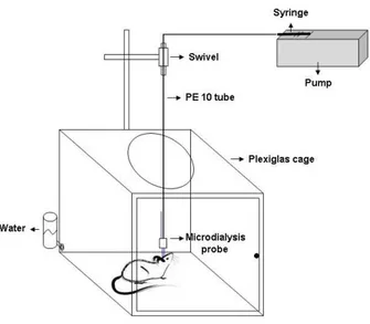
4.9IN VIVO BRAIN MICRODIALYSIS 85
4.10HIGH PERFORMANCE LIQUID CHROMATOGRAPHY (HPLC) 86
4.11IMMUNOHISTOCHEMISTRY 87
5.2BINOCULAR DECORRELATION IMPAIRS VISUAL DEPTH PERCEPTION IN EE ANIMALS 94
5.35-HT IS CRUCIALLY INVOLVED IN THE REOPENING OF OD PLASTICITY 99
5.4EXCITATION-INHIBITION BALANCE REGULATES PLASTICITY 101
5.5A ROLE FOR BDNF IN THE RECOVERY OF ADULT OD PLASTICITY 105
5.6REDUCING 5-HT LEVELS PREVENTS ENVIRONMENTAL MODULATION OF GABAERGIC
INHIBITION AND BDNF EXPRESSION 108
5.7EE HOUSING PROMOTES THE RECOVERY IN ADULT AMBLYOPIC RATS 109
CHAPTER 6: DISCUSSION 113
6.1REJUVENATING THE VISUAL CORTEX 113
6.2MONOCULAR DEPRIVATION AFFECTS DEPTH PERCEPTION IN ADULT EE ANIMALS 115
6.3THE CRUCIAL ROLE OF SEROTONIN IN EE-INDUCED CORTICAL PLASTICITY 117
6.4INTRACORTICAL INHIBITION REGULATES PLASTICITY RECOVERY IN EE ANIMALS 120
6.5BDNF IN EE-INDUCED PLASTICITY 122
6.6A MODEL OF MOLECULAR MECHANISMS UNDERLYING EE-INDUCED PLASTICITY 124
6.7EE PROMOTES THE RECOVERY OF VISUAL FUNCTIONS IN AMBLYOPIC ANIMALS 127
6.8CONCLUSIONS 130
REFERENCES 131 ACKNOWLEDGEMENTS 181
INTRODUCTION
CHAPTER 1
EXPERIENCE-DEPENDENT PLASTICITY: CRITICAL PERIOD OF
OCULAR DOMINANCE PLASTICITY
The word ‘plasticity’ refers to the ability of the nervous system to reorganize its connections functionally and structurally in response to changes in environmental experience, underlying the adaptive development of neuronal circuitry, but also learning and memory.
It has been widely demonstrated the existence of time windows in early postnatal life, named ‘sensitive periods’, during which neural circuits display a heightened sensitivity to external stimuli. ‘Critical periods’ are a special class of sensitive periods wherein proper experience is required at fixed developmental times and results in irreversible changes in brain function (Knudsen, 2004). The question of what mechanisms underlie the activation and regulation of central nervous system (CNS) critical periods is of great interest in the field of neuroscience. Manipulation of such mechanisms may potentially allow reactivation of neural circuit plasticity during times when the adult brain is normally less plastic. Furthermore, understanding the mechanisms involved in the development and plasticity of neural connections is also important for specifying possible deviations from the proper developmental plan, and hence the aetiology of developmental brain disorders.
It is now understood that many regions of the brain have critical periods that occur at different times and are activated and regulated by distinct mechanisms (for a review, see Hensch, 2004), but the visual cortex has long been a proving ground for studying
experience-dependent plasticity because visual experience can be easily manipulated and the consequences of manipulations can be readily measured at the anatomical, physiological and molecular levels. Classic experiments have been performed in cats and primates, but recent findings regarding the cellular and molecular mechanisms underlying the experience-induced changes in visual cortical function have been derived predominantly from rodents, owing to the simplicity of the rodent visual system and the relative ease of genetic manipulation.
1.1 Survey of the mammalian visual system with reference to rodents
This paragraph addresses the basic aspects of visual system organization with particular reference to similarities and differences between the visual system of rodents and that of other mammalian species.
The retina is a sensory structure responsible for converting light into neural signals that are relayed to the brain through the optic nerve. The basic functional organization of the mouse retina is largely similar to that of other mammalian species (Adams, 1964; Rodieck, 1971). In all mammals the optic nerves from each eye join at the optic chiasm, where fibers destined for one or the other side of the brain are sorted out. The majority of retinal ganglion cell (RGC) axons carrying information from a given retina cross at the chiasm and project to the contralateral retinorecipient nuclei.
The proportion of RGC axons that do not cross and thus project to the ipsilateral retinorecipient nuclei, however, varies considerably among species and is closely related to the position of the eyes in the skull. In species with laterally positioned eyes and therefore a relatively small binocular field, the proportion of ipsilaterally projecting RGCs tends to be small. In particular, in rodents the proportion of RGCs projecting ipsilaterally is 2-3%
(Dräger and Olsen, 1980) against the 25-40% observed in cats and primates (Perry et al., 1984; Tassinari et al., 1997). Retinal fibers then enter each optic tract, which projects mainly to three subcortical targets, i.e. the superior colliculus, several nuclei in the pretectal complex of the epithalamus and the lateral geniculate nucleus (LGN) of the thalamus (Dräger, 1974; Hofbauer and Dräger, 1985; Provencio et al., 1998). The LGN is the thalamic nucleus that relays visual information from the retina to the visual cortex. RGC project in an orderly manner to points in the LGN, so that in each LGN there is a retinotopic representation of the contralateral half of the visual field (Wagner et al., 2000). In all studied species of carnivores and primates the LGN cells that receive contralateral retinal inputs and those receiving ipsilateral retinal inputs are clustered into cellular layers (Kaas et al., 1972; Hickey and Guillery, 1974). By contrast, in rodents there is not evidence of a layered organization of LGN; contralateral and ipsilateral retinal fibers are segregated in a patchy fashion (Reese and Jeffery, 1983; Godement et al., 1984; Reese, 1988).
The projections of LGN neurons reach the primary visual cortex (V1), in the occipital portion of the brain (Caviness and Frost, 1980; Simmons et al., 1982). The visual cortex consists of six layers of cells (layers I-VI) between the pial surface and the underlying white matter and contains a complete representation of the contralateral visual hemifield. The principal layer of inputs from the LGN is layer IV; then information flows systematically from one cortical layer to another, to higher visual cortical regions, to visual regions of the other hemisphere, to midbrain regions and also back to the LGN (Ruiz-Marcos and Valverde, 1970; Caviness, 1975; Yorke and Caviness, 1975; Frost and Caviness, 1980; Wagor et al., 1980; Cusick and Lund, 1981; Simmons et al., 1982; Olavarria and Montero, 1989). In carnivores and primates V1 is clustered into narrow columns, running from the pial surface to the white matter, devoted to the analysis of orientation (orientation columns) and binocular interactions (ocular dominance columns)
(Hubel and Wiesel, 1963; 1972; LeVay et al., 1975; Hubel et al., 1978; Shatz and Stryker, 1978; Ts’o et al., 1990). These regularly spaced columnar systems communicate with each other by means of long-range horizontal connections that link cells within a layer, allowing the integration of sensory information over the cortex (Livingstone and Hubel, 1984; Kisvarday et al., 1989; Gilbert, 1992).
In rodents, the binocular segment of V1, in which the binocular part of the visual field (nasal 30-40° of the visual field) is represented, occupies the lateral third of area V1. Not surprisingly, the proportion of cells in the binocular region that can be activated preferentially by stimuli presented through the contralateral eye is much larger than the proportion of cells activated preferentially by stimuli presented through the ipsilateral eye (Dräger, 1975; 1978; Métin et al., 1988; Porciatti et al., 1999; Kalatsky and Stryker, 2003). There is no evidence of cell clustering into radial columns based on eye preference. Indeed, monocular cells responding only to the stimuli presented to one eye, or binocular cells dominated by one eye, are intermingled with cells that preferentially (or solely) respond to stimuli presented to the other eye (Dräger, 1975; 1978; Fagiolini et al., 1994; Antonini et al., 1999). Even though rodents do not have anatomically distinct ocular dominance columns, they do, nevertheless, have a critical period during early postnatal life in which the relative representations of the two eyes in the binocular region of the cortex are sensitive to visual experience (Dräger, 1978; Fagiolini et al., 1994; Gordon and Stryker, 1996).
1.2 Critical period for ocular dominance plasticity in the visual cortex
Although the maturation of visual system circuitry starts well before the time of eye opening, and the targeting of thalamocortical connections occurs at very early developmental stages controlled by genetic programmes and spontaneous activity
(Crowley and Katz, 1999; Sur and Leamey, 2001; Crowley and Katz, 2002; Sur and Rubenstein, 2005), a proper development of the visual system requires sensory experience.
There is evidence that the total absence of sensory input leads to a delay in the functional and anatomical maturation of the visual cortex, that appears still immature far beyond the end of the critical period. The visual cortex of adult animals reared in darkness from birth (dark rearing, DR) displays serious physiological deficits including reduced orientation and direction tuning, lower cell responsiveness and increased latency, larger receptive field sizes, altered spontaneous activity, rapid habituation to repeated stimulus presentation, immature ocular dominance distribution and lower visual acuity (Fregnac and Imbert, 1978; Timney et al., 1978; Benevento et al., 1992; Fagiolini et al., 1994; Pizzorusso et al., 1997). A total lack of visual experience also affects the fine structure of visual cortical neurons, measured as alterations in the size, morphology and density of dendritic spines (Valverde, 1971; Wallace and Bear, 2004). Regular developmental processes seem to be restored once the animals are exposed to light, thus allowing the recovery of both neuronal response properties (Buisseret et al., 1978; 1982) and normal anatomical features (Wallace and Bear, 2004).
A classic model used for understanding how experience-dependent activity shapes the maintenance and refinement of brain circuitry is ocular dominance (OD) plasticity, i.e. the rapid changes in visual cortex circuitry resulting from unbalanced inputs from the two eyes.
Hubel and Wiesel first reported that thalamocortical inputs from the two eyes segregate in V1 of cats to form OD columns and that these neural circuits are influenced by manipulations of the animal visual experience. Reducing input from one eye by lid suture (monocular deprivation, MD) during development dramatically affects the binocularity properties of cortical neurons, leading to a loss of cortical physiological responses to that
eye and an increase in the number of neurons responding preferentially to stimuli presented to the open eye. As a direct behavioural consequence, the deprived eye becomes amblyopic: its visual acuity is strongly reduced and its contrast sensitivity blunted (Wiesel and Hubel, 1963a; Hubel and Wiesel, 1970; Olson and Freeman 1975; Movshon and Dürsteler, 1977; Olson and Freeman, 1980). Remarkably, physiological responses in the deprived retina and thalamus remained completely unaffected (Wiesel and Hubel, 1963b; Sherman and Stone, 1973; Kratz et al., 1979; Baro et al., 1990). Hubel and Wiesel observed that in kittens the susceptibility to the effects of MD begins suddenly near the start of the fourth week of life, remains robust until some time between the sixth and eighth weeks, and then declines completely after the third month, thus defining a critical period for MD effectiveness. MD starting in adulthood produces no detectable outcome (Hubel and Wiesel, 1970; Olson and Freeman, 1980).
The effects of MD and the relative existence of a critical period have been subsequently described also in several species of mammals, including primates (Hubel et al., 1977; Blakemore et al., 1978; LeVay et al., 1980; Horton and Hocking, 1997), rabbits (Van Sluyters and Stewart, 1974), hamsters (Emerson et al., 1982) and ferrets (Issa et al., 1999). The effects of deprivation can be reversed to a limited extent during the critical period by reversing the visual deprivation, but they later become irreversible (Wiesel and Hubel, 1965a; Movshon, 1976; van Sluyters, 1978; Blakemore et al., 1981; Antonini et al., 1998).
Anatomical tracing studies revealed that the imbalance of activity between the two eyes results in the actual loss of synaptic inputs from the thalamic regions representing the closed eye, and in the expansion of those representing the open eye, both in cats and primates. While in normal animals monocular cells responding only or binocular cells responding preferentially to visual stimuli presented via a particular eye cluster into
alternating, equal-sized OD columns, closure of one eye reduces the cortical territory of the closed eye to shrunken broken stripes, with its former territory now invaded by inputs representing the open eye (Hubel et al., 1977; Shatz and Stryker, 1978; LeVay et al., 1980). A more detailed morphological analysis has been shown that the thalamo-cortical axonal arbors driven by the deprived eye are less branched, have reduced length and decreased maximal innervation density, and show a reduction in spine density and size, while non-deprived thalamic arbors display an increase in all these parameters (Tieman, 1991; Antonini and Stryker, 1993; 1996). Due to the excellent correlation between the functional changes induced by MD and the reorganization of the geniculocortical afferents, it was believed that the latter was the structural correlate of OD shift. The finding that a functional OD shift occurs first in the extragranular layers and not in layer IV, and that the reorganization of the geniculo-cortical projections follows several days later came as a surprise (Trachtenberg et al., 2000). In search of a better structural correlate, Trachtenberg and Stryker (2001) analyzed the reorganization of horizontal connections in the upper layers of the cortex after inducing strabismus in cats during the critical period. They found that rapid plasticity of binocular responses in the upper layers of the cortex is mirrored by similarly rapid anatomical changes in the long-range horizontal connections between OD columns in these layers. Thus, horizontal connections seem to be a better structural correlate for the functional changes than geniculo-cortical afferents. Several question remain, however, with respect to the involvement of the connections between neurons of layer IV and II/III in OD plasticity and to the mechanism by which thalamocortical remodelling is guided by higher cortical stages (see Bence and Levelt, 2005).
Similar to higher mammals, MD in rodents shifts the physiological responsiveness of neurons in the binocular zone of V1 towards the open eye, and this plasticity is confined to a well-defined critical period (Dräger, 1978; Fagiolini et al., 1994; Gordon and Stryker,
1996). At least in the mouse, this is at first due to a rapid weakening of deprived-eye response, and later to strengthening of open-eye response (Frenkel and Bear, 2004). Interestingly, the OD shift is found in all layers, but it is more pronounced in extragranular layers than in layer IV, with the greatest shift in infragranular cells (Gordon and Stryker, 1996), suggesting that in rodents, as in other species, intracortical as well as geniculo-cortical synapses undergo plasticity with MD. Anatomical changes accompany functional plasticity in the developing visual cortex of the mouse, as they do in higher mammals (Antonini et al., 1999). Moreover, the advent of two-photon microscopy allowed the in vivo study of visual cortex spine dynamics during development and after visual deprivation: Oray et al. (2004) showed that spine motility in the binocular region of V1, contralateral to the deprived eye, is 35% higher than motility in control, nondeprived animals, indicating that sensory deprivation in a plastic cortex is able to initiate a rapid sequence of events that lead to increased structural dynamics at the level of individual spines. Such an increase in spine dynamics may reflect structural destabilization of a population of spines whose function is affected by visual deprivation. This, in turn, could precede a robust pruning of spine protrusions, probably correlated to the rapid reduction in the deprived-eye drive (Mataga et al., 2004).
It is worth to stress that also in humans vision during a definite time period is required for normal development of spatial acuity, global motion detection, and other visual system characteristics. Lasting visual impairments can result from several conditions that degrade or unbalance vision prior to adolescence, including strabismus, uncorrected refractive errors and cataracts (Lewis and Maurer, 2005). In particular, amblyopia is clinically important because, aside from refractive errors, it is the most frequent cause of vision loss in infants and young children, occurring naturally in about 2-4% of the population (Levi, 2006).
1.3 Physiological mechanisms of plasticity expression
Once it has been defined the crucial role of experience and the existence of a critical period during the development of visual system, the next step is to understand what is changing in the nervous system following alterations in visual experience. There are several caveats, however, to consider in establishing that variations in cortical responsiveness are due to particular synaptic modifications, because changes in environmental experience can affect the sensory systems at multiple levels (deprivation may also affect orientation and direction selectivity) and the synaptic modifications underlying a shift in OD may occur at multiple synapses. Furthermore, there may be multiple forms of synaptic plasticity (Hooks and Chen, 2007). Wiesel and Hubel proposed a mechanism in which OD plasticity operates through a competitive interaction between inputs from the two eyes for the control of cortical neurons, depending on the activity state of the postsynaptic neurons. This hypothesis was supported by the fact that binocular lid suture is not effective to shift OD columns in mammals (Wiesel and Hubel, 1965b; Gordon and Stryker, 1996; Antonini and Stryker, 1998). An experiment performed by Stryker’s laboratory showed that patterned vision is not necessary for visual cortical plasticity, and that an imbalance in spontaneous retinal activity alone can produce a significant OD shift, thus supporting the competitive view (Chapman et al., 1986). In addition, a reversible blockade of the discharge activities of cortical neurons by intracortical infusion of tetrodotoxin (TTX) or muscimol completely prevents the OD shift that would normally be seen after MD, or causes a paradoxical shift in favour of the deprived eye (Reiter et al., 1986; Reiter and Stryker, 1988; Hata and Stryker, 1994; Hata et al., 1999). However, the mechanism underlying binocular competition has remained elusive. The classic competition-based model is related to heterosynaptic mechanisms, where open eye inputs drive down the synaptic efficacy of the deprived inputs (Miller et al., 1989; Harris et al., 1997). Previous studies have implicated
activity-dependent uptake of neurotrophins, as the mediator of binocular competition (Maffei et al., 1992; Cabelli et al., 1995), but subsequent experiments have shown that neurotrophins actually have cell-specific effects, such as the regulation of inhibitory circuitry, which may provide an alternative explanation of their importance for OD plasticity (Berardi and Maffei, 1999; Huang et al., 1999). Chronic electrophysiological recordings in mice at the peak of the critical period indicate that binocular competition may actually be the consequence of separable processes with distinct time courses mediating depression of deprived-eye and potentiation of non deprived-eye responses (Frenkel and Bear, 2004; Mrsic-Flogel et al., 2007; Kaneko et al., 2008a,b).
The homosynaptic view It is tempting to speculate that the loss or gain of visual
responsiveness of neurons in V1 during the critical period is simply the result of homosynaptic long-term depression (LTD) or potentiation (LTP) of excitatory connections somewhere in the visual circuit (Smith et al., 2008). However, the role of LTP and LTD in OD plasticity is hotly debated. The induction of LTP has been extensively demonstrated at multiple synapses of the visual cortex ex vivo, although the mechanism appear to vary across layers (Wang and Daw, 2003). Additionally, in rats, NMDA receptor (NMDAR)-dependent LTP can be induced in layers II/III and IV in vivo following tetanic stimulation of LGN, and this LTP is sufficient to increase the magnitude of visually evoked responses (Heynen and Bear, 2001), suggesting that homosynaptic LTP, possibly at thalamortical synapses, can mimic the effects of open-eye potentiation after MD. Many manipulations known to disrupt homosynaptic LTP have been applied during OD plasticity. One example of this is the finding that OD plasticity is disrupted in mice with either disrupted αCaMKII autophosphorylation or lacking the protein entirely, which suggests a role for LTP (Gordon et al., 1996; Taha et al., 2002). Similarly, open-eye potentiation is absent in mice with a postnatal deletion of NR1 targeted to cortical layers II-IV (Sawtell et al., 2003). Further
suggestion comes from the recently discovered phenomenon of stimulus-selective response potentiation: in juvenile mice, the magnitude of visually driven thalamo-cortical responses in layer IV increases following repeated presentation of an oriented stimulus and this potentiation is dependent on NMDAR activation. Moreover, it has been shown that GluR1 delivery to synapses, that is crucial for LTP, is required for visual experience-dependent plasticity (Frenkel et al., 2006). Stronger evidence exists that LTD-like mechanisms influence depression of deprived-eye responses. The biochemical signature of LTD (in terms of AMPA receptor phopshorylation and cell-surface expression) has been used as a ‘molecular fingerprint’ to ask whether similar changes occur in visual cortex following a period of MD. To date, this has been examined in the rat visual cortex and the results support the hypothesis that MD induces this type of LTD in visual cortex (Heynen et al., 2003; Yoon et al., 2009). A second approach to address whether LTD is induced by MD is to ask whether naturally occurring synaptic depression in vivo occludes LTD ex vivo. This issue has been recently examined in rodents: LTD measured in slices is reduced (occluded) by 3 days of MD in vivo in both layer IV and II/III (Heynen et al., 2003; Crozier et al., 2007). Furthermore, the reduction in deprived-eye responses after lid suture is likely due to hebbian processes, as monocular inactivation with TTX (which prevents decorrelated inputs) blocks this depression (Frenkel and Bear, 2004). However, the question of the relative contribution of this synaptic modification to the functional consequences of MD is still controversial. An approach to this question has been to correlate deficits in LTD and OD plasticity in genetically or pharmacologically modified mice. A mutation that disrupts LTD dependent on metabotropic glutamate receptor (mGluR) does not alter the normal OD shift in response to MD (Renger et al., 2002). GAD65 knockout mice, which lack normal OD plasticity, show no deficit in induction of LTP or LTD in layer II/III of mouse binocular visual cortex (Hensch et al., 1998a), while similar studies at younger ages show
an impairment of LTD (Choi et al., 2002). A dissociation of LTD and OD plasticity has been suggested also by the study of several protein kinase A (PKA) regulatory subunit mutants. For example, the RIβ knockout mouse has a deficit in layer II/III LTD but exhibits a normal OD shift after 4 days of MD (Hensch et al., 1998b). However, two additional studies deleting both of the two RII subunits of PKA further complicate this story. RIIα knockout mice display normal LTD in layer II/III, whereas both LTP and OD plasticity are reduced (Rao et al., 2004). By contrast, RIIβ knockout mice exhibit normal LTP at the same synapse, but lack both LTD and OD plasticity (Fischer et al., 2004). Also calcineurin, the only known Ca2+/calmodulin-activated protein phosphatase in the brain, has been identified as a molecular constraint involved in OD plasticity, but a transient increase in calcineurin activity, that prevents the shift of responsiveness in the visual cortex following MD, does not impair LTD induction (Yang et al., 2005).
Given that many different plasticity mechanisms exist in the visual cortex (Wang and Daw, 2003; Rao and Daw, 2004), it is likely that a large portion of these seemingly conflicting results may be attributable to laminar differences between the molecular pathways supporting LTD and LTP, but essentially the LTD/LTP mechanisms alone are unlikely to account for the OD plasticity. Indeed, several alternative hypotheses have also been advanced to account for the phenomenology of OD plasticity. For example, balanced levels of excitation and inhibition have shown to be critical for enabling plasticity (Hensch, 2005a; Hensch and Fagiolini, 2005).
Excitatory-inhibitory balance In all species tested so far anatomical and physiological
evidences indicate that synaptic inhibition matures later than excitatory transmission in the neocortex (Blue and Parnavelas, 1983; Luhmann and Prince, 1991; Benevento et al., 1992; Guo et al., 1997; Micheva and Beaulieu, 1997; Gao et al., 2000; Mower and Guo, 2001; Murphy et al., 2005). By controlling excitation, GABAergic circuits are ideally posed to
control the engagement of activity-dependent synaptic modification. Thus, the mismatch in the maturation of excitation and inhibition may define a window of opportunity for activity-dependent plasticity to occur. Taking advantage of gene-targeting technology, this hypothesis has been directly tested by reducing GABA synthesis or by prolonging glutamatergic synaptic responses, both adjustements yielding a similar shift of balance in favour of excitation in vivo.
Mice carrying a targeted disruption of the GAD65 gene show and identical OD distribution to wild-type mice; however, the response to a brief period of MD during the critical period is strikingly different. These mice, indeed, show no shift in their responsiveness in favour of the open eye and the cortical neurons continue to respond better to the contralateral eye input. The enhancement of inhibition obtained by local delivering of diazepam produces a complete OD shift in the infused mutant visual cortex (Hensch et al., 1998a). Equally, in NR2A knockout mice, OD distribution is similar to control animals. Unlike GAD65 knockout mice, brief MD is able to induce a slight shift in favour of the open eye but, interestingly, the overall magnitude of this plasticity is significantly weakened. Long-term MD (> 2 weeks) produces no further shift, confirming that saturation is reached within 4 days. Also in this case, diazepam infusion concomitant with MD fully rescues OD plasticity (Fagiolini et al., 2003). A direct physiological consequence of excitatory-inhibitory unbalance in GAD65 and NR2A KO mice is enhanced activation in response to visual stimulation, as assessed by the observation that visual cortical neurons display a tendency for prolonged discharge outlasting the visual stimulus (Hensch et al., 1998a; Fagiolini et al., 2003). Whereas a robust prolonged discharge appears throughout life in both mutants, it is only evident early in the life of wild-type animals before the critical period, when intrinsic inhibition is weak and OD plasticity is absent. With the natural appearance of OD plasticity during the critical period
in wild-type mice, the prolonged discharge drops off sharply (Fagiolini and Hensch, 2000). Taken together, these results indicate that a delicate equilibrium between excitation and inhibition intrinsic to visual cortical circuits is necessary to detect the imbalanced activity between competing inputs from the deprived and non-deprived eyes. Consistent with this view, the onset of the critical period can be accelerated in wild-type animals by premature enhancement of GABA-mediated transmission (Huang et al., 1999; Fagiolini and Hensch, 2000). Moreover, feedforward inhibition can enhance the precise timing of postsynaptic firing (Pouille and Scanziani, 2001). Specific spike timing-dependent windows for synaptic plasticity have been elucidated in developing and neocortical structures (Bi and Poo, 2001). Spike-timing forms of plasticity rely upon physiologically realistic, millisecond-scale changes in the temporal order of pre- and post-synaptic action potentials. Prolonged discharge in both NR2A and GAD65 knockout mice would impair plasticity by altering the pattern of neural activity encoding visual input. The normal development of inhibitory circuitry, as well as diazepam infusion in transgenic mice, improve temporal processing of sensory input, allowing OD shift in response to MD to take place (Hensch and Fagiolini, 2005).
Among the vast diversity of GABAergic interneurons in neocortex, two major sub-classes of parvalbumin-containing cells target the axon initial segment and soma. Both are ideally situated to control either spike initiation (chandelier cells) or back-propagation (basket cells) required for synaptic plasticity in the dendritic arbour (DeFelipe, 1997; Somogyi et al., 1998). Because distinct GABAA receptor subunits are enriched at these two
parvalbumin-cell synapses, their individual contributions to visual cortical processing and plasticity have been identified by point mutations that selectively remove diazepam sensitivity: systematic use of the mouse knock-in technique showed that only one of these subtypes, the α1-subunit-containing interneurons (i.e. basket cells), drives cortical
plasticity (Fagiolini et al., 2004). Two scenarios centred on the parvalbumin-positive basket cells have been proposed (Hensch, 2005a). One is an ‘instructive’ model, in which powerful, fast somatic inhibition edits one-by-one the action potentials that can pass into the dendritic arbour by back-propagation through the cell body. Sloppy gating by weak inhibition at the soma would prevent a competitive outcome by allowing excess back-propagation and spurious coincident activity with infrequent inputs from the deprived retina. Consistently with this model, large-basket cells extend a wide, horizontal axonal arbour that can span ocular dominance columns in cat visual cortex and, receiving input from one eye, inhibits targets of the other eye (Buzas et al., 2001). A second ‘permissive’ model is based on the observation that basket cells are organized in electrically-coupled networks, endowed with the ability to detect synchrony (Galarreta and Hestrin, 2001a,b; Meyer et al., 2002). Whereas simultaneous inputs (for example, from the same eye) rapidly co-excite cells through gap junctions, even a 2 ms input jitter (for example, between opposite eyes) is sufficient to dampen the coupling by reciprocal GABAA synapses, which
are also enriched in α1 subunits (Gao and Fritschy, 1994). As a result, these neurons are maximally active on a columnar scale, time-locked to release growth or plasticity factors when strong synchronous activity arrives in the neocortex.
It is worth to point out that during development the inhibitory tone surpasses two functional thresholds in the visual cortex: the first one enables OD plasticity and the second one causes the end of the critical period. A recent study shows that pharmacological reduction of intracortical inhibition obtained through the infusion of either MPA (an inhibitor of GABA synthesis) or picrotoxin (a GABAA antagonist) directly
into the visual cortex reactivates OD plasticity in response to MD in adult rats (Harauzov et al., in press). Moreover, also other manipulations resulting in reductions of cortical inhibition promote adult plasticity (He et al., 2006; Maya-Vetencourt et al., 2008).
Homeostatic synaptic plasticity Homeostatic synaptic plasticity mechanisms are emerging
as important complements to Hebbian forms of plasticity in the activity-dependent refinement of synaptic connectivity (Turrigiano and Nelson, 2004; Davis, 2006; Turrigiano, 2008). Homeostatic plasticity acts to stabilize the activity of a neuron or neuronal circuit against perturbations that alter excitability, providing a robust mechanism for generating stability in network function in the face of experience-related changes in synaptic input. Plasticity phenomena that conform to this definition of homeostatic plasticity include the activity-dependent regulation on intrinsic neuronal firing properties (Desai, 2003; Marder and Prinz, 2003; Zhang and Linden, 2003); pre- and post-synaptic forms of excitatory synaptic plasticity, such as synaptic scaling, that adjust all of a neuron’s excitatory synapses up or down in the right direction to stabilize firing (Turrigiano and Nelson, 2004; Davis, 2006); the balancing of excitation and inhibition within neuronal networks (Maffei et al., 2004); compensatory changes in synapse number (Kirov et al., 2004; Wierenga et al., 2006); and meta-plastic mechanisms that adjust the relative threshold of LTP and LTD induction (Bienenstock et al., 1982; Abraham and Bear, 1996). The best studied mechanism of homeostatic regulation is synaptic scaling of excitatory synapses, which was first described in dissociated rat cortical cultures, where blockade of activity with TTX increases and blocking GABA-mediated inhibition decreases the amplitude of miniature excitatory postsynaptic currents (mEPSCs) (Turrigiano et al., 1998). Interestingly, the rules for synaptic scaling depend on the synapse type: inhibitory synapses onto pyramidal neurons are scaled in the opposite direction from excitatory synapses, suggesting that the firing rate is regulated through reciprocal changes in excitation and inhibition (Kilman et al., 2002; Swanwick et al., 2006). Homeostatic adjustements in synaptic strength include post-synaptic and pre-synaptic modifications in synaptic function (Turrigiano et al., 1998; Murthy et al., 2001; Thiagarajan et al., 2005;
Wierenga et al., 2005; 2006) and require that neurons sense and translate changes in activity into compensatory changes in synaptic strength, but the nature of the activity signal that controls synaptic scaling is still debated. Neurons could sense changes in their own firing rate through intracellular calcium levels and the modification of one or more intracellular signalling pathways (e.g. the calcium/calmodulin-dependent protein kinase family, the immediate early gene Arc, the polo-like kinase 2 and the cyclin-dependent kinase 5) globally scales synaptic weights up or down (Thiagarajan et al., 2002; 2005; Rial Verde et al., 2006; Shepherd et al., 2006; Ibata et al., 2008; Seeburg et al., 2008). Recently, several molecules important for trans-synaptic signalling and cell adhesion have been implicated in synaptic scaling (Goddard et al., 2007; Cingolani et al., 2008). Finally, synaptic scaling could require widespread changes in network activity, perhaps through activity-dependent release of a soluble factor by many neurons or glia simultaneously, such as BDNF and TNFα (Rutherford et al., 1998; Stellwagen and Malenka, 2006; Kaneko et al., 2008a).
Homeostatic plasticity appears to stabilize circuit function in vivo in a number of organisms and brain areas (Turrigiano, 1999; Davis and Bezprozvanny, 2001; Marder and Goaillard, 2006). Synaptic scaling has been most thoroughly studied in vivo in the visual system, using standard visual deprivation paradigms to mimic in vivo the activity blockade in culture. There is now increasing evidence that synaptic scaling in excitation and inhibition plays important roles during various critical periods of visual system development (Desai et al., 2002; Maffei et al., 2004; Maffei and Turrigiano, 2008a). In particular, it has been suggested that the potentiation of non deprived-eye responses following MD might arise through homeostatic mechanisms that boost the excitability of cortical neurons in response to a drop of sensory input. A recent study using in vivo calcium imaging to monitor eye-specific activation of individual neurons within binocular
layer II/III of visual cortex reported that binocularly driven neurons maintain their overall level of responsiveness to the two eyes, so that the decrease in the responsiveness to the eye stimulation is compensated by an increase in responsiveness to non deprived-eye stimulation. Interestingly, in monocular visual cortex, the population of neurons driven only by the deprived eye has homeostatic-mediated stronger responses after deprivation, as do all neurons after binocular deprivation (Mrsic-Flogel et al., 2007). In support of the notion that synaptic scaling underlies gain of responsiveness to the non-deprived eye, blocking TNFα signalling in visual cortex either pharmacologically or genetically has no effect on the loss of responsiveness to the deprived eye but prevented the gain of responsiveness to the non-deprived eye (Kaneko et al., 2008a). Complicating the interpretation of these studies is the recent report that the mode of homeostatic plasticity within layer II/III of the visual cortex during the critical period depends strongly on the method of visual deprivation: lowering visual drive through TTX or dark rearing induces synaptic scaling, whereas eyelid suture causes an increase in the intrinsic excitability of monocular cortex pyramidal neurons (Desai et al., 2002; Maffei and Turrigiano, 2008b). This suggests that also the homeostatic response observed after MD is likely due to homeostatic intrinsic plasticity rather than synaptic scaling, but further studies will be necessary to elucidate this point.
In conclusion, these studies highlight the notion that experience-dependent plasticity is unlikely to be explained by a single form of synaptic plasticity, but rather arises through a complex interplay between multiple forms of change in synaptic strength, including modifications in inhibitory circuitry, homosynaptic depression and potentiation, and global changes in circuit gain.
1.4 Molecular substrates of ocular dominance plasticity
A complete understanding of critical period plasticity requires linking the change in circuit function with the molecular mechanisms that make circuit changes possible. The molecular mechanisms that control the developmental plasticity of visual cortical connections are not fully understood. This paragraph reviews evidences establishing some factors as determinant for visual cortex plasticity.
Glutamatergic receptors The properties of NMDA receptors (NMDARs) suggest that these
molecules might play a central role in visual cortex plasticity, acting as ‘coincident detectors’ for Hebbian plasticity. The involvement of NMDARs in OD plasticity has been repeatedly proposed by pharmacological experiments (Kleinschmidt et al., 1987; Gu et al., 1989; Bear et al., 1990), but such manipulations have potent suppressive effects upon normal synaptic transmission. Recently, the use of different NMDAR antagonists or antisense oligonucleotides to reduce expression of NR1 subunit of the NMDA receptor has overcome this problem, showing that it is possible to block the effects of MD without affecting visual responses (Roberts et al., 1998; Daw et al., 1999a). The direct dependence of OD plasticity on NR1 subunits has been further demonstrated using conditional NR1-knockout mice (Sawtell et al., 2003). An interesting property of NMDARs is that their subunit expression, determining the calcium influx, is developmentally and activity regulated. In particular, subunit composition varies in the visual cortex, from low to high NR2A/NR2B ratio, with a time course paralleling that of functional visual cortical development and the critical period (Roberts and Ramoa, 1999). It has been shown that in dark-reared animals the NR2A/NR2B ratio is lower than in light-reared animals (Quinlan et al., 1999 a,b; Tongiorgi et al., 2003). However, recent results have demonstrated that NR2B over-expressing animals don’t show an increased susceptibility to plasticity (Philpot
et al., 2001) and in mice with the deletion of NR2A subunit the sensitivity to MD is weakened, even if restricted to the normal critical period (Fagiolini et al., 2003). Interestingly, a very recent study highlights a co-regulation of OD plasticity and NMDAR subunit expression in GAD65 knockout mice. In the visual cortex of these animals there are reduced NR2A levels and slower NMDA currents. In addition, application of benzodiazepines, which rescues OD plasticity, also increases NR2A levels, suggesting that changes in inhibition would engage mechanisms that converge to regulate NMDA receptors, thereby enabling plasticity (Kanold et al., 2009).
Further results establish a role for AMPA receptors (AMPARs) in the deprived-eye response depression following MD, reporting that a brief MD during the critical period alters AMPAR phosphorylation and reduces the expression of AMPARs on the surface of visual cortical neurons (Heynen et al., 2003). Finally, there is also direct evidence that metabotropic glutamate receptors (mGluRs) are involved in visual cortex plasticity, with distinct roles depending on the receptor subtype and cortical layer (Daw et al., 1999b; Wang and Daw, 2003; Rao and Daw, 2004). Recently, using the molecular genetic approach, it has been shown an important role for mGluRs in the regulation of OD plasticity during development, since a 50% reduction in mGluR5 expression prevents OD plasticity induced by 3 days of MD (Dolen et al., 2007; Dolen and Bear, 2008).
Neurotrophins There is a conspicuous number of observations suggesting that
neurotrophins control visual cortical plasticity during the critical period. Early studies in the rat demonstrated that intraventricular as well as intracortical infusion of NGF prevents OD shift following MD (Domenici et al., 1991; 1992; Lodovichi et al., 2000). Moreover, infusion of antibodies that specifically activate the NGF receptor trkA equally blocks OD plasticity (Pizzorusso et al., 1999). With the exception of NT-3, exogenous supply of all neurotrophins affects the outcome of MD. However, the effects of neurotrophins on OD
plasticity are sometimes accompanied by alteration of other properties of visual cortical neurons, such as their pattern of discharge and orientation selectivity (Gillespie et al., 2000; Lodovichi et al., 2000). Other studies, which followed the opposite course of antagonizing the action of endogenous neurotrophins, have also shown that neurotrophins are important for normal visual cortical development and plasticity (Berardi et al., 1994; Domenici et al., 1994a; Cabelli et al., 1997). In addition, neurotrophin production and release is developmentally regulated and depend on electrical activity, in particular on visual activity (Castren et al., 1992; Bozzi et al., 1995; McAllister et al., 1999). In turn, neurotrophins can modulate electrical activity and synaptic transmission at both presynaptic and postsynaptic levels (Carmignoto et al., 1997; Berardi and Maffei, 1999; Poo, 2001). They can have both fast actions, for instance by increasing transmitter release (Sala et al., 1998; Jovanovic et al., 2000) or by directly depolarizing neurons (Kafitz et al., 1999), and slow actions, by modulating gene expression (Poo, 2001). This reciprocal regulation between neurotrophins and neural activity might provide a means by which active neuronal connections are selectively strengthened. Indeed, neurotrophins seem to require the presence of electrical activity to exert their actions (Sala et al., 1998; Caleo et al., 1999; McAllister et al., 1999). The classic hypothesis (‘neurotrophic hypothesis’) states that competition for limited amounts of neutrotrophins is the effector of activity-dependent plasticity in the cortex, and the conventional explanation for OD plasticity is that the deprived eye does not activate cortical cells as well as the open eye, thereby failing to stimulate them to release sufficient neurotrophins to sustain the deprived-eye pathway (Thoenen, 1995; Bonhoeffer, 1996; McAllister et al., 1999). The possibility of an anterograde action of neurotrophins as opposed to target-derived action has also emerged from literature (Caleo et al., 2000; Kohara et al., 2001; von Bartheld, 2004). This significantly changes the frame of thought: in addition to thinking that cortex-derived
factors guide stabilization of thalamic afferents on cortical neurons, we may have to consider that thalamic fibers themselves release factors which promote and guide the formation and maintenance of their synapses onto cortical neurons and that corticothalamic afferents may contribute to the development of the pattern of thalamocortical connectivity. However, some recent experiments show that a possible mechanism of action of neurotrophins on OD plasticity is an orchestrated modulation of synaptic efficacy, rather than a direct effect on thalamocortical afferents alone. In visual cortex synaptosomes, both NGF and BDNF potentiate glutamate and acetylcholine release, while only BDNF does so for GABA release. Like BDNF, NT4 potentiates GABA and glutamate release but is much less effective in enhancing acetylcholine release (Sala et al., 1998). Putting this information together with data on the expression of trk receptors in the visual cortex and with data on retrograde transport of cortically injected NGF (Domenici et al., 1994b), it can be concluded that NGF is likely to act directly on cholinergic afferents from the basal forebrain and on a population of glutamatergic cortical neurons; BDNF targets are principally cortical glutamatergic pyramidal cells and inhibitory interneurons, whereas NT4 acts on glutamatergic thalamic afferents and probably pyramidal neurons and inhibitory interneurons (Berardi and Maffei, 1999). In line with this, it has been shown that infusion of exogenous NGF in the cat has little or no effect on MD outcome and this seems related to a different cholinergic arborisation in the visual cortex of the two species. Similarly, BDNF infused in the cat visual cortex paradoxically results in the expansion of connections subserving the deprived eye, as previously observed with the intracortical infusion of the GABA receptor agonist muscimol (Reiter et al., 1986; Carmignoto et al., 1993; Fiorentini et al., 1995; Galuske et al., 2000; Gillespie et al., 2000; Silver et al., 2001). The relationship between neurotrophins and the development of inhibitory processes has been investigated, using an elegant transgenic mouse with postnatal
overexpression of BDNF in the forebrain. In these animals, BDNF overexpression accelerates the maturation of intracortical GABA-mediated inhibition and this is paralleled by a precocious development of visual acuity with respect to wild type and an accelerated time course of the critical period, resulting in an early shift of the critical period of about one week (Huang et al., 1999).
It should be noted that recent studies of a mutant mouse heterozygous for the null allele of BNDF demonstrate that a 50% reduction in the BDNF levels has no effect upon OD plasticity (Bartoletti et al., 2002). Similarly, Stryker and colleagues, using a conditional transgenic mouse, show that TrkB inactivation does not affect the induction of OD plasticity following MD (Kaneko et al., 2008b). However, since the redundancy of neurotrophin action on the modulation of synaptic transmission, these data do not exclude that neurotrophic factors play a fundamental part in the plasticity of visual cortex: the compensating action of other neurotrophins could account for the absence of alterations in visual cortex plasticity in these mutant mice.
Neuromodulatory systems Several studies have aimed to uncover the contribution of
neuromodulators to cortical plasticity (Gu, 2002). As with many other molecules involved in cortical plasticity, the distribution of different receptors and fibres is developmentally regulated and dependent on cortical input (Foote and Morrison, 1984; Stichel and Singer, 1987; Prusky et al., 1988; Mower, 1991; Nakazawa et al., 1992; Vu and Törk, 1992; Mechawar and Descarries, 2001; Latsari et al., 2002). The involvement of these transmitters in visual cortex plasticity was first investigated by Kasamatsu and Pettigrew who showed that depletion of noradrenaline in kitten visual cortex disrupts OD plasticity (Kasamatsu and Pettigrew, 1976; 1979; Kasamatsu et al., 1979; 1981) and infusion of noradrenaline in kitten visual cortex enhances plasticity (Kasamatsu et al., 1979; 1981; Kuppermann and Kasamatsu, 1984; Imamura and Kasamatsu, 1988; 1991). Further
experiments demonstrated that intracortical infusion of noradrenaline combined with MD reduces the proportion of binocular neurons in adult cat visual cortex, restoring neuronal plasticity to the normally aplastic visual cortex of adult animals (Kasamatsu et al., 1979). In addition, OD changes are inducible in adult cat visual cortex by electrical stimulation of the locus coeruleus (Kasamatsu et al., 1985) or peripheral administration of an exogenous precursor of noradrenaline (Mataga et al., 1992). Experiments using osmotic minipumps to infuse β-adrenergic antagonists in kitten visual cortex indicated that activation of β1
-receptors seems to be mostly involved in regulation of OD plasticity (Kasamatsu and Shirokawa, 1985). Noradrenaline may support cortical plasticity through a NMDA receptor-gated mechanism. In vitro slice experiments have provided evidence that noradrenaline facilitates synaptic plasticity by enhancing NMDA receptor-mediated response component (Bröcher et al., 1992; Kirkwood et al., 1999). Noradrenaline could increase the probability of activation of NMDA receptors by its action on membrane K+ conductance (Foehring et al., 1989) and second messengers, such as cAMP (Imamura et al., 1999).
Also the involvement of acetylcholine in OD plasticity has been examined in kittens through the lesion of basal forebrain. This alone results not sufficient to prevent OD shift in the visual cortex following MD, but combining this kind of lesion with depletion of cortical noradrenergic innervation reduces the physiological response to MD (Bear and Singer, 1986). Further experiments using pharmacological compounds to chronically block cholinergic receptors in visual cortex showed that blockade of muscarinic but not nicotinic receptors disrupts OD shifts in visual cortex of monocularly deprived kittens. In particular the chronic blockade of muscarinic M1 but not M2 receptor subtypes prevents OD shift (Gu
could be attributed to a facilitation of NMDA receptor activation (McCormick and Prince, 1985; Markram and Segal, 1992).
The role of serotonin in OD plasticity has been investigated chronically infusing a specific neurotoxin into the visual cortex of kittens undergoing MD: the results showed that serotonin depletion prevents the susceptibility to experience-dependent modifications. Equally, the combined infusion of two broad serotoninergic receptor antagonists reduces OD plasticity (Gu and Singer, 1995). In addition, it has been demonstrated that the serotonin 5-HT2c receptor subtype plays a key role in activity-dependent synaptic
modifications in visual cortex (Wang et al., 1997). To explain the facilitatory action of serotonin in OD plasticity it has been proposed also in this case a mechanism associated with NMDA receptors (Nedergaard et al., 1987; Reynolds et al., 1988; Panicker et al., 1991; Hoyer and Martin, 1997). It is worth to point out, however, that administration of the selective serotonin reuptake inhibitor fluoxetine has been recently shown to restore OD plasticity in adult animals. The effects induced by fluoxetine are associated with a marked reduction of GABAergic inhibition, thus suggesting that serotonin could affect visual cortical plasticity also modulating intracortical inhibition (Maya Vetencourt et al., 2008).
Intracellular signalling of cortical plasticity Experiments using transgenic mice and/or
pharmacological manipulations have identified three signalling kinases that can modulate synaptic strength and are critical for inducing OD plasticity: extracellular signal-regulated kinase 1,2 (ERK-1,2), cAMP-dependent protein kinase (PKA), and calcium/calmodulin-dependent protein kinase II alpha (CaMKIIα; Beaver et al., 2001; Di Cristo et al., 2001; Taha et al., 2002; Taha and Stryker, 2005). These kinases may rapidly promote OD plasticity by directly phosphorylating plasticity-regulating molecules at the synapse (such as glutamate or GABA receptors) or cytoplasmatic substrates crucial for synaptic transmission, neuronal excitability and morphological stabilization (e.g. synapsin I,
potassium channels, MAP2), or they may signal to the nucleus to mediate changes in gene transcription (Berardi et al., 2003). The intracellular mechanisms mediated by kinase signalling can lead to the activation of cAMP-responsive element-binding protein (CREB), which in turn controls CRE-mediated gene expression of proteins essential for establishment and maintenance of plastic changes (Cancedda et al., 2003; Suzuki et al., 2004). Indeed, it has been recently demonstrated that CRE-mediated transcription is upregulated by MD during the critical period in the visual cortex contralateral to the deprived eye and that CREB is necessary for OD plasticity (Pham et al., 1999; Liao et al., 2002; Mower et al., 2002). As with many other molecules that mediate changes in plasticity, CREB levels also decreases with age (Pham et al., 1999).
Recently, the regulation of chromatin structure as emerged as one of mechanisms regulating visual cortex plasticity, since it has been demonstrated the involvement of histone phosphoacetylation in OD plasticity. In juvenile mice, visual stimulation that activates CREB-mediated gene transcription also induces ERK-dependent MSK and histone H3 phosphorylation and H3-H4 acetylation, an epigenetic mechanism of gene transcription activation. In adult animals, ERK and MSK are still inducible; however, visual stimulation induces weak CREB-mediated gene expression and H3-H4 posttranslational modifications. Finally, stimulation of histone acetylation in adult animals by means of trichostatin is able to promote OD plasticity (Putignano et al., 2007). The gene expression modifications deriving from the induction of histone acetylation could explain the way by which long-term changes of brain circuitry take place.
Additional classes of molecules are also likely to be important for calcium-dependent cellular processes that may mediate brain plasticity. For example, a link between calcium signalling and cytoskeletal dynamics comes from a recent microarray screen, which has found that the calcium sensor cardiac troponin C (part of a complex that
mediates calcium-dependent actin-myosin interaction) is elevated in visual cortex during the critical period, and is regulated by visual activity (Lyckman et al., 2008). Additionally, calcineurin, a calcium/calmodulin-activated phosphatase, has proven to be an effective negative regulator of OD plasticity: indeed, calcineurin overexpression reversibly prevents an OD shift during the critical period in mouse (Yang et al., 2005), suggesting that the balance between protein kinases and phosphatases is critical for visual cortex plasticity.
Extracellular environment Downstream effectors that implement the program initiated by
the signalling mechanisms described in the preceding section are largely unknown. However, it is becoming clear that the extracellular environment, and in particular the extracellular matrix (ECM), plays an important part in controlling spine dynamics and visual cortical plasticity. Recent studies have shown a key role in OD plasticity for the major components of brain ECM, the chondroitin-sulfate proteoglycans (CSPGs). During development CSPGs condense at high concentration in lattice-like structures, called perineuronal nets (PNNs), which completely ensheath visual cortical neurons, in particular parvalbumin-positive neurons. The time course of PNN condensation in the visual cortex tightly matches the visual cortex critical period for the effects of MD (Hartig et al., 1992; Köppe et al., 1997; Brükner et al., 2000; Pizzorusso et al., 2002). In addition, the development of CSPGs is regulated by visual activity, since the process of PNN condensation is prolonged by dark rearing (Hockfield et al., 1990). The enzymatic degradation of CSPGs from the adult visual cortex reactivates OD plasticity in monocularly deprived adult animals, suggesting that adult ECM exerts a powerful inhibitory control on OD plasticity (Pizzorusso et al., 2002). The outcome of the study of ECM influence on OD plasticity led to analyse the role of endogenous extracellular proteases in the visual cortical plasticity during the critical period. It has been shown that pharmacological inhibition of tPA hampers visual cortical plasticity (Mataga et al., 1996;
Muller and Griesinger, 1998), and MD is ineffective in mice with deletion of the tPA gene both at the functional and structural level. Plasticity can be rescued in tPA knockout mice by the exogenous administration of tPA during the period of MD. Moreover, the link between tPA and experience-dependent plasticity is strengthened by the observation that in wild type animals MD elicits a fast and transient increase of tPA activity during the critical period but not in the adult (Mataga et al., 2002; 2004). The released tPA increases extracellular proteolysis directly or by the activation of plasmin. These proteases have a wide spectrum of targets and the available information is not sufficient to dissect which of these targets must be cleaved for plasticity to proceed. However, converging data point to an important role for tPA in ‘freeing up’ the extracellular matrix to promote the structural reorganization of connections during deprivation (Mataga et al., 2004; Oray et al., 2004).
Another candidate for plasticity regulation has been revealed by a recent study highlighting the critical role of myelin, particularly via its interaction with the Nogo receptor (NgR). The authors first characterized the density and laminar distribution of NgR and its ligands in mouse visual cortex: while total levels of myelin as well as of NgR increase only slightly during the critical period, layer IV shows the greatest increase in myelin. The main result is that the absence of either Nogo or NgR prevents the closure of the critical period and preserves plasticity: indeed, these transgenic mice exhibit an undiminished OD plasticity, even when MD is imposed in four month-old animals. Interestingly, the Nogo/NgR-dependent regulation of visual cortical plasticity does not seem to involve a change in GABAergic inhibition or tPA activity, as parvalbumin and tPA immunoreactivity are normal in NgR knockout mice. Therefore, Nogo/NgR must act either independently or further downstream in the signalling cascade, presumably converging to regulate cortical anatomical rearrangements (McGee et al., 2005).
In addition to the results described up to here, in the past few years several studies have investigated the molecular mechanisms of visual cortex plasticity using genetic screens, and have opened the door for examination of new families of molecules in plasticity (e.g proteins related to IGF-I pathway or immune/inflammation system signals). Expression of most of these molecules is developmentally regulated and differentially altered by sensory experience (Ossipow et al., 2004; Majdan and Shatz, 2006; Tropea et al., 2006; Lyckman et al., 2008). These studies further highlight that OD plasticity invokes a complex, interrelated set of mechanisms, involving a large number of molecules of different classes. An important goal for the field of cortical plasticity is to understand how the many molecular mechanisms guiding plasticity are recruited, how they interact and converge to permit and instruct plasticity, and over which time scale they act.
1.5 Experience-dependent plasticity in adult visual cortex
It is widely accepted that experience-dependent plasticity is a prominent feature of the developing visual cortex. However, adult cortical circuits can be still modified by a variety of manipulations, such as perceptual learning and visual deprivation (Karmarkar and Dan, 2006).
Perceptual learning refers to a robust gain in performance on basic perceptual tasks that are induced by sensory experience and are dependent on practice. Studies in both humans and animals have shown that in adults with normal vision practice can improve performance in various aspects of visual perception, including stimulus spatial frequency, orientation, luminance contrast, motion-speed and motion-direction discrimination (Fine and Jacobs, 2002; Fahle, 2005). The characteristics of such learning processes suggests that they involve early stages along sensory pathways, in some instances primary sensory cortices. Despite this recent progress in localizing the visual areas involved in perceptual
learning, elucidation of the underlying mechanisms at the cellular level remains a challenge (Karmarkar and Dan, 2006). Interestingly, a number of studies over the last years suggest that perceptual learning may provide an important new method for treating ambyopia (Levi, 2005; Levi and Li, 2009).
While the cortical modifications mediating perceptual learning appear to be induced by increased exposure to certain visual stimuli, significant changes can also be caused by deprivation of inputs in part or all of the visual field. Although it is induced by abnormal visual experience, the capacity of the adult cortex for such reorganization is functionally advantageous, since it allows the neuronal machinery rendered inactive by peripheral injury to be reused for processing other inputs. This could in turn facilitate functional recovery of perception. One form of visual deprivation is caused by lesioning a portion of the retina and thus causing a scotoma in the visual field. Retinal lesions initially silence the visual cortical region retinotopically mapped to the scotoma, but most cells recorded in the cortical projection zone of the retinal lesion exhibit 'ectopic' excitatory visual receptive fields which are displaced in the immediate vicinity of the lesion already after a few hours from the placement of lesions. The presence of ectopic receptive fields, combined with the presence of normal cortical representation of the retinal region surrounding the lesion, indicate a clear expansion of the cortical representation of the part of the retina near the lesion (Kaas et al., 1990; Heinen and Skavenski, 1991; Gilbert and Wiesel, 1992). To determine the loci along the visual pathway at which the reorganization takes place, the course of topographic alterations in the primary visual cortex and dorsal lateral geniculate nucleus (LGN) have been compared. At a time when the cortical reorganization is complete, the silent area of LGN persists, indicating that changes in cortical topography are due to alterations of long-range intrinsic horizontal connections (Darian-Smith and Gilbert, 1994; 1995; Calford et al., 2003).
Another form of deprivation-related plasticity is OD plasticity. Even if this issue is extremely controversial, recent studies showed that in adult mice a long period of MD can cause a shift in cortical OD. The 2-3 days of MD effective in juveniles must be extended to at least 5 days in order to induce OD shift in adult mouse V1 (Sawtell et al., 2003; Tagawa et al., 2005). However, adult plasticity involves different functional changes in cortical circuits: while developmental OD plasticity is due to a rapid reduction of responses to the deprived inputs followed by a later enhancement of non-deprived inputs (Frenkel and Bear, 2004), adult OD shifts in mice are primarily accounted for by increased responses to the non-deprived eye (Sawtell et al., 2003; Tagawa et al., 2005). Recent studies showed that prior experience can facilitate adult OD plasticity: indeed, inducing a saturating OD shift by brief eye closure in juvenile or adult mouse visual cortex enables faster and more persistent OD changes in response to a second MD several weeks later (Hofer et al., 2006). Similarly, housing adult animals in the dark for a brief period allows strong plasticity in the visual cortex, probably restoring a state similar to that in juveniles (He et al., 2006; 2007).
The advancement of our understanding of the mechanisms responsible for plasticity will offer the potential for novel strategies aimed to promote the reorganization of neuronal circuits in the mature nervous system.
CHAPTER 2
THE INFLUENCE OF ENVIRONMENT ON BRAIN AND
BEHAVIOUR: NEURAL CONSEQUENCES OF ENVIRONMENTAL
ENRICHMENT
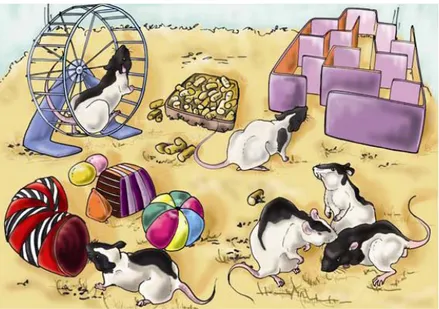
Within the hoary ‘nature versus nurture’ debate a relevant progress in understanding the influence of environmental experience on the development, refinement and maintenance of appropriate nervous system connections was obtained by introducing environmental enrichment (EE) as experimental protocol. EE is an alteration of the standard laboratory conditions that modifies the quality and intensity of environmental stimulation, reaching an optimization of the rearing environment. A comprehensive definition of EE was provided for the first time by Rosenzweig et al. (1978) as ‘a combination of complex inanimate and social stimulation’. Enriched animals are reared in large groups (6-10 individuals can be considered the most common used condition) and housed in wide stimulating environments where a variety of objects differently shaped (e.g. running wheels, platforms, boxes, toys, tunnels, shelters, stairs and nesting material) are present and changed frequently (specifically, the objects are completely replaced at least once a week). The goal of EE is to improve the animals’ quality of life by providing them with high levels of multi-sensory stimulation, increasing physical activity and social interactions, stimulating natural behaviours and cognitive abilities (since the novelty due to frequent objects’ replacement attracts the explorative curiosity of most laboratory animals). The significance of EE is based on the comparison of the enriched conditions with the standard environment condition (SC), that consists in housing 2-5 individuals in laboratory standard cages where no particular objects are present except for food, water and litter, and with the
impoverished environment condition (IC) that consists in housing the animals singly in cages identical to those used for SC or even smaller.
Although EE research has been mostly done upon rodents, similar effects occur in several species of mammals (gerbils, ground squirrels, rabbits, cats and primates), but also in some avian species (Rosenzweig and Bennett, 1969; Cornwell and Overman, 1981; Hansen and Berthelsen, 2000; Kozorovitskiy et al., 2005; Lazic et al., 2007; Jansen et al., 2009).
Since EE is a rather mild and non-invasive treatment, the results obtained are of great interest and applicability also for humans in many different fields, from psychology to medical clinic. EE is likely to affect the human brain, but direct research has been predominantly limited to the study of behavioural effects. All results are consistent with an increasing body of knowledge that implicates an enriched, stimulating environment in beneficial psychological and behavioral outcomes. The important work of Ramey and Ramey has shown that, starting at an early age, the use of a comprehensive enriched environment can increase IQ by a mean of 15 points in children from disadvantaged homes (Ramey and Ramey, 1998). Moreover, it has been demonstrated that subjects who participated in an enrichment program (nutritional, educational, and physical enrichment) at the age of 3-5 years have lower scores for schizotypal personality and antisocial behavior at the age of 17 years and for criminal behaviour at the age of 23 years, compared with control subjects (Raine et al., 2003).
2.1 Environmental enrichment affects animals’ behaviour
In the 40s Hebb was the first to introduce the idea of the ‘enriched environment’ as experimental concept. He described that rats that he took home as family pets, and that he reared in an extremely more complex environment than a laboratory rodent’s setting,
showed behavioural improvements in learning task. Subsequently, the finding that a more complex and stimulating rearing environment enhances performance on learning tasks was repeated by Hebb’s students (Bingham and Griffiths, 1952; Forgays and Read, 1962). These pioneering observations have been followed by a large body of studies which highlighted that environmental enrichment is able to modify animals’ behaviour leading to a sensitive improvement of complex cognitive functions, particularly learning and memory (for an exhaustive review, see Rampon and Tsien, 2000). Independently on the gender and age of tested animals, EE effects are especially evident in hippocampal-dependent tasks involving spatial memory, such as the Hebb-Williams maze (Kobayashi et al., 2002), the Lashley III maze (Greenough et al., 1972), the radial maze (Leggio et al., 2005) the Morris water maze (Tees et al., 1990; Falkenberg et al., 1992; Paylor et al., 1992; Moser et al., 1997; Kempermann et al., 1998a; Nilsson et a., 1999; Williams et al., 2001; Lee et al., 2003; Leggio et al., 2005; Fréchette et al., 2009) and the discrimination between different spatial contexts (Mitra and Sapolsky, 2008). EE animals have better performances also in nonspatial tasks, exhibiting enhanced learning and memory retention in the object recognition test, contextual and cued-fear conditioning (Rampon and Tsien, 2000; Duffy et al.,. 2001; Lee et al., 2003). However, Rampon and Tsien noticed that enrichment has no effect on the social transmission of food preference test, a task that is also dependent on the hippocampus and measures olfactory discrimination memory.
The consequences of EE upon emotional reactivity are less documented and remained controversial for a long time. While some authors reported no or inconsistent effects (Huck and Price, 1975; Fernandez-Teruel et al., 1997), others have observed modifications of emotional and stress reactions in EE animals (Chamove, 1989; Escorihuela et al., 1994). A more careful examination of the animals’ behaviours in prenatally or postnatally stressed rats (Francis et al., 2002), aged mice (Thouvarecq et al.,