Int. J. Mol. Sci. 2015, 16, 5510-5516; doi:10.3390/ijms16035510
International Journal of
Molecular Sciences
ISSN 1422-0067 www.mdpi.com/journal/ijms Short CommunicationImpact of Soluble HLA-G Levels and Endometrial NK Cells
in Uterine Flushing Samples from Primary and Secondary
Unexplained Infertile Women
Roberta Rizzo 1,†, Giuseppe Lo Monte 2,3,†,*, Daria Bortolotti 1, Angela Graziano 2,3,
Valentina Gentili 1, Dario Di Luca 1 and Roberto Marci 2,3
1 Department of Medical Sciences, Section of Microbiology and Genetics, University of Ferrara,
44100 Ferrara, Italy; E-Mails: [email protected] (R.R.); [email protected] (D.B.); [email protected] (V.G.); [email protected] (D.D.L.)
2 Department of Morphology, Surgery and Experimental Medicine, Section of Gynecology and
Obstetrics, University of Ferrara, 44100 Ferrara, Italy; E-Mails: [email protected] (A.G.); [email protected] (R.M.)
3 University Hospital “S. Anna”, 44100 Ferrara, Italy † These authors contributed equally to this work.
* Author to whom correspondence should be addressed; E-Mail: [email protected]; Tel.: +39-3470-824499; Fax: +39-0532-974470.
Academic Editor: Robert J. Norman
Received: 11 December 2014 / Accepted: 4 March 2015 / Published: 10 March 2015
Abstract: The aim of this research was to determine the levels of human leukocyte antigen G (HLA-G) and endometrial Natural Killer ((e)NK) cell percentages in uterine flushing samples from primary and secondary infertile women. sHLA-G levels were lower in the uterine flushing samples from primary infertile women in comparison with women with secondary infertility. Lower CD56+KIR2DL4+ (e)NK cell percentages were detected in
primary infertile women compared with secondary infertile women. This is the first study demonstrating that primary and secondary unexplained infertilities are characterized by different basal sHLA-G levels and CD56+KIR2DL4+ (e)NK cell percentages.
Keywords: primary infertility; secondary infertility; soluble human leukocyte antigen (sHLA-G); uterine flushing; endometrial natural killer; CD158d (KIR2DL4)
1. Introduction
A complex, highly coordinated sequence of structural and biochemical changes culminate in the generation of a “window” of uterine receptivity during the mid-luteal phase of each menstrual cycle. Compromised receptivity of the endometrium is believed to be a primary cause of unexplained infertility characterized by implantation failure and subclinical pregnancy loss. In women, unexplained infertility has been associated with a range of cellular and molecular defects of the endometrium and immunological factors. Since the fetus is semi-allogenic respect to the mother, the maternal immune-modulation is crucial in protecting the fetus.
Natural Killer (NK) cells are the dominant immune cell type in the endometrium and play a major role in determining pregnancy outcome [1]. Human decidual (d)NK cells are a distinct
CD56brightCD16− NK cell subset with a reduced cytotoxicity. They express specific markers such as
CD9 and CD49a [2] and inhibitory receptors (i.e., KIR2DL4, LILRB1 and LILRB2), known to interact with Human Leukocyte Antigen (HLA)-G molecules [3]. HLA-G antigens are non classical HLA class I molecules characterized by seven isoforms obtained by mRNA alternative splicing: four membrane-bound (HLA-G1–G4) and three soluble isoforms (HLA-G5–G7) [4] and a tolerogenic function during pregnancy. In particular lower levels of sHLA-G in maternal blood were associated with pregnancy complications as pre-eclampsia and recurrent pregnancy loss [4]. Moreover, HLA-G expression is important also during oocytes maturation and embryos implantation [5]. HLA-G molecules are expressed by cytotrophoblast cells at the maternal-fetus interface where they bind KIR2DL4 receptor expressed by the uterine NK cells [3]. This interaction controls the activation of the uterine NK cells and promotes the formation of the placenta [1]. Interestingly, sHLA-G was detected both in testis and in seminal fluid with significant differences among fertile and infertile couples [6]. Moreover, the decrease in CD56brightCD16− dNK cells and HLA-G expression have been associated
with recurrent miscarriage, suggesting their relevance in pregnancy outcome [7]. (d)NK cells have been extensively investigated during pregnancy, while endometrial (e)NK cells still lacks comprehensive researches.
We evaluated the role of sHLA-G in uterine flushing from women affected by primary or secondary infertility and the possible correlation with (e)NK presence.
2. Results
All patients were characterized by years of sterility (Table 1). Hormonal and demographic parameters (Follicle-stimulating hormone (FSH), Luteinizing hormone (LH), 17-β-Estradiol, progesterone, prolactin, Thyroid-stimulating hormone (TSH) and free thyroxine (FT4) levels, smoke habits, age and weight) proved not to be related with primary and secondary infertility (Table 1). The only difference was in TSH levels, with lower values in secondary infertile women (p = 0.0041; Student t test). However, the TSH concentrations ranged between the normal values (0.27–4.20 µUI/mL) in both the group of women, excluding any pathological condition. Moreover, we observed no correlation between TSH levels and the condition of primary or secondary infertility (r = −0.212; p = 0.252; Spearman correlation test). The evaluation of sHLA-G levels in the uterine flushing samples from primary and secondary infertile women revealed higher levels of sHLA-G in secondary
infertile women (mean ± standard deviation: 4.53 ± 2.78 ng/mL) compared with primary infertile women (0.14 ± 0.31 ng/mL) (p < 0.0001; Student t test) (Figure 1). Interesting, we observed a significant correlation between sHLA-G levels in uterine flushing samples and the condition of primary or secondary infertility (r = 0.80; p = 7.3 × 10⁻⁸). The analysis of cell content in uterine
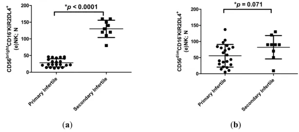
flushing samples (Table 2) showed a lower amount of (e)NK cells in the uterine flushing samples of primary infertile women compared with secondary infertile women (p < 0.0001; Student t test) (Table 2). In particular, when we analyzed (e)NK cell subpopulations, we observed a lower percentage of
CD56brightCD16−KIR2DL4+ (e)NK cells in the uterine flushing samples of primary infertile women
compared with secondary infertile women (p < 0.0001; Student t test) (Figure 2a). On the contrary, no differences were observed between CD56dimCD16−KIR2DL4+ (e)NK cells in the two groups of
women (p = 0.071; t test) (Figure 2b). The uterine flushing samples presented no CD3+ cells and
a low percentage of CD14+ cells (Table 2), with no significant differences between primary infertile
and secondary infertile women (p = 0.74; Student t test) (Table 2).
Table 1. Hormonal and demographic parameters of patients.
Items Primary
Infertility
Secondary
Infertility p Value
Age 34.7 ± 3.5 35.6 ± 3.4 0.53 *
Duration of Infertility (years) 2.7 ± 2.0 3.1 ± 2.3 0.61 * Length of menstrual cycle (days) 29.0 ± 4.0 28.2 ± 3.3 0.58 * FSH (mUI/mL) day 3 of the menstrual cycle 7.6 ± 2.7 6.9 ± 2.9 0.59 * LH (mUI/mL) day 3 of the menstrual cycle 6.9 ± 3.4 5.0 ± 2.2 0.13 * Estradiol (pg/mL) day 3 of the menstrual cycle 73.8 ± 65.5 54.9 ± 42.8 0.43 * TSH (µUI/mL) 3.8 ± 4.1 2.1 ± 1.4 0.0041 *
FT4 (pg/mL) 2.8 ± 3.9 2.2 ± 3.0 0.45
Progesterone (pg/mL) day 21 of the menstrual cycle 13.7 ± 9.6 12.4 ± 2.7 0.76 * Smoke habits (percentage) 22.7% 11.1% 0.074 ** Day (menstrual cycle) of sample collection 8.9 ± 1.9 9.1 ± 1.8 0.84 *
* Student t test; ** Fisher exact test; Follicle-stimulating hormone (FSH); Luteinizing hormone (LH); Thyroid-stimulating hormone (TSH), free thyroxine (FT4).
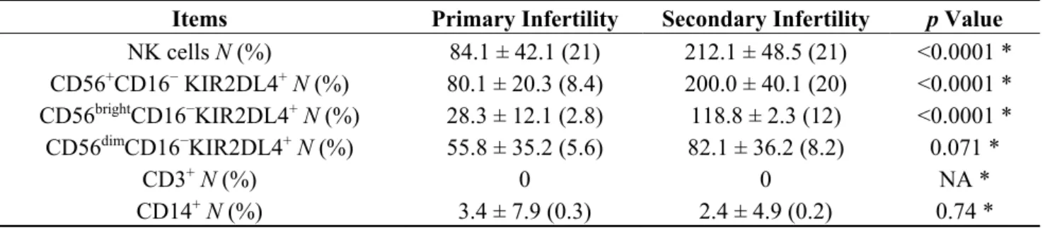
Table 2. Cell count in uterine flushing samples.
Items Primary Infertility Secondary Infertility p Value
NK cells N (%) 84.1 ± 42.1 (21) 212.1 ± 48.5 (21) <0.0001 * CD56+CD16− KIR2DL4+ N (%) 80.1 ± 20.3 (8.4) 200.0 ± 40.1 (20) <0.0001 * CD56brightCD16−KIR2DL4+ N (%) 28.3 ± 12.1 (2.8) 118.8 ± 2.3 (12) <0.0001 * CD56dimCD16−KIR2DL4+ N (%) 55.8 ± 35.2 (5.6) 82.1 ± 36.2 (8.2) 0.071 * CD3+ N (%) 0 0 NA * CD14+ N (%) 3.4 ± 7.9 (0.3) 2.4 ± 4.9 (0.2) 0.74 * * Student t test; Not applicable (NA).
Prim ary In fertil e Seco ndar y In fertil e 0 5 10 15 *p < 0.0001 sH LA -G ; ng/ m L
Figure 1. sHLA-G levels (mean ± SD) in primary and secondary infertility. sHLA-G levels were normalized for flushing volume and total protein content. * Student t test.
Prim ary Infer tile Sec onda ry Inf ertile 0 50 100 150 200 *p < 0.0001 CD 56 br ig htCD 16 -KI R2 D L 4 + (e )N K ; N Prim ary I nfertile Seco ndary Infe rtile 0 50 100 150 200 *p = 0.071 CD 56 di mCD 16 -KI R2 D L 4 + (e )N K ; N (a) (b)
Figure 2. (a) Number of CD56brightCD16−KIR2DL4+ (e)NK cells (mean ± SD) in flushing
samples from primary and secondary infertile women. * Student t test; (b) Number of CD56dimCD16−KIR2DL4+ (e)NK cells (mean ± SD) in flushing samples from primary and
secondary infertile women. * Student t test. 3. Discussion
The data obtained in this study support the hypothesis of an implication of sHLA-G molecules in infertility condition. We found different profiles of sHLA-G expression between primary and secondary infertile women. In particular, primary infertile women presented lower levels of sHLA-G in uterine flushing samples if compared with secondary infertile women. Several evidences support the major role of HLA-G in the physiopathology of infertility and the evaluation of sHLA-G expression was suggested as a future tool to select single embryos for transfer in order to reduce the risk of multiple pregnancy and to increase clinical pregnancy outcomes during in vitro fertilization protocols [5,8]. Recently, HLA-G was evaluated also in male reproductive system: sHLA-G has been detected in testis and also in semen and semen plasma with significant differences among different
men [6] suggesting that a favorable environment for pregnancy could be induced by the presence of sHLA-G molecules in the female reproductive tract that could be also carried by semen after coitus.
Moreover, the analysis of the amount of (e)NK cells showed a lower percentage of
CD56brightCD16−KIR2DL4+ (e)NK cells in primary infertile women. It is known that HLA-G antigens
act as immune-inhibitory molecules interacting with immune-inhibitory receptors (ILT2, ILT4 and KIR2DL4). During pregnancy, HLA-G molecules interact with (d)NK cells inducing a polarization towards cytokine production [9] and sustaining a correct placentation and embryo implantation. For this, the observation of a different characterization of primary and secondary unexplained infertility with regard to sHLA-G levels and CD56brightCD16−KIR2DL4+ (e)NK cell percentages in
uterine flushing samples is of extreme interest. These differences sustain different mechanisms at the basis of these two infertile conditions. Our results suggest that lower sHLA-G levels could prevent the creation of an appropriate tolerogenic uterine environment and a consequent lower presence of
CD56brightCD16−KIR2DL4+ (e)NK cells in primary infertility. On the contrary, the high levels of
sHLA-G and CD56brightCD16−KIR2DL4+ (e)NK cells found in secondary infertile women uterine
flushing samples suggest a different cause at the basis of this condition. In fact, secondary infertile women, unlike primary infertile ones, were able to carry out almost one previous pregnancy. It is interesting to note that the modulation in the number of circulating NK cells seems to be a primary event during inflammatory/autoimmune processes rather than a consequence of inflammation and drug administration, playing a fundamental role in the pathogenesis of a number of autoimmune diseases [10] and, on the basis of our results, also in female infertility.
4. Experimental Section 4.1. Patient Recruitment
The study was carried out in an inpatient setting following the ethical rules of the Azienda Ospedaliero Universitaria Ferrara, Italy. All patients signed an informed consent for the specimen collection. We enrolled 31 women affected by unexplained infertility (primary infertility, n = 22; secondary infertility, n = 9). Women were diagnosed with “primary infertility” in case they were both unable to conceive and to carry pregnancy to live birth. Otherwise, the failure to conceive following a previous pregnancy (spontaneous/voluntary abortions or childbirths) was referred to as “secondary infertility”. Patients were recruited at admission for tubal patency assessment by Hystero-sono contrast sonography 7–9 days after menstruation. Inclusion criteria for the study group were: 21–38 years old, regular menstrual cycle (24–35 days), body mass index (BMI) ranging between 18 and 26 kg/m2,
FSH (day 2–3 of the menstrual cycle) <10 mUI/mL, 17-β-Estradiol < 50 pg/mL (day 2–3 of the menstrual cycle), normal karyotype. Women with endometritis, endometriosis, tubal factor, ovulatory dysfunction, anatomical uterine pathologies and recurrent miscarriage were excluded.
4.2. Samples Collection
Uterine flushing was performed with a 14-gauge Foley 3-way balloon catheter (Eschmann) inflating an appropriate (5 mL) amount of sterile physiologic saline solution, as previously described [11]. All samples were stored at −20 °C until the analysis.
4.3. sHLA-G Analysis by ELISA
sHLA-G quantification in endometrial flushing was performed by ELISA [12] using anti-HLA-G (G233) and anti-β2-microglobulin HRP-conjugated moAbs (Exbio, Praha, Czech). sHLA-G levels were normalized for flushing volume and total protein content ((sHLA-G, ng/mL/total flushing sample, mL)/total proteins, ng/mL). Standard supernatants of HLA-G/721.221 were utilized for the generation of standard calibration curves. The limit of sensitivity was 1 ng/mL.
4.4. (e)NK Cell Analysis by Flow Cytometry
(e)NK cells were obtained from flushing sample pellets and were analyzed by flow cytometry with CD3-PerCp, CD14-PE, CD56-PE, CD16-FITC (BD Pharmigen, Erembodegem, Belgium), CD158d-APC (KIR2DL4) (Biolegend, San Diego, CA, USA) monoclonal antibodies.
4.5. Statistical Analysis
The data were compared by Student t test, as normally distributed according with Kolmogorov-Smirnov test, Fisher exact test and Spearman Correlation test.
5. Conclusions
Even though this study is based on a limited number of samples, and no mechanistic data are presented, we report, for the first time, a possible biological difference between primary and secondary unexplained infertility, based on sHLA-G and CD56brightCD16−KIR2DL4+ (e)NK cells in uterine
flushing samples. Further investigations on a larger cohort of subjects, including a control group, different time points during the menstrual cycle, and the evaluation of the biological relationship between HLA-G and (e)NK cells, would be necessary to confirm our data.
Acknowledgments
This study was funded by Young Scientist Grant-Regione Emilia Romagna-Alessandro Liberati 2013 with the title: “Prognostic markers in women with primary unexplained infertility”.
Author Contributions
Roberta Rizzo, Giuseppe Lo Monte and Roberto Marci conceived and designed the experiments; Giuseppe Lo Monte, Angela Graziano, Roberto Marci collected the samples; Roberta Rizzo, Daria Bortolotti, Valentina Gentili, Dario Di Luca performed the experiments; Roberta Rizzo, Daria Bortolotti and Giuseppe Lo Monte analyzed the data; and Giuseppe Lo Monte, Daria Bortolotti and Roberta Rizzo wrote the paper.
Conflicts of Interest
References
1. Moffett-King, A. Natural killer cells and pregnancy. Nat. Rev. Immunol. 2002, 2, 656–663.
2. Geiselhart, A.; Dietl, J.; Marzusch, K.; Ruck, P.; Ruck, M.; Horny, H.P.; Kaiserling, E.; Handgretinger, R. Comparative analysis of the immunophenotypes of decidual and peripheral blood large granular lynphocytes and T cells during early human pregnancy. Am. J. Reprod. Immunol. 1995, 33, 315–322.
3. Gonen-Gross, T.; Goldman-Wohl, D.; Huppertz, B.; Lankry, D.; Greenfield, C.; Natanson-Yaron, S.; Hamani, Y.; Gilad, R.; Yagel, S.; Mandelboim, O. Inhibitory NK receptor recognition of HLA-G: Regulation by contact residues and by cell specific expression at the fetal-maternal interface. PLoS One 2010, 5, e8941.
4. Rizzo, R.; Bortolotti, D.; Baricordi, O.R.; Fainardi, E. New insights into HLA-G and inflammatory diseases. Inflamm. Allergy Drug Target 2012, 11, 448–463.
5. Rizzo, R.; Stignani, M.; Melchiorri, L.; Baricordi, O.R. Possible role of human leukocyte antigen-G molecules in human oocyte/embryo secretome. Hum. Immunol. 2009, 70, 970–975. 6. Dahl, M.; Perin, T.L.; Djurisic, S.; Rasmussen, M.; Ohlsson, J.; Buus, S.; Lindhard, A.;
Hviid, T.V. Soluble human leukocyte antigen-G in seminal plasma is associated with HLA-G genotype: Possible implications for fertility success. Am. J. Reprod. Immunol. 2014, 72, 89–105. 7. Emmer, P.M.; Steegers, E.A.; Kerstens, H.M.; Bulten, J.; Nelen, W.L.; Boer, K.; Joosten, I.
Altered phenotype of HLA-G expressing trophoblast and decidual natural killer cells in pathological pregnancies. Hum. Reprod. 2002, 17, 1072–1080.
8. Kotze, D.; Kruger, T.F.; Lombard, C.; Padayachee, T.; Keskintepe, L.; Sher, G. The effect of the biochemical marker soluble human leukocyte antigen G on pregnancy outcome in assisted reproductive technology—A multicenter study. Fertil. Steril. 2013, 100, 1303–1309.
9. Le Bouteiller, P. HLA-G in human early pregnancy: Control of uterine immune cell activation and likely vascular remodeling. Biomed. J. 2015, 38, 32–38.
10. Perricone, R.; Perricone, C.; de Carolis, C.; Shoenfeld, Y. NK cells in autoimmunity: A two-edg’d weapon of the immune system. Autoimmun. Rev. 2008, 7, 384–390.
11. Mikołajczyk, M.; Skrzypczak, J.; Szymanowski, K.; Wirstlein, P. The assessment of LIF in uterine flushing: A possible new diagnostic tool in states of impared fertility. Reprod. Biol. 2003, 3, 259–270.
12. Gonzalez, A.; Alegre, E.; Arroyo, A.; LeMaoult, J.; Echeveste, J.I. Identification of circulating nonclassic human leukocyte antigen G (HLA-G)-like molecules in exudates. Clin. Chem. 2011, 57, 1013–1022.
© 2015 by the authors; licensee MDPI, Basel, Switzerland. This article is an open access article distributed under the terms and conditions of the Creative Commons Attribution license (http://creativecommons.org/licenses/by/4.0/).