Regular Article
CLINICAL TRIALS AND OBSERVATIONSGIMEMA AML1310 trial of risk-adapted, MRD-directed
therapy for young adults with newly diagnosed acute
myeloid leukemia
Adriano Venditti,1,2Alfonso Piciocchi,3Anna Candoni,4Lorella Melillo,5Valeria Calafiore,6Roberto Cairoli,7Paolo de Fabritiis,8Gabriella Storti,9
Prassede Salutari,10Francesco Lanza,11Giovanni Martinelli,12,13Mario Luppi,14Patrizio Mazza,15Maria Paola Martelli,16Antonio Cuneo,17
Francesco Albano,18Francesco Fabbiano,19Agostino Tafuri,20Anna Chierichini,21Alessia Tieghi,22Nicola Stefano Fracchiolla,23
Debora Capelli,24Robin Fo `a,25Caterina Alati,26Edoardo La Sala,3Paola Fazi,3Marco Vignetti,3Luca Maurillo,2Francesco Buccisano,1,2
Maria Ilaria Del Principe,1,2Maria Irno-Consalvo,1Tiziana Ottone,1Serena Lavorgna,1Maria Teresa Voso,1,2Francesco Lo-Coco,1,2
William Arcese,1,2and Sergio Amadori3
1Hematology, Department of Biomedicine and Prevention, University Tor Vergata, Rome, Italy;2Fondazione Policlinico Tor Vergata, Rome, Italy;3GIMEMA
Foundation, Rome, Italy;4Hematology, Azienda Sanitaria Universitaria Integrata di Udine, Udine, Italy;5Fondazione IRCCS Casa Sollievo della Sofferenza, UO di
Ematologia, San Giovanni Rotondo (FG), Italy;6Ospedale Ferrarotto, Catania, Italy;7Ospedale Niguarda Ca Granda, Milan, Italy;8Ospedale S. Eugenio, Rome,
Italy;9Azienda Ospedaliera S. G. Moscati, Avellino, Italy;10Azienda USL di Pescara, Pescara, Italy;11Ospedale S. Maria delle Croci, Ravenna, Italy;12Istituto Tumori
della Romagna, Meldola, Italy;13Policlinico S. Orsola-Malpighi, Bologna, Italy;14Ematologia, Dipartimento di Scienze Mediche e Chirurgiche Materno-Infantili e
dell’Adulto, Universit `a degli Studi di Modena e Reggio Emilia, Modena, Italy;15A. O. SS Annunziata-P. O. S. G. Moscati, Taranto, Italy;16Ospedale S. Maria della
Misericordia, Perugia, Italy;17Azienda Ospedaliero Universitaria Arcispedale Sant’Anna, Ferrara, Italy;18Ematologia, Dipartimento dell’Emergenza e dei Trapianti
di Organi, Universit `a degli Studi di Bari Aldo Moro, Bari, Italy;19Ospedali Riuniti Villa Sofia-Cervello, Palermo, Italy;20Azienda Ospedaliera Sant’ Andrea, Rome,
Italy;21San Giovanni Addolorata, Rome, Italy;22S. C. Ematologia AUSL-IRCCS Reggio Emilia, Meldola, Italy;23Fondazione IRCCS Ca’ Granda Ospedale Maggiore
Policlinico, Milan, Italy;24Ospedali Riuniti Umberto I G. M. LANCISI, Ancona, Italy;25Ematologia, Dipartimento di Biotecnologie Cellulari ed Ematologia,
Ematologia Universit `a degli Studi Sapienza, Rome, Italy; and26A. O. Bianchi-Melacrino-Morelli, Reggio Calabria, Italy
K E Y P O I N T S
lA risk-adapted, MRD-driven transplant strategy is a feasible approach for the treatment of younger adults with AML. lPretransplant MRD
positivity should not contraindicate delivery of an allogeneic stem cell transplant.
We designed a trial in which postremission therapy of young patients with de novo acute myeloid leukemia (AML) was decided combining cytogenetics/genetics and postconsolidation levels of minimal residual disease (MRD). After induction and consolidation, favorable-risk patients (FR) were to receive autologous stem cell transplant (AuSCT) and poor-risk patients (PR) allogeneic stem cell transplant (AlloSCT). Intermediate-risk patients (IR) were to receive AuSCT or AlloSCT depending on the postconsolidation levels of MRD. Three hundred sixty-one of 500 patients (72%) achieved a complete remission, 342/361 completed the consolidation phase and were treatment allocated: 165 (48%) to AlloSCT (122 PR, 43 IR MRD-positive) plus 23 rescued after salvage therapy, for a total of 188 candidates; 150 (44%) to AuSCT (115 FR, 35 IR MRD-negative) plus 27 IR patients (8%) with no leukemia-associated phenotype, for a total of 177 candidates. Overall, 110/177 (62%) and 130/188 (71%) AuSCT or AlloSCT candidates received it, respectively. Two-year overall (OS) and disease-free survival (DFS) of the whole series was 56% and 54%, respectively. Two-year OS and DFS were 74% and 61% in the FR category, 42% and 45% in the PR category, 79% and 61% in the IR MRD-negative category, and 70% and 67% in the IR MRD-positive category. In conclusion, AuSCT may still have a role in FR and IR MRD-negative categories. In the IR MRD-positive category, AlloSCT prolongs OS and DFS to equal those of the FR category. Using all the available sources of stem cells, AlloSCT was delivered to 71% of the candidates.This trial was registered at www.clinicaltrials.gov as #NCT01452646 and EudraCT as #2010-023809-36. (Blood. 2019;134(12):935-945)
Introduction
Despite the continuously growing knowledge about the genetic
and molecular landscape of acute myeloid leukemia (AML),1-6
the paradigm of treatment of young adults with AML is still largely
based on the“one-size-fits-all” approach, with postremission
strategies still depending on donor availability rather than on the
actual risk of disease relapse.7In the short term, this has led to
satisfactory rates of complete remission (CR) (70%-80%), but in
the long-term survival, estimates are still disappointing, with,30%
to 40% of patients becoming long-term survivors.8,9
Indeed, dealing with the high propensity for relapse and the considerable genetic heterogeneity of AML requires either development of new agents or adoption of modern, risk-adapted therapeutic programs. Risk-adapted approaches may consist of integrating pretreatment prognosticators, such as cytogenetics and molecular genetics, with posttreatment parameters, such as
assessment of minimal (or measurable) residual disease (MRD).10
Even though in AML cytogenetic is a historical and robust de-terminant of outcome, the modern stratification of the patients
in“favorable-risk" (FR), “intermediate-risk” (IR), or “adverse-risk”
categories relies ever more increasingly on the baseline
mo-lecular pattern.11,12Based on this, FR patients achieve overall
survival (OS) and disease-free survival (DFS) rates of 50% to 60% at 3 to 5 years with standard chemotherapy, whereas those with adverse risk show OS and DFS rates of 5% to 20% at 3 to 5 years if not submitted to allogeneic stem cell transplantation
(AlloSCT).7,11,13Therefore, it appears that in FR and adverse-risk
patients, the sole genetic/cytogenetic profile, regardless of the MRD levels, is helpful enough to guide decisions for the delivery of AlloSCT in the postremission phase. On the other hand, there are no accepted criteria to direct the decision-making process after consolidation for patients in the IR category: for these patients, evaluation of the MRD status appears appropriate to extrapolate those at high (MRD positive) or low (MRD negative) risk of relapse, for whom differentiated treatments may be adopted.
Although MRD assessment in AML is prognostic,14-18still,50%
of relapses are detected by MRD; thus, the false negative rate is
still high, resulting in low specificity. Moreover, MRD is assessed
exploiting disparateflow cytometry or molecular protocols so
that its use for treatment decisions in AML is still at an early stage. Depending on the technical platforms and targets, a sensitivity
of 1023to 1026is reported.19 In particular, we observed that
the integrated evaluation of baseline prognosticators and MRD improves risk assessment and helps optimizing postremission
therapy.20 In fact, directing MRD-positive patients toward
in-tensified therapy like AlloSCT while sparing those MRD-negative
patients the procedure-related morbidity and mortality may be
highly beneficial in terms of toxicity minimization.10
Considering all the above, the Gruppo Italiano Malattie
EMatologiche dell’Adulto (GIMEMA) Foundation has developed
a risk-adapted, MRD-oriented, prospective clinical trial, the strategy of which consisted of the prognostic integration of pretreatment cytogenetics and genetics with postconsolidation
MRD, as detected by multiparametric flow cytometry (MFC).
Based on this strategy, patients were to receive postconsolidation autologous stem cell transplantation (AuSCT) or AlloSCT, re-spectively, depending on their risk profile. We report here the final analysis of this multicenter study.
Patients and methods
Patients
Previously untreated patients with a diagnosis of de novo AML according to the World Health Organization diagnostic
criteria21were recruited to the GIMEMA AML1310 Study (EudraCT
#2010-023809-36; www.clinicaltrials.gov Identifier #NCT01452646) provided they met the criteria for eligibility (see supplemental
Material, available on the Blood Web site). The study was ap-proved by the ethics committees of the participating hospitals/ academic institutions and was conducted in accordance with the Declaration of Helsinki. All participants gave their informed consent.
Study design
The main objective of the study was to verify whether the de-livery of a postremission therapy, the intensity of which was risk driven, improved the outcome of adult patients with AML in terms of increased antileukemic efficacy. The primary endpoint of the study was OS at 24 months from treatment start; for comparative purposes, we included a historical control con-sisting of patients recruited to the previous LAM99P GIMEMA
trial.1Secondary endpoints were CR or complete remission
complete (CRi) rate after induction, DFS, and cumulative in-cidence of relapse (CIR) from CR. Upfront evaluation included bone marrow (BM) aspirate for morphology, cytogenetics, molecular genetics, and MFC analysis. The baseline MFC as-sessment was a necessary step, not only for diagnostic purposes but also to identify leukemia-associated immunophenotypes
(LAIPs). Identification of baseline LAIPs was the essential
re-quirement for monitoring MRD after therapy; at the established time point, BM MRD was determined by a high-sensitivity 8-color MFC assay. Based on several retrospective validations in the context of former European Organization for the Research
and Treatment of Cancer/GIMEMA protocols,22the threshold
for discriminating MRD-negative from MRD-positive cases was
set at 3.53 1024residual leukemic cells, and the selected time
point was the postconsolidation phase, once the hematologic recovery was complete. Patients were studied at diagnosis for the presence of RUNX1-RUNX1T1 or CBFb/MYH11
rearrange-ments, defining core binding factor (CBF) leukemias, and for
NPM1, FLT3, and c-KIT mutations. In CBF or NPM1-positive
AML, MRD was investigated as reported elsewhere.14,23,24
Mo-lecular analysis, LAIPs assessment, and postconsolidation MRD determinations were centralized at Laboratorio di Diagnostica
Integrata Oncoematologica“OPPO”, at Tor Vergata University
Hospital of Rome, whereas conventional karyotype was carried out at local institutions. Response to treatment was assessed on BM and peripheral blood, according to the recommendations of
an international working group.25Patients who did not achieve
CR/CRi or PR after the first induction course or CR/CRi after
2 induction courses were considered treatment failures. The AML1310 trial was designed at a time when European Leuke-miaNet 2010/2017 and National Comprehensive Cancer Net-work (NCCN) 2018 recommendations were not yet published. Therefore, when the trial regulatory path was concluded, we started recruiting and stratifying patients according to
con-temporary classification, that was the NCCN 2009, version 1.26
For the purpose of our study, 4 categories of risk were identified (Table 1): favorable-risk (NCCN-FR) or poor-risk (NCCN-PR) patients, who were submitted to AuSCT or AlloSCT, respectively; intermediate-MRD negative IR-Neg) or positive (NCCN-IR-Pos) patients, who were to receive AuSCT or AlloSCT,
respectively. Moreover, we enucleated afifth group of patients
belonging to the IR category, in whom we failed to identify any LAIP (NCCN-IR-no-LAIP category); these patients were allocated to the AuSCT postconsolidation option. AlloSCT and AuSCT were to be performed within 3 months of the end of the consolidation course.
Treatment
Induction consisted of IV daunorubicin 50 mg/m2daily on days 1,
3, and 5; IV etoposide 50 mg/m2daily on days 1 to 5; and IV
cytarabine 100 mg/m2as a daily continuous infusion, days 1 to
10. All patients in CR/CRi, after 1 to 2 induction cycles, received
1 consolidation course consisting of IV daunorubicin 50 mg/m2
daily on days 4,×5, and 6 and IV cytarabine 500 mg/m2every
12 hours on days 1 to 6. In patients belonging to NCCN-FR and NCCN-IR categories, peripheral blood stem cell collection was attempted by initiating, on day 20 from the start of consolidation therapy, granulocyte colony-stimulating factor until completion
of stem cell collection. In the case of failure to collect a sufficient
number of peripheral blood stem cells, BM was used as a source. In the case of poor BM harvest, instead of AuSCT, patients were to receive a second consolidation course with high-dose cytarabine (HDARAC). Postconsolidation therapy was based on risk allocation: NCCN-FR patients were to receive AuSCT; NCCN-PR patients were to receive AlloSCT; NCCN-IR patients were to receive AuSCT or AlloSCT depending on the levels of BM MRD as measured by MFC, after consolidation therapy. Allocation to AlloSCT required the procedure to be performed whatever the source of stem cells (identical sibling, HLA-identical unrelated donor, cord blood, HLA-haploHLA-identical sibling).
Salvage therapy consisted of 1 or 2 courses of IVfludarabine
30 mg/m2daily, on days 1 to 5; cytarabine 2000 mg/m2daily,
on days 1 to 5; idarubicin 8 mg/m2daily, on days 1 to 3. Whatever
the original NCCN risk category of assignment, patients with resistant disease after 1 to 2 cycles of induction therapy were considered PR and allocated to the AlloSCT procedure once CR/CRi was achieved.
Statistical analysis and sample size calculation
The primary objective was the percentage of OS at 2 years. An estimated number of 213 subjects were initially required to accomplish this primary objective. This sample size was to achieve a 90% power to detect a difference of 10% between the null hypothesis that OS at 2 years is 50% and the alternative hypothesis that OS is 60%, using a single-stage phase 2 design with a 5% significance level (based on data of the historic
con-trol group GIMEMA LAM99P).1Based on the historical control
group, we also considered that;70% of the observed patients
would have been classified as IR, therefore allowing attainment
of thefigure of 150 patients available for MRD-driven treatment
allocation. However, after 173 subjects were enrolled, only 56 belonged to the IR category (32% vs 70% expected). Therefore, to reach the target of 150 subjects belonging to the IR category,
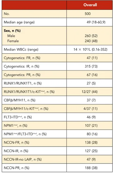
Table 2. Patients demographics and clinicobiologic characteristics
Overall
No. 500
Median age (range) 49 (18-60,9)
Sex, n (%) Male 260 (52) Female 240 (48) Median WBCc (range) 143 109/L (0.16-352) Cytogenetics: FR, n (%) 47 (11) Cytogenetics: IR, n (%) 315 (73) Cytogenetics: PR, n (%) 67 (16) RUNX1/RUNX1T1, n (%) 27 (5) RUNX1/RUNX1T1/c-KITmut, n (%) 12/27 (44) CBFb/MYH11, n (%) 37 (7) CBFb/MYH11/c-KITmut, n (%) 4/37 (11) FLT3-ITDmut, n (%) 46 (9) NPM1mut, n (%) 107 (21)
NPM1mut/FLT3-ITDmut, n (%) 80 (16)
NCCN-FR, n (%) 138 (28)
NCCN-IR, n (%) 127 (25)
NCCN-IR-no LAIP, n (%) 47 (9)
NCCN-PR, n (%) 188 (38)
WBCc, white blood cell count.
Table 1. Risk categories in which the patients were stratified
1. NCCN-FR Inv(16) t(8;21) t(16;16)
RUNX1/RUNX1T1 without c-Kit mutations CBFb/MYH11 without c-Kit mutations NPM1 mutation without FLT3 mutations 2. NCCN-IR postconsolidation MRD negative
Normal karyotype 18 only t(9;11) only
Other karyotypic abnormalities not listed as FR or PR RUNX1/RUNX1T1 with c-Kit mutation
CBFb/MYH11 with c-Kit mutation No NPM1 mutations
No FLT3-ITD mutations
3. NCCN-IR postconsolidation MRD positive
As in 2 but with measurable MRD after the consolidation course 4. NCCN-PR
Complex karyotype ($3 abnormalities) 25/5q- 27/7q-Abnormalities of 11q23, excluding t(9;11) inv(3) t(3;3) t(6;9) FLT3-ITD mutations 5. NCCN-IR-no LAIP
Patients belonging to the IR category in whom no leukemia associated
Immunophenotype (LAIP) was identified, at diagnosis
an amendment to the protocol was adopted in 2013, and the sample size was adjusted to 515 subjects to recruit. The efficacy analysis was performed as per treatment received, including individuals who commenced induction therapy and censoring patients at the time when they received a nonassigned treat-ment. OS (time elapsed from treatment start to death) and DFS (time from CR to relapse or death in remission) were calculated using the Kaplan-Meier product limit estimator. Differences in terms of OS and DFS were evaluated by means of Log-Rank test in univariate analysis and by means of Cox regression model in multivariate analysis, after assessment of proportionality of
hazards. All variables with a P value, .15 in univariate analysis
were considered in the multivariate models. The influence of the
transplant (AuSCT and AlloSCT) on the survival outcome was evaluated in the Cox model by means of a time-dependent covariate. CIR was estimated by cumulative incidence curves
using the proper nonparametric method. Patients’ and disease
characteristics were summarized by means of cross-tabulations for categorical variables or by quintiles for continuous variables. Differences between categorical variables or response rates in
subgroups were tested by the x2or Fisher exact tests, as
ap-propriate. Confidence intervals (CIs) were calculated at 95%
level, and all tests were 2 sided, accepting P# .05 as indicating
a statistically significant difference. All analyses were performed
using the SAS (version 9.4) and R (R Foundation for Statistical Computing, Vienna, Austria) system software. Study data were collected and managed using the REDCap20 electronic data capture tools hosted at GIMEMA Foundation.
Results
Between January 2012 and May 2015, 515 patients with de novo AML, seen at 55 GIMEMA institutions, were registered to the trial. Fifteen patients did not commence induction because of pretherapy death, infections, or ineligibility, and 500 started treatment and were available for the analysis. Demographic characteristics are summarized in Tables 2 and 3. Median age was 49 (18-60.9) years, and 52% were men. For 429 evaluable patients, cytogenetic distribution was favorable, intermediate, and poor in 11%, 73%, and 16%, respectively. Among 500 cases, RUNX1/RUNX1T1 was detected in 27 (5%) with 12 (44%) also
c-KIT mutated; CBFb/MYH11 was positive in 37 (7%) with 4 (11%)
also c-KIT mutated, and FLT3-ITD and NPM1 mutations were detected in 46 (9%) and in 107 (21%), respectively. Finally, concomitant mutations of NPM1 and FLT3-ITD were observed in 80 cases (16%). We found no instances of FLT3 mutations in
CBF-positive AML. Based on these data, patients’ distribution within
the risk categories was as follows: 138 (28%) were NCCN-FR, 127 (25%) were NCCN-IR, 47 (9%) were NCCN-IR-no-LAIP, and
188 (38%) were NCCN-PR. Patients’ disposition is illustrated in
Figure 1. After thefirst induction cycle, 333 (67%) and 21 (4%)
patients achieved a CR and CRi, respectively. A second in-duction course was delivered to 10 of 13 patients in PR, with 7 entering CR. Therefore, after 1 to 2 cycles of induction, 361 (72%) patients obtained a CR: 88% in the NCCN-FR category, 65% and 69% in the NCCN-IR and NCCN-PR category, respectively
(P, .001). Eighty-four (17%) patients had a refractory AML and
63 of them received a salvage therapy; 23 of these 63 (37%) achieved a CR. Three hundred forty-two of 361 (95%) patients started the consolidation phase and were treatment allocated: 177 (52%) to AuSCT (115 [65%] NCCN-FR, 35 [20%] NCCN-IR-Neg, 27 [15%] NCCN-IR-no-LAIP) and 165 (48%) to AlloSCT (122 [74%] NCCN-PR, 43 [26%] NCCN-IR-Pos). Of the 177 AuSCT candidates, 110 (62%) were transplanted (78 [71%] NCCN-FR, 20 [18%] NCCN-IR-Neg, 12 [11%] NCCN-IR-no-LAIP). Of the 165 AlloSCT candidates, 110 (67%) were transplanted (78 [71%] NCCN-PR, 32 [29%] NCCN-IR-Pos). If we include also the 23 patients who achieved a CR after salvage therapy, the group of AlloSCT candidates enlarges to 188. Because 20 of these 23 patients were given AlloSCT, the number of AlloSCT candidates who received it was 130/188 (71%). For the 78 patients belonging to the NCCN-PR category, the source of stem cells was a HLA-identical sibling in 26, a HLA-identical unrelated donor in 34, umbilical cord blood in 1, and HLA-haploidentical sibling in 17; for the 32 belonging to the NCCN-IR-Pos category, the source of stem cells was a HLA-identical sibling in 12, a HLA-HLA-identical unrelated donor in 9, umbilical
cord blood in 1, and HLA-haploidentical sibling in 10. By physicians’
decision, 1 patient belonging to the NCCN-PR category received AuSCT and 1 belonging to NCCN-FR category received AlloSCT.
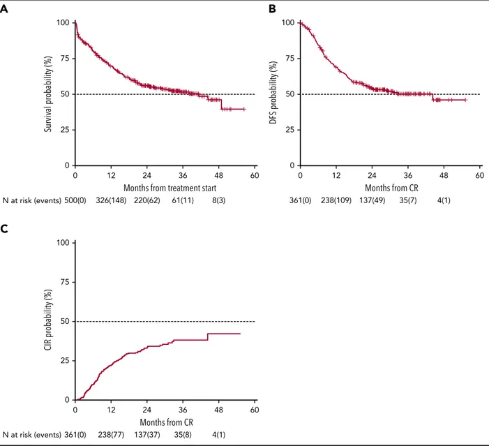
OS, DFS, and CIR
OS and DFS rates at 24 months of our historical control were
49% (95% CI 47-52) and 55% (95% CI 52-59), respectively.1In the
Table 3. Patients demographics and clinico-biologic characteristics according to treatment received
AlloSCT AuSCT HDARAC P
n 131 111 19
Median age (range) 46.7 (18-60.9) 48.4 (18-60.8) 54.7 (27-59.5) .033
Sex, n (%) Male 66 (50) 59 (53) 10 (53) .909 Female 65 (50) 52 (47) 9 (47) Median WBC (range) 12.93 109/L (0.16-352) 16.73 109/L (0.90-186) 11.63 109/L (1.24-102) .462 Risk category, n (%) NCCN-FR 1 (1) 78 (71) 18 (95) ,.001 NCCN-IR 41 (32) 20 (18) 1 (5) NCCN-PR 87 (66) 1 (1) 0 (0) NCCN-IR-no LAIP 2 (1) 12 (10) 0 (0)
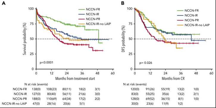
present trial, after a median follow-up of 28.8 months, 2-year OS was 56% (95% CI 52-61) with a median duration of 38 months (Figure 2), and DFS was 54% (95% CI 49-60) with a median duration of 32.4 months (Figure 2). The estimated OS at 24 months of 56% was less than the alternative hypothesis of 60%. However, the upper value of 95% CI included also the alternative hypothesis of 60% 2-year survival. Therefore, we considered the trial as not conclusive with regards to the pri-mary endpoint. CIR, considering death in CR as a competing risk, was 33% (95% CI 28-38) (Figure 2). When splitting the survival analysis according to the identified categories of risk, 2-year OS was 42% (95% CI 36-50) for NCCN-PR patients, 58% (95% CI 50-68) for NCCN-IR patients, 74% (95% CI 67-82) for NCCN-FR patients, and 50% (95% CI 37-67) for NCCN-IR-no
LAIP patients (P , .0001) (Figure 3). Two-year DFS was
45% (95% CI 37-55) for NCCN-PR patients, 61% (95% CI 52-73) for NCCN-IR patients, 61% (95% CI 52-71) for NCCN-FR patients, and
48% (95% CI 33-70) for those belonging to the NCCN-IR-no LAIP
category (P 5 0×026) (Figure 3). Using this risk-adapted
ap-proach, DFS duration of NCCN-FR and NCCN-IR was super-imposable, whereas the NCCN-IR-no LAIP one was the shortest. When we focused on the NCCN-IR patients, whose post-consolidation choice was MRD-driven, no significant differ-ences were observed in terms of 2-year OS between those MRD negative (79% [95% CI 66-94]) and MRD positive (70% [95% CI
57-86]) (P5 .713) (Figure 4). The same was observed regarding
the 2-year DFS (MRD negative5 61% [95% CI 47-80]; MRD
positive5 67% [95% CI 53-83]) (P 5 .773) (Figure 4). The
mul-tivariate analysis confirmed the independent role of risk category
in affecting CR rate, duration of OS, and DFS. The transplant procedure (AuSCT plus AlloSCT), analyzed as a time-dependent variable, affected independently the duration of OS. Age affected independently the duration of OS and DFS, whereas WBCc achievement of CR (supplemental Table 2).
NCCN-FR
• Lost to follow-up during the two years of follow-up (n=9)
NCCN-IR
• Lost to follow-up during the two years of follow-up (n=12)
NCCN-PR
• Lost to follow-up during the two years of follow- (n=12)
NCCN-IR-no-LAIP
• Lost to follow-up during the two years of follow-up (n=2) NCCN-FR: 138 NCCN-IR: 127 NCCN-PR: 188 NCCN-IR-no-LAIP: 47 Enrollment Follow-up Allocation Analysis
Assessed for eligibility (n=515)
Start treatment (n=500) Excluded (n=15) • Toxicity (n=1) • Withdrawal (n=3) • Death (n=3) • Ineligibility (n=3) • Lost fo Follow-up (n=2) • Other reasons (n=3) NCCN-FR Allocated to intervention (n=138) • Consolidation received: 115 • Second induction course: 2 • Salvage received: 2
• Did not Received post induction therapy Resistant (n=1) Toxicity (n=7) Withdrawal (n=1) Death (n=8) Other (n=4) • Received AUTO-graft after consolidation (n=78) • Received High dose of cytarabine (n=18) • Received ALLO-graft after salvage (n=1)
NCCN-IR Allocated to intervention (n=127) • Consolidation received: 78 (35 NCCN-IR-Neg, 43 NCCN-IR-Pos) • Second induction course: 2 • Salvage received: 23 • Did not Received post induction therapy: Resistant (n=5) Toxicity (n=11) Death (n=5) Lost to follow up (n=1) Other (n=4) • Received AUTO-graft after consolidation (n=20) • Received High dose of cytarabine (n=1) • Received ALLO-graft after consolidation (n=32) • Received ALLO-graft after salvage (=9)
NCCN-PR
Allocated to intervention (n=188) • Consolidation received: 122 • Second induction course: 5 • Salvage received: 32 • Did not Received post induction therapy: Resistant: (n=10) Toxicity (n=13) Withdrawal (n=1) Death (n=4) Other (n=6) • Received ALLO-graft after consolidation (n=78) • Received AUTO-graft after consolidation (n=1) • Received ALLO-graft after salvage (n=9)
NCCN-IR-no-LAIP
Allocated to intervention (n=47) • Consolidation received: 27 • Second induction course: 1 • Salvage received: 6
• Did not Received post induction therapy:
Resistant (n=2) Toxicity (n=5) Death (n=4) Other (n=3) • Received AUTO-graft after consolidation (n=12) • Received ALLO-graft after consolidation (n=1) • Received ALLO-graft after salvage (n=1)
Figure 1. Consort diagram of patients’ disposition.
MFC and molecular integrated evaluation of MRD
As an ancillary activity of the protocol, of 251 patients whose AML was characterized by the presence of a molecular marker useful for MRD assessment, we received 112 BM samples
(RUNX-RUNX15 9, CBFB-MYH11 5 9, and NPM1 5 94) at the
post-consolidation time point. In 60 of these, we had the opportunity to combine the postconsolidation results of MFC and reverse transcription quantitative polymerase chain reaction (RT-qPCR)
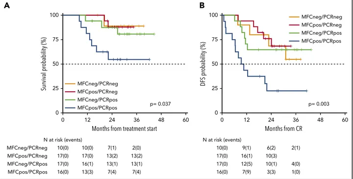
MRD studies. This integrated analysis identified 4 categories of
patients: double negative (MFCneg/PCRneg), double positive (MFCpos/PCRpos), and single positive (MFCpos/PCRneg or MFCneg/PCRpos). Patients who were double negative had a 2-year OS and DFS of 89% (95% CI 71-100) and 69% (95% CI 44-100), respectively. Patients who were MFCpos/PCRneg had a 2-year OS and DFS of 88% (95% CI 73-100) and 76% (95% CI 58-100), respectively. Patients who were MFCneg/PCRpos had a 2-year OS and DFS of 87% (95% CI 72-100) and 65% (95% CI 45-92), respectively. Finally, patients who were double positive
had a 2-year OS and DFS of 55% (95% CI 34-87) and
22% (95% CI 9-58), respectively (Figure 5A-B; P 5 .037 and
.003, respectively).
AuSCT vs HDARAC consolidation
As per protocol, 19 patients (18 NCCN-FR and 1 NCCN-IR) received HDARAC, because they did not have enough stem cells collected. Figure 6 shows OS and DFS of these patients com-pared with those who were submitted to AuSCT. OS was
83% (95% CI 67-100) and 85% (95% CI 78-93), respectively (P5 .753);
DFS was 68% (95% CI 50-93) and 63% (95% CI 54-73), respectively
(P5 .595). Of these 19 patients, 15 were NCCN-FR MFCneg/
PCRneg, MFCpos/PCRneg, or MFCneg/PCRpos, 3 were NCCN-FR MFCpos/PCRpos, and 1 was NCCN-IR MRD negative.
Discussion
The role of molecular and cytogenetic abnormalities in pre-dicting response to therapy and survival in patients with AML has
N at risk (events) 100 75 50 CIR pr obability (%) 25 0 0 361(0) 238(77) 137(37) 35(8) 4(1) 12 24 Months from CR 36 48 60 100 75 50 DFS pr obability (%) 25 0 0 361(0) 238(109) 137(49) 35(7) 4(1) 12 24 Months from CR 36 48 60 100
A
C
B
75 50 Survival pr obability (%) 25 0 0 500(0) N at risk (events) 326(148) 220(62) 61(11) 8(3) 12 24Months from treatment start
36 48 60
Figure 2. Survival estimates and CIR of the whole patient population.With a median follow-up of 28.8 months, 2-year-OS (A) and DFS (B) were 56% (median duration 38 months) and 54% (median duration 32.4 months), respectively. (C) CIR, considering death in CR as a competing risk, was 33%.
been extensively documented.24-26Indeed, genetic/cytogenetic abnormalities are powerful prognosticators, so that obtaining information about their presence is essential for an optimal decision-making process. The clinical implication is that, based on their genetic status, patients would benefit from more or less aggressive postconsolidation strategy such as AuSCT and AlloSCT or, in a more modern vision, from targeted new agents. However, prognostic models barely based on pretreatment covariates, such as genetic status, have a limited predictive
ability.11,27-29This highlights not only the need to expand
fur-ther our knowledge about the genetic and molecular pattern of
AML but also the potential role of“factors after diagnosis,”
such as MRD monitoring. Therefore, integrating baseline factors and monitoring of MRD appears a promising tool to refine and possibly customize our outcome prediction ability in AML. This philosophy was at the basis of the GIMEMA AML1310 protocol
in which, deviating from the classical“one-size-fits-all” approach,
we applied a risk-adapted and MRD-driven approach. AlloSCT is
N at risk (events) 0 NCCN-IR-no LAIP 47(0) 28(16) 20(6) 5(1) NCCN-FR 138(0) 108(23) 80(11) 18(2) 3(1) NCCN-IR 127(0) 80(40) 56(11) 21(6) 3(0) NCCN-PR 188(0) 110(69) 64(34) 17(2) 2(2) 0 25 50 Survival pr obability (%) 75 100 12 24
Months from treatment start
36 48 60 p<0.0001 NCCN-FR NCCN-IR NCCN-PR NCCN-IR-no LAIP
A
23(6) 11(9) 1(2) 30(0) 120(0) 91(26) 55(19) 13(2) 1(0) 55(25) 35(6) 13(2) 2(1) 83(0) 69(52) 36(15) 8(1) 1(0) 128(0) p= 0.026 N at risk (events) 0 0 25 50 75 100 12 24 Months from CR 36 48 60 DFS pr obability (%) NCCN-FR NCCN-IR NCCN-PR NCCN-IR-no LAIPB
Figure 3. Survival estimates plotted by NCCN categories of risk.(A) Two-year OS was 42%, 58%, 74%, and 50%, respectively, for patients belonging to the PR, NCCN-IR, NCCN-FR, and NCCN-IR-no LAIP categories. (B) Two-year DFS was 45%, 61%, 61% and 48%, respectively, for patients belonging to the NCCN-PR, NCCN-NCCN-IR, NCCN-FR and NCCN-IR-no LAIP categories.
A
N at risk (events) 0 0 25 50 Survival pr obability (%) 75 100 12 24Months from treatment start
36 48 60 NCCN-IR MRD neg NCCN-IR MRD pos 35(0) 29(5) 29(5) 29(5) 29(5) 43(0) 33(6) 24(6) 10(1) 1(0) p= 0.713 NCCN-IR MRD neg NCCN-IR MRD pos
B
N at risk (events) 0 25 50 DFS pr obability (%) 75 100 Months from CR 0 12 24 36 48 60 35(0) 23(12) 15(1) 6(0) 1(0) 43(0) 30(9) 20(5) 7(2) 1(1) p= 0.773 NCCN-IR MRD neg NCCN-IR MRD posFigure 4. Survival estimates of NCCN-IR category, plotted by the status of MRD after consolidation therapy.(A) Two-year OS of NCCN-IR MRD negative and MRD positive patients were 79% and 70%, respectively. (B) Two-year DFS were 61% and 67%, respectively.
generally recommended when the risk of relapse exceeds 35% to
40% if the procedure is not performed.7,10In this view, NCCN-PR
category represents a priority and, in these patients, AlloSCT should be performed as soon as CR is achieved. However, an
HLA-identical sibling is available for,30% of the patients,30and,
in reality, even,30% receive it, due to disease recurrence.11In
our study, utilization of any available source of stem cells resulted
in 71% of AlloSCT candidates receiving it. Adoption of this strategy also translated to a 2-year OS and DFS of 42% and 45%,
respectively, for the NCCN-PR category (Figure 2). Suchfigures
compare very favorably with the 2-year OS and DFS of 20% to
30% currently reported for this category.27,29Based on our study
design, patients belonging to the NCCN-FR category were given AuSCT as a postconsolidation therapy. The role of AuSCT is
N at risk (events) 0 0 25 50 Survival pr obability (%) 75 100 12 24
Months from treatment start
36 48 p= 0.037 60 MFCneg/PCRneg 10(0) 10(0) 7(1) 2(0) 17(0) 16(1) 13(1) 13(1) MFCneg/PCRpos 16(0) 13(3) 7(4) 7(4) MFCpos/PCRpos 17(0) 17(0) 13(2) 13(2) MFCpos/PCRneg MFCpos/PCRpos MFCneg/PCRpos MFCpos/PCRneg MFCneg/PCRneg
A
N at risk (events) 0 25 50 DFS pr obability (%) 75 100 Months from CR p= 0.003 0 12 24 36 48 60 10(0) 9(1) 6(2) 2(1) 17(0) 12(5) 10(1) 4(0) 16(0) 7(9) 3(3) 1(0) 17(0) 16(1) 10(3) MFCpos/PCRpos MFCneg/PCRpos MFCpos/PCRneg MFCneg/PCRnegB
Figure 5. Survival estimates of 60 patients whose MRD was analyzed integrating MFC and RT-qPCR.By integrating MFC and RT-qPCR, 4 categories of distinct MRD status were identified with different duration of OS (A) and DFS (B). Patients who were double negative had a 2-year OS and DFS of 89% and 69%, respectively. Patients who were MFCpos/PCRneg had a 2-year OS and DFS of 88% and 76%, respectively. Patients who were MFCneg/PCRpos had a 2-year OS and DFS of 87% and 65%, respectively. Finally, patients who were double positive had a 2-year OS and DFS of 55% and 22%, respectively.
0 0 N at risk (events) 12 98(0) AuSCT 93(5) 70(9) 17(5) 3(0) 19(0) High dose of cytarabine 18(1) 13(2) 1(0) 24 36 48 60 100 75 50 Survival pr obability (%) 25 p= 0.753 AuSCT
High dose of cytarabine AuSCT
High dose of cytarabine
Months from treatment start
A
0 N at risk (events) 0 98(0) 75(23) 50(13) 14(2) 1(0) 8(3) 16(3) 19(0) 12 24 36 48 60 DFS pr obability (%) p= 0.595 100 75 50 25 Months from CRB
Figure 6. Survival estimates of the 19 patients who received high dose of cytarabine versus those who received AuSCT.(A) Patients who received high dose of cytarabine or an autologous stem cell transplant had an OS of 83% and 85%, respectively. (B) DFS was 68% and 63%, respectively.
controversial; in 1 randomized study, it provided better DFS and
similar OS as conventional consolidation chemotherapy.31,32 In
our NCCN-FR category, 2-year OS and DFS were 74% and 61%, respectively (Figure 3). We believe there is still a role for AuSCT; indeed, this option has the advantage of sparing patients mul-tiple courses of postconsolidation chemotherapy (usually high-/ intermediate-dose cytarabine). In fact, the recently revised Euro-pean LeukemiaNet classification suggests that limiting AuSCT
to MRD-negative AML might improve the results.11Based on our
limited experience, HDARAC might represent the choice for
patients with very“high-quality” CR such as those NCCN-FR
MFCneg/PCRneg (Figure 6). Management of patients belonging to the NCCN-IR category is still controversial. For these patients,
the relapse rate after AuSCT can be as high as 50% to 55%,10so
that this option appears as a suboptimal approach. Indeed, AlloSCT is recommended for patients within this category. However, in selected patients with MRD-negative CR, there might
still be a room for AuSCT.10In the present study, we planned
AlloSCT or AuSCT for NCCN-IR patients, based on the level of MRD after the postconsolidation course. By making this choice, we observed that the 2-year OS and DFS were 58% and 61%,
respectively (Figure 3). Thisfigure compares very favorably with
recent analyses showing, for these patients, a 2-year OS and DFS
of;35% and 50%.27,29Using this strategy, we also noted that the
2-year DFS of NCCN-IR patients was prolonged to equal that of NCCN-FR patients (Figure 3). Finally, within the NCCN-IR
category, we focused on outcome as influenced by the
post-consolidation MRD status. By delivering AuSCT to NCCR-IR-Neg and AlloSCT to NCCN-IR-Pos patients, we observed no difference
in terms of 2-year OS or DFS (Figure 4). The stratification role of
MRD determination in IR patients has been recently suggested in
a prospective survey of the NCRI-AML17 trial.33According to the
authors, an MRD-positivefinding helps selecting patients who can
benefit from AlloSCT. An indirect confirmation of the importance
of MRD determination in IR category was that our 47 NCCN-IR-no-LAIP patients who were submitted to AuSCT had the shortest duration of 2-year OS and DFS (Figure 2). A reasonable ex-planation is that these patients harbored significant post-chemotherapeutic levels of MRD, meaning that AlloSCT would have been the most appropriate choice. Our results highlight the potent antileukemic effect exerted by AlloSCT in NCCN-IR-Pos patients and the minimization of toxicity after AuSCT in NCCN-IR-Neg ones. This interpretation can be extended to include the overall population we had under investigation; indeed, gener-ating the maximum antileukemic effort in high-risk patients
(NCCN-PR1 NCCN-IR-Pos) and preserving from excess of
tox-icity those who are at low risk (NCCN-FR 1 NCCN-IR-Neg)
appears a very plausible goal. In this view, the integration of different techniques for MRD monitoring may offer the chance to improve even further our capability to discriminate prognostically discrete subsets of patients, directing treatment more precisely. Combining MFC and RT-qPCR for cases carrying a molecular signature, we demonstrated that double-positive patients had the worst prognosis. For these patients, a front-line intensified pro-gram appears a reasonable option (Figure 5). Although there is evidence that AlloSCT is not able to reverse the unfavorable
long-term impact of MRD positivity,34-37we believe that a pretransplant
MRD-positive status should not be a contraindication for
per-forming it.38-40In the study by Walter35and Araki,37patients who
were MRD positive before the transplant had an outcome com-parable to the one of patients with active disease. However, these studies were retrospective; the patient population was
heterogeneous in terms of age and conditioning regimens received, and there was a concentration of adverse karyotype and secondary AML in the group of MRD-positive patients. Our experience takes advantage of a prospective and
homoge-neous context in terms of therapy delivered and risk strati
fi-cation. A recent, retrospective analysis of 547 patients enrolled in the Hemato-Oncology Foundation for Adults in the Neth-erlands and the Swiss Group for Clinical Cancer Research
protocols indicates that, although all categories benefit from
AlloSCT, the absolute benefit was greater in pretransplant
MRD-positive than MRD-negative patients.41Our present
expe-rience adds a piece of information favoring the use of AlloSCT in MRD-positive patients, and future trials should possibly explore
the prognostic role of different levels of pretransplant MRD38and
the value of posttransplant maintenance.
In conclusion, we recognize that the study suffers from some intrinsic limitations due to the changes occurring over the time (more modern biologic knowledge, new AML classifications, and an ever more frequent MRD monitoring) that make the historical control and the study population not fully superimposable.
However, this is one of thefirst attempts to apply a prospective
program of risk-adapted, MRD-driven therapy, integrating upfront genetics and postconsolidation MRD status, in AML of adults. In the NCCN-FR category, AuSCT guarantees the same survival expectation as multiple courses of cytarabine. In the NCCN-IR category, AlloSCT can be avoided if MRD is not measurable; if MRD is positive, AlloSCT can prolong OS and raise the DFS duration to the level of NCCN-FR patients. Finally, using all the available sources of stem cells allowed AlloSCT to be delivered to a large proportion of the candidates, emphasizing the fea-sibility of the trial transplant policy.
Acknowledgments
The authors thank the following contributors: Giorgia Giuliani and Mariangela Iodice, for data collection at the GIMEMA Data Center; Laura Di Donato, for pharmacovigilance activity (GIMEMA); and Stefano Soddu, for assistance in statistical analysis (GIMEMA). The authors thank the following centers for their contribution to patient recruitment: Ospedale A. Businco, Cagliari (La Nasa Giorgio), Azienda Ospedaliera Pugliese Ciaccio, Catanzaro (Molica Stefano), Ospedale Santa Maria Goretti, Latina (De Blasio Angelo), Azienda Ospedaliera Universitaria Federico II, Napoli (Pane Fabrizio), A.U. Policlinico Paolo Giaccone, Palermo (Siragusa Sergio), CTMO Ospedale San Salvatore, Pesaro (Visani Giuseppe), Policlinico A. Gemelli, Roma (De Stefano Valerio), Azienda Ospedaliera San Camillo Forlanini, Roma (Pierelli Luca), Dipartimento di Medicina Clinica e Sperimentale, Sassari (Fozza Claudio), IRCCS AOU San Martino IST, Genova (Angelucci Emanuele), CTMO Universita’ degli Studi di Parma, Parma (Aversa Franco), C.T.M.O. Istituti Ospitalieri, Cremona (Molteni Alfredo), Ospedali Riuniti, Foggia (Capalbo Silvana Franca), Ospedale S. Luigi Gonzaga, Orbassano (Guerrasio Angelo), Azienda Ospedaliera SS. Antonio e Biagio e Cesare Arrigo, Alessandria (Ladetto Marco), Azienda Ospedaliera Pisana, Pisa (Petrini Mario), Cancer Center Humanitas, Rozzano (Santoro Armando), Universita’ degli Studi di Padova, Padova (Semenzato Pietro), A. Tortora di Pagani, Pagani (SA) (Califano Catello), I.F.O. Istituto Nazionale Tumori Regina Elena, Roma (Mengarelli Andrea), Ist. Nazionale Tumori, Milano (Corradini Paolo), Azienda Ospedaliera Ospedali Riuniti Papardo Piemonte, Messina (Mannina Donato), Ospedale Binaghi, Cagliari (La Nasa Giorgio), Ospedale Ca’ Foncello, Treviso (Gherlinzoni Filippo), Ospedale Infermi, Rimini (Tosi Patrizia), A.O. S.Anna e S.Sebastiano, Caserta (Frigeri Ferdinando), Azienda Ospedaliera Pia Fondazione di Culto e di Religione Card. G.Panico, Tricase (LE) (Pavone Vincenzo), ASL Le/1 P.O. Vito Fazzi, Lecce (Di Renzo Nicola), Ospedale Monsignor Raffaele Dimiccoli, Barletta (Tarantini Giuseppe), Istituto Scientifico Romagnoli per lo Studio e la Cura dei Tumori IRST, Meldola (Martinelli Giovanni), AUSL Ospedale G. da Saliceto, Piacenza (Vallisa Daniele), Ospedale Civile, Civitanova Marche (Centurioni Riccardo).
This study was conducted thanks to the partial contribution of the Associazione Italiana Contro le Leucemie-Linfomi e Mieloma. This work is dedicated to the memory of Francesco Lo-Coco, a dear friend and a valuable colleague.
Authorship
Contribution: A.V., A.P., P.F., M.V., F.L.-C., W.A., and S.A. created the concept and design; A.V., A. Candoni, L. Melillo, V.C., R.C., P.d.F., G.S., P.S., F.L., G.M., M.L., P.M., M.P.M., A. Cuneo, F.A., F.F., A. Tafuri, A. Chierichini, A. Tieghi, N.S.F., D.C., R.F., C.A., L. Maurillo, F.B., M.I.D.P., W.A., and S.A. provided the study materials or patients; A.V., A.P., A. Candoni, L. Melillo, V.C., R.C., P.d.F., G.S., P.S., F.L., G.M., M.L., P.M., M.P.M., A. Cuneo, F.A., F.F., A. Tafuri, A. Chierichini, A. Tieghi, N.S.F., D.C., R.F., C.A., E.L.S., L. Maurillo, F.B., M.I.D.P., M.I.-C., T.O., S.L., and M.T.V. collected and assembled the data; A.V., A.P., E.L.S., L. Maurillo, F.B., M.I.D.P., M.T.V., F.L.-C., W.A., and S.A. analyzed and interpreted the data; L. Maurillo, F.B., M.I.D.P., M.I.-C., T.O., S.L., and M.T.V. performed the laboratory testing and monitoring; and all authors contributed to the writing of the manuscript and to thefinal approval of the manuscript.
Conflict-of-interest disclosure: A.V. reports personal fees from Pfizer, Celgene, Novartis, Daiichi-Sankyo, and Jazz Pharmaceuticals outside the submitted work. S.A. reports personal fees from Amgen, Celgene, Novartis, and Daiichi-Sankyo outside of the submitted work. R.F. reports personal fees from Roche, Genentech, Janssen, Gilead, Celgene, Novartis, Ariad, and Amgen outside of the submitted work. M.L. reports
grants from Novartis and MSD, personal fees from Novartis, Abbvie, and Gilead outside of the submitted work. The remaining authors declare no competingfinancial interests.
Francesco Lo-Coco died on 3 March 2019.
ORCID profiles: A.V., 0000-0002-0245-0553; A.P., 0000-0001-8648-885X; F.A., 0000-0001-7926-6052; M.V., 0000-0003-1278-604X; F.B., 0000-0003-4320-9253; T.O., 0000-0003-1936-3592; M.T.V., 0000-0002-6164-4761.
Correspondence: Adriano Venditti, Fondazione Policlinico Tor Vergata, Viale Oxford 81-00133, Rome, Italy; e-mail: adriano.venditti@ uniroma2.it.
Footnotes
Submitted 29 November 2018; accepted 1 August 2019. Prepublished online as Blood First Edition paper, 8 August 2019; DOI 10.1182/ blood.2018886960.
Individual participant data will not be shared.
The online version of this article contains a data supplement. The publication costs of this article were defrayed in part by page charge payment. Therefore, and solely to indicate this fact, this article is hereby marked“advertisement” in accordance with 18 USC section 1734.
R E F E R E N C E S
1. Lo-Coco F, Cuneo A, Pane F, et al; Acute Leukemia Working Party of the GIMEMA group. Prognostic impact of genetic charac-terization in the GIMEMA LAM99P multicenter study for newly diagnosed acute myeloid leukemia. Haematologica. 2008;93(7): 1017-1024.
2. D ¨ohner H, Estey EH, Amadori S, et al; Euro-pean LeukemiaNet. Diagnosis and manage-ment of acute myeloid leukemia in adults: recommendations from an international ex-pert panel, on behalf of the European Leu-kemiaNet. Blood. 2010;115(3):453-474. 3. Slovak ML, Kopecky KJ, Cassileth PA, et al.
Karyotypic analysis predicts outcome of pre-remission and postpre-remission therapy in adult acute myeloid leukemia: a Southwest Oncol-ogy Group/Eastern Cooperative OncolOncol-ogy Group Study. Blood. 2000;96(13):4075-4083. 4. Grimwade D, Hills RK, Moorman AV, et al;
National Cancer Research Institute Adult
Leukaemia Working Group. Refinement of
cytogenetic classification in acute myeloid
leukemia: determination of prognostic signif-icance of rare recurring chromosomal abnor-malities among 5876 younger adult patients treated in the United Kingdom Medical Re-search Council trials. Blood. 2010;116(3): 354-365.
5. Papaemmanuil E, Gerstung M, Bullinger L,
et al. Genomic classification and prognosis in
acute myeloid leukemia. N Engl J Med. 2016; 374(23):2209-2221.
6. Metzeler KH, Herold T, Rothenberg-Thurley M, et al; AMLCG Study Group. Spectrum and prognostic relevance of driver gene mutations in acute myeloid leukemia. Blood. 2016; 128(5):686-698.
7. Koreth J, Schlenk R, Kopecky KJ, et al. Allogeneic stem cell transplantation for acute
myeloid leukemia infirst complete remission:
systematic review and meta-analysis of pro-spective clinical trials. JAMA. 2009;301(22): 2349-2361.
8. Mandelli F, Vignetti M, Suciu S, et al. Daunorubicin versus mitoxantrone versus idarubicin as induction and consolidation chemotherapy for adults with acute myeloid leukemia: the EORTC and GIMEMA Groups Study AML-10. J Clin Oncol. 2009;27(32): 5397-5403.
9. Burnett AK, Hills RK, Milligan DW, et al. Attempts to optimize induction and consoli-dation treatment in acute myeloid leukemia: results of the MRC AML12 trial. J Clin Oncol. 2010;28(4):586-595.
10. Cornelissen JJ, Gratwohl A, Schlenk RF, et al. The European LeukemiaNet AML Working Party consensus statement on allogeneic HSCT for patients with AML in remission: an integrated-risk adapted approach. Nat Rev Clin Oncol. 2012;9(10):579-590.
11. D ¨ohner H, Estey E, Grimwade D, et al. Diagnosis and management of AML in adults: 2017 ELN recommendations from an in-ternational expert panel. Blood. 2017;129(4): 424-447.
12. NCCN. NCCN Clinical Practice Guidelines in Oncology. Acute Myeloid Leukemia. http:// www.nccn.org/professionals/physician_gls/ pdf/aml.pdf Accessed 6 May 2018. Version 1. 2018. 2018
13. Greenberg PL, Al-Kali A, Bennett JM, et al. NCCN Guidelines Version 2.2018 Myelodys-plastic Syndromes Continue NCCN Guide-lines Panel Disclosures. 2018;
14. Ivey A, Hills RK, Simpson MA, et al; UK Na-tional Cancer Research Institute AML Working Group. Assessment of minimal residual
disease in standard-risk AML. N Engl J Med. 2016;374(5):422-433.
15. Jongen-Lavrencic M, Grob T, Hanekamp D, et al. Molecular minimal residual disease in acute myeloid leukemia. N Engl J Med. 2018; 378(13):1189-1199.
16. Hourigan CS, Gale RP, Gormley NJ, Ossenkoppele GJ, Walter RB. Measurable residual disease testing in acute myeloid leukaemia. Leukemia. 2017;31(7):1482-1490. 17. Terwijn M, van Putten WLJ, Kelder A, et al.
High prognostic impact offlow cytometric
minimal residual disease detection in acute myeloid leukemia: data from the HOVON/ SAKK AML 42A study. J Clin Oncol. 2013; 31(31):3889-3897.
18. Freeman SD, Virgo P, Couzens S, et al. Prognostic relevance of treatment response
measured byflow cytometric residual disease
detection in older patients with acute myeloid leukemia. J Clin Oncol. 2013;31(32): 4123-4131.
19. Schuurhuis GJ, Heuser M, Freeman S, et al. Minimal/measurable residual disease in AML: consensus document from the European LeukemiaNet MRD Working Party. Blood. 2018;131(12):1275-1291.
20. Buccisano F, Maurillo L, Spagnoli A, et al. Cytogenetic and molecular diagnostic char-acterization combined to postconsolidation
minimal residual disease assessment byflow
cytometry improves risk stratification in adult
acute myeloid leukemia. Blood. 2010;116(13): 2295-2303.
21. Vardiman JW, Thiele J, Arber DA, et al. The 2008 revision of the World Health
Organiza-tion (WHO) classification of myeloid
neo-plasms and acute leukemia: rationale and important changes. Blood. 2009;114(5): 937-951.
22. Maurillo L, Buccisano F, Del Principe MI, et al. Toward optimization of postremission therapy for residual disease-positive patients with acute myeloid leukemia. J Clin Oncol. 2008; 26(30):4944-4951.
23. Gorello P, Cazzaniga G, Alberti F, et al. Quantitative assessment of minimal residual disease in acute myeloid leukemia carrying nucleophosmin (NPM1) gene mutations. Leukemia. 2006;20(6):1103-1108.
24. Gabert J, Beillard E, van der Velden VHJ, et al. Standardization and quality control studies of “real-time” quantitative reverse transcriptase polymerase chain reaction of fusion gene transcripts for residual disease detection in leukemia - a Europe Against Cancer program. Leukemia. 2003;17(12):2318-2357. 25. Cheson BD, Bennett JM, Kopecky KJ, et al;
International Working Group for Diagnosis, Standardization of Response Criteria, Treat-ment Outcomes, and Reporting Standards for Therapeutic Trials in Acute Myeloid Leukemia. Revised recommendations of the International Working Group for diagnosis, standardization of response criteria, treatment outcomes, and reporting standards for therapeutic trials in acute myeloid leukemia [published correction appears in J Clin Oncol. 2004;22(3):576]. J Clin Oncol. 2003;21(24):4642-4649. 26. Acute Myeloid Leukemia. Version 1.2009, NCCN
Clinical Practice Guidelines in Oncology. http:// www.nccn.org/professionals/physician_gls/pdf/ aml.pdf Accessed 6 May 2018.
27. Patel JP, G ¨onen M, Figueroa ME, et al. Prognostic relevance of integrated genetic
profiling in acute myeloid leukemia. N Engl
J Med. 2012;366(12):1079-1089.
28. Bullinger L, D ¨ohner K, D ¨ohner H. Genomics of Acute Myeloid Leukemia Diagnosis and Pathways. J Clin Oncol. 2017;35(9):934-946. 29. Estey EH. Acute myeloid leukemia: 2019
up-date on risk-stratification and management.
Am J Hematol. 2018;93(10):1267-1291. 30. Di Bartolomeo P, Santarone S, De Angelis G,
et al. Haploidentical, unmanipulated, G-CSF-primed bone marrow transplantation for patients with high-risk hematologic malignancies. Blood. 2013;121(5): 849-857.
31. Estey E. Acute myeloid leukemia: 2016
up-date on risk-stratification and management.
Am J Hematol. 2016;91(8):824-846. 32. Vellenga E, van Putten W, Ossenkoppele GJ,
et al; Swiss Group for Clinical Cancer Research Collaborative Group (SAKK). Autologous pe-ripheral blood stem cell transplantation for acute myeloid leukemia. Blood. 2011;118(23): 6037-6042.
33. Freeman SD, Hills RK, Virgo P, et al. Measurable residual disease at induction
redefines partial response in acute myeloid
leukemia and stratifies outcomes in patients at
standard risk without NPM1 mutations. J. Clin. Oncol. 2018;36(15):1486-1497.
34. Buckley SA, Wood BL, Othus M, et al. Minimal residual disease prior to allogeneic hemato-poietic cell transplantation in acute myeloid leukemia: a meta-analysis. Haematologica. 2017;102(5):865-873.
35. Walter RB, Buckley SA, Pagel JM, et al.
Significance of minimal residual disease
be-fore myeloablative allogeneic hematopoietic
cell transplantation for AML infirst and second
complete remission. Blood. 2013;122(10): 1813-1821.
36. Thol F, Gabdoulline R, Liebich A, et al. Measurable residual disease monitoring by NGS before allogeneic hematopoietic cell transplantation in AML. Blood. 2018;132(16): 1703-1713.
37. Araki D, Wood BL, Othus M, et al. Allogeneic hematopoietic cell transplantation for acute myeloid leukemia: Time to move toward
a minimal residual disease-based definition of
complete remission? J Clin Oncol. 2016;34(4): 329-336.
38. Buccisano F, Maurillo L, Piciocchi A, et al. Pre-transplant persistence of minimal residual disease does not contraindicate allogeneic stem cell transplantation for adult patients with acute myeloid leukemia. Bone Marrow Transplant. 2017;52(3):473-475.
39. Leung W, Pui C-H, Coustan-Smith E, et al. Detectable minimal residual disease before hematopoietic cell transplantation is prog-nostic but does not preclude cure for children with very-high-risk leukemia. Blood. 2012; 120(2):468-472.
40. Wayne AS, Radich JP. Pretransplant MRD: the light is yellow, not red. Blood. 2012;120(2): 244-246.
41. Versluis J, Kalin B, Zeijlemaker W, et al. Graft-versus-leukemia effect of allogeneic stem-cell transplantation and minimal residual disease
in patients with acute myeloid leukemia infirst
complete remission. JCO Precis. Oncol. 2017; 1(1):1-13.