UNIVERSITY OF CALABRIA
Department of Biology, Ecology and Heart Sciences
PhD Program in Operation Research
‘La presente tesi è cofinanziata con il sostegno della Commissione Europea, Fondo Sociale Europeo e della Regione Calabria. L’autore è il solo responsabile di questa tesi e la Commissione Europea e la Regione Calabria declinano ogni responsabilità sull’uso che potrà essere fatto delle informazioni in essa contenute.’
CYCLE XXVI
Inositol pyrophosphates in Dictyostelium discoideum:
Developing the model
(Disciplinary field MAT/09)
Co-ordinator: Prof. Lucio Grandinetti
Supervisors: Prof. Giuseppina Rose
Dr. Adolfo Saiardi
Candidate: Francesca Pisani
INDEX
Sommario……….I Summary………...III
CHAPTER I – Introduction………1
1. Inositol Poly- and Pyro-phosphates………2
2. Synthesis: kinases and phosphatases………..8
3. Functions of inositol pyrophosphates………...15
4. Mechanism of action……….19
5. Physiological role………..22
6. D. discoideum as a model to study inositol pyrophosphates……….23
7. Plan of the thesis………...27
References………...28
CHAPTER II Analysis of Dictyostelium discoideum inositol pyrophosphate metabolism by gel electrophoresis………..36
Pisani F, Livermore T, Rose G, Chubb J R, Gaspari M, Saiardi A. CHAPTER III Conclusive remarks………...64
APPENDIX Common polymorphisms in nitric oxide synthase (NOS) genes influence quality of aging and longevity in humans………..66
Montesanto A, Crocco P, Tallaro F, Pisani F, Mazzei B, Mari V, Corsonello A, Lattanzio F, Passarino G, Rose G.
I
SOMMARIO
Il lavoro di tesi qui presentato è stato realizzato in collaborazione con il Dr. Adolfo Saiardi che attualmente lavora al Medical Research Council Laboratory for Molecular Cell Biology (LMCB), all'interno del campus della University College London (UCL), a Londra. Da diversi anni il Dr. Saiardi si occupa dello studio degli inositoli polifosfati, importanti molecole di segnale che svolgono un ruolo sostanziale in diverse patologie umane come il cancro, il diabete e l'obesità. Il più descritto tra queste molecole è il fattore di rilascio del calcio I(1,4,5)P3 o (IP3) che rappresenta un classico esempio di secondo messaggero utilizzato nella
trasduzione del segnale cellulare. Negli ultimi anni il ruolo fisiologico degli inositoli polifosfati ha suscitato un forte interesse da parte della comunità scientifica in quanto sarebbe interessante capire quali funzioni svolgono gli inositoli che contengono nel loro interno legami pirofosforici ovvero legami ad alta energia. I più caratterizzati e descritti tra gli inositoli pirofosfati sono l’IP7 o PP-IP5 e l’IP8 o [PP]2-IP4. I legami ad alta energia presenti in
queste molecole costituiscono un importante potenziale energetico per diverse reazioni molecolari e la loro particolare struttura suggerisce come queste molecole possano rappresentare una nuova classe di secondi messaggeri con funzioni importanti ma ancora non completamente note. Inoltre, la recente scoperta che il gruppo fosfato, presente nella frazione pirofosforica della molecola, possa essere donato direttamente alle proteine prefosforilate ha permesso di ipotizzare l’esistenza di un nuovo tipo di modificazione post-traduzionale e potrebbe aprire un altro campo in quello che è il già così vasto settore di ricerca della trasduzione del segnale.
Considerato che l’inositolo è stato utilizzato nel corso dell'evoluzione per produrre diverse molecole di segnale metabolicamente interconnesse, l'obiettivo principale della ricerca è stato quello di comprendere come molecole antichissime quali gli inositoli polifosfati, con funzioni limitate, si possano essere evolute fino a generare il sofisticato sistema di molecole di segnale presente nelle nostre cellule. Utilizzando un’ameba unicellulare, Dictyostelium discoideum, che condivide molti percorsi metabolici con gli organismi superiori e possiede caratteristiche peculiari che lo rendono un buon organismo modello, abbiamo cercato di sviluppare un sistema in cui analizzare il metabolismo degli inositoli pirofosfati e le vie di segnalazione in cui queste molecole sono implicate. Nel complesso, i risultati ottenuti ci permettono di affermare che Dictyostelium non solo possiede IP6 ma anche gli inositoli pirofosfati che da
esso derivano; la sintesi di IP7 e IP8 non è però indotta dall’cAMP come precedentemente
II
Per di più, i livelli di IP8 riscontrati nella fase vegetativa del ciclo vitale di Dictyostelium sono
molto più alti rispetto a quanto pubblicato in precedenza. Siamo anche riusciti ad identificare altre due importanti molecole di segnale: IP5 (Dictyostelium ne possiede tre diverse isoforme)
e IP9. Questi risultati sono riportati in un articolo scientifico ‘Analysis of Dictyostelium
discoideum inositol pyrophosphate metabolism by gel electrophoresis’ (PlosOne – In corso di stampa).
Nella prima parte del mio programma di Dottorato ho inoltre collaborato ad uno studio rivolto a valutare il ruolo svolto dalla variabilità dei geni che codificano per le ossido nitrico sintasi (NOS) nell’invecchiamento umano. I risultati di tale studio hanno portato ad una pubblicazione scientifica, ‘Common polymorphisms in nitric oxide synthase (NOS) genes influence quality of aging and longevity in humans’ (Biogerentology), riportata in appendice.
III
SUMMARY
This thesis has been realized in collaboration with Dr. Adolfo Saiardi who currently works at the Medical Research Council Laboratory for Molecular Cell Biology (LMCB), University College London (UCL), in London. For many years, Dr. Saiardi has studied the inositol polyphosphates, important signaling molecules playing a substantial role in several human diseases such as cancer, diabetes and obesity. Among the inositol polyphosphates the best characterized is the calcium releasing factor I(1,4,5)P3 (IP3), which represents a classical
example of a secondary messenger molecule used in cellular signal transduction. In recent years, the physiological role played by inositol polyphosphates has fascinated the scientific community because it would be interesting to understand which functions are carried out by inositols that contain high-energy pyrophosphate bonds. The best characterized inositol pyrophosphates are diphosphoinositol pentakisphosphate (IP7 or PP-IP5) and
bis-diphosphoinositol tetrakisphosphate (IP8 or [PP]2-IP4). The high-energy bonds present in
these molecules have the potential energy for many molecular reactions and their distinctive structure suggests that inositol pyrophosphates could represent a new class of second messengers with basic and not yet completely characterized functions. Moreover, the discovery that the high-energy phosphate of the pyrophosphate moiety can be directly donated to pre-phosphorylated proteins provided a novel type of post-translational modification (protein pyro-phosphorylation) and could possibly open a new field in signal transduction.
Considering that evolution used the inositol module to create different signaling entities that are metabolically interconnected, the current research was focused on understanding how ancient inositol polyphosphates with restricted functions have evolved into the sophisticated system of signaling molecules present in our cells. Using an unicellular amoeba, Dictyostelium discoideum, which shares many metabolic pathways with higher organisms and possesses characteristics that make it a good model organism, we attempted to develop a system in which to analyse inositol pyrophosphate metabolism and the signaling pathways involving these molecules. On the whole, these results allowed us to assert that Dictyostelium possesses IP6 and its derivative inositol pyrophosphates IP7 and IP8. We also demonstrated
that cAMP does not induce inositol pyrophosphates synthesis as previously reported. Furthermore, our study revealed much higher levels of IP8 in the vegetative state of D.
discoideum than previously detected. In addition, we also identified two other important signaling molecules namely IP5 (three different isoforms are present in Dictyostelium) and
IV
IP9. In detail the work is reported in a scientific paper entitled 'Analysis of Dictyostelium
discoideum inositol pyrophosphate metabolism by gel electrophoresis' (PlosOne – In Press). In the first part of my PhD program, I participated in a study conducted to evaluate the role of the genetic variability of nitric oxide synthase (NOS) genes in the human aging. The results of this study are summarized in the published paper ‘Common polymorphisms in nitric oxide synthase (NOS) genes influence quality of aging and longevity in humans’ (Biogerontology) that is presented in the appendix.
1
CHAPTER I
2
1. Inositol Poly- and Pyro-phosphates
The inositol polyphosphates are molecules that have a well-known place in the field of cell signaling and in many other important areas of cell biology. Recently, the di-phosphoinositol phosphates or inositol pyrophosphates have been added to the inositide family.
The name inositol derives from the Greek ‘ino’ that means ‘sinew’, indicating its role as a vitamin. Using myo-inositol as a starting point, the creation of an enormous range of molecules with different cellular functionsis enabled.
Myo-inositol (1,2,3,4,5,6-cyclohexanol), the most copious inositol in nature, was discovered by a German medical scientist, Johann Joseph Scherer. It represents just one of the nine possible inositol isomeric isoforms; the other isoforms are: cis-, epi-, allo-, muco-, neo-, D-chiro(+)-, L-chiro(-)-, and scyllo-inositol (Scherer, 1850). All isomers are mesomeric forms aside from the enantiomeric chiro-inositol.
The special chair conformation of myo-inositol contains one single axial (assigned to position 2) and five equatorial hydroxyl moieties. The inner mirror symmetry of the molecule is due to the presence of this isomerism. The mirror plane contains the 2- and the 5-hydroxyl C-O bonds and normally stands perpendicular to the ring with a chair conformation (Figure 1). A large set of mesomeric and enantiomeric derivates is produced when carrying substituents of the ring hydroxyls in this particular structure.
Figure 1. Nomenclature of inositol phosphates (Wundenberg and Mayr, 2012).
The success of this vast family is founded on the stereochemistry of myo-inositol itself. The orientation of the hydroxyls around the myo-inositol permits the phosphorylation of the
3
cyclohexane ring in a series of combinations which can give up to 63 stereo-chemically distinctive forms (Table 1). A great part of them has been shown to be of biological significance.
Table 1. Number of all mathematically possible myo-inositol phosphate isomers (Wundenberg and
Mayr, 2012).
Agranoff was the first to suggest the use, for the relaxed rule for numbering of substituted inositols, of a ‘turtle with head up’ concept to symbolize the inositol ring, where the right forelimb is assigned to 1-, the head to 2-position, and, by moving counterclock-wise around the turtle’s limbs, the tail is at the 5-position (Agranoff, 1978) (Figure 2).
Until that time, the standard IUPAC system for carbohydrates was used with the D/L-notation for pairs of enantiomers following the ‘lowest number rule’ (NC-IUB, 1989).
Figure 2. Agranoff’s turtle. (a) Shows myo-inositol as a Haworth projection and (b) shows a more
accurate representation of the ‘chair’ structure that Agranoff noted is similar to a turtle (c) (Irvine and Schell, 2001).
4
The history of inositol phosphates starts at the beginning of the 20th century, however, this research area almost disappeared in the successive decades. A noticeable breakthrough came in the 1980s. Inositol-1,4,5-trisphosphate, Ins(1,4,5)P3 or IP3, was established as a
phosphoinositide-derived second messenger that regulates the release of Ca²+ from intracellular stores (Streb et al., 1983).
This discovery was the first to demonstrate the importance of inositol phosphates. Subsequently, other inositol phosphate isomers were identified whose functions have been partially characterized. The variety of soluble inositols varies from unphosphorylated myo-inositol to the fully phosphorylated myo-inositol hexakisphosphate (also named IP6 or phytic acid)
and beyond (Irvine and Schell, 2001).
The signaling roles for inositol polyphosphates are very complex in all eukaryotic organisms; in fact IP3 represents the predecessor of a large range of inositol polyphosphates (Irvine and
Schell, 2001) (Resnick and Saiardi, 2008). A characteristic of these molecules is their duality of function, acting as transient metabolites in the synthetic pathways of further or less phosphorylated inositol species and in addition as signaling molecules themselves (Resnick and Saiardi, 2008) (Saiardi and Cockcroft, 2008).
Although the inositol polyphosphates are implicated in different aspects of cell biology, from plasma membrane ion channel regulation to nuclear mRNA export, it has been hard to identify detailed signaling pathways for these molecules (Bennett et al., 2006).
Just 10 years after the discovery of the function of IP3, the field was ignited again by the
discovery that more than six phosphates can be bound to the six-carbon inositol ring, demonstrating the existence of pyrophosphate groups. This finding has been attributed to two papers, from two different research groups, both published in the Journal of Biological Chemistry in early 1993 (Menniti et al., 1993) (Stephens et al., 1993). In the years since their discovery, these molecules have been found in every eukaryote, from yeast to mammalian neurons (Bennett et al., 2006).
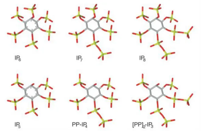
IP6 is the precursor of the best characterized inositol pyrophosphates IP7 (or PP-IP5;
5-diphosphoinositol pentakisphosphate) and IP8 (or (PP)2-IP4; bisdiphosphoinositol
tetrakisphosphate). Respectively, these molecules have seven or eight phosphate groups attached to the six-carbon inositol ring and as a result have one and two pyrophosphate moieties (Figure 3).
5
Figure 3. The figure shows the chemical structure of IP6 with its pyrophosphate derivates IP7 and IP8,
as well as IP5, with its derived pyrophosphates PP-IP4 and [PP]2-IP3 (Bennett et al., 2006).
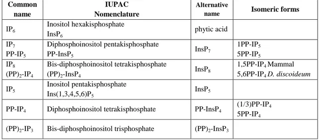
At the moment, even if the pyro-nomenclature is generally used, it is important to note that the correct IUPAC definition for this moiety is diphospho-, so the correct chemical name of IP7 is diphosphoinositol pentakiphosphate (PP-IP5). The different nomenclatures that indicate
6 Common name IUPAC Nomenclature Alternative
name Isomeric forms IP6 Inositol hexakisphosphate InsP6 phytic acid IP7 PP-IP5 Diphosphoinositol pentakisphosphate PP-InsP5 InsP7 1PP-IP5 5PP-IP5 IP8 (PP)2-IP4 Bis-diphosphoinositol tetrakisphosphate (PP)2-InsP4 InsP8 1,5PP-IP4 Mammal 5,6PP-IP4 D. discoideum IP5 Inositol pentakisphosphate Ins(1,3,4,5,6)P5 InsP5
PP-IP4 Diphosphoinositol tetrakisphosphate PP-InsP4
(1/3)PP-IP4
5PP-IP4
(PP)2-IP3 Bis-diphosphoinositol trisphosphate (PP)2-InsP3
Table 2. This table summarizes the nomenclature of the inositol pyrophosphates, with the correct
IUPAC nomenclature, the alternative names infrequently present in the literature, and the identified biological isomers (Wilson et al., 2013).
In eukaryotic cells, IP6 is the most abundant inositol polyphosphate with a concentration in
mammalian cells in the range of 10-60µM (Pittet et al., 1989) (Szwergold et al., 1987) and up to 0.7mM in slime moulds such as Dictyostelium discoideum (D. discoideum) (Martin et al., 1987) (Laussmann et al., 2000). In general, IP6 has housekeeping functions as a phosphate
storage molecule and as an antioxidant (Raboy, 2003) (Shears, 2001), and in addition is a signaling molecule that is involved in the regulation of vesicular trafficking (Shears, 2001) as well as quite in a lot of nuclear events (York et al., 1999) (Shears, 2001).
IP7 and IP8 are present in all eukaryotic cells. In mammalian cells, these molecules are present
in sub-micromolar amounts, representing less than 5% of their precursor IP6 (Bennett et al.,
2006). Though the inositol pyrophosphate species levels are low and constant, this concentration hides an extraordinary turnover that has been calculated (using fluoride inhibition of phosphatases in mammalian cells) to convert up to 50% of the IP6 pool every
hour to its pyrophosphorylated derivatives (Menniti et al., 1993). This most probably reveals an important signaling function in the cell.
In contrast to mammalian cells, D. discoideum has IP7 and IP8 levels in the 100–200mM
range (Laussmann et al., 2000), and the structures of these molecules has been elucidated by
1
H-31P 2D NMR spectroscopy (Laussmann et al., 1996) and isomeric analysis using stereo-specific inositol phosphatases (Laussmann et al., 1997).
7
The results revealed a single IP8 isomer present as 5,6-[PP]2-IP4, and two IP7 isomers,
5-PP-IP5 and 6-PP-IP5 (Albert et al., 1997), with a relatively higher abundance in vivo of the
6-pyrophosphorylated species. The IP8 species from another amoeba, Polysphondylium
pallidum, has been identified as 1,5-[PP]2-IP4 (Laussmann et al., 1998), while an IP7 species
from Entamoeba histolyticaas was found to be 5-PP-IP5 (Martin et al., 2000).
Consequently, the results from structural analyses have shown that the inositol pyrophosphates are structurally different and may also suggest that organisms possess various inositol pyrophosphate species with diverse functions.
8
2. Synthesis: kinases and phosphatases
In Saccharomyces cerevisiae (S. cerevisiae), the yeast experimental model, the synthesis of inositol phosphates begins with the well-known second messenger IP3 (Figure 4) that is the
product of cleavage of the lipid PI(4,5)P2 by phospholipase C (Mikoshiba et al., 1993).
Subsequent to liberation from its diacyl glycerol (DAG) tail, this molecule can be dephosphorylated to myo-inositol, or it can be additionally phosphorylated to the completely phosphorylated IP6 ring and further, to form the inositol pyrophosphates IP7 and IP8 (Figure
4) (Laussmann et al., 1998).
While S. cerevisiae synthesizes inositol phosphates by this lipid path (Stephens and Irvine, 1990), D. discoideum synthesizes IP6 by the cytosolic path. In this amoeba phospholipase C
activity is not necessary to synthesize the higher phosphorylated inositols, because the synthesis happens directly from myo-inositol to IP6 (Figure 4) (Stephens and Irvine, 1990).
Figure 4. This linear pathway shows the production of inositol pyrophosphates starting from
myo-inositol. In S. cerevisiae this occurs via the lipid route, whereas in D. discoideum they are produced by the cytosolic route (Wilson et al., 2013).
9
Two different classes of enzymes are able to synthesize inositol pyrophosphates: the IP6
kinases (IP6K) and the PP-IP5 kinases (PPIP5K), Kcs1 and Vip1 respectively in S. cerevisiae.
The difference between these enzymes consist of a difference in catalytic activity against different positions on the ring, IP6Ks place a phosphate group at the β position of carbon 5 on the fully phosphorylated IP6 ring (Draskovic et al., 2008), whereas the PPIP5Ks
phosphorylate position 1 (Lin et al., 2009) (Figure 4).
In mammalian cells, the isomeric form of IP8 that is present has been identified as 1,5-PP-IP4
(Chakraborty et al., 2011) whereas in D. discoideum the 5,6-PP-IP4 isomer of IP8 is present
(Chakraborty et al., 2011), probably because the two metabolisms are different (Figure 4).
In vitro, PPIP5Ks do not act on the main inositol pentakisphosphate isomer, I(1,3,4,5,6)P5,
while the IP6K enzymes are capable of phosphorylating this isomer, mostly but not completely at the enantiomeric positions (1,3), producing (1/3)PP-IP4. Further
phosphorylation by IP6K or PPIP5K can generate (PP)2-IP3 (Wang et al., 2011).
However, in vivo, it has been shown that yeast Kcs1 generates inositol pyrophosphate even using IP3 and IP4 (Seeds et al., 2005). Meanwhile, in vitro the IP6Ks are also able to generate
a tri-phosphate PPP-IP5 form of ‘IP8’ (Draskovic et al., 2008).
The high conservation of IP6Ks has facilitated not only the identification of these enzymes from different organisms, but also helped to identify another family of inositol polyphosphate kinases, known as inositol polyphosphate multi-kinases (IPMKs). These enzymes are able to phosphorylate IP3 and IP4 isoforms as well as phosphorylating the phosphoinositide lipid
PtdIns(4,5)P2 to PtdIns(3,4,5)P3.
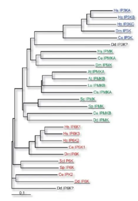
Consequently, the IP6Ks, IMPKs and IP3-3Ks belong to an inositol polyphosphate kinase
family, the IPKs that generated from a common ancestor. The most important characteristic of this family is the presence of the conserved PxxxDxKxG motif in the inositol-binding region (Bennett et al., 2006). Phylogenetic analysis of their sequences indicates that in fact IP6Ks are the most ancient members of this family (Figure 5).
10
Figure 5. Phylogenetic tree of IPK proteins family members (Bennett et al., 2006).
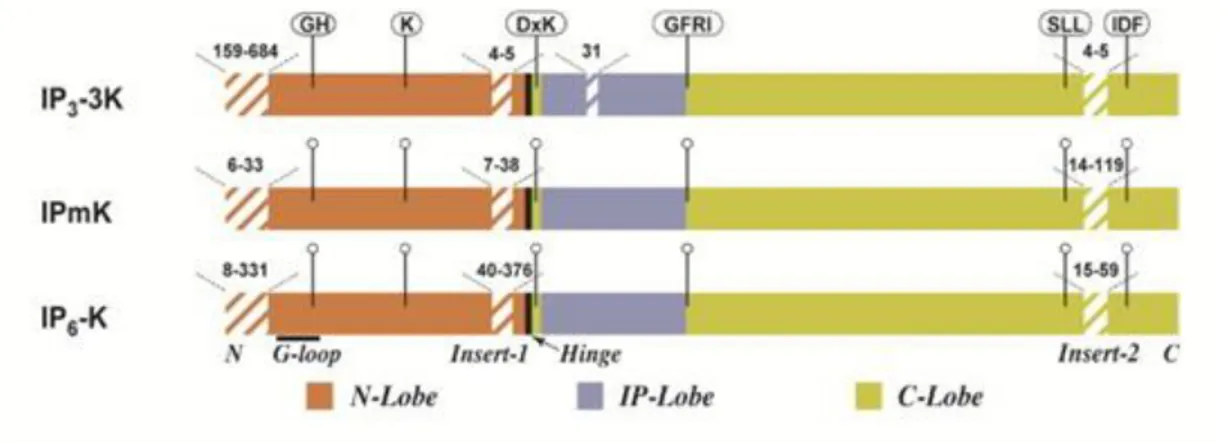
In addition to the consensus PxxxDxKxG motif, all three family members share a common carboxy-terminal catalytic domain containing the conserved SSLL and IDF motifs (El Bakkoury et al., 2000) (Irvine and Schell, 2001). There is an extra 30 amino acid residue insertion in the inositol binding domain of IP3-3Ks over IPMK and IP6Ks, while IPMK has a
more open binding domain which corresponds to its greater substrate versatility (Holmes and Jogl, 2006). There is also a large 129 amino acid insert in the N-terminal region of the IP6
11
Figure 6. Schematic diagram of common IPK catalytic domain structural elements. The three major
subdomains of the IPKs are indicated with different colors. Hatched lines signify non-conserved insert regions, and the numbers over these regions indicate the range in the number of residues observed among all identifiable IPKs. Flagged regions show conserved motifs present in most IPK members (Gonzáles et al., 2004).
The IP6Ks were first purified in Solomon Snyder’s laboratory (Voglmaier et al., 1996). Three mammalian genes IP6K1, 2 and 3 were identified, one of which, IP6K2, had previously been identified as PiUS (inorganic phosphate uptake stimulator) (Voglmaier et al., 1996) (Saiardi et al., 2001). In vitro, all three mammalian IP6Ks as well as the yeast Kcs1 are capable of phosphorylating IP6 to IP7, as well as IP5 to PP-IP4 (Saiardi et al., 1999).
IP6K1 possesses high amino acid similarity in a number of different species, for example 99% between mouse and rat and 95% between rat-human and mouse-human. The human gene is found on chromosome 3 (3p21.3) and codes for a 50-kDa protein. Studies have shown strong levels of expression in different mouse tissues such as brain and testis in addition to pancreatic β-cells (Illies et al., 2007), with a weak expression in heart, kidney, liver, lung and spleen (Saiardi et al., 1999).
IP6K2 has also been mapped to chromosome 3 (3p21.31) and this gene encodes for a protein of ~49-kDa. By alternative splicing two different variants have been identified that lack most of the C-terminal part of the whole protein. Northern blot analysis has shown a tissue distribution which is highest in brain and lung and lower in liver, kidney, and testis (Saiardi et al., 1999).
IP6K3 is located on chromosome 6 (6p21.31), has a smaller mass (46-kDa) and is most enriched in the brain where its levels resemble that of IP6K1 and IP6K2 (Saiardi et al., 2001).
12
IP6K3 shows 50 and 45% sequence identity to IP6K1 and IP6K2, respectively and possess a less acidic character than the other two enzymes. It has been shown by over-expression studies that IP6K2 is exclusively nuclear, IP6K3 is predominant in the cytoplasm, and IP6K1 displays comparable nuclear and cytosolic densities (Saiardi et al., 2001).
All these proteins possess a similar structure nevertheless showing some differences (Figure
6). In fact, it is not clear how protein-protein interactions occur and/or the regions where these
connections happen. These proteins possess diverse binding partners such as GRAB (Luo et al., 2001), heat-shock protein 90 (HSP90) (Chakraborty et al., 2008) and tumor necrosis factor receptor-associated factor 2 (TRAF2) (Figure 7).
Figure 7. IP6Ks showing basic structure of proteins. In blue the similarities between the proteins are
shown, while purple shows the differences (Barker et al., 2009).
In 2007 the York laboratory identified the second class of kinases: the yeast enzyme Vip1 (Fridy et al., 2007). It is able to phosphorylate at the 1/3 enantiomeric positions generating 1/3PP-InsP5 or 1/3,5(PP2)-InsP4 from IP6 and 5PP-InsP5 respectively (Lin et al., 2009). Two
mammalian homologues, PPIP5K1 and PPIP5K2, were also identified (Shears et al., 2013). These proteins do not belong to the inositol phosphate kinase family; they possess two different domains: kinase domain and histidine acid phosphatase-like domain in the C terminal portion of the protein.
13
It has been supposed that the phosphatase domain is responsible for the allosteric regulation of PPIP5K by IP6 (Shears et al., 2013) but it is catalytically inactive. However, truncated
PPIP5K constructs lacking the phosphatase domain show increased activity when over-expressed, indicating that this domain is active. It may antagonize the kinase domain, specifically dephosphorylating 1PP-IP5, the IP7 isomer produced by the kinase domain.
Although in vitro, PPIP5K can phosphorylate IP6 to 1PP-IP5 (Shears et al., 2013), while in
vivo, this activity is masked by phosphatase activity and cannot be easily detected, suggesting that the major physiological target of the PPIP5Ks is IP7. This is further supported by the
increase in levels of IP7 in vip1Δ mutants, whilst levels of IP6 remain unchanged (Shears et
al., 2013).
Phosphatases are enzymes that show the reverse capability of kinases; in fact they are able to hydrolyze pyrophosphate groups. Two different proteins have been discovered: MIPP (multiple inositol polyphosphate phosphatase) and DIPP (diphosphoinositol phosphate phosphohydrolase). DIPPs were first cloned (Safrany et al., 1998) before the IP6Ks but there is at the moment no more than modest information on their physiological role.
MIPP is located in the endoplasmic reticulum (Ali et al., 1993) and is able to hydrolyze the 5β-phosphates in vitro, however it does not play a role in inositol pyrophosphate degradation in vivo (Shears et al., 1995).
In human and mouse, there are five different DIPP isoforms (1, 2α, 2β, 3α, 3β) while there is only one DIPP in S. cerevisae (Ddp1p/Yor163w) (Safrany et al., 1999) and one in S. pombe (Aps1) (Ingram et al., 2003).
DIPP1 is the product of the NUDT3 gene. DIPP2α and DIPP2β, only differing by one amino acid (glutamine Q86), are the products of the NUDT4 gene (Caffrey et al., 2001). DIPP3α and DIPP3β are the products of the NUDT10 and NUDT11 genes, respectively (Leslie et al., 2002) (Hidaka et al., 2002).
DIPP3α is also called hAsp2 whilst DIPP3β is called hAsp1. These two proteins are identical in mouse while in humans differ through one amino acid (Hua et al., 2003).
DIPP1 and DIPP2 are expressed in a large range of tissues (Chu et al., 2004) (Hua et al., 2001) whereas DIPP3 shown a more restricted expression in fact has been found in testis, liver, kidney and brain (Hua et al., 2003).
14
DIPPs have a common catalytic site used to hydrolyze inositol pyrophosphates and diadenosine polyphosphates, as well as PRPP (5-phosphoribosyl 1-pyrophosphate pyrophosphatase) (Fisher et al., 2002) that is not a substrate for these enzyme in vivo (Hidaka et al., 2002) (Figure 8).
Between these enzymes, DIPP1 is the most catalytically active (Hua et al., 2003), DIPP2α is more active than DIPP2β, probably because they differ by one amino acid that has a strong influence on catalytic function (Caffrey et al., 2000), whereas DIPP3α and DIPP3β are the least active against inositol pyrophosphates (Leslie et al., 2002).
Figure 8. Metabolic interrelationships between inositol pyrophosphates and enzymes responsible for
their synthesis and degradation (Barker et al., 2009).
The DIPPs hydrolyze their substrate in a site-specific order, indeed in 1,5PP2-IP4 they
hydrolyze first position 1 and then position 5 (Caffrey et al., 2000). For complete activity, DIPPs require a free Mg2+ of 1.5-2mM (Safrany et al., 1999), on the contrary fluoride inhibits their activity with a Ki around 10μM (Leslie et al., 2002).
15
3. Functions of inositol pyrophosphates
Inositol pyrophosphates are recognized to control a variety of cellular activities such as apoptosis (Morrison et al., 2005), telomere maintenance (Saiardi et al., 2005) (York et al., 2005) and vesicular trafficking (Saiardi et al., 2002). This variety of activities underlines the biological significance of these molecules though until recently it was not clear how these molecules controlled all these cellular functions.
It was published that inositol pyrophosphates play a role as metabolic messengers (Shears, 2009), regulators of cell homoeostasis (Wunderberg and Mayr, 2012) and regulators of cellular energetic metabolism (Szijgyarto et al., 2011). These functions are summarized in the following figure (Figure 9) in which is shown the link between inositol pyrophosphates and the metabolism at molecular, cellular and organismal levels.
Figure 9. Diverse controls that inositol pyrophosphates (IP7) perform on the metabolism at different
levels (Wilson et al., 2013).
The synthesis of inositol pyrophosphates is linked to ATP levels for the reason that the IP6K enzymes show high affinity for ATP. Startlingly, it has been demonstrated that in cells that do not possess or cells that have low levels of inositol pyrophosphates that they have an increased cellular ATP concentration while ADP and AMP levels are reduced, producing an increase in the adenylate pool and to an increase in adenylate energy charge (AEC) (Szijgyarto et al., 2011).
16
On the contrary, in the wild-type yeast cells that shown increased levels of IP7 since there is
an over-expression of mammalian IP6K1, the concentration of ATP is decreased. In the yeast system it was revealed that protein pyrophosphorylation was influencing the interactions of the crucial yeast glycolytic transcription factors Gcr1, Gcr2 and Rap1 (Barbara et al., 2007). Pyrophosphorylation of Gcr1 decreases its capability to bind Gcr2, thus inhibiting the transcription of genes encoding glycolytic enzymes. Thus, the kcs1Δ yeast consumed glucose more rapidly than wild-type yeast (Szijgyarto et al., 2011).
Unexpectedly, in the kcs1Δ yeast given the increase in cellular ATP concentration, the mitochondria were malfunctioning. Generally, yeast produce energy and intermediates by fermentation, however it is possible to achieve mitochondrial metabolism/oxidative phosphorylation by providing just a non-fermentable carbon source. The kcs1Δ yeast are unable to grow in these conditions.
Instead, the high ATP level and the reduced oxygen consumption of IP6K1-/- MEFs compared with wild-type MEFs evidence that the regulation of cellular metabolism by inositol pyrophosphate is evolutionarily conserved. ATP availability is underpins virtually all other cellular activity.
In D. discoideum it has been shown that inositol pyrophosphates furthermore control chemotaxis (Luo et al., 2003). In wild-type cells, starvation and successive cAMP signaling and chemotaxis induces formation of a multicellular fruiting body. The ip6k knockout was more responsive to cAMP and aggregated more rapidly than wild-type cells responding to starvation (Luo et al., 2003).
These results suggest that in wild-type cells there is an antagonism between IP7 and PIP3 in
binding the PH domain of Crac, a protein related to cAMP-dependent chemotaxis. During D. discoideum aggregation PIP3 is not able to be the principal regulatory molecule, as deletion of
every PI3K capable of producing PIP3 did not inhibit chemotaxis to cAMP (Hoeller and Kay,
2007). Furthermore, as IP7 influences energetic metabolism in addition to the adenine
nucleotide level, the high level of ATP could lead to an increased cAMP concentration and then the more rapidly chemotaxic response observed in D. discoideum ip6k knockout.
In addition, it has been shown that inositol pyrophosphates are molecules involved also in the regulation of organismal metabolism. In pancreatic β-cells, the overexpression of IP6K1 or the addition of IP7 generate induction of exocytosis (Illies et al., 2007). This result is
17
hypersensitivity to insulin and decrease in fatty tissue levels (defective mitochondria cannot synthesize the intermediates for fatty acid synthesis) (Hiltunen et al., 2010).
However, it has been shown that the interaction between the inositol pyrophosphates levels and AEC generates a link that regulates the connections between insulin and plasma glucose concentration.
Additionally, the IP6K2-/- mouse characterization has shown that IP6K2 could be the most important isoform in cell death regulation by inositol pyrophosphates. Particularly, seems that IP6K2 interacts with two different proteins, HSP90 and p53, by a via unknown and independent of inositol pyrophosphates synthesis (Koldobskiy et al., 2010).
Another important link is established between metabolism and aging (Passarino et al., 2010). It has been shown that high levels of IP6 and IP7 are present in hepatocytes from old wild-type
mice but not in younger mice. Also, one probable link between metabolism and aging has been found in the telomere length; indeed shorter telomeres are associated with aging in mammals (Mather et al., 2011).
The kcs1Δ yeast has longer telomeres than wild-type whilst ipk1Δ yeast has shorter telomeres; (Saiardi et al., 2005) perhaps these results are due to Tel1 which is a telomere regulator inhibited by inositol pyrophosphates. Furthermore it has been supposed that inositol pyrophosphates can also influence other important proteins involved in DNA damage signaling/DNA repair (Lovejoy and Cortez, 2009) such as Ku protein (Ma and Lieber, 2002).
In recent years, it has been shown that inositol pyrophosphates are also able to regulate the process of DNA repair. In yeast, ROS signaling is generally studied using the exposure to exogenous agents for example H2O2, which causes DNA base modification, single- and
double-strand breaks, and the formation of apurinic/apyrimidinic lesions (Letavayova et al., 2006). To generate DNA-damage, it is usually appropriate to use sub-lethal concentrations of H2O2 (Leroy et al., 2001).
Yeast mutants which are deficient in inositol pyrophosphates have a higher threshold of resistance to the lethal effects of H2O2, but not to other DNA-damaging agents. This specific
resistance to H2O2 connects with a constant activation of Rad53 and as a result promotion of
DNA repair mechanisms. Further it has been reported that H2O2 regulates higher inositide
metabolism, producing a decrease in cellular levels of inositol pyrophosphates (Onnebo and Saiardi, 2009).
18
Inositol pyrophosphates also possess the ability to regulate phosphate metabolism (Saiardi, 2012). Inositol pyrophosphates regulate the access of phosphate into the cells (Norbis et al., 1997), suggesting that they could affect phosphate uptake either directly, for example stimulating a transporter, or indirectly by helping ‘fixing’ free phosphates in organic molecules.
Additionally, many hypotheses have been formulated to elucidate the biological link between phosphate, inositol pyrophosphates and polyP that is a linear polymer of phosphodiester bound. Indeed, the synthesis of polyP may be related to phosphate ingress into the cell. Inositol pyrophosphate control of energy metabolism (Szijgyarto et al., 2011) influences not only ATP levels but it can also change the total cellular balance of adenine nucleotides. The phosphate transfer reactions generally use ATP as a means of transport for the phosphate groups, so inositol pyrophosphate could influence phosphate metabolism by regulating the adenylate cellular pool. Moreover, the existence of a feedback mechanism that coordinates the metabolic balance between ATP, phosphate and inositol pyrophosphates has been suggested.
Inositol pyrophosphates could either contribute to the regulation of polyP synthesis, play a role in polyP degradation, or both. The analysis of yeast mutant that are not able to synthesize inositol pyrophosphates has shown a striking correlation between the lack of inositol pyrophosphates and the absence of polyP (Auesukaree et al., 2005) (Lonetti et al., 2011).
It has even been reported that increasing cellular IP7 levels augments cell sensitivity to cell
death. The phosphorylation of mammalian target of rapamycin (mTOR) was also depressed in cells that over-express IP6Ks, suggesting that the mTOR pathway regulates autophagosomes generated by IP6Ks. These results suppose that IP6Ks support autophagy and provoke caspase-independent cell death (Nagata et al., 2011).
Different experiments have been conducted to demonstrate the existence of a link between inositol pyrophosphates and autophagy in yeast. These results indicate that inositol phosphates are implicated in the regulation of autophagy, however the precise role of each inositol phosphate species in this process is not clear (Taylor et al., 2012).
19
4. Mechanism of action
To transduce signals, the inositol pyrophosphates may use two different mechanisms of action: binding (Lemmon, 2008) and pyro-phosphorylation (Bhandari et al., 2007). In cells, these two mechanisms are not exclusive but coexist. Usually, the first mechanism happens through the binding of small molecules to specific protein targets, such as cAMP to protein kinase A otherwise lipid hormone to specific receptors.
Inositol polyphosphates bind to several proteins containing specific domains such as PH (pleckstrin homology), PX (phagocyte oxidase homology) or FYVE (for Fab1, YOTB, Vac1 and EEA1) domains (Alcázar-Román and Wente, 2008). The best characterized mechanism of action in this area is the binding of IP3 to its receptor (IP3-receptor) that leads to an alteration
of the tridimensional structure of the channel, which in turn allows Ca2+ efflux (Mikoshiba et al., 1993) (Irvine, 2003).
The similar mechanism of action has been theorized for inositol pyrophosphates that may also signal through allosteric interactions with proteins indeed, in vitro, IP7 is able to bind several
proteins such as AP3/AP180 (Saiardi et al., 2002), the Golgi coatomer (Ali et al., 1995) and the clathrin-assembly adaptors AP2 (Voglmaier et al., 1992). Further, IP7 competes for
binding to the D. discoideum chemotaxis protein Crac (cytosolic regulator of adenylate cyclase) and mammalian Akt (Luo et al., 2003).
The proteins that bind to IP7 also bind to IP6, though to a more modest extent, and IP6 has
been proposed to be the more essential ligand in vivo (Cremona and De Camilli, 2001). As the concentration of inositol pyrophosphates is low, a severe specificity of binding for IP7 over
IP6 is required. To solve this problem it is possible to induce the IP6 kinase activity inducing
increase in the IP7/IP6 ratio.
However, the importance of binding as a mechanism of action has been confirmed by IP7
-mediated competition for the binding of PI(3,4,5)P3 to the PH domain-containing CRAC that
is consequently inhibitory to chemotaxis (Luo et al., 2003).
It was also revealed that IP6 is only 1–2% as potent as IP7, and then IP7 is more likely to be
the physiological binding partner during aggregation (Laussmann et al., 2000). IP7 was found
to bind to a variety of other PH-domain containing proteins, including, in vitro, mammalian Akt (Luo et al., 2003).
20
In addition, a further recent study demonstrated the inability of the PH-domain of phosphoinositide-dependent protein kinase 1 (PDK1) to bind IP7 (Komander et al., 2004).
Allosteric interaction has been also suggested like a mechanism for controlling the Pho85/Pho80/Pho81 cyclin dependent kinase/cyclin/cyclin-dependent kinase inhibitor complex of the yeast S. cerevisiae (Lee et al., 2008). The binding of 1/3PP-IP5, the IP7 isomer
generated by Vip1, to the Pho85/Pho80/Pho81 complex inhibits its action: the transcription factor Pho4 is now not hyper-phosphorylated by Pho85 so is able to go into the nucleus to activate the PHO pathway (Lee et al., 2007).
The second mechanism of action is the pyro-phosphorylation that occur by donation of the β-phosphate from the pyroβ-phosphate group to proteins (Saiardi et al., 2004). The process of pyro-phosphorylation is due to the presence of a functional group: the pyro-phosphate group. Furthermore the mechanism of action is shown in the following figure (Figure 10) (Saiardi et al., 2004).
Figure 10. The proposed mechanisms of action for inositol pyrophosphates: the direct binding
mechanism when a protein target has a particular pocket able to distinguish IP7 (upper panel) and the
pyro-phosphorylation modification of a protein carried out by IP7 (lower panel) (Wilson et al., 2013).
In recent years, the pyrophosphate constituent of IP7 has been shown to act as a phosphate
donor to proteins, in a non-enzymatic and temperature-dependent reaction (Saiardi et al., 2004).
In yeast, have been identified three different potential IP7 substrates: NSR1 (a nuclear protein
21
SRP40 (protein that acts as a ribosomal chaperone). All these proteins have regions of similar sequence, as everyone contains regions of serine residues surrounded by acidic residues.
Generally, proteins that are phosphorylated by IP7 in eukaryotic cells are no longer
phosphorylated if expressed in bacteria, indicating that substrate priming, across a post-translation modification, is an necessary requisite for IP7-driven phosphorylation (York and
Hunter, 2004).
Pyro-phosphorylation mediated by IP7 may cause functional alterations in protein
conformation and then may give rise to variations in the protein’s interactions, activity or localization (Azevedo et al., 2009) (Szijgyarto et al., 2011). Further investigation into the mechanism of action of IP7 will confidently provide insight into its regulation in the cell and
22
5. Physiological role
To study the physiological role of inositol pyrophosphates yeast cells that don’t have IP6K enzymes (kcsI∆) and consequently don’t possess inositol pyrophosphates are generally used (Saiardi et al., 2002). Morphologically, these mutants are abnormal, suggestive of important functions for inositol pyrophosphates. The kcsI∆ mutants are bigger and grow slower than wild-type cells, and appear to be hypersensitive to salt stress however appear unaffected by osmotic challenge with sorbitol (Dubois et al., 2002).
Problems in cell wall integrity have also been observed as well as a disjointed vacuolar morphology (Saiardi et al., 2002). These findings suggest that inositol pyrophosphates play important functions in central cellular processes such as growth, endocytosis and response to stress.
In mammalian cells, where three different isoforms of IP6K and two different isoforms of PP-IP5K are present, it is more complicated to establish the effects of a lack of inositol pyrophosphates.
Therefore to elucidate which phenotypic abnormalities are related to the deficiency of inositol pyrophosphates further studies are needed to comprehend if these molecules are truly involved also in complex signaling functions in mammalian cells.
23
6. D. discoideum as a model to study inositol pyrophosphates
D. discoideum is a small soil-living amoeba that possesses a haploid genome having six completely sequenced chromosomes (Eichinger et al., 2005). Commonly referred to as slime mold, is a eukaryote that shifts from a collection of unicellular amoebae into a multicellular slug and then into a fruiting body within its lifetime. This organism also contains many proteins and molecular processes that were previously thought to be present only in metazoa. For example, the D. discoideum genome possesses 24 classes of protein kinase that are not present in the yeast S. cerevisiae (Annesley and Fisher, 2009), so the presence of these proteins in this amoeba proposes that they are derived from ancestral pathways abandoned in the yeast heredity. D. discoideum has an exclusively asexual lifecycle including four different stages: vegetative, aggregation, migration, and culmination (Figure 11).
Figure 11. The life cycle of D. discoideum begins as a vegetative amoeba and ends with the formation
of the mature fruiting body (Fey et al., 2007).
Developmental morphogenesis starts from single and vegetative amoebae and ends with the formation of the mature fruiting body, which contains diverse spores on top of a stalk. Myxamoebae hatch from the spores under warm and moist conditions. Throughout the vegetative stage, the myxamoebae divide by mitosis as they feed on bacteria. The bacteria secrete folic acid, attracting the myxamoebae. When the supply of bacteria is depleted, the myxamoebae go into the aggregation stage.
24
During aggregation, starvation starts the formation of a biochemical process involving glycoproteins and adenylyl cyclase (Gilbert, 2006). The glycoproteins permit cell-cell adhesion, and adenylyl cyclase produces cAMP that is secreted by the amoebas to attract adjacent cells to an inner location. Moving towards the signal, they impact into each other and stick together by the use of glycoprotein adhesion molecules.
The migration phase starts once the amoebas have produced a tight aggregate; the amoebas work together as a motile pseudo-plasmodium also known as a slug. The slug is about 2–4 mm long, composed of up to 100,000 cells (Cooper and Geoffrey, 2000), and is capable of movement by producing a cellulose scabbard in its anterior cells through which the slug shifts (Tyler, 2000).
The formation of different cell types is helped by cAMP and DIF (differentiation-inducing factor) (Tyler, 2000). The slug gets differentiated into pre-stalk and pre-spore cells that go to the anterior and posterior ends, respectively. Once established in an appropriate environment, the anterior end of the slug will create the stalk of the fruiting body and the posterior end will produce the spores of the fruiting body (Tyler, 2000). The culmination stage begins when the slug settles into one spot, the posterior end spreads out with the anterior end raised in the air, forming a structure looking like a ‘hat’.
The anterior end of the ‘hat’ forms a cellulose tube, which allows the cells to move (Tyler, 2000). To obtain the mature fruiting body takes 8–10 hours (Tyler, 2000). The fruiting body is then capable of starting the entire cycle over again by releasing the mature spores that can be converted into myxamoebae.
Usually D. discoideum are capable of reproducing asexually but are still able to reproduce sexually if there are certain conditions. During aggregation, if two amoebae of different mating types are present in a dark and wet environment, they can combine to create an enormous cell that will phagocytizes the other cells in the aggregate furthermore collect the entire aggregate in a cellulose wall to defend it. This is also known as a macro-cyst where inside the enormous cell divides first through meiosis, then through mitosis to produce many haploid amoebae.
D. discoideum is commonly used as a model organism not just because has a simple life cycle but also because it possesses a restricted number of cell types and the growth is typically very rapid (Tyler, 2000).
25
It is used to study different cellular processes such as cell differentiation, chemotaxis and programmed cell death or other aspects of development including phagocytosis and signal transduction. These processes and aspects of development are not present or too complex to view easily in other model organisms, so these features make D. discoideum a good candidate model organism (Nag and Tikhonenko, 2008).
Furthermore, D. discoideum is often used as a model to study inositol polyphosphate signaling for the reason that it has high levels of IP7 and IP8, important in the initial
identification of their structures and functions (Stephens et al., 1993). The inositol pyrophosphate levels are not highduring the vegetative state but they increase dramatically in the aggregation state and chemotaxis, which depend on cAMP signaling (Luo et al., 2003), reaching the equivalent levels of IP6 (Laussmann et al., 2000).
In D. discoideum 7 genes belonging to the IPK family are present. One of these genes, namely I6KA was identified as an IP6K (Luo et al., 2003). The activity of this enzyme has not been tested in vitro, however destroying I6KA by homologous recombination produced a dramatically decrease in the inositol pyrophosphate levels. Regarding the phenotype, mutant cells possess similar shape and size; in addition they show equal growth rate comparate to wild-type cells. However during starvation, aggregation occurs that is faster in the mutants than in wild-type cells. Mutants exhibit high sensitivity to chemotactants, responding rapidly to low concentrations of cAMP. These results suggest that in the mutants there is an alteration of chemotactic pathways (Luo et al., 2003).
The translocation of CRAC to the membrane, mediated by the PH domain, is an indispensable step for directional sensing, since either decreasing the amount of PI(3,4,5)P3, by disrupting
PI3K genes, or abnormally increasing PI(3,4,5)P3 levels by disruption of PTEN decreases the
ability of cells to orientate and to move in response to chemotactic gradients (Iijima and Devreotes, 2002). The lack of IP7 in the null mutant would free the CRAC-PH domain to bind
to PI(3,4,5)P3, and its translocation to the membrane would consequently increase.
In addition to being an excellent experimental model, during D. discoideum development the most dramatic modulation of the levels of inositol pyrophosphates has been seen (Stephens et al., 1993).
In mammalian cells, it has been possible to use fluoride to dramatically modulate the levels of inositol pyrophosphates, and in addition to discover that 50% of IP6 is converted, every hour,
26
many times each hour (Glennon and Shears, 1993) (Menniti et al., 1993). These results suggest that inositol pyrophosphates are signaling molecules with ‘molecular switch activity’ though their physiological significance remains unknown. Fluoride inhibits the DIPP phosphohydrolases and is a common phosphatase inhibitor with many effects on cell signaling (Bollen and Stalmans, 1988); the specific effect on inositol metabolism is unclear. Overall these results suggest that in mammalian cells inositol pyrophosphates are dynamic molecules that turnover very rapidly, and that cells spend a lot of energy to keep their levels constant.
However, the best example of modulation of inositol pyrophosphate levels has been observed in D. discoideum, where during starvation-induced aggregation it is possible to observe a considerable increase in the levels of IP7 and IP8 (Laussmann et al., 2000). During starvation
cAMP is secreted that induces elevation in the IP7 and IP8 levels (Luo et al., 2003).
In fact, a decrease in extracellular inorganic phosphate concentration led to a dramatic rise in the intracellular levels of inositol pyrophosphates, in particular IP7 (Mulugu et al., 2007).
More studies are needed to elucidate this result that is controversial as usually reduction of phosphate causes a reduction of ATP, which is fundamental for the synthesis of inositol pyrophosphates (Boer et al., 2010).
27
7. Plan of the thesis
Chapter II reports one manuscript (PlosOne – In Press) aimed at investigating the inositol pyrophosphate metabolism in D. discoideum. In particular, in the manuscript ‘Analysis of Dictyostelium discoideum inositol pyrophosphate metabolism by gel electrophoresis’ we report results obtained using polyacrylamide gel electrophoresis (PAGE) to resolve and detect inositol pyrophosphate species in this amoeba. We have achieved three major findings:
D. discoideum possesses three bands correspond to IP6 and its derivative inositol
pyrophosphates, IP7 and IP8;
there is another band that correspond to IP9;
this amoeba possesses three different IP5 isomers;
during development there is a three times increase of inositol pyrophosphates and not the 25 times previous reported;
cAMP does not induce inositol pyrophosphates synthesis.
In the thesis is also included an appendix which reports the integral version of the published paper (Montesanto et al., 2013), ‘Common polymorphisms in nitric oxide synthase (NOS) genes influence quality of aging and longevity in humans’ Biogerontology 14(2):177-86.
28
REFERENCES
1. Agranoff, B. W., (1978) Cyclitol confusion. Trends Biochem Sci 3, N283-N285.
2. Alcázar-Román, A. R., Wente, S. R., (2008) Inositol polyphosphates: a new frontier for regulating gene expression. Chromosoma 117, 1-13.
3. Albert, C., Safrany, S. T., Bembenek, M. E., Reddy, K. M., Reddy, K., Falck, J., Bröcker, M., Shears, S. B., Mayr, G. W., (1997) Biological variability in the structures of diphosphoinositol polyphosphates in Dictyostelium discoideum and mammalian cells. Biochem J 327, 553-560.
4. Ali, N., Craxton, A., Shears, S. B., (1993) Hepatic Ins(1,3,4,5)P4 3-phosphatase is compartmentalized
inside endoplasmic reticulum. J Biol Chem 268, 6161-6167.
5. Ali, N., Duden, R., Bembenek, M. E., Shears, S. B., (1995) The interaction of coatomer with inositol polyphosphates is conserved in Saccharomyces cerevisiae. Biochem J 310, 279-284.
6. Annesley, S. J., Fisher, P. R., (2009) Dictyostelium discoideum – a model for many reasons. Mol Cell Biochem 329, 73-91.
7. Auesukaree, C., Tochio, H., Shirakawa, M., Kaneko, Y., Harashima, S., (2005) Plc1p, Arg82p, and Kcs1p, enzymes involved in inositol pyrophosphate synthesis, are essential for phosphate regulation and polyphosphate accumulation in Saccharomyces cerevisiae. J Biol Chem 280, 25127-25133.
8. Azevedo, C., Burton, A., Ruiz-Mateos, E., Marsh, M., Saiardi, A., (2009) Inositol pyrophosphate mediated pyrophosphorylation of AP3B1 regulates HIV-1 Gag release. Proc Natl Acad Sci U S A 106, 21161-21166.
9. Barbara, K. E., Haley, T. M., Willis, K. A., Santangelo, G. M., (2007) The transcription factor Gcr1 stimulates cell growth by participating in nutrient-responsive gene expression on a global level. Mol Genet Genomics 277, 171-188.
10. Barker, C. J., Illies, C., Gaboardi, G. C., Berggren, P. O., (2009) Inositol pyrophosphates: structure, enzymology and function. Cell Mol Life Sci 66, 3851-3871.
11. Bhandari, R., Saiardi, A., Ahmadibeni, Y., Snowman, A. M., Resnick, A. C., Kristiansen, T. Z., Molina, H., Pandey, A., Werner, J. K. Jr, Juluri, K. R., Xu, Y., Prestwich, G. D., Parang, K., Snyder, S. H., (2007) Protein pyrophosphorylation by inositol pyrophosphates is a posttranslational event. Proc Natl Acad Sci U S A 104, 15305-15310.
12. Bennett, M., Onnebo, S. M., Azevedo, C., Saiardi, A., (2006) Inositol pyrophosphates: metabolism and signaling. Cell Mol Life Sci 63, 552-564.
13. Boer, V. M., Crutchfield, C.A., Bradley, P. H., Botstein, D., Rabinowitz, J. D., (2010) Growth-limiting intracellular metabolites in yeast growing under diverse nutrient limitations. Mol Biol Cell 21, 198-211.
14. Bollen, M., Stalmans, W., (1988) Fluorine compounds inhibit the conversion of active type-1 protein phosphatases into the ATP Mg-dependent form. Biochem J 255, 327-333.
15. Caffrey, J. J., Darden, T., Wenk, M. R., Shears, S.B., (2001) Expanding coincident signaling by PTEN through its inositol 1,3,4,5,6-pentakisphosphate 3-phosphatase activity. FEBS Lett 499, 6-10.
29
16. Caffrey, J. J., Safrany, S. T., Yang, X., Shears, S. B., (2000) Discovery of molecular and catalytic diversity among human diphosphoinositol-polyphosphate phosphohydrolases. An expanding Nudt family. J Biol Chem 275, 12730-12736.
17. Chakraborty, A., Kim, S., Snyder, S. H., (2011) Inositol Pyrophosphates as Mammalian Cell Signals. Sci Signal 4, 188.
18. Chakraborty, A., Koldobskiy, M. A., Sixt, K. M., Juluri, K. R., Mustafa, A. K., Snowman, A. M., van Rossum, D. B., Patterson, R. L., Snyder, S. H., (2008) HSP90 regulates cell survival via inositol hexakisphosphate kinase-2. Proc Natl Acad Sci U S A 105, 1134-1139.
19. Chu, C., Alapat, D., Wen, X., Timo, K., Burstein, D., Lisanti, M., Shears, S., Kohtz, D. S., (2004) Ectopic expression of murine diphosphoinositol polyphosphate phosphohydrolase 1 attenuates signaling through the erk 1/2 pathway. Cell Signal 16, 1045-1059.
20. Cooper, G. J., Geoffrey, M., (2000) Chapter 1. An Overview of Cells and Cell Research. The Cell (Work in NCBI Bookshelf) Part I.
21. Cremona, O., De Camilli, P., (2001) Phosphoinositides in membrane traffic at the synapse. J Cell Sci 114, 1041-52.
22. Draskovic, P., Saiardi, A., Bhandari, R., Burton, A., Ilc, G., Kovacevic, M., Snyder, S. H., Podobnik, M., (2008) Inositol hexakisphosphate kinase products contain diphosphate and triphosphate groups. Chem Biol 15, 274-286.
23. Dubois, E., Scherens, B., Vierendeels, F., Ho, M. M., Messenguy, F., Shears, S. B., (2002) In Saccharomyces cerevisiae, the inositol polyphosphate kinase activity of Kcs1p is required for resistance to salt stress, cell wall integrity, and vacuolar morphogenesis. J Biol Chem 277, 23755-23763.
24. Eichinger, L., Pachebat, J. A., Glöckner, G., Rajandream, M. A., Sucgang, R., Berriman, M., Song, J., Olsen, R., Szafranski, K., Xu, Q., Tunggal, B., Kummerfeld, S., Madera, M., Konfortov, B. A., Rivero, F., Bankier, A. T., Lehmann, R., Hamlin, N., Davies, R., Gaudet, P., Fey, P., Pilcher, K., Chen, G., Saunders, D., Sodergren, E., Davis, P., Kerhornou, A., Nie, X., Hall, N., Anjard, C., Hemphill, L., Bason, N., Farbrother, P., Desany, B., Just, E., Morio, T., Rost, R., Churcher, C., Cooper, J., Haydock, S., van Driessche, N., Cronin, A., Goodhead, I., Muzny, D., Mourier, T., Pain, A., Lu, M., Harper, D., Lindsay, R., Hauser, H., James, K., Quiles, M., Madan Babu, M., Saito, T., Buchrieser, C., Wardroper, A., Felder, M., Thangavelu, M., Johnson, D., Knights, A., Loulseged, H., Mungall, K., Oliver, K., Price, C., Quail, M. A., Urushihara, H., Hernandez, J., Rabbinowitsch, E., Steffen, D., Sanders, M., Ma, J., Kohara, Y., Sharp, S., Simmonds, M., Spiegler, S., Tivey, A., Sugano, S., White, B., Walker, D., Woodward, J., Winckler, T., Tanaka, Y., Shaulsky, G., Schleicher, M., Weinstock, G., Rosenthal, A., Cox, E. C., Chisholm, R. L., Gibbs, R., Loomis, W. F., Platzer, M., Kay, R. R., Williams, J., Dear, P. H., Noegel, A. A., Barrell, B., Kuspa, A., (2005) The genome of the social amoeba Dictyostelium discoideum. Nature 435, 43-57.
25. El Bakkoury, M., Dubois, E., Messenguy, F., (2000) Recruitment of the yeast MADS-box proteins, ArgRI and Mcm1 by the pleiotropic factor ArgRIII is required for their stability. Mol Microbiol 35, 15-31.
26. Fey, P., Kowal, A. S., Gaudet, P., Pilcher, K. E., Chisholm, R. L., (2007) Protocols for growth and development of Dictyostelium discoideum. Nat Protoc 2, 1307-1316.
30
27. Fisher, D. I., Safrany, S. T., Strike, P., McLennan, A. G., Cartwright, J. L., (2002) Nudix hydrolases that degrade dinucleoside and diphosphoinositol polyphosphates also have 5-phosphoribosyl 1-pyrophosphate (PRPP) pyrophosphatase activity that generates the glycolytic activator ribose 1,5-bisphosphate. J Biol Chem 277, 47313-47317.
28. Fridy, P. C., Otto, J. C., Dollins, D. E., York, J. D., (2007) Cloning and characterization of two human VIP1-like inositol hexakisphosphate and diphosphoinositol pentakisphosphate kinases. J Biol Chem 282, 30754-30762.
29. Gilbert, S. F., (2006) Developmental Biology. 8th ed. Sunderland (MA) Sinauer p. 36-39.
30. Glennon, M. C., Shears, S. B., (1993) Turnover of inositol pentakisphosphates, inositol hexakisphosphate and diphosphoinositol polyphosphates in primary cultured hepatocytes. Biochem J 293 (Pt 2), 583-590.
31. Gonzalez, B., Schell, M.J., Letcher, A.J., Veprintsev, D.B., Irvine, R.F., and Williams, R.L., (2004) Structure of a human inositol 1,4,5-trisphosphate 3-kinase: substrate binding reveals why it is not a phosphoinositide 3-kinase. Mol Cell 15, 689–701.
32. Hidaka, K., Caffrey, J. J., Hua, L., Zhang, T., Falck, J. R., Nickel, G. C., Carrel, L., Barnes, L. D., Shears, S. B., (2002) An adjacent pair of human NUDT genes on chromosome X are preferentially expressed in testis and encode two new isoforms of diphosphoinositol polyphosphate phosphohydrolase. J Biol Chem 277, 32730-32738.
33. Hiltunen, J. K., Autio, K. J., Schonauer, M.S., Kursu, V.A., Dieckmann, C. L., Kastaniotis, A. J., (2010) Mitochondrial fatty acid synthesis and respiration. Biochim Biophys Acta 1797, 1195-1202.
34. Hoeller, O., Kay, R. R., (2007) Chemotaxis in the absence of PIP3 gradients. Curr Biol 17, 813-817.
35. Holmes, W., Jogl, G., (2006) Crystal structure of inositol phosphate multikinase 2 and implications for substrate specificity. J Biol Chem 281, 38109-16.
36. Hua, L.V., Green, M., Warsh, J.J., Li, P.P., (2001) Molecular cloning of a novel isoform of diphosphoinositol polyphosphate phosphohydrolase: a potential target of lithium therapy. Neuropsychopharmacology 24, 640-651.
37. Hua, L. V., Hidaka, K., Pesesse, X., Barnes, L. D., Shears, S. B., (2003) Paralogous murine Nudt10 and Nudt11 genes have differential expression patterns but encode identical proteins that are physiologically competent diphosphoinositol polyphosphate phosphohydrolases. Biochem J 373, 81-89.
38. Iijima, M., Devreotes, P., (2002) Tumor suppressor PTEN mediates sensing of chemoattractant gradients. Cell 109, 599-610.
39. Illies, C., Gromada, J., Fiume, R., Leibiger, B., Yu, J., Juhl, K., Yang, S. N., Barma, D. K., Falck, J. R., Saiardi, A., Barker, C. J., Berggren, P. O., (2007) Requirement of inositol pyrophosphates for full exocytotic capacity in pancreatic beta cells. Science 318, 1299-1302.
40. Ingram, S. W., Safrany, S.T., Barnes, L. D., (2003) Disruption and overexpression of the Schizosaccharomyces pombe aps1 gene, and effects on growth rate, morphology and intracellular diadenosine 5',5"'-P1,P5-pentaphosphate and diphosphoinositol polyphosphate concentrations. Biochem J 369, 519-528.
31
42. Irvine, R. F., Schell, M. J., (2001) Back in the water: the return of the inositol phosphates. Nat Rev Mol Cell Biol 2, 327-338.
43. Koldobskiy, M. A., Chakraborty, A., Werner, J. K. Jr, Snowman, A. M., Juluri, K. R., Vandiver, M.S., Kim, S., Heletz, S., Snyder, S. H., (2010) p53-mediated apoptosis requires inositol hexakisphosphate kinase-2. Proc Natl Acad Sci U S A 107, 20947-51.
44. Komander, D., Fairservice, A., Deak, M., Kular, G. S., Prescott, A. R, Peter Downes, C., Safrany, S. T., Alessi, D. R., van Aalten, D. M., (2004) Structural insights into the regulation of PDK1 by phosphoinositides and inositol phosphates. Embo J 23, 3918-3928.
45. Laussmann, T., Eujen, R., Weisshuhn, C. M., Thiel, U., Vogel, G., (1996) Structures of diphospho-myo-inositol pentakisphosphate and bisdiphospho-diphospho-myo-inositol tetrakisphosphate from Dictyostelium resolved by NMR analysis. Biochem J 315, 715-720.
46. Laussmann, T., Hansen, A., Reddy, K. M., Reddy, K. K., Falck, J. R., Vogel, G., (1998) Diphospho-myo-inositol phosphates in Dictyostelium and Polysphondylium: identification of a new bisdiphospho-myo-inositol tetrakisphosphate. FEBS Lett 426, 145-150.
47. Laussmann, T., Pikzack, C., Thiel, U., Mayr, G. W., Vogel, G., (2000) Diphospho-myo-inositol phosphates during the life cycle of Dictyostelium and Polysphondylium. Eur J Biochem 267, 2447-2451.
48. Laussmann, T., Reddy, K. M., Reddy, K. K., Falck, J. R., Vogel, G., (1997) Diphospho-myo-inositol phosphates from Dictyostelium identified as D-6-diphospho-myo-inositol pentakisphosphate and D-5,6-bisdiphospho-myo-inositol tetrakisphosphate. Biochem J 322, 31-33.
49. Lee, Y. S., Huang, K., Quiocho, F. A., O'Shea, E. K., (2008) Molecular basis of cyclin-CDK-CKI regulation by reversible binding of an inositol pyrophosphate. Nat Chem Biol 4, 25-32.
50. Lee, Y. S., Mulugu, S., York, J. D., O'Shea, E. K., (2007) Regulation of a cyclin-CDK-CDK inhibitor complex by inositol pyrophosphates. Science 316, 109-112.
51. Lemmon, M. A., (2008) Membrane recognition by phospholipid-binding domains. Nat Rev Mol Cell Biol 9, 99-111.
52. Leroy, C., Mann, C., Marsolier, M. C., (2001) Silent repair accounts for cell cycle specificity in the signaling of oxidative DNA lesions. Embo J 20, 2896-2906.
53. Leslie, N. R., McLennan, A. G., Safrany, S. T., (2002) Cloning and characterisation of hAps1 and hAps2, human diadenosine polyphosphate-metabolising Nudix hydrolases. BMC Biochem 3: 20.
54. Letavayová, L., Marková, E., Hermanská, K., Vlcková, V., Vlasáková, D., Chovanec, M., Brozmanová, J., (2006) Relative contribution of homologous recombination and non-homologous end-joining to DNA double-strand break repair after oxidative stress in Saccharomyces cerevisiae. DNA Repair (Amst) 5, 602-610.
55. Lin, H., Fridy, P. C., Ribeiro, A. A., Choi, J. H., Barma, D. K., Vogel, G., Falck, J. R., Shears, S. B., York, J. D., Mayr, G. W., (2009) Structural analysis and detection of biological inositol pyrophosphates reveal that the family of VIP/diphosphoinositol pentakiphosphatase kinases are 1/3-kinases. J Biol Chem 284, 1863-1872.