A
A
l
l
m
m
a
a
M
M
a
a
t
t
e
e
r
r
S
S
t
t
u
u
d
d
i
i
o
o
r
r
u
u
m
m
–
–
U
U
n
n
i
i
v
v
e
e
r
r
s
s
i
i
t
t
à
à
d
d
i
i
B
B
o
o
l
l
o
o
g
g
n
n
a
a
DOTTORATO DI RICERCA
ONCOLOGIA E PATOLOGIA SPERIMENTALE
PROGETTO 2 – PATOLOGIA SPERIMENTALE
Ciclo XXI
Settore scientifico disciplinare di afferenza: MED / 04
TITOLO TESI
MEMBRANE LIPIDOMICS:
THE REORGANIZATION OF FATTY ACIDS
AS A BIOMARKER OF CELL CONDITION
Presentata da:
ALEXANDROS CHATGILIALOGLU
Coordinatore di Dottorato Char.mo ProfessorSANDRO GRILLI
Relatore Char.mo ProfessorANDREA BOLOGNESI
“...the mighty whales which swim in a sea of water,
“...the mighty whales which swim in a sea of water,
“...the mighty whales which swim in a sea of water,
“...the mighty whales which swim in a sea of water,
and have a sea of oil swimming in them...”
and have a sea of oil swimming in them...”
and have a sea of oil swimming in them...”
and have a sea of oil swimming in them...”
MOBY DICK
MOBY DICK
MOBY DICK
MOBY DICK
Herman Melville
Herman Melville
Herman Melville
Herman Melville
INTRODUCTION... 1
CHAPTER 1: THE PLASMA MEMBRANE ... 3
1.1 An historical perspective... 3
1.2 Where we stand today ... 7
1.2.1 Lipid repertoire ... 7
1.2.2 Structure ... 10
1.2.3 Functions ... 14
CHAPTER 2: MEMBRANE FATTY ACIDS ... 21
2.1 Physico-chemical characterization... 21
2.1.1 Nomenclature ... . 22
2.1.2 Classification... 24
2.1.3 The geometry of the double bond: cis vs trans ... 30
2.1.4 Membrane properties ... 35
2.2 Biological characterization ... 47
2.2.1 Metabolism and human biosynthetic pathways ... 47
2.2.2 Second-messenger like actions of PUFA ... 56
2.2.3 Composition of human cell membranes... 62
2.2.4 Pathological conditions ... 66
2.2.5 Evolution of the fatty acid biosynthetic machinery, of the pre- and post-biosynthetic homeostasis control: ... 77
• Archaeabacteria ... 80
• Eubacteria ... 84
• Eukaryots (Protists, Fungi, Plants)... 91
CHAPTER 3: RIBOSOME-INACTIVATING PROTEINS ... 101
3.1 General characteristics ... 101
3.2 Biological activities... 104
3.3 Uptake and routing ... 107
3.4 Cell fate: necrosis vs apoptosis ... 109
CHAPTER 4: HYDROGEN SULPHIDE... 115
4.1 Overview of signalling gaseous compounds ... 115
4.2 Hydrogen Sulphide as a new signalling molecule ... 116
4.3 Sulphur compounds and radical stress ... 121
AIM of RESEARCH ... 123
MATERIALS and METHODS... 129
RESULTS ... 135
DISCUSSION ... 161
ABBREVIATIONS
AA Arachidonic Acid
ALA α-Linolenic Acid
CL Cardiolipin
DGLA di-homo-γ-Linolenic Acid
DHA Docosaesanoic Acid
EPA Eicosapentaenoic Acid
FA Fatty Acids
GLA γ-Linolenic Acid
LA Linoleic Acid
MUFA Monounsaturated Fatty Acids
OA Oleic Acid
PA Palmitic Acid
PA Phosphatidic Acid
PC Phosphatidylcholine
PE Phosphatidylethanolamine
PI Phosphatidylserine
POA Palmitoleic Acid
PS Phosphatidylserine
PUFA Polyunsaturated Fatty Acids
SA Stearic Acid
SFA Saturated Fatty Acids
UFA Unsaturated Fatty Acids
VA Vaccenic Acid
CHAPTER 1
THE PLASMA MEMBRANE
1.1 An istorical perspective
Given their biological importance, membranes have been surprisingly neglected until recently. Perhaps this is understandable in view of the technical hurdles that working with them presents. Most methods require purification and observation in aqueous environments alien to the molecular design of a membrane, and so the field had to rely on oversimplified views that still dominate the texts and teaching in this area. But now we have a rising number of high-resolution structures, an abundance of functional data and an evolving conceptual basis for framing more pointed questions. This is leading to a great expansion of interest in the area, after the very tough path that traces the way in which the lipid-bilayer model developed over the past one-hundred years.
At the beginning, cells had only an ‘end layer’ — an outer layer of protoplasm of unknown composition and properties, which was often described in nineteenth century literature as a precipitate (Overton E, 1899-1968). This end layer was explored by physiologists, chemists and morphologists. Physiologists characterized the cell surface in terms of its functions; they measured the ease or difficulty with which migrant molecules and ions crossed the frontier. These physiological measurements showed that fat-soluble molecules generally crossed the frontier more easily than water-soluble molecules and ions. The cell-surface barrier was therefore inferred to be a lipid of some sort — in the words of the pioneering study, a “fatty oil” — rich in cholesterol and phospholipids (Overton E, 1899-1968). Later, physiological and biophysical experiments developed this initial model into a combined chemical and morphological model that was a layer, just a few lipids thick, which was coated with proteins. In the 1920s and 1930s measurements of cell-membrane capacitance by Fricke H (1923) indicated that the plasma cell-membrane was only 4-nm thick, and measurements of the surface tension of many kinds of cells (Cole KS, 1932) indicated that the surface was covered with proteins rather than being naked lipid. The model was elaborated in a 1935 review by Danielli JN and Davson H.
Membrane chemistry and physics as we know them today began with observations of the spreading of oils and fats on water. In 1917, Langmuir I developed a method for measuring the pressure that is exerted by molecular films as they spread on water; he showed that lipids that spread in this way form a monomolecular layer on the surface of water. Simple arithmetic gave the area per lipid molecule and also showed that the hydrocarbon chains of the lipids were flexible; they did not extend straight out from the surface of the water, but were bent (Langmuir I, 1917).
This work paved the way for the resolution of the bilayer structure of the plasma membrane. The first step in this resolution came when Langmuir’s methods for measuring the area per lipid molecule were applied to lipid extracts of erythrocyte membranes by Gorter E and Grendel F(1925). Using Langmuir’s method, they measured the area occupied by lipids that were extracted from a known number of erythrocytes. Then, they measured the surface area of whole erythrocytes and calculated that the lipids of a single erythrocyte could be accommodated by a lipid bilayer. After summarizing their measurements and calculations for the erythrocytes of six different mammalian species, they concluded that,“It is clear that all our results fit in well with the supposition that the erythrocytes are covered by a layer of fatty substances that is two molecules thick”. So, the lipid-bilayer membrane was born.
Optical imaging of membrane morphology had to wait for the advent of electron microscopy and the resolution that it can obtain. However, once a structure that corresponded to a bilayer had been imaged, it became clear that it was not only the plasma membrane that had this 75-Å-thick structure and, by 1959, it was being argued by Robertson that all cell-organelle membranes had a common structure (Robertson JD, 1959). Even ten years later, though, the bilayer was not accepted as the basic structure of cell membranes, and an important review by Stoeckenius W and Engelman DM (1969) was devoted to weighing up the evidence for the bilayer structure against the possibility that cell membranes were made of discrete, globular subunits. However, within a few more years, the reinterpretation of older work on the X-ray diffraction patterns of membranes (Fernandez-Morán H and Finean JB, 1957) and the accumulation of new evidence on the physical state of membrane lipids (Blasie JK and Worthington CR, 1969) consolidated the bilayer model for membranes. Rapidly evolving magnetic resonance methods — NMR and electron spin resonance — showed that bilayer lipids were in motion over numerous scales of time and distance, flexing and diffusing in the plane of the membrane. In short, the bilayer was more like a fluid than a solid. This work, which was mainly from the laboratories of McConnell and Chapman, is summarized in a contemporary review (Chapman D, 1975). The review also mentions the possibility that bilayer lipids are asymmetrically distributed — that is, that the two membrane leaflets have a different lipid composition and fluidity . Studies of erythrocyte membrane proteins (Bretscher MS, 1973; Fairbanks G et al., 1971) and surveys of proteins that were extracted from various other membranes led Singer SJ and Nicolson GL (1972) to make a crucial distinction between integral and peripheral membrane proteins in 1972. This took us to the model that is still the way most of us see membranes — the fluid mosaic model. The mosaic is made of proteins that are inserted into the fluid, which is the lipid bilayer. The model is more of a cartoon than a predictive model, but it
successfully managed to capture and integrate diverse experiments on membrane physics and chemistry.
In the bilayer membrane model of the 1980s, cell membranes were based on a largely fluid lipid bilayer in which proteins were embedded. The bilayer was highly dynamic; lipids (Smith RL and Oldfield E, 1984) and proteins (Edidin M, 1974) could flex, rotate and diffuse laterally in a two-dimensional fluid. The fluid was isotropic, that is, the diffusion of proteins and lipids was random unless it was constrained by the cytoskeleton or by the high concentration of membrane proteins. The lipids immediately surrounding a membrane protein could affect the function of the protein, which might be one explanation for the large number of lipid species (some 500–1,000 different kinds of lipids) that are present in a single membrane. There were numerous ideas about the coupling of reactions by diffusion (Gupte S. et al.,1984; Jans DA, 1977), but often the diffusion measurements were made on a µm scale, when the relevant reactions occurred on a scale of 10s of Å. The 1980s model captures the complexity of the fluid bilayer and the possibilities for molecular interactions in it by diffusion and collision. Although there had been a brief interest in detecting lipid-phase transitions in cell membranes, by 1980 the model largely neglected the possibility that lipids might not be randomly distributed in the bilayer and also understated the degree of local order that might be possible in membranes.
As the fluid mosaic picture was being assimilated by cell biologists, another picture was being sketched in which membranes contained patches of lipids, the composition and physical state of which differed from the average for the bilayer. This sketch by Jain and White started with model membranes (Jain MK and White HB, 1977), and was followed by a lot of work on the formation of lipid patches in model membranes. The lipids were said to form ‘domains’, which implies that the patches are not at equilibrium and so are not as stable or as long-lived as separated phases, which are at equilibrium. Some measurements on whole cells and intact membranes also detected lipid domains. Lipid domains were proposed to solve the problem of sorting and trafficking lipids and lipid-anchored proteins in polarized epithelial cells (Simons K and van Meer G, 1988). These molecules are differentially presented on the apical surface of morphologically polarized cells, which indicates that the cytoplasmic cell-sorting machinery can recognize them, even though they are on the inner surface of trafficking vesicles (Rodriguez-Boulan E and Nelson WJ, 1989). The ‘lipid-raft’model proposed that lipids that are to be sorted segregate into a raft, which is rich in cholesterol and sphingolipids. The entire raft is then recognized for trafficking either because it also contains transmembrane proteins or because the state of the raft lipids is somehow detected by cytoplasmic proteins. In 1992, the first, careful test of the raft hypothesis by Brown and Rose showed that a lipid-anchored protein could indeed enter a cholesterol- and sphingolipid-rich lipid
domain, which could be isolated in cold detergent (Brown DA and Rose JK, 1992). Later work found that many other molecules, such as signalling kinases, could be isolated in this detergent insoluble complex and attention therefore shifted from lipid rafts as trafficking units to lipid rafts as signalling platforms (Simons K and Ikonnen E, 1997).
1.2 Where we stand today
1.2.1 Lipid repertoire
From the ongoing cataloguing of lipid structures (lipidomics), it is clear that eukaryotic cells invest substantial resources in generating thousands of different lipids. Why do cells use ~5% of their genes to synthesize all of these lipids? The fundamental biological maxim that ‘structure subserves function’ implies that there must be evolutionary advantages that are dependent on a complex lipid repertoire. Although we now understand the specific functions of numerous lipids, the full definition of the utility of the eukaryotic lipid repertoire remains elusive.
The major structural lipids in eukaryotic membranes are the glycerophospholipids: phosphatidylcholine (PC), phosphatidylethanolamine (PE), phosphatidylserine (PS), phosphatidylinositol (PI) and phosphatidic acid (PA). Their hydrophobic portion is a diacylglycerol (DAG), which contains saturated or cis-unsaturated fatty acyl chains of varying lengths (Figure 2). PC accounts for >50% of the phospholipids in most eukaryotic membranes. It self-organizes spon-taneously as a planar bilayer in which each PC has a nearly cylindrical molecular geometry, with the lipidic tails facing each other and the polar headgroups interfacing with the aqueous phase. Most PC molecules have one cis-unsaturated fatty acyl chain, which renders them fluid at room temperature. PE assumes a conical molecular geometry because of the relatively small size of its polar headgroup. The inclusion of PE in PC bilayers imposes a curvature stress onto the membrane, which is used for budding, fission and fusion (Marsh D, 2007). Non-bilayer lipids like PE and cardiolipin (CL) may also be used to accommodate membrane proteins and modulate their activities (Marsh D, 2007; Dowhan W and Bogdanov M, 2002). An additional factor that contributes to curvature stress in biomembranes is the asymmetric distribution of various lipids between the two bilayer leaflets.
The sphingolipids constitute another class of structural lipids. Their hydrophobic backbone is ceramide (Cer). The major sphingolipids in mammalian cells are sphingomyelin (SM) and the glycosphingolipids (GSLs), which contain mono-, di- oligosaccharides based on glucosylceramide (GlcCer) and sometimes galactosylceramide (GalCer)(Van Meer G and Lisman Q, 2002). Gangliosides are GSLs with terminal sialic acids. Sphingolipids have saturated (or trans-unsaturated) tails so they are able to form taller, narrower cylinders than PtdCho lipids of the same chain length and they pack more tightly, adopting the solid ‘gel’ or so phase; they are fluidized by sterols. Sterols are the major non-polar lipids of cell membranes: cholesterol predominates in mammals whereas ergosterol predominates in yeast. According to the umbrella model (Huang J and Feigenson GW, 1999; Ali MR et al., 2006), the preferential mixing of sterols with sphingolipids is
caused by shielding of the non-polar sterol by the sphingolipid headgroup rather than being caused by preferential intermolecular interactions.
Signalling-induced hydrolysis of glycerolipids and sphingolipids produces parallel series of messenger lipids: lysoPC (LPC), lysoPA (LPA), PA and DAG, versus sphingosylphosphorylcholine (SPC), sphingosine (Sph), sphingosine-1-phosphate (S1P), ceramide-1-phosphate (C1P) and Cer (Figure 2). LPC, LPA, SPC, Sph and S1P carry only one aliphatic chain and readily leave membranes; they signal through related membrane receptors (Meyer zu Heringdorf D and Jakobs KH, 2007). By contrast, PA, DAG, C1P and Cer remain in the membrane and can recruit cytosolic proteins (Fernandis AZ and Wenk MR, 2007). Interestingly, when signalling lipids such as Cer are generated in large quantities (Kolesnick R and Hannun YA, 1999; Tepper AD et al., 2000), they can also affect membrane physical properties such as lipid phase behaviour. Cer can displace cholesterol from lipid umbrellas (Megha Sawatzki P, 2007) and drive its esterification. Phosphorylated derivatives of PtdIns (Figure 1) participate in signalling and recognition. These phosphoinositides are important in defining organelle identity and in recruiting both soluble and membrane proteins to specific membranes.
Figure 1. The main eukaryotic membrane lipids are the glycerophospholipids such as phosphatidylcholine (PC). Their diacylglycerol (DAG) backbone carries a phosphate (phosphatidic acid; PA) esterified to either a choline (forming PC), ethanolamine (forming phosphatidylethanolamine (PE); not shown), serine (forming phosphatidylserine (PS); not shown), or inositol (forming phosphatidylinositol (PI)). The prototypical phospholipid, dipalmitoyl-PC, exhibits nearly cylindrical molecular geometry with a cross-sectional surface area of 64 Å2 and a head-to-tail length of 19 Å. The phosphosphingolipid sphingomyelin (SM) and the glycosphingolipid glucosylceramide (GlcCer) have a ceramide (Cer) backbone, consisting of a sphingoid base (such as sphingosine; Sph), which is amide-linked to a fatty acid. Yeast sphingolipids carry a C26 fatty acid and have phosphoinositol-X substituents that contain additional mannoses and phosphates. Breakdown products of membrane lipids serve as lipid second messengers. The glycerolipid-derived signalling molecules include lysoPC (LPC), lysoPA (LPA), PA and DAG. The sphingolipid-derived signalling molecules include sphingosylphosphorylcholine (SPC), Sph, sphingosine-1-phosphate (S1P), Cer-1-phosphate (C1P) and Cer. Arachidonic acid (AA) yields the signalling eicosanoids and endocannabinoids (not shown). The various phosphorylated PtdIns molecules (PIPs; also known as the phosphoinositides) mark cellular membranes and recruit cytosolic proteins. They are interconverted by the actions of kinases and phosphatases. (van Meer G et al., 2008).
1.2.2 Strucrure
An influential step in the study of membranes was taken with the development of the “fluid mosaic model” (Singer SJ and Nicolson GL, 1972), which pulled together findings and ideas from the preceding decade. The model has become the standard conceptualization of membrane architecture and is shown redrawn in Figure 2 as it appears in virtually all biochemistry texts. As important and insightful as this model has been, the emergence of new findings during the passage of 36 years has weakened the generalizations it contains. The model includes the ideas that the proteins of a membrane are dispersed, are at low concentration and that they match the hydrophobic dimension of an unperturbed lipid bilayer with peripheral belts of exposed hydrophobic side chains. The lipid is seen as a sea in which mainly monomeric proteins float unencumbered, and the bilayer surface is exposed directly to the aqueous environment. Each of these ideas is misleading. Most of the authors of the following reviews write of the preferential associations of molecules in the membrane plane; such associations are expected and membranes are typically crowded with their bilayers varying considerably in thickness. Is a membrane a random two-dimensional liquid? In the Singer– Nicholson model, molecules are distributed randomly in two dimensions. But we know from first principles and from experimental observation that non-randomness is the rule. Considering a mixture of n lipid and protein components in a membrane, the planar distribution can be random only if all pairwise interaction energies of the n different molecular species are within thermal energies (about 0.6 kcal mol-1 at room temperature) of each other. In a plasma membrane there are many species of lipids and proteins. The E. coli genome, for example, codes for more than a thousand putative helical transmembrane proteins (Liu Y et al., 2002), giving more than half a million kinds of pairwise combinations. A narrow range of interaction energies is a highly improbable condition given the range of known intermolecular interactions from hydrogen bonds, packing, electrostatics and the hydrophobic effect. Indeed, simply rotating a pair of identical helices against each other or changing a single interfacial side chain can result in interaction variations of several times kT (Doura AK et al., 2004; Adams PD et al., 1996). Thus, it should have been expected that regions of biased composition would exist and that the environments of proteins should vary, because it is highly improbable that interaction energies will match each other across all protein and lipid species in a membrane. Time-invariant complexes, transient associations and biased distributions should be the norm. Evolution, ever seeking to exploit the natural tendencies of molecules, has seized the opportunity to craft functional associations, and it is clear that there are functional protein complexes, separated lipid compositional areas and regions of functional specialization, although we do not yet know their extent. Views of the participation of membrane
proteins in organizing large functional complexes are beginning to emerge (Wong W and Scott JD., 2004). In common with proteins, lipids also tend to group together, forming lipid–lipid and lipid– protein interactions. Many lipids are seen in crystal structures to form specific complexes with proteins, most famously in the only structure of an entire membrane that we know — the purple membrane from Halobacterium salinarium (Grigorieff N et al., 1996). A large body of literature shows that lipids on their own form regions of separated composition in the plane of pure lipid vesicles (Reviewed by Maxfield FR and Tabas I, 2005). So, it would seem that patchiness is the order of the day.
The fluid mosaic model posits that “the structures of the lipid in the membrane and of the lipid in isolated aqueous dispersion are closely similar” and that “hydrophobic and hydrophilic interactions are to be maximized and the lowest free energy state is to be attained for the intact membrane in an aqueous environment” (Singer SJ and Nicolson GL, 1972). It follows that membrane proteins should have evolved their hydrophobic regions to fit the lipid dimension perpendicular to the membrane plane, since the exposure of hydrophobic surface area to water is unfavourable by about 25 cal Å-2. Inspection of the known structures shows that they vary in hydrophobic dimension around their peripheries and also from one to another. Something must give — either the protein distorts to match the bilayer or the lipid distorts to match the protein, or both. The fluidity of the lipid and the relative rigidity of the proteins (Zaccai G, 2000) suggest that it is the main lipid that distorts to match the protein, and both modelling and experiment support this view (Mitra K et al., 2004), although protein distortion may occur in extreme cases of protein–lipid mismatch (Williamson IM et al., 2002). If the lipid distorts to cover the hydrophobic area of a membrane protein, the transmembrane dimension of a bilayer in membranes with high protein-to-lipid ratios must be variable. Further, if the distortion is asymmetrical across a bilayer, curvature can result (Reviewed by McMahon HT and Gallop JL, 2005). Local distortion of the bilayer is likely to influence protein interactions; for example, the peripheral energy of distorting the bilayer may enhance interactions that reduce the peripheral contour length. What effect the energy of distorting the bilayer might have on the protein itself is not known, but might have functional relevance for cases where the protein varies its transmembrane conformation in the course of activity (Perozo E et
al., 2002).
In general it is not known how much of the membrane bilayer area is occupied by protein the answer to this question; yet the answer strongly influences concepts of membrane organization. Further, proteins may occupy small areas at the bilayer level but have large ectodomains covering lipid and creating steric restrictions. Most drawings of the fluid mosaic model greatly exaggerate the lipid area in both senses — the area occupied by protein and the area covered by protein are
shown as small and zero, respectively. Membrane protein shapes vary greatly. Some are largely contained within the bilayer (Abramson J et al., 2003; Fu D et al., 2000; Luecke H et al., 1999; Zheng L et al., 2004; Abramson J et al., 2000), while many protein complexes, such as the ATP synthase, have large structures outside the lipidic region that will create steric contacts and other interactions outside the bilayer (Iverson TM et al., 1999; Long SB et al., 2005; Kurisu G et al., 2003; Miyazawa A et al., 2003; Stock D et al., 1999; Stroebel D et al., 2003; Xia D et al., 1997; Zouni A et al., 2001). These may occupy larger areas in projection onto the membrane than do the transmembrane regions. Proteins anchored by single helices or by lipidic anchors such as fatty acids can cover large regions of a membrane with protein surfaces (Binda C et al., 2002; Bracey MH et
al., 2002). Interactions of the ectodomain structures are known to be functionally important in many
cases, such as the tyrosine kinase receptors (Ferguson KM et al., 2003). The exposure of membrane lipid surface may be rather small, for example when a plasma membrane is viewed from either the cytoplasm or the extracellular milieu. However, some proteins associate and dissociate with lipid as part of their function (Newton AC, 2003), so some lipid exposure must be maintained. Whether lipid exposure to the cytoplasm might be used to control or focus such interactions is at present unexplored.
Figure 2. a, The Singer–Nicholson “fluid mosaic model”. b, An amended and updated version. (Engelman DM, 2005).
Although most membranes exhibit fluidity, with rapid diffusion observed for some lipid and protein species in the plane of a membrane, recent measurements using single-particle tracking reveal a complex set of restrictions on protein lateral mobility. These include directed motion, confined motion and anomalous diffusion (Kusumi A et al., 2005). Although the observation that some proteins can move relatively freely suggests that a subset may not be in larger assemblies, crowding, ectodomain collisions, transbilayer interactions, adhesion sites and cytoskeletal structure produce a variety of restrictions on the motion of most proteins and lipids (Saxton MJ and Jacobson K, 1997). Not yet considered are the additional constraints imposed by lipid–protein interactions through complex formation and thickness perturbation. Fluidity must be reconciled with order. It follows that the patchiness of many membranes must be local enough for there to be channels of lipid separating regions of protein assemblies, but this constraint would still allow large segregated regions. The sizes and variability of segregated regions are still to be established.
The concepts developed above lead to a view of a membrane that has variable patchiness, variable thickness and higher protein occupancy than has generally been considered. But, while we await improved measurements and models, the modified view sketched in figure 2b is suggested as a guide to thinking.
1.2.3 Functions
All membranes of eukaryotic cells separate functional compartments, but the plasma membrane is an extreme. It is the frontier between the cell and its environment. The composition of the plasma membrane regulates frontier crossings by molecules between cell’s surroundings and its interior, and the properties of the bilayer are different from those of any of its components alone. The reductionist view of biology, to which many adhere, rests in part on the structure–function hypothesis: that the structures we find are there for specific functional reasons selected by evolution. In the case of plasma membrane, it is meaningful starting with the origin of life. The matrix of cellular membranes is formed by amphipathic lipids, which consist of a hydrophobic and a hydrophilic portion. The propensity of the hydrophobic moieties to self-associate entropically driven by water, and the tendency of the hydrophilic moieties to interact with aqueous environments and with each other is the physical basis of the spontaneous formation of membranes. This fundamental principle of amphipathic lipids is a chemical property that enabled the first cells to segregate their internal constituents from the external environment. This same principle is recapitulated within the cell to produce discrete organelles. The cell membrane surrounds the cytoplasm of a cell and, in animal cells, physically separates the intracellular components from the extracellular environment. In fungi, some bacteria, and plants, an additional cell wall forms the outermost boundary; however, the cell wall plays mostly a mechanical support role rather than a role as a selective boundary. The compartmentalization enables segregation of specific chemical reactions for the purposes of increased biochemical efficiency and restricted dissemination of reaction products. In addition to the barrier function, lipids provide membranes with the potential for budding, tubulation, fission and fusion, characteristics that are essential for cell division, biological reproduction and intracellular membrane trafficking. Compartmentalization is essential for an organism, and that with compartmentalization must come specific ways to surmount the barrier defining the boundary of the compartment — the membrane. Thus, the lipid bilayer, which spontaneously forms permeability barriers surrounding aqueous interiors, must be modified by macromolecules for the uptake of nutrients and the disposal of waste. Further refinements led to the use of the barrier for its energy-storage properties and to the creation of ways to pass information between a cell and its environment. The cell membrane also plays a role in anchoring the cytoskeleton to provide shape to the cell, and in attaching to the extracellular matrix to help group cells together in the formation of tissues. The barrier is selectively permeable and able to regulate what enters and exits the cell, thus facilitating the transport of materials needed for survival. The movement of substances across the membrane can be either passive, occurring without the input of
cellular energy, or active, requiring the cell to expend energy in moving it. Moreover, membranes play a big role in tissues organization, with some receptors on the external surface of the cell membrane that participate in the grouping of cells to form tissues (cellular adhesion).
The processing of informations is one of the best goals obtained by cells through evolution, and enables them to communicate with the sorrounding envinronment, exploiting the very powerful tools that membranes developed during the history of life. Specific proteins embedded in the cell membrane can act as molecular signals that allow cells to communicate and protein receptors are found ubiquitously and function to receive signals from both the environment and other cells. These signals are transduced and passed in a different form into the cell. The movement of bacteria toward food and the response of target cells to hormones such as insulin are two examples of processes that hinge on the detection of a signal by a specific receptor in a cell membrane. Cell membranes can also serve as an assembly that organizes the specific enzymes involved in a given metabolic pathway. Binding the enzymes to the membrane in sequential order enables the series of chemical reactions in the pathway to be carried out efficiently. Some membranes have the capacity to generate chemical or electrical signals; for example cell membranes of nerve cells, muscle cells, and some eggs are excitable electrically. Other proteins on the surface of the cell membrane can serve as "markers" that identify a cell to other cells. The interaction of these markers with their respective receptors forms the basis of cell-cell interaction in the immune system. Lipids can also act as first and second messengers in signal transduction and molecular recognition processes. The degradation of amphipathic lipids allows for bipartite signalling phenomena, which can be transmitted within a membrane by hydrophobic portions of the molecule and also propagated through the cytosol by the soluble polar portion of the molecule. In addition, some lipids function to define membrane domains, which recruit proteins from the cytosol that subsequently organize secondary signalling or effector complexes.
In the last decades, great discoveries regarding lipid rafts, caveole, phospholipids asimmetry in leaflets and protein recruitment through membrane charge have contribute to open a very huge field of research in this area and to deeply modify our understanding of membrane dinamics.
Lipid rafts are defined as liquid ordered (lo) lipid domains enriched with sphingolipids and cholesterol segregated from bulk liquid disordered (ld) membranes (reviewed by London E, 2005). In the lo phase, the lateral mobility of the lipid molecules is similar to that in the ld phase, whereas the conformational order of the lipid hydrocarbon chains in the lo phase is similar to that in the solid ordered (so) phase. Cholesterol plays a crucial role in lo phase formation via interaction with the hydrocarbon chains of phospholipids or sphingolipids. Rafts have been implicated in a number of processes and systems both physiological and pathological. These include cell signalling, molecular
trafficking, the function of the immune, vascular, digestive and reproductive systems. The pathogenesis of diseases such as HIV (viral), Salmonella (bacterial) and malaria (eukaryotic) has been linked to the role of rafts. Typically this involves the exploitation of the host cell raft function by the pathogen for its own purposes, that is to gain access to the interior of a host cell. Certain proteins associated with cellular signaling processes have been shown to associate with lipid rafts (Brown D and Rose JK, 1992). Proteins that have shown association to the lipid rafts include glycosylphosphatidylinositol (GPI)-anchored proteins, doubly-acylated tyrosine kinases of the Src family and transmembrane proteins. This association can at least be partially contributed to the acylated, saturated tails of both the tyrosine kinases and the GPI-anchored proteins, which matches the properties of sphingolipids more than the rest of the membrane (Simons K and Ikonen E, 1997). While these proteins tend to continuously be present in lipid rafts, there are others that associate with lipid rafts only when the protein is activated. Some examples of these include, but are not limited to, B cell receptors (BCRs), T cell receptors (TCRs), thyrotropin receptor (TSHR), PAG, and an enzyme called CD39 (Horejsí V et al., 1999; Matkó J and Szöllõsi J, 2002; Latif R et al., 2003; Papanikolaou A et al., 2005; Petrie RJ et al., 2000). Other proteins are excluded from rafts, such as transferrin-receptor and a member of the Ras family. Sphingolipid and cholesterol-rich liquid ordered lipid domains have been studied in both eukaryotic cells and model membranes. However, while the coexistence of ordered and disordered liquid phases can now be easily demonstrated in model membranes, the situation in cell membranes remains ambiguous. Unlike the usual situation in model membranes, under most conditions, cell membranes rich in sphingolipid and cholesterol may have a "granular" organization in which the size of ordered and/or disordered domains is extremely small and domains may be of borderline stability. After 20 years since the proposal of the original concept though, structure and functions of lipid rafts are still obscure. The cell biology of caveolae is a rapidly growing area of biomedical research. Caveolae are small (50–100 nanometer) invaginations of the plasma membrane in many vertebrate cell types, especially in endothelial cells and adipocytes. These flask-shaped structures are rich in proteins as well as lipids such as cholesterol and sphingolipids and have several functions in signal transduction (Anderson RG, 1998). They are known primarily for their ability to transport molecules across endothelial cells, but modern cellular techniques have dramatically extended our view of caveolae. They form a unique endocytic and exocytic compartment at the surface of most cells and are capable of importing molecules and delivering them to specific locations within the cell, exporting molecules to the extracellular space, and compartmentalizing a variety of signaling activities. They are not simply an endocytic device with a peculiar membrane shape but constitute an entire membrane system with multiple functions essential for the cell. They are also believed to
play a role in oncogenesis and in the uptake of pathogenic bacteria and certain viruses (Frank P and Lisanti M, 2004; Li X et al., 2005; Pelkmans L, 2005). Caveolae are one source of clathrin-independent endocytosis involved in turnover of adhesive complexes. Formation and maintenance of caveolae is primarily due to the protein caveolin. This protein has both a cytoplasmic C-terminus and a cytoplasmic N-terminus, linked together by a hydrophobic hairpin that is inserted into the membrane. The presence of caveolin leads to the local change in morphology of the membrane. Because of their specific lipid content, caveolae are sometimes considered as a caveolin-positive subset of lipid rafts.
The plasma membrane of mammalian cells contains about 20 mol % of anionic lipids on the inner leaflet (Figure 3). The preferential accumulation of negative charges creates an electric field, estimated at 105 V/cm. The negative surface charge of the inner leaflet determines the targeting of proteins containing polycationic motifs, including peripheral membrane proteins (Olivotto M et al., 1996). This electrostatic interaction has been best documented for the myristoylated alanine-rich C kinase substrate (MARCKS), which interacts with the plasmalemma through a polycationic domain, in conjunction with a myristoyl anchor (McLaughlin S and Aderem A, 1995). Hydrolysis of phosphoinositides and displacement of phosphatidylserine accounted for the change in surface potential at the phagosomal cup (Young et al., 2005); signaling molecules such as K-Ras, Rac1, and c-Src that are targeted to the membrane by electrostatic interactions were rapidly released from membrane subdomains where the surface charge was altered by lipid remodeling during phagocytosis. The realization of this charge-dependent anchorage led to the postulation of an "electro-static switch” model (McLaughlin S and Aderem A, 1995), which predicts that the formation and stability of electrostatic associations can be regulated by changes in the charge of either the cationic protein complex or the anionic lipid layer. The unique negativity of the plasmalemmal inner leaflet has been attributed, in part, to its high polyphosphoinositide content. Phosphatidylinositol-4,5-bisphosphate [PI(4,5)P2] and phosphatidylinositol-3,4,5-trisphosphate [PI(3,4,5)P3] are required to target and retain polycationic proteins to the plasma membrane (Heo WD et al., 2006). Although they are polyvalent, polyphosphoinositides represent only a minor fraction of the phospholipids of the plasma membrane and are less abundant than phosphatidylserine (PS), the predominant anionic species, which represents 10 to 20% of all surface lipid (Vance JE and Steenbergen R, 2005). The accumulation of this anionic lipid produces the accretion of negative surface charge. As a result, polycationic proteins, particularly those bearing a hydrophobic anchorage site, associate with PS-enriched compartments, including endosomes and/or
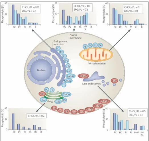
Figure 3. The lipid composition of different membranes varies throughout the cell. The lipid compositional data (shown in graphs) are expressed as a percentage of the total phospholipid (PL) in mammals (blue) and yeast (light blue). As a measure of sterol content, the molar ratio of cholesterol (CHOL; in mammals) and ergosterol (ERG; in yeast) to phospholipid is also included. The figure shows the site of synthesis of the major phospholipids (blue) and lipids that are involved in signalling and organelle recognition pathways (red). It should be appreciated that the levels of signalling and recognition lipids are significantly below 1% of the total phospholipid, except for ceramide (Cer). The major glycerophospholipids assembled in the endoplasmic reticulum (ER) are phosphatidylcholine (PC), phosphatidylethanolamine (PE), phosphatidylinositol (PI), phosphatidylserine (PS) and phosphatidic acid (PA). In addition, the ER synthesizes Cer, galactosylceramide (GalCer), cholesterol and ergosterol. Both the ER and lipid droplets participate in steryl ester and triacylglycerol (TG) synthesis. The Golgi lumen is the site of synthesis of sphingomyelin (SM), complex glycosphingolipids (GSLs) and yeast inositol sphingolipid (ISL) synthesis. PC is also synthesized in the Golgi, and may be coupled to protein secretion at the level of its diacylglycerol (DAG) precursor. Approximately 45% of the phospholipid in mitochondria (mostly PE, PA and cardiolipin (CL)) is autonomously synthesized by the organelle. BMP (bis(monoacylglycero)phosphate) is a major phospholipid in the inner membranes of late endosomes. PG, phosphatidylglycerol; PI(3,5)P2, phosphatidylinositol-(3,5)-bisphosphate; PI(4,5)P2, phosphatidylinositol-(4,5)-bisphosphate; PI(3,4,5)P3, phosphatidylinositol-(3,4,5)-trisphosphate; PI4P, phosphatidylinositol-4-phosphate; R, remaining lipids; S1P, sphingosine-1-phosphate; Sph, sphingosine. (van Meer G et al., 2008).
lysosomes. Electrostatic binding will occur in a manner dependent on both the charge of the membrane and that of the ligand, such that the most-negative membrane (i.e., the plasmalemma) will overwhelmingly accumulate the most cationic proteins, whereas less-positive proteins associate with the plasma membrane and, to a substantial degree, also with membranes of intermediate charge. Because the interaction is dynamic, changes in charge can redirect proteins from one target membrane to another. Thus, diminution of the plasmalemmal charge caused by phospholipid redistribution or metabolism, or phosphorylation of proteins like K-Ras can relocalize them to endocytic membranes, where they could catalyze a different set of reactions. Clearly, interaction with the surface charge of membranes is but one of the determinants of protein targeting, and other types of interactions must not be neglected.
To ensure the numerous functions we have told before can work properly, cells have to preserve their membranes fluidity, trying to respond to the variability of the external envinronment. The “homeoviscous adaptation” is the ability of cells and organisms to regulate the fluidity of their cell membranes by altering lipid composition. The maintenance of proper cell membrane fluidity is of critical importance for the function and integrity of the cell, essential for the mobility and function of embedded proteins and lipids, diffusion of proteins and other molecules laterally across the membrane for signaling reactions, and proper separation of membranes during cell division. A fundamental biophysical determinant of membrane fluidity is the balance between saturated and unsaturated fatty acids. Regulating membrane fluidity is especially important in poikilothermic organisms such as bacteria, fungi, protists, plants, fish and other ‘cold-blooded’ animals that cannot regulate their own body temperatures. The general trend is an increase in unsaturated fatty acids at lower growth temperatures and an increase in saturated fatty acids at higher temperatures. This compositional adaptation of membrane lipids serves to maintain the correct membrane fluidity at the new conditions. Although it is known and well defined its importance, few is already known about the mechanisms that cells exploit to follow membrane fluidity, and to respond in a fast way to changes that can happen in the outer envinronment. But only one seems to be the answer: to modify their fatty acid balance.
CHAPTER 2
MEMBRANE FATTY ACIDS
2.1 Physico-chemical characterization
Phospholipids, the main constituents of membranes, are composed by an hydrophilic polar head and two hydrophobic non-polar fatty acid tails (Figure 4), which properties will be discussed in this chapter. In chemistry, a fatty acid (FA) is a carboxylic acid often with a long unbranched aliphatic chain; it has a carboxylic acid at one end and a methyl group at the other end (Figure 5). Carbon atoms in a FA are identified by greek letters on the basis of their distance from the carboxylic acid. The carbon atom closest to the carboxylic acid is the alpha carbon, the next adjacent carbon is the beta carbon, etc. In a long-chain FA the carbon atom in the methyl group is called the omega carbon, because omega is the last letter of the greek alphabet. Carboxylic acids as short as butyric acid (4 carbon atoms) are considered to be FA, whereas FA derived from natural fats and oils may be assumed to have at least 8 carbon atoms. Most of the natural FA have an even number of carbon atoms, because their biosynthesis involves acetyl-CoA, a coenzyme carrying a two-carbon-atom group.
Figure 5. Chemical structure of a fatty acid.
2.1.1 Nomenclature
To describe precisely the structure of a FA molecule, it should be given the length of the carbon chain (number of carbons), the number of double bonds and also the exact position of these double bonds. This will define not only each FA physico-chemical properties, but also its biological reactivity and even of the lipid containing the FA studied. There are several different systems of nomenclature in use for FA; table 1 describes the most common systems. Hereinafter in this thesis, FA will be designated with their common names where possible, despite the accuracy of informations that a systematic nomenclature would give.
System Example Explanation
Trivial nomenclature
α-Linolenic acid
Common names are non-systematic historical names which are the most frequent naming system used in literature. Most common FA have trivial names in addition to their systematic
names. These names do not follow any pattern, but are concise
and generally unambiguous.
Systematic nomenclature
(9E,12E,15E)-Octadecatrien
oic acid
Systematic names (or IUPAC names) derive from the standard
IUPAC Rules for the Nomenclature of Organic Chemistry,
published in 1979, along with a recommendation published specifically for lipids in 1977. Counting begins from the carboxylic acid end. Double bonds are labelled with cis-/trans- notation or E-/Z- notation, where appropriate. This notation is generally more verbose than common nomenclature, but has the advantage of being more technically clear and descriptive.
∆x nomenclature
cis,cis,cis-∆9,∆12,∆15
In ∆x nomenclature, each double bond is indicated by ∆x, where the double bond is located on the xth carbon–carbon bond, counting from the carboxylic acid end. Each double bond is preceded by a cis- or trans- prefix, indicating the conformation of the molecule around the bond.
n−x
nomenclature
n−3 Ω-3
n−x (n minus x; also ω−x or omega-x) nomenclature does not
provide names for individual compounds, but is a shorthand way to categorize FA by their physiological properties. Holman RT proposed in 1964 a new numbering system for the unsaturation of FA, the "omega nomenclature". A double bond is located on the xth carbon–carbon bond, counting from the terminal methyl carbon (designated as n or Ω) toward the carbonyl carbon. For example, α-linolenic acid is classified as a
n−3 or omega-3 FA, and so it shares properties with other
compounds of this type. The Ω −x or omega-x notation is common in popular literature, but IUPAC has deprecated it in favor of n−x notation in technical documents.
Lipid
numbers 18:3
Lipid numbers take the form C:D, where C is the number of carbon atoms and D is the number of double bonds in the FA. This notation is ambiguous, as different FA can have the same numbers. Consequently, this notation is usually paired with either a ∆x or n−x term.
Table 1. Different systems of nomenclature in use for FA. Examples of the different nomenclature of α-Linolenic acid are highlighted. (Fahy E et al., 2005).
2.1.2 Classification
Because FA are composed by a huge number of different families, there are many classifications available. The following FA classification is first based on the type of carbon chain: either straight (or normal), or branched, or containing a carbon ring. In each category, subdivisions are created according to the functional groups substituted on the carbon chain.
A - Normal fatty acids (straight chain)
Carbon chain without substituent 1 - Saturated fatty acids
2 - Monoenoic fatty acids 3 - Polyenoic fatty acids
Methylene-interrupted Polymethylene-interrupted Conjugated
Allenic acids Cumulenic acids 4 - Acetylenic fatty acids
Carbon chain with substituent 1 - Hydroxy fatty acids 2 - Dicarboxylic acids 3 - Fatty acid carbonates 4 - Divinyl ether fatty acids 5 - Sulfur containing fatty acids 6 - Fatty acid amides
7 - Methoxy and acetoxy fatty acids 8 - Keto fatty acids
9 - Aldehydic fatty acids 10 - Halogenated fatty acids 11 - Nitrated fatty acids
12 - Arsenic containing fatty acids
B - Branched-chain fatty acids
1 - Mono or multibranched chain fatty acids 2 - Branched methoxy fatty acids
3 - Branched hydroxy fatty acids (Mycolic acids)
C - Ring containing fatty acids
1 - Cyclopropane acids 2 - Cyclobutane acids 3 - Cyclopentenyl acids 4 - Furanoid acids 5 - Cyclohexyl acids 6 - Phenylalkanoic acids 7 - Epoxy acids
8 - Cyclic fatty peroxides 9 - Lipoic acid
Despite this huge variety, few of them are actually used by the human body and by vertebrates in general. Analysis of all these groups would require a long discussion, and is beyond the scope of this thesis. More informations are available online (http://www.cyberlipid.org/fa, http://www.lipidbank.jp). In the next paragraphs, properties of normal FA with straight chains, commonly used by human cells, will be anayzed. When different ones will come into discussion, they will be characterized at the moment.
Saturated Fatty Acids (SFA)
SFA do not contain any double bond or other functional groups along the chain. The term "saturated" refers to hydrogen, in that all carbons (apart from the carboxylic acid [-COOH] group) contain as many hydrogens as possible. In other words, the omega (ω) end contains 3 hydrogens (CH3-), and each carbon within the chain contains 2 hydrogen atoms. They have commonly straight chains and even carbon number (4-30) (Figure 6). They have the general formula: CH3(CH2)nCOOH. They are named from the saturated hydrocarbon with the same number of carbon atoms, with the final -e changed to -oic. Up to 6 (or 4) carbon atoms, organic acids are considered "short-chain organic acids", they have substantial solubility in water. Biochemically, they are more closely related to carbohydrates than to fats. From 8 (or 6) to 10 (or 12) carbon atoms, fatty acids are said to have a medium chain. FA which have 14 (or 12) and more carbon atoms are considered as long-chain FA.
Figure 6. A two-dimensional representation and a space-filling model of the SFA myristic acid (14:0).
Most commonly-occurring SFA are found in table 2. With the exception of the short chain ones, all SFA have a high melting point, meaning that at normal temperature they are in solid state; this physico-chemical characteristic is due to their straight shape structure that cause the high packing that these FA are able to obtain. Normal FA exhibit appreciable solubility in water compared to the corresponding hydrocarbons due to the presence of the polar carboxyl group. The first members of the SFA series are miscible with water in all proportions at room temperature. The normal saturated FA are generally more soluble in chloroform and less soluble in acetonitrile than in any of the organic solvents investigated.
Common name Systematic name Shorthand
designation Melting point (°C)
Butyric acid Butanoic acid C4:0 -8 Caproic acid Hexanoic acid C6:0 -3 Caprylic acid Octanoic acid C8:0 16-17
Capric acid Decanoic acid C10:0 31 Lauric acid Dodecanoic acid C12:0 44-46 Myristic acid Tetradecanoic acid C14:0 58.8 Palmitic acid Hexadecanoic acid C16:0 63-64
Stearic acid Octadecanoic acid C18:0 69.9 Arachidic acid Eicosanoic acid C20:0 75.5 Behenic acid Docosanoic acid C22:0 74-78 Lignoceric acid Tetracosanoic acid C24:0 87.5-88.0
Table 2. The most commonly-occurring SFA.
Mono-Unsaturated Fatty Acids (MUFA)
Monounsaturated fats are FA that have a single double bond in the FA chain and all the other carbon atoms in the chain are single-bonded. They have the general structure: CH3(CH2)xCH=CH(CH2)yCOOH. The most frequently they have an even number of carbon atoms and the unique double bond may be in a number of different positions. FA fluidity increases with the increasing number of double bonds, because the double bond in the cis configuration insert a bend in the straight chain. This bend leads to a total spatial width of 0.72 nm for a cis-MUFA that narrows to 0.32 nm in saturated structures. The consequence in a three dimensional membrane is a higher molecular disorder due to the inhability of unsaturated FA to pack as SFA do. Therefore, MUFA have a higher melting temperature than PUFA but lower than SFA. MUFA are liquids at room temperature and semisolid or solid when refrigerated. Over a hundred naturally occurring monoene FA have been identified; common MUFA are very few though, being palmitoleic acid (16:1,cis-∆7), cis-vaccenic acid (18:1,cis-∆7) and oleic acid (18:1,cis-∆9). Palmitoleic acid has 16
carbon atoms with the first double bond occurring 7 carbon atoms away from the methyl group (and 9 carbons from the carboxyl end). It can be lengthened to the 18-carbon cis-vaccenic acid. Oleic acid has 18 carbon atoms with the first double bond occurring 9 carbon atoms away from the methyl group (Figure 7).
Figure 7. A two-dimensional representation and a space-filling model of the MUFA oleic acid (18:1).
Common name Systematic name Shorthand
designation Melting point (°C)
Palmitoleic acid 9-hexadecenoic 16:1 0.5
Vaccenic acid 11-octadecenoic 18:1 14.5-15.5
Oleic acid 9-octadecenoic 18:1 16.2
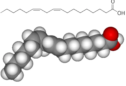
Poli-Unsaturated Fatty Acids (PUFA)
These FA have two or more cis double bonds which are the most frequently separated from each other by a single methylene group (methylene-interrupted polyenes), with a typical structure of the type -C-C=C-C-C=C-. Linoleic acid is a typical member of this group (Figure 8). Some other PUFA undergo a migration of one of their double bonds which are not again
Figure 8. A two-dimensional representation and a space-filling model of the poliunsaturated fatty acid linoleic acid (18:2).
methylene-interrupted and are known as conjugated FA (-C-C=C-C=C-C-). Some unusual FA have not the regular structure with a methylene group between two double bonds but are polymethylene-interrupted polyenes (known also as non-methylene-polymethylene-interrupted FA, -C=C-C-C-C-C=C-). They are found in certain classes of bacteria, plants, marine invertebrates and insects. Rare FA have allenic (-C=C=C-) or cumulenic (-HC=C=C=CH-) double bonds; they are found in some higher plants. The greater the degree of unsaturation in a FA, the more vulnerable it is to lipid peroxidation. Antioxidants can protect unsaturated fat from lipid peroxidation. The differences in geometry between the various types of unsaturated FA, as well as between saturated and unsaturated fatty acids, play an important role in biological processes, and in the construction of biological structures such as cell membranes. Unsaturated fats have a more enlarged shape than saturated fats, because the double bond structures increase the bend of the acyl chain; moreover, the lack of the extra hydrogen atoms on the molecule's surface typically reduces the strength of the compound's
intermolecular forces. All this therefore causes the melting point of the compound to be significantly lower (Table 4). The most important FA can be grouped into three series with a common structural feature: CH3(CH2)xCH=R. x=4 for the n-6 series, x=1 for the n-3 series and x=7
for the n-9 series.
Common name Systematic name designation Shorthand Family Melting point (°C)
Linoleic acid
9,12-octadecadienoic 18:2 n-6 -5
γ-linolenic acid octadecatrienoic 6,9,12- 18:3 n -6 -11.3 to -11 Dihomo-γ-linolenic acid 8,11,14-eicosatrienoic 20:3 n -6
/
Arachidonic acid 5,8,11,14-eicosatetraenoic 20:4 n -6 -50α-linolenic acid octadecatrienoic 9,12,15- 18:3 n -6 -11 Stearidonic acid 6,9,12,15-octadecatetraenoic 18:4 n -6 -57 EPA eicosapentaenoic 5,8,11,14,17- 20:5 n -6 -54 DPA docosapentaenoic 7,10,13,16,19- 22:5 n -6
/
DHA 4,7,10,13,16,19-docosahexaenoic 22:6 n -6 -44 Mead acid eicosatrienoic 5,8,11- 20:3 n -6/
2.1.3 The geometry of the double bond: Cis vs Trans
Carbon atoms are tetravalent, forming four covalent bonds with other atoms, while hydrogen atoms bond with only one other atom. In SFA, each carbon atom is connected to its two neighbour carbon atoms as well as two hydrogen atoms. In unsaturated FA the carbon atoms that are missing a hydrogen atom are joined by double bonds rather than single bonds, so that each carbon atom participates in four bonds. The same molecule, containing the same number of atoms, with a double bond in the same location, can be either a trans or a cis FA, depending on the conformation of the double bond. For example, oleic acid and elaidic acid (Figure 9) are both unsaturated FA with the chemical formula C9H17C9H17O2 (Taylor and Francis, 2007). They both have a double bond located
Figure 9. A space-filling model and a two-dimensional representation comparison between the
midway along the carbon chain. It is the conformation of this bond that sets them apart. The conformation has implications for the physical-chemical properties of the molecule. A cis configuration means that adjacent hydrogen atoms are on the same side of the double bond. The rigidity of the double bond freezes its conformation and, in the case of the cis isomer, causes the chain to bend and restricts the conformational freedom of the fatty acid. When a chain has many cis bonds, it becomes quite curved in its most accessible conformations. For example, OA, with one double bond, has a kink in it, whereas LA, with two double bonds, has a more pronounced bend. ALA, with three double bonds, favors a hooked shape. The effect of this is that, in restricted environments, such as when FA are part of a phospholipid in a lipid bilayer, cis bonds limit the ability of FA to be closely packed, and therefore could affect the melting temperature of the membrane. A trans configuration, by contrast, means that the next two hydrogen atoms are bound to opposite sides of the double bond. As a result, they do not cause the chain to bend much, and their shape is similar to straight SFA (Figure 8). The trans FA elaidic acid has different chemical and physical properties owing to the slightly different bond configuration. Notably, it has a much higher melting point, 45 °C rather than OA's 13.4 °C (Table 5), due to the ability of the trans molecules to pack more tightly, forming a solid that is more difficult to break apart (Taylor and Francis, 2007). This notably means that it is a solid at human body temperatures.
Common name Systematic name Shorthand
designation Melting point (°C)
Oleic acid cis-9-octadecenoic 18:1 16.2
Elaidic acid trans-9-octadecenoic tr18:1 43.7 Vaccenic acid cis-11-octadecenoic 18:1 14.5-15.5 C
Trans-vaccenic acid
trans-11-octadecenoic acid tr18:1 44
Table 5. The comparison between the most commonly-occurring trans-fatty acids and their respective cis-configured ones.
A type of trans fat occurs naturally in the milk and body fat of ruminants (such as cattle and sheep) at a level of 2–5% of total fat. Natural trans fats, which include conjugated LA (CLA) and vaccenic acid, originate in the rumen of these animals. However, CLA is also a cis fat. Animal-based fats were once the only trans fats consumed, but by far the largest amount of trans fat consumed today
is created by the processed food industry as a side-effect of partially hydrogenating unsaturated plant fats (generally vegetable oils). These partially hydrogenated fats have displaced natural solid fats and liquid oils in many areas, notably in the fast food, snack food, fried food and baked good industries. In food production, the goal is not to simply change the configuration of double bonds while maintaining the same ratios of hydrogen to carbon. Instead, the goal is to decrease the number of double bonds and increase the amount of hydrogen in the FA. This changes the consistency of the FA and makes it less prone to rancidity, in which free radicals attack double bonds. Production of trans FA is therefore a side-effect of partial hydrogenation. Catalytic partial hydrogenation necessarily produces trans-fats, because of the reaction mechanism. In the first reaction step, one hydrogen is added, with the other coordinatively unsaturated carbon being attached to the catalyst. The second step is the addition of hydrogen to the remaining carbon, producing a saturated fatty acid. The first step is reversible, such that the hydrogen is readsorbed on the catalyst and the double bond is re-formed. Unfortunately, the intermediate with only one hydrogen added contains no double bond, and can freely rotate. Thus, the double bond can re-form as either cis and trans, of which trans is favored, regardless the starting material. Complete hydrogenation also hydrogenates any produced trans fats to give saturated fats. Most trans fats consumed today are created industrially in partial hydrogenation of plant oils — a process developed in the early 1900s and first commercialized as Crisco in 1911. Commercial hydrogenation is typically partial in order to obtain a malleable mixture of fats that is solid at room temperature, but melts upon baking or consumption. In most naturally occurring unsaturated fatty acids, the hydrogen atoms are in cis configuration. However, partial hydrogenation reconfigures most of the double bonds that do not become chemically saturated, twisting them to the trans configuration, that has the lower energy form, and is favored when catalytically equilibriated as a side reaction in hydrogenation.
An increasing number of studies have explored the presence of trans FA residues in living systems. This is a very lively field of interdisciplinary research spanning from chemistry to microbiology, pharmacology, biology, and, of course, medicine. The configuration of isolated double bonds in naturally occurring lipids of eukaryotes is cis. In modified fats, the structures of trans FA residues consist of geometrical and positional isomers having un-shifted and shifted double bonds, respectively, compared to natural cis compounds. Some trans geometrical isomers found in living organisms can only arise via an endogenous transformation of the naturally occurring cis structures and are correlated with radical stress produced during physiological and pathological processes. (Ferreri C et al., 2002;Zghibeh CM et al., 2004; Ferreri C et al., 2005; Kermorvant-Duchemin E et
al., 2005; Zambonin L et al., 2006). Several free radicals, including the biologically relevant thiyl
and Ferreri C, 2005; Ferreri C and Chatgilialoglu C, 2005). Figure 10 shows the reaction mechanism, involving reversible addition of a thiyl radical to the double bond to form a radical-adduct. The reconstitution of the double bond is obtained by β-elimination of RS•, and the result favours trans geometry, the most thermodynamically stable configuration, by 0.6-1 Kcal/mol. It should be noted that (i) RS• acts as a catalyst for cis–trans isomerization and (ii) positional isomers cannot be formed because the mechanism does not allow a double-bond shift. The cis–trans isomerization by RS• is an efficient process and detailed kinetic data for the reactions are available in the case of methyl oleate with HOCH2CH2S• radical (Chatgilialoglu C et al., 2002, 2005).
Figure 10. Reaction mechanism for the cis–trans isomerization catalysed by thyl radical. (Ferreri C
et al., 2005).
Recently, using a biomimetic model of vesicle suspension which mimics the aqueous and membrane compartments of a cell, Lykakis IN et al. (2007) demonstrated the potential of sulfhydryl radicals (HS•/S•-) derived from H2S to access the hydrophobic fatty acid chains and attack the
double bonds. The phospholipids produced in this way contained a high proportion of trans fatty acid residues. This model offers some insight into the chemical basis of the biological activity of H2S, that will be discussed later on in this thesis. This chemistry is important in view of the
intriguing role of the sulfhydryl radical induced cis-trans conversion of lipids, either as a damaging or a signaling pathway. On the other hand, the available kinetic data for the cis–trans isomerization by NO2• suggest that this radical cannot be very efficient as an isomerizing species, and in a
biological environment this reaction should not play a role (Chatgilialoglu C and Ferreri C, 2005). In the n-6 series of PUFA, cis–trans isomerization of methyl linoleate (Ferreri C et al., 2001), γ-linolenate (Ferreri C et al., 2001) and arachidonate (Ferreri C et al., 2002) catalysed by RS• has been studied in some detail. Each isolated double bond in PUFA behaves independently. Moreover, isomerization is a stepwise process with the formation of mono-trans isomers, followed by di-trans isomers and so on, the isomeric composition being regulated by the relative thermodynamic stability. It was also possible to demonstrate that the double bonds closest to the membrane polar region are the most reactive towards attack by diffusing thiyl radicals (Ferreri C et al., 2001). For
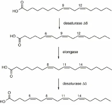
example, AA residues in vesicles were more reactive than OA and LA; two positions, the double bonds at positions 5 and 8 out of the four present in this FA, were transformed preferentially. From studies to date, AA residues in membrane phospholipids emerged as very important pointers to help distinguish endogenous trans isomers, formed by radical processes, from the exogenous trans isomers derived from dietary contributions (Ferreri C et al., 2002; 2004). The interplay between exogenous and endogenous pathways for isomerization of arachidonic double bonds is shown in Figure 11, where the biosynthesis from the precursor LA is detailed: the two double bonds of positions 11 and 14 are provided by dietary LA (which can be cis or trans, depending on the food), whereas the other two double bonds (positions 5 and 8) are formed by desaturase enzymes, which produce selectively cis unsaturation. In vivo, double bonds at positions 5 and 8 of AA, stored in membrane phospholipids, can only have a cis configuration, unless these positions are involved by an isomerization process occurring in membranes by diffusible thiyl radicals.
Figure 11. Enzymatic FA transformations from linoleic acid to arachidonic acid. (Ferreri C et al., 2002).