INDEX
• ABSTRACT……… 1
• INTRODUCTION……….. 2
• RESULTS AND DISCUSSION………...………. 12
All-trans retinoic acid binds and inhibits 2-oxoglutarate carrier……….. 12
Adrenal glands, as steroidogenic tissue, are affected by retinoylation reaction……… 18
The activity of the adenine nucleotide translocator is affected by a component of the retinoylation buffer……….…... 24
Combined low doses of PPARγ and RXR ligands trigger an intrinsic apoptotic pathway in human breast cancer cells……..……… 33
• METHODS……… 41
• REFERENCES……….. 47
1
ABSTRACT
Vitamin A (Retinol) plays a central role in many essential biological processes such as vision, immunity, reproduction, growth, development, control of cellular proliferation and differentiation. The main active forms of retinol, not primary involved in vision, are
all-trans retinoic acid and 9-cis retinoic acid, both able to act at nuclear level by binding their
receptors RAR and RXR and modulating many physiological processes. However, the nuclear action of vitamin A derivatives is not the only mechanism of retinoic acid (RA) acting on cells. RA is able to modify covalently proteins via a post-translational modification, named retinoylation that has been shown to occur at physiological concentration on pre-existing proteins and localized mainly in the mitochondrial compartment. The present study has been focused on the non genomic action of RA on steroidogenic tissues, testes and adrenal glands, giving further details on the ability of RA to influence protein activity and therefore cell physiology. In particular RA effects on mitochondria from the adrenal glands and the 2-oxoglutarate carrier protein from testes and TM-3 Leydig cell line were studied, providing new data on the peculiarity of steroidogenic tissues to incorporate RA at dietary levels and demonstrating how the shuttling of reducing equivalent across the mitochondrial membrane is influenced by RA treatment. Looking for the biochemical mechanism of RA action on the Adenine Nucleotide Translocator, that exchanges ATP for ADP between mitochondria and cytosol, for the first time, it was possible to demonstrate how the activity of this carrier protein is positively modulated by the Coenzyme A, a fundamental component of the retynoilating buffer. At pharmacological levels, retinoids are also active compounds in the treatment of cancer due to the capability to promote cell differentiation and their pro-apoptotic activity. In this latter concern, the mechanisms of nutriceutical concentration of 9-cis RA, together with nanomolar concentration of the selective PPARγ ligand, rosiglitazone, to promote apoptosis in breast cancer cell lines, have been investigated. The data lay the basis for a potential use of the combined therapy with low doses of both BRL and 9-cis RA as novel therapeutic tool particularly for breast cancer patients who develop resistance to antiestrogen therapy.
INTRODUCTION
The retinol (Vitamin A) belongs to the “retinoids” family including both the compounds possessing one of the biological activities of the retinol (ROH) and the many synthetic analogues related structurally to the retinol, with or without biological activity. Provitamin A is the dietary source of retinol and is supplied as carotenoids (mainly β-carotene) in vegetables and preformed retinyl-esters (long chain fatty acids esters of retinol: palmitate, stearate, oleate, linoleate) in animal meat (Blomhoff 1994). Retinol plays a central role in many essential biological processes such as vision, immunity, reproduction, growth, development, control of cellular proliferation and differentiation. The main active forms of retinol are retinoic acids, except for vision and reproduction where retinal and retinol play important roles. Two vehicles are described for mammalian blood transport of retinoids. First the retinyl esters and carotenoids can be incorporated in intact or remnant chylomocrons or very low density lipoproteins (Debier and Larondelle, 2005). Second, the main form of retinol blood transport is the association with a specific binding protein (RBP), which is itself complexed with transthyretin (ratio 1 mol/1 mol). The constitution of this ternary complex prevents the glomerular filtration of the small RBP-retinol form (21KDa) and increases the affinity of RBP for retinol (Bellovino et al. 2003). A binding to other plasma proteins, such as albumin or lipocalins, is also described for retinol. Albumin could serve as transport for RA, which circulates in very small levels in the blood. The transfer of retinol to target cells involves a specific membrane-bound receptors (Sivaprasadarao et al., 1998). It has been recently demonstrated that cellular retinol penetration is based on the interaction between the RBP-retinol complex and a membrane receptor STRA6. STRA6 is a multitransmembrane domain protein not homologous to any other proteins with known function. It functions as the high-affinity receptor for plasma retinol binding protein (RBP) and mediates cellular uptake of vitamin A from the vitamin A-RBP complex. It is widely expressed in embryonic development and in adult organ
systems. Consistent with the diverse roles of vitamin A and the wide tissue expression pattern of STRA6, mutations in STRA6 are associated with severe pathological phenotypes in humans (Kawaguchi et al., 2007). The uptake of remnant chilomicrons and very low density lipoproteins containing retinyl esters and carotenoids is realized by target tissues using, respectively, the lipoprotein lipase and low density lipoproteins receptor pathways. Bound to albumin, RA can be transferred into the tissues by passive diffusion, with an efficiency of transfer, which is cell type and tissue specific. To be biologically active retinol must be first oxidized to retinaldehyde and then to RA. A large number of enzymes catalyzes the reversible oxidation of retinol to retinaldehyde: the alcohol dehydrogenas (ADH), the retinol dehydrogenases (RDH 1-3) of the microsomal fraction, cis-retinol:androgen dehydrogenase 1 and 2 (CRAD), retSDRs1–4, 9:11-cis-retinol dehydrogenase, and 17β-hydroxysteroid dehydrogenase (17β-HSD) types 6 and 9, and some members of the cytochrome P450 family (Napoli, 2001). Several enzymes are able to catalyze irreversibely the oxidation of retinaldehyde to RA: the retinaldehyde dehydrogenases (RALDH 1-4) are also member of the cytochrome P450 family (Liden and Eriksson, 2006). Specific isomerization reactions are also likely to occur within the cells, since there are at least two RA stereoisomers in vivo (all-trans and 9-cis RA) exhibiting distinct biochemical activities. The catabolism of all-trans and 9-cis RA is also an important mechanism for controlling RA levels in cells and tissues and is carried out by three specific members of cytochrome P450s, CYP26A1, B1, and C1. RA is catabolized to products such as 4-oxo-RA, 4-hidroxy-RA, 18-hydroxy-RA, and 5,18-epoxy-RA, which are finally excreted. These compounds can also undergo glucuronidation (Marill et a.l 2003). The complex of microsomal UDP-glucuronosyl transferases (UGT; EC 2.4.1.17) of rat liver catalyzes the formation of retinoyl beta-glucuronide (RAG) from retinoic acid (RA) and retinyl beta-beta-glucuronide (ROG) from retinol (ROL) in the presence of UDP-glucuronic acid (UDPGA). Rates of glucuronidation of retinoids clearly depend both on their isomeric states and on their chemical structures. Different UGT enzymes might well act on different geometric
isomers of RA (Genchi et al. 1996). Numerous reports have indicated that the biological activity of trans retinoyl-glucuronide (RAG) is similar to that of all-trans-retinoic acid (RA), but without the toxic side effects of RA. An alternative metabolic pathway was present for intracellular retinol: the formation and storage of retinyl esters. Indeed retinol may be esterified by two enzymes: the first is lecithin:retinol acyltransferase (LRAT), which catalyzes the transfer of a fatty acid from the snK1 position of phosphatidylcholine (PC) to retinol. The products of this reaction are 1-lyso-phosphatidylcholine (lyso-PC) and a retinyl ester, usually retinyl palmitate. The mRNA for LRAT has been cloned and shown to encode an integral-membrane protein of 230 residues. LRAT is present in multiple tissues including liver, small intestine, testis, mammary gland, and retinal pigment epithelium. Although LRAT preferentially esterifies retinol (atROL) to yield all-trans-retinyl esters (atREs), it also utilizes 11-cis-retinol (11cROL) as a substrate to form 11-cis-retinyl esters (11cREs). The second retinyl-ester synthase is acyl CoA:retinol acyltransferase (ARAT), which uses a fatty-acyl coenzyme A, such as palmitoyl coenzyme A (palm CoA), as an acyl donor. ARAT has not been purified or cloned. Palm CoA-dependent retinyl-ester synthase activity has been observed in multiple tissues including liver, small intestine, mammary gland, epidermis, testis, and cone-dominant retinas. Palm CoA-dependent synthesis of retinyl esters has also been observed in retinal pigment epithelium. However, it has been suggested that in this tissue, the palm CoA stimulation effect is due to regeneration of PC from lyso-PC, and is thus mediated by LRAT. (Catherine et al., 2006 and references therein). These esters are then stored in cytosolic lipid droplets. The mobilization of these retinyl esters and the release of retinol esters are realized by a etinyl ester hydrolase. Since retinol, retinaldehyde and RA are lipids they lack appreciable water solubility and consequentely must be bound to protein within cells. Several intra-cellular binding proteins for retinol, retinaldehyde and RA have been identified and extensively characterized. They include cellular retinol-binding proteins (CRBP I and II) and cellular retinoic acid-binding protein (CRABP I and II). The CRBPI is a key protein
to regulate the metabolism of retinol by orientating to storage, export of retinol or conversion into RA (Ghyselinck et al., 1999). Both the CRABPs bind RA controlling the intracellular levels of retinoids, acting as cofactors for RA-metabolizing enzymes, and/or participating in the cytoplasmic-nuclear transport of RA (Napoli, 1999; Ong, 1994). The 15.5 kDa protein, cellular RA binding protein has been shown to specifically bind RA with nanomolar affinities. There are several reports that suggest CRABP may protect the cell from the genomic action of RA by sequestering the compound and accelerating its catabolism. RA bound to CRABP has been demonstrated to be a substrate for oxidation by members of the P450 family. A potential key to attenuation of the RA signal may be found in the specific location of CRABP within the cell. Immunohistochemical experiments performed on tissue sections has revealed that this protein is restricted to the cytoplasmic compartment of cells, even though its size should enable free entry into the nucleus (In contrast, CRBP, a related protein of the same family, was shown to be present in both the cytoplasm and the nucleus, as expected for a protein of this size). This nuclear restriction might create a barrier that prevents RA from reaching the RARs in the nucleus. However, the mechanism(s) by which this restriction is accomplished has not been established. Moreover, Ruff and Ong demonstrated that CRABP is localized to the mitochondria of bovine adrenal gland. This association with the mitochondria effectively restricts it to the cytosolic compartment of the cell and suggests a role for mitochondria in retinoid metabolism (Ruff and Ong, 2000, and references therein). Although RA functions mainly by binding to nuclear receptor proteins, this is not the only mechanism of retinoids action on cells. Retinoic acid, is capable to modify covalently pre-existing proteins via a post-translational modification (PTM) termed retinoylation (acylation by RA of protein). Single PTMs are capable of regulating protein function, either through creating new protein binding sites, by abrogating protein-protein interactions, or through allosteric effects. However, many proteins are multiply modified, and a significant increase in information content would be obtained if PTMs acted combinatorially. There are several established principles
through which PTMs mediate crosstalk. In this context, crosstalk can be either positive or negative in nature. Positive crosstalk is defined as a situation in which one PTM serves as a signal for the addition or removal of a second PTM or for recognition by a binding protein that carries out a second modification. In the case of negative crosstalk, there can be direct competition for modification of a single residue in a protein, or indirect effects, wherein one modification masks the recognition site for a second PTM. Histone tails provide one of the most remarkable examples of PTM density and variety, with Lys acetylation, mono-, di-, or trimethylation, biotinylation, ubiquitinylation, NEDDylation, SUMOylation; Arg methylation; Ser/Thr/Tyr phosphorylation; and Glu ADP ribosylation all occurring within 50–100 residues on the N-terminal and C-terminal tails of H2A, H2B, H3, and H4 (Bhaumik et al., 2007; Latham and Dent, 2007). These ‘‘marks’’ are proposed to represent a code that is read by a series of transcriptional regulators and chromatin modifiers that affect local chromatin structure, leading to activation or repression of adjacent transcription units. Although there is little direct information regarding which PTMs occur simultaneously on a single histone molecule, functional analysis of histone tail PTMs has revealed many examples of crosstalk between different PTMs. Nuclear coreceptors are another family of proteins in which PTM crosstalk is beginning to be uncovered (Lonard and O’Malley, 2007). About RA Acylation, via retinoylation RA is incorporated into proteins of cells in culture (Breitman and Takahashi, 1996; Takahashi and Breitman, 1989, 1990, 1994; Tournier et al., 1996) and into proteins of rat tissues, both in vivo (Myhre et al., 1996) and in vitro (Genchi and Olson, 2001; Myhre et al., 1998; Renstrom and DeLuca,1989). The retinoylation reaction involves the intermediate formation of retinoyl-CoA (Wada et al., 2001) and subsequent transfer and covalent binding of the retinoyl moiety to protein(s) (Renstrom and DeLuca, 1989). The covalent linkage between RA and protein(s) is a thioester bond (Genchi and Olson, 2001; Myhre et al., 1996). Retinoylated proteins that have been identified so far include cyclic AMP-binding proteins, vimentin, the cytokeratins, some nuclear proteins and a component of the mitochondrial carrier family (Breitman
and Takahashi, 1996; Takahashi and Breitman, 1989, 1990, 1994; Tournier et al., 1996, Cione et al. 2009). Several types of lipid modifications have received considerable attention over the last few years. It has been demonstrated that palmitic, myristic, acetic and phosphoric acids, as well as isoprens farnesol and geranilgeranol, bind to a number of proteins (Schultz et al., 1988; Towler et al., 1988). Retinoylation is one of these covalent modification reactions occurring on proteins. Biochemical similarities exist between retinoylation and acylation (i.e., palmitoylation and myristoylation). In these processes, RA, MA, and PA covalently bind to preformed protein via a thioester bond after the intermediate formation of acyl-CoA and retinoyl-CoA (Renstrom and DeLuca, 1989; Wada et al., 2001). Such covalent modification reactions regulate the interactions of the proteins with cellular membranes, as well as the interactions with other proteins. Physiologically, retinoids, bound to proteins or lipoproteins in the extracellular fluids, are taken up by cell surface receptors, are transferred into the cytoplasm and subsequently bound to intracellular proteins (Blomhoff et al., 1991). Therefore, retinoids do not normally
equilibrate in phospholipids of cellular membranes. Retinoylation appear to be tissue specific: steroidogenic tissues (adrenal glands and
testes) resulted to be the most active tissues in incorporating RA with significative differences in tha necessity of ATP among the two tissues (Cione and Genchi, 2004; Pingitore et al. 2009). In general it is known that RA is essential for the normal growth of testes (Chaudhary et al., 1989). The steroid biosynthesis is very active into testes mitochondria and moreover is stimulated also by RA (Tucci et al, 2008); therefore this so active testes mitochondrial process of retinoylation should be bound to steroidogenesis. The retinoylation reaction of rat testes mitochondrial proteins
occurred in the presence of ATP, CoASH, and Mg++. Either the omission of ATP
and/or CoASH in the incubation buffer or the omission of Mg++, or its substitution
with Ca++ reduced the radioactivity bound to the mitochondrial proteins in
considerable manner. Moreover the retinoylation reaction exhibited substrate specificity. In fact the reaction occurred only in the presence of ATP, ADP (50% of
the control), and GTP (30% of the control). In testes derived cell lines, RA was able to bind pre-existing protein and RA supplementation was able to stimulate testosterone biosynthesis in these cells in the same doses where the retinoylation reaction was found to be enhanced suggesting a putative role in testicular steroidogenesis. RA in mouse testes is present constitutively at a concentration of 7–
14 pmol/g or approximatively 7–14 nM (Deltour et al. 1997; Kane et al. 2005). At
nuclear level it is well known that RA functions by binding to ligand-inducible transcription factors (nuclear receptor proteins belonging to the steroid/thyroid hormone receptor superfamily) that activate or repress the transcription of downstream target genes (Chambon, 1996; Soprano and Soprano, 2003). Six nuclear receptors, termed RARα, RARβ, RARγ, RXRα, RXRβ and RXRγ, encoded by different genes, have been demonstrated to mediate the actions of RA. The natural metabolite all-trans RA (atRA) and 9-cis RA are high affinity ligands for RARs, whereas 9-cis RA, phytanic acid, docosahexanoic acid, and unsaturated fatty acids, have been suggested to bind RXRs. These proteins, as heterodimers (RAR/RXR) or homodimers (RXR/RXR), function to regulate transcription by binding to DNA sequences located within the promoter of target genes called retinoic acid response elements (RARE) or retinoid X response elements (RXRE), respectively. RAREs consist of direct repeats of the consensus half-site sequence AGGTCA separated most commonly by five nucleotides (DR-5) while RXREs are typically direct repeats of AGGTCA with one nucleotide spacing (DR-1). The RAR/RXR heterodimer binds to the RARE with RXR occupying the 5’ upstream half-site and RAR occupying the 3’ downstream half-site. atRA and 13-cis RA bind to and transactivate only RXR/RAR complexes, while 9-cis RA interacts with both RXR/RAR heterodimers and RXR/RXR homodimers. RXR/RAR dimers are believed to be nonpermissive complexes, in which RXRs act as silent partner. In other words, when the cognate ligand is bound to the RAR moiety, the RXR counterparts loses the ability to bind its corresponding ligand (Mangelsdorf and Evans, 1995; Mangelsdorf et al. 1993). In basal conditions, the RXR/RAR heterodimer is bound to the cognate DNA sequence
(RARE) and interacts with a multiprotein complex known as the corepressor (Wei, 2004; Weston et al. 2003). The corepressor (RIP140 receptor interacting protein 140) contains protein endowed with the histone deacetylase (HDAC) and DNA-methylating activity that concur to keep the surrounding chromatin structure in a “closed” state, effectively suppressing the transcriptional activity of RNA polymerase II. On ligand binding the corepressor is released from the RXR/RAR dimer and substituted by the “coactivator”, which consists of a multiprotein complex with histone acetylase and demethylase activity (Westin et al. , 2000; Xu et al., 1999a) opening the chromatine structure and favouring transcription. The activity of the RXR/RAR dimer is also controlled by a number of accessory signals in which phosphorylation events stand out (Gianni et al. 2002, 2006) A further layer of control is represented by the rate of proteolitic degradation of the RXR/RAR dimer and the various components of the corepressor and coactivator complexes (Gianni et al. 2006). Little is known about the RXR/RXR pathway which is far less studied than the RXR/RAR counterpart. Retinoids are promising agents in the chemoprevention and treatment of the neoplastic disease. Fenretinide, a synthetic retinoid, has shown efficacy in the secondary chemoprevention of breast cancer (Veronesi et al. 1999). Indeed, this class of agents finds clinical application or is proposed also in the treatment of various neoplastic diseases, among which, acute promyelocytic leukemia (APL) is the most prominent example. The observation that atRA induces remission in APL through a mechanism of action that is dinstinct from cytotoxicity, is regarded as a milestone in the history of medicine (Huang et al. 1988). The retinoid is the first and only example of clinically succesfull cytodifferentiating agent. Cytodifferentiation is a particularly attractive modality of treatment and differentiating agents promise to be less toxic and more specific than conventional chemotherapy. Currently, the promise of differentiation therapy is only partially met and a more general use of atRA and other retinoids as differentiating agents in oncology is hampered by a number of problems including natural and acquired resistance as well as local and systemic toxicity. Therefore, retinoids are
often used as part of a combined therapy. atRA and derivatives modulate the activity of numerous genes and intracellular pathways. On the other hand, the activity of nuclear RARs is controlled by various signals, including different types of kinase cascades. Often the cross talk between atRA-dependent and other intracellular pathways modulates the cytodifferentiating activity of the retinoid. The knowledge on the molecular mechanism underlying this cross talk has increased tremendously over the course of the last few years. In cell cultures, the cytodifferentiating activity of retinoids is almost invariably accompanied by growth inhibition and the two processes are difficult to dissociate (Gianni et al. 2000). On the other hand, retinoid-dependent cytodifferentiation is not necessarily associated with cell death or apoptosis. Indeed, classical retinoids are relatively weak apoptotic agents and, in some cases, they even exert a prosurvival action (Lomo et al. 1998). These aspects of retinoids pharmacology need to be considered when discussing the use of such agents in oncology. An antiproliferative effect superimposed to cytodifferentiation is highly desirable, whereas an antiapoptotic action should be avoided. A generalized use of retinoids in oncology is hampered by a number of unresolved problems including natural and induced resistance as well as toxicity issues are of particular evidence, given the fact that differentiation therapy with retinoids requires prolonged administration of the agents. Chronic exposure to retinoids is accompanied by serious effects at the level of central nervous and hepatic systems, as well as the well-know teratogenic problems associated with the administration of these compounds. Clearly these problems call for strategies aimed at increasing the efficacy and the therapeutic index of retinoids developing novel, more powerful and less toxic syntethic retinoids and identifing non-retinoid agents capable of potentiating the pharmacological activity of retinoids without affecting their toxicity. This second approach is based on the experimental evidence that the nuclear receptor pathway interacts with numerous other intracellular pathways, some of which are of obvious significance from a therapeutic point of view. Retinoids have been proposed in the adjuvant treatment of brest carcinoma for their ability to inhibit growth and
induce morphological or phenotypic deifferentiation of breast carcinoma cell lines (Paik et al. 2003; Yang et al., 2002) and most of the studies have focused on the antiproliferative activity of retinoids (del Rincon et al. 2003). It is generally accepted that breast cancer cells express RARα, RARγ and RXRα but also Peroxisome Proliferator-Activated Receptor gamma (PPARγ) that can regulate cell proliferation, differentiation and survival (Lemberger et. Al., 1996; Lefebvre et al. 2006). PPARγ functions as a transcription factor by heterodimerizing with RXR, after which this complex binds to specific DNA sequence elements called Peroxisome Proliferator Response Elements (PPREs). PPREs are direct repeats of the consensus sequesnce with a spacing of one nucleotide (AGGTCA-N-AGGTCA) (Palmer et al. 1995). The heterodimer PPAR/RXR activated with their ligands, can bind to PPREs in promoter regions of target genes recruiting coactivator or corepressor to this complex to modulate gene expression (DiRenzo et al. 1997; McIerney et al. 1998; Yuan et al. 1998). Previous data show that PPARγ, poorly expressed in normal breast epithelial cells (Elstenr et al., 1998), is present at higher levels in breast cancer cells (Tontonoz, P., et al. 1994) and its synthetic ligands, such as thiazolidinediones (TZDs), induce growth arrest and differentiation in breast carcinoma cells in vitro and in animal models (Mueller et al., 1998; Suh et al., 1999).
RESULTS AND DISCUSSION
All-trans retinoic acid binds and inhibits 2-oxoglutarate carrier
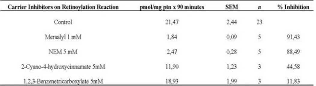
Previously it was demonstrated that mitochondria from rat testes (Genchi and Olson, 2001; Cione and Genchi, 2004) and TM-3 cells (Cione et al., 2005) were extremely active in incorporating retinoic acid. Moreover, it was highlighted how the retinoylation reaction and testosterone biosynthesis are positively correlated when Leydig cell cultures are incubated with atRA at 100 nM (Tucci et al., 2008). As the biosynthetic steps that lead to testosterone production are mainly NADH/NADPH dependent, the 2-Oxoglutarate carrier (OGC) activity from mitochondria extracted both from whole rat testes and Leydig TM-3 cells, was chosen as the experimental target for its involvement in the malate-and aspartate oxoglutarate–isocitrate shuttle to provide for the necessary exchange of reducing equivalents between the mitochondria and the cytosol. The efforts were focused on the OGC from rat testes as the retinoylation process is more efficient in this tissue and testosterone production in TM-3 positively correlates to atRA supplementation. In addition, the strong inhibitory effect of 2-cyano-4-hydroxycinnamate (an inhibitor of OGC) but not 1,2,3-benzentricarboxylate (an inhibitor of citrate carrier) on the retinoylation processes (as highlighted in Table 1) was the start point for further investigation in this work. 2-cyano-4-hydroxycinnamate shows 45% inhibition when used at a concentration of 5 Mm, whilst, at the same concentration 1,2,3-benzentricarboxylate has a very weak
effect (12% inhibition). In proteoliposomes OGC has been shown to exist as a
homodimer and to function according to a sequential antiport mechanism, catalyzing the transport of 2-oxoglutarate in electroneutral exchange for some other dicarboxylates to which malate is bound with the highest affinity. These results have been interpreted by assuming two separate and coordinated substrate translocation pathways, one in each monomer.
Table 1. Effect of Mitochondrial Carrier Inhibitors on Retinoylation Reaction.
Mitochondria from testes were incubated 90 minutes in a buffer as decribed in Materials and Methods with 3HatRA, 100 nM final concentration, at 37 °C. Then the reaction was stopped with TCA and the radioactivty detected in a liquid scintillation counter. Results are presented as Mean ± SEM of three indipendent experiments. **P< 0.01 compared to the control. 1,2,3-Benzenetricarboxylate P >0.05
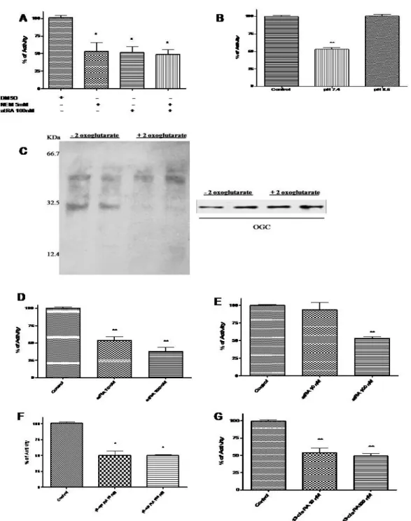
Our results showed that transport activity of OGC from rat testes mitochondria was strongly influenced by the sulphydryl group reagent N-ethil-maleimmide (NEM) and atRA. NEM, at 5 mM, markedly reduced the OGC activity by 47%. A similar inhibition of 51%was highlighted for 100 nM atRA, and when the two compounds are co-incubated the activity was reduced to 49%, equal to RA alone as shown in Fig. 1A. In humans, cows and rats there is only one gene encoding OCG: according to the amino acid sequence the bovine OGC protein contains three cysteines: Cys184 located in TMS IV and Cys221 and Cys224 in TMS V. Mercurials and maleimides interact only with Cys184 of the purified and reconstituted OGC, as Cys221 and Cys224 are linked by a disulphide bridge (Palmieri, 2004). Therefore we propose that atRA, via retinoylation reaction, could bind the OGC on the same residue (Cys184), as the inhibitory effect of atRA is still the same when NEM is present concomitantly as shown in Fig. 1A, leading us to hypothesize the existence of a putative amino acid sequence related to the atRA binding site in OGC. For what concerns OGC and its involvement in testosterone biosynthesis, the first enzymatic step is to convert cholesterol in pregnenolone: the reaction occurs in the mitochondrial matrix and
requires reducing equivalents mainly as NADPH; conversely the role of the OGC is to carry out reducing equivalents from the mitochondria to the cytoplasm. Previously it was demonstrated that there is a positive correlation between retinoylation reaction and testosterone biosynthesis (Tucci et al., 2008): the action of RA to slow down the OGC transport activity is in agreement with the testosterone synthetic process as reducing equivalents are more necessary to convert cholesterol in the matrix rather than in the cytoplasm (Stocco, 2001). At the same time the retinoylation reaction is tightly dependent on the pH: in fact the inhibitory effect of atRA on OGC is lost when the pH is higher than 7.5 (Fig. 1B) as predicted by the general condition of retinoyletion described by Cione and Genchi (2004). To gain insight into the interaction of atRA and OGC two separate assays could have been performed: the first through photolabeled testes mitochondrial protein with 3HatRA, because atRA binds covalently to proteins under UV light exposure (Bernstein et al., 2005), and the
second via retinoylation reaction with 3HatRA (Takahashi and Breitman, 1990).
Performing the latter we observed how the 3HatRA binding to a 31.5 KDa protein
was prevented by 2-oxoglutarate, the specific OGC substrate (Fig. 1C). Fluorography of the electrophoresed proteins revealed the labeling of very few mitochondrial proteins. Indeed it was observed that the labeling of the 31.5-kDa protein was prevented when 2-oxo-glutarate, the specific OGC substrate, was added demonstrating that OGC was labeled by 3HatRA. It is presumed that the binding is covalent on the basis of the work of Takahashi and Breitman (1989). Under normal conditions, atRA is present in the testes at nanomolar concentrations (Kane et al., 2008). Our results show that, only in mitochondria derived from the Leydig TM-3 cell line, does atRA have effects on OGC at concentration of 10 nM and in a stronger manner inhibit the OGC activity at a concentration of 100 nM: OGC activity decreased to 54% of control values with 10 nM atRA and 38% of control values when atRA was used at a concentration of 100 nM (Fig. 1D). Interestingly, the concentrations of atRA required for producing this effect in steroidogenic cells are lower than those required with mitochondria isolated from the whole organ,
supporting the above-mentioned view that steroidogenic cells can be more sensitive to atRA than isolated mitochondria as no effect was highlighted at 10 nM of atRA on OGC extracted from whole tissue (Fig. 1E). In addition 13-cis RA has been shown as a competitive inhibitor of atRA in the retinoylation process (Ki =13.50 nM)(Cione and Genchi, 2004). In this case 13-cis RA exerts its effects of reducing OGC transport activity on mitochondria from whole tissue at a lower concentration than atRA (Fig. 1F): 10 nM 13-cis RA was more active in inhibiting OGC activity than atRA, most likely thanks to the altered conformation of this isomer that may allow it to better interact with OGC both in mitochondria from cultured cells or whole tissue (Fig. 1G). Our study, along with others (Notario et al., 2001; Rial et al., 1999; Radominska-Pandya et al., 2000), suggests that specific interactions among retinoids and non-nuclear receptor proteins, such as PKC, ANT and OGC, which are different from nuclear receptors, take place. Thus, the extra-nuclear action of retinoids seems to be a more general and important phenomena leading to both physiological and also pharmacological relevance.
It is known that retinoids play an essential role in spermatogenesis in rodents. In fact, a vitamin A-deficient diet causes the cessation of spermatogenesis, loss of mature germ cells and a reduction in testosterone level in mice and rat testes (Wolbach and Howe, 1925; Appling and Chytil, 1981). There is argument in favour of biological action of atRA through OGC binding and inhibition. AtRA does not exist in the cell in free form but is bound to proteins such as cellular retinoic acid binding protein (CRABP). The unexpected discovery of the existence of a CRABP associated with mitochondria that binds and keeps retinoic acid in the organelle has been described (Ruff and Ong, 2000). CRABP had been studied for 25 years and has always been presented as a soluble, presumably cytosolic, protein and mitochondria had not previously been considered to have any role in RA function or metabolism. The only demonstrated function for CRABP is to bind RA. Since this protein is associated with mitochondria, this implies that mitochondria participate in RA management. This mitochondrial CRABP could explain how retinoic acids could concentrate and
regulate OGC in the mitochondrial compartment in vivo. The influence of atRA on OGC via retinoylation might therefore be another level of control in steroidogenesis.
Figure 1.Mitochondria from testes were incubated for 90 minutes , at 37 °C , in buffer with: (A) ATP, CoA (control) supplemented with atRA, 100 nM final concentration and atRA + NEM 5mM and(B) ATP, CoA and atRA 100nM at different pH values. (C) Testes mitochondrial protein labeled with 3HatRA by retinoylation process as described in Materials and Methods. In fluorography, the lanes 1 and 2 correspond to 20 µg of mitochondrial testes protein labeled with 3HatRA. In lanes 3 and 4 10 mM of α-ketoglutarate was added to 20 µg of mitochondrial testes protein together
with 3HatRA. OGC presence was verified by immunoblotting.(D)OGC activity from TM-3 cell line after atRA
supplementation (E)Effect on OGC activity of different concentrations of atRA in mitochondria from testes incubated at 37 °C for 90 min. Effect of on OGC after treatment of mitochondria from TM-3 (F) and testes (G) with 13-cis RA. OGC was extracted as decribed in Materials and Methods. After extraction and reconstitution into liposomes the exchange activity was assayed by adding 14C 2-oxo-glutarate 0.1 mM. Results are presented as Mean ± SEM of three indipendent experiments. * P< 0.05 .**P<0.01
Adrenal glands, as steroidogenic tissue, are affected by retinoylation reaction
Adrenal glands and gonads synthesize and secrete steroid hormones in response to pituitary hormones such as corticotrophin (ACTH) or luteotropin (LH) (Waterman, 1994; Saez, 1994). The binding of these peptide hormones to their cognate receptors is coupled to the formation of cAMP and activation of the protein kinase A signaling pathway (Richards and Hedin, 1988; Waterman and Bischof, 1997). In particular, adrenal glands are vital to health and have an important role in development and reproduction (Harvey and Everett, 2003). The adrenal glands serve a number of important purposes. They help to regulate glucose levels through cortisol, also known for their natural anti-inflammatory activity, and supply the organisms with sex hormones. Among them the dehydroepiandrosterone (DHEA) can be converted into sex hormones, including estrone and testosterone. In this latter concern, it is known that retinoids are important in maintaining testes function and testosterone production is found to be positively correlated to retinoylation in Leydig cells (Tucci et al., 2007). Therefore we aimed to examine the retinoylation process (as post-translational modification) on adrenal glands mitochondria to better understand whether the physiological role of retinoylation process is steroidogenic tissues dependent as the adrenal glands are the second main steroidogenic tissue in males. In the mitochondrial compartment of adrenal glands, after 90 minutes at 37°C, the value of
3HRA incorporated reached that one of testes (Cione and Genchi, 2004), in the
standard assay, with no need of ATP supplementation in the incubation buffer (Fig. 2A). In addition, the concentration of CoA required for the retinoylation process in adrenal glands mitochondria in the absence of ATP, was lower than testes, as shown in (Fig. 2B-C). The retinoylation extent (about 20 pmol/mg protein, 90 min) for adrenal glands mitochondria is time dependent: the incorporation rate was essentially linear for 20 minutes but then fell off at 30 to 90 minutes, decreasing at 150. In adrenal glands, the process reaches the maximum value found for testes mitochondria (Cione and Genchi, 2004) after the same incubation time (90 minutes), without any need of ATP supplementation in the incubation buffer (Fig. 2D). As happened for
testes, among the cellular sub-fractions mitochondria were 2.5-fold more active than adrenal glands homogenate (Table 2), providing a further evidence of a mitochondrial localization of the retinoylation system in steroidogenic tissues. Indeed, all fractions other than the mitochondria showed relative activities lower than that of the homogenate. In addition endogenous ATP quantification in both mitochondrial preparations showed a very significant difference between testes and adrenal glands: the great difference in the endogenous ATP levels between the two organs, 13 x 10-3 M for adrenal glands and 52 x 10-9 M for testes, is the explanation because no extra ATP is required for the retinoylation reaction to occur (Table 3). In our case, the Arrhenius plot of the retinoylation process showed that changes exist in the functioning of the transferring of the retinoyl moiety to the protein(s) between adrenal glands and testes: the adrenal glands activation energy was found equal to 19.36 KJ/mol in contrast with the 43.5 KJ/mol value determined for testes (Cione and Genchi, 2004). Most likely our retinoylated protein(s), such as the enzymatic complex that allows the transfer of the retinoyl moiety, is (are) embedded within the inner membrane and/or is localized on both side of the membrane because the retinoylation does not require external ATP supplementation to occur. A further evidence of it comes from Genchi and Olson (2001) as mitoplasts from testes are still labelled with 3HRA. Several proteins in Leydig cells are regulated by a cAMP-dependent pathway and are involved in steroidogenesis; first of all, cytochrome P450scc (side chain cleavage) (Mellon and Vaisse, 1989) that catalyzes the conversion of cholesterol to pregnenolone. In our experimental procedures the aminoglutethimide (3-(4-aminophenyl)-3-ethyl-piperidine-2,6-dione), a specific inhibitor of cytochrome P450scc, has no effect on the retinoylation process in both testes and adrenal glands mitochondria giving us the proof that this enzyme is not involved in the retinoylation process as shown in Fig. 2E. The membrane lipid composition, the degree of unsaturation and the lenght of the fatty acid chains play important roles in determining the influence of membranes lipids with respect to the specific enzyme activities in the mitochondria. Analyzing the composition of the
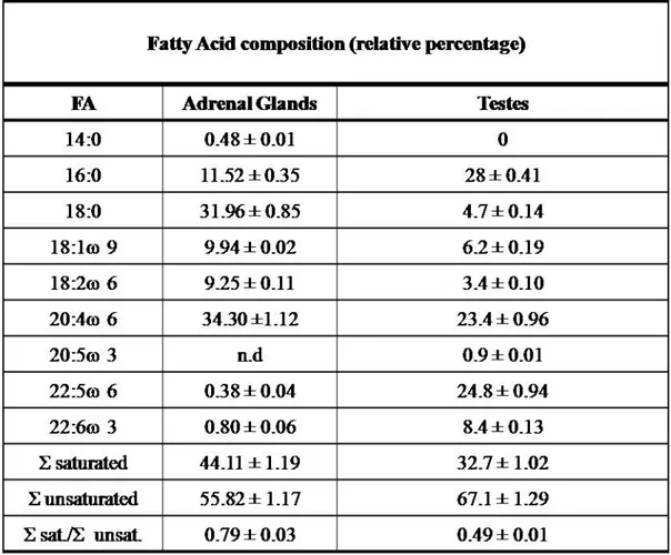
mitochondrial membranes of the two organs important differences in fatty acid composition were detected. Significant changes were measured in the ratio of total saturated/unsaturated fatty acids between the two tissues as shown in Table 4 and Fig. 2 (F-M). A lower percentage of myristic acid (14:0) (Fig. 2F) 10-fold less in testes as well as stearic acid (18:0) about 7-fold less were found in the testes mitochondrial membranes than adrenal gland as shown in Fig. 2G. In addition, about 3-fold higher amount of palmitic acid (16:0) was found only in testes mitochondria (Fig. 2H). Fig. 2 (I-M) shows the differences of unsaturated fatty acid composition. A much lower percentage of docosapentaenoic acid (DPA, C22:5 ω6) about 60-fold less and a lower percentage of docosahexaenoic acid (DHA, C22:6 ω3) about 10-fold less were found in the adrenal glands membranes of mitochondria compared to testes as shown in Fig. 2L. In addition, about 30% higher amount of arachidonic acid (ARA, C20:4 ω6) was found in adrenal gland mitochondria (Fig. 2M). Due to the important physiological functions of arachidonic acid that serve to maintain membrane content and can permit the activity of enzymes of the respiratory chain (Vázquez-Memije et al., 2005) the same statement could not be valid the retinoylation process as the differences could contribute to the enhanced incorporation of RA. Therefore, about 2-fold changes in the ratio of total saturated/unsaturated fatty acids between the two tissues lead us to suppose a relationship through fatty acids and retinoylation process (Table 4). In conclusion RA action by retinoylation process on protein(s) of rat adrenal glands and testes mitochondria is steroidogenic tissue dependent; cytochrome P450scc is not affected by the process and probably docosapentaenoic acid (DPA, C22:5 ω6) should play a fundamental role to allow the transfer of the retinoyl moiety. However, since adrenal glands are arguably the neglected organ in endocrine toxicology (Harvey et al., 2007), it can’t be excluded that perhaps the retinoylation process is toxic/protective for that organ.
Figure 2. (A) Incorporation of [3H]RA (100 nM) into proteins vi Retinoylation reaction on rat adrenal glands mitochondria at 37°C for 90 min in the absence and in the presence of 5 and 10 mM ATP. Retinoylation activity of mitochondria from rat testes (B) and adrenal glands (C) plotted as function of indicated CoA concentrations under the standard conditions for 90 min at 37°C, with 10 mM (B) and 5 mM ATP (C) respectively. (D) Time-dependent incorporation of [3H]RA (100 nM) into protein from rat adrenal glands mitochondria, incubated under standard assay conditions but without ATP at 37°C for the times indicated. (E) Mitochondria were incubated with 10 mM AMG in the presence of 10 mM ATP (testes) and in the absence of ATP (adrenal glands). Saturated(F-H) and unsaturated (I-M) fatty acids composition (mol %) and of rat mitochondrial membrane phospholipids from adrenal glands and testes determined by gas-liquid chromatography as Muci et al (20); other conditions as in Table 4;
Table 2. Retinoylation reaction on adrenal glands cellular sub-fractions Incorporation of [3H]RA (100 nM) into proteins by cellular fractions of adrenal glands in duplicate, incubated in a buffer without ATP for 90 min at 37°C, as described in Materials and Methods
Table 3. Mitochondrial ATP quantification
Table 4. Fatty Acid composition (mol %) of rat mitochondrial membrane phospholipids from adrenal glands and testes.
The fatty acid composition of adrenal glands and testes mitochondrial membranes was determined by gas-liquid chromatography as Muci et al (20). The data are the mean ± S.D. of three independent determinations. Σ saturated = sum of saturated fatty acids; Σ unsaturated = sum of unsaturated fatty acids.
The activity of the adenine nucleotide translocator is affected by a component of the retinoylation buffer.
Indeed, the OGC is not the only mitochondrial carrier protein influenced by the atRA. A previous study by Notario et al. (2001) demonstrated how micromolar concentrations of atRA are capable to bind the Adenine Nucleotide Translocator (ANT) via photo-labelling and inhibit its activity. The ANT translocator exchanges ADP/ATP across the inner mitochondrial membrane. Discovered about four decades ago (Bruni et al., 1964; Pfaff et al., 1965), ANT is a well-studied protein (Klingenberg, 1989; Fiore et al. 1998) due to its abundance, its sensitivity to transport inhibitors and the robustness of the reconstitution method of transport activity in liposomes (Klingenberg, 1985). Consisting of two identical subunits of about 32 kDa, the ANT is present in two distinct conformational states, namely the cytosolic (c) and the matrix (m) states. The inhibitors carboxyatractyloside (CATR) and atractyloside (ATR) bind to c-state, while bongkrekic acid (BKA) binds to the m-state (Klingenberg et al., 1983). It is also assumed that the ANT is the rate-limiting step in energy metabolism (Heldt and Klingenberg, 1968). It has been demonstrated that Mg2+ inhibits the ADP/ATP exchange and Mg2+-nucleotides are not recognized by the ANT carrier in reconstituted proteoliposomes (Brandolin et al., 1980; Brandolin et al., 1981). To provide insight to the biochemical mechanism of atRA we studied the ANT exchange activity under retinoylating conditions (Genchi and Olson, 2001; Cione and Genchi, 2004). Instead of confirming the negative regulation of the ATP/ADP exchange (Notario et al.,2003) we found a positive modulation of the ANT activity due to the presence of Coenzyme A (CoA) in the buffer rather than RA. CoA is an enzyme cofactor in various reactions: it functions as an acyl group carrier and carbonyl activating group in numerous reactions central to cellular metabolism, and provides the 4- phosphopantetheine prosthetic group to proteins that play key roles in fatty acid, polyketide and non-ribosomal peptide biosynthesis. As acetyl-CoA, it is essential to the citric acid cycle, to the synthesis and oxidation of ketone bodies and to cholesterol synthesis. The importance of CoA is reflected in conserved
biosynthetic pathway across animals, plants and micro-organisms (Genschel, 2004). Palmitoyl-CoA and long chain acyl-CoA have been shown as potent inhibitors of the ATP/ADP exchange, when added to a suspension of liposomes reconstituted with ANT from rat or bovine heart or liver mitochondria, while CoA and palmitic acid have no effects (Brandolin et al.,1980; Woldegiorgis et al., 1981; Ruoho et al., 1989; Faergeman and Knudsen, 1997). The role of the ANT in energy-linked respiration is an important factor in assessing the potential significance of the acyl-CoA esters as inhibitors and implies that the ANT carrier, like other rate-limiting enzymes, must be carefully regulated. In order to better understand whether CoA is involved in ANT interaction, we used a novel experimental model that pre-incubates mitochondria in an isotonic buffer to mimic the cellular milieu rather than pre-loading vesicles with CoA or simultaneous addition of CoA with ADP or ATP. It seems reasonable to consider the possibility that the CoA might represent a natural ligand of the translocator; fatty acyl-esters share a common structural backbone with the ANT substrates. Therefore, we studied the activity of ANT after pre-incubation of isolated liver mitochondria into Tris-HCl buffer, pH 7.4, supplied with ATP-Mg2+ and CoA. Our results indicate that CoA is responsible for an enhanced transport activity of the ANT carrier protein suggesting a new role of CoA in energy-requiring processes. The ANT represents the most abundant protein of the inner mitochondrial membrane, spans the inner mitochondrial membrane and allows the exchange of cytosolic ADP for mitochondrial ATP. In all the transport assays performed, the liposomal reconstituted system consisted of partially purified protein. In order to study whether CoA has any influence on the ANT, we used liver mitochondria in isotonic Tris-HCl
buffer, pH 7.4, supplied with ATP-Mg2+ and CoA as experimental model. The
[3H]ATP/ATP exchange was carried out at 25°C and the transport activity of ANT
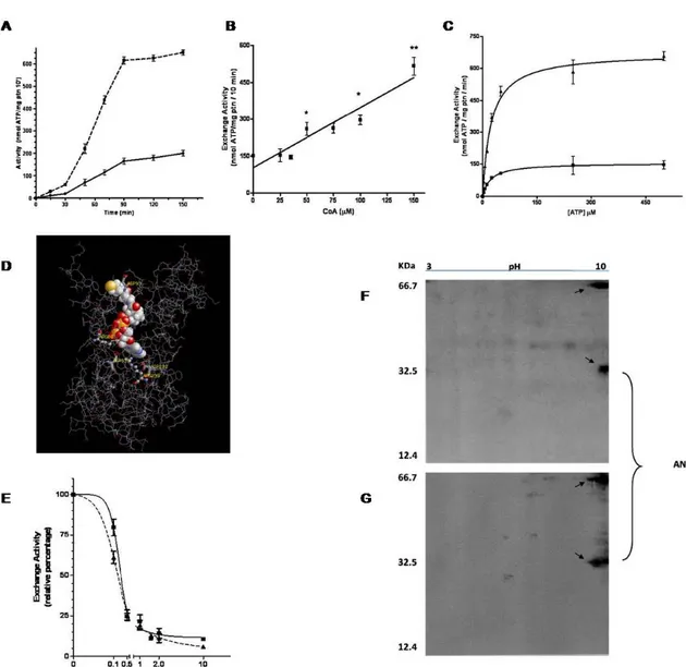
versus mitochondria pre-incubation time with or without CoA is shown in Fig. 3A. In the first section of the graph (0-30 minutes), there’s a linear behaviour with slopes of 0.6 and 2.0, respectively for control and CoA incubation. In the second section (30-90 minutes), CoA pre-incubation shows a 3.8 fold greater increase in activity compared
to control. Both CoA pre-incubated and control samples reach maximum activity at 90 minutes, that remains constant till 150 minutes. Therefore a pre-incubation time of 90 minutes was selected for all subsequent studies. In order to study how different concentrations of CoA affect the ANT activity, mitochondria were pre-incubated with the coenzyme in a range of 0-150 µM. The treatment induced activation of the ANT in a dose-dependent manner as shown in Fig. 3B. Linear regression analysis resulted in a R2 value of 0.8951. To obtain Km and Vmax values with or without CoA, the
dependence of the [3H]ATP/ATP exchange rate on ATP concentration at 25°C was
studied increasing the concentration of external [3H]ATP (in a range of 0-500 µM), while the internal concentration of ATP was constant at 20 mM. Michaelis-Menten equation was used to interpolate our data. In control experiments, Km and Vmax were
22.19 ± 0.98 µM and 155 ± 1.9 nmol ATP/mg protein/min; while with CoA, Km and
Vmax were 22.85 ± 2.52 µM and 673.3 ± 20.74 nmol ATP/mg protein/min,
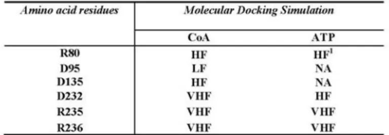
respectively (Fig. 3C). 3D imaging shows that the interaction between CoA and ANT is located within the cavity (Fig. 3D) and molecular docking revealed that the R80, D135, D232 and R235 residues are all involved in the interaction between CoA and ATP on ANT protein (Table 5). All the above-mentioned amino acids are tightly conserved among species, including humans, and are key positions for CATR interaction resulting in inhibitory effects (Pebay-Peroula et al., 2003). To investigate the influence of CoA on the inhibitory effect of CATR on ANT activity, we used 150 μM CoA as described and a range of CATR concentrations from 0 to 10 µM preloaded into liposomes. The most prominent effect occurs at 0.1 µM CATR, where the transport activity of the control was less inhibited compared to the CoA treatment (Fig. 3E). At 0.1 µM of inhibitor the ANT transport activity of the CoA treated mitochondria was reduced of a further 20% compared to the control (60% and 80% of relative exchange activity, respectively). No significant differences were evident at 0.5 and 1 µM CATR, while a slight influence of CoA could be observed at 2 and
10 µM CATR. IC50 resulted changed with a value of 0.198 ± 0.011 µM for control
of the observed CoA-induced activation on a specific ANT isoform, mitochondria prepared from different tissues were tested. ANT1 is mainly expressed in heart and skeletal muscle and ANT2 is expressed in tissues able to undergo proliferation, such as liver, while ANT3 is expressed ubiquitously. The highest transport activation was found in heart (6.7 activation fold), followed by liver (3.4 activation fold), and skeleton muscle (2.9 activation fold), while the lowest enhancement of transport activity was detected in brain (2.5 activation fold), as shown in Table 6. To assess any modification on the protein between CoA treated and untreated samples, 2D electrophoresis of purified ANT were run on 18% AA/bisAA gel, resulting in a slightly shift towards the positive side of the IPG strip after CoA treatment (Fig. 3F-G). The demonstration that ADP and ATP are the actual substrates for the ANT comes from experiments carried out either with isolated mitochondria or after incorporation of the purified carrier into liposomes where the effectors were pre-loaded into the vesicles or added simultaneously with the substrates when starting the exchange. In our case, pre-incubation of mitochondria into an isotonic buffer supplemented with CoA led to completely unique results, which are opposite to the general behaviour provided by the literature (Brandolin et al., 1980; Woldegiorgis et al., 1981; Ruoho et al., 1989; Faergeman and Knudsen, 1997). When mitochondria are pre-incubated at 37°C, the exchange activity of ANT during the first 30 minutes is almost equal to zero in control sample (20.0 ±4.5 nmol/mg protein/10 min) and three times higher (60.0± 6.3 nmol/mg protein/10 min), when CoA is present (Fig. 3A), suggesting that the relationship between ANT and CoA should be enzyme related. Activity increases rapidly between 30 and 90 min for CoA-treated (10.2 fold) and control mitochondria (8.25 fold) further supporting an enzymatic ANT/CoA relationship. The increase in control activity is probably due to endogenous mitochondrial CoA. Both CoA-treated and control reach maximum activity at 90 minutes (615.0 ± 16.1 nmol/mg protein/10 min and 165.0 ± 13.0 nmol/mg protein/10 min, respectively) leading to the maximum difference in CoA-mediated ANT exchange activity over control. In the 90 minutes period both curves are sigmoidal
with the CoA-mediated activity curve 3~4-fold greater than the control curve additionally supporting a possible enzymatic mechanism. Hence, the incubation time of 90 minutes, the first point of maximum activity, was chosen for subsequent experiments. Once the system overcomes the lag phase (0-30 minutes) ANT activity is directly proportional to CoA concentration in the range 0-150 μM as shown in Fig. 3B. Kinetic properties of ANT were modified in the presence of CoA (Fig. 3C). Vmax,
determined in the presence of CoA was more than fourfold greater than control. No differences in Km were observed in the presence of CoA. Enhanced Vmax indicates a
positive modulation of the carrier, suggesting that ANT could be poised to exchange when both anabolic and catabolic CoA-mediated biochemical events, requiring more energy to be transferred from the mitochondrial matrix to the cytoplasm, take place in the cell. Partial purification of ANT by chromatographic passage through an HTP column does not decrease the transport activity of the CoA treated samples. CoA mediated enhancement of ANT transport activity is observed in HTP eluates and in crude Triton X-100 extracts (with 4 and 9 nmol ATP /mg protein /10 min for mitochondria pre-incubated in control buffer, and control buffer plus 150 µM CoA, respectively). Exchange activity of freshly prepared mitochondria, in the presence of CoA was almost tenfold higher than the control (1412.2 ± 52.8 nmol ATP/mg protein/10min versus 153.7±13.5 nmol ATP/mg protein/10 min). Moreover, because the Km value is not affected by the CoA treatment, it is reasonable to affirm that the
ATP binding site on ANT is not involved in the CoA interaction. In order to identify the region involved in the CoA-ANT interaction, we performed a molecular docking simulation. Results of the molecular docking simulation indicate that R80, D135, D232 and R235 are all involved in the interaction with CoA as shown in Table 5 and Fig. 3D. In addition the ATP binds to the same region and the same amino acids that are also responsible for CATR interaction (Pebay-Peroula et al., 2003). As these simulated results are in contrast with the ones provided by the kinetic profile, we investigated the infuence of CoA on CATR effect. The stronger inhibition given at 0.1 µM (20% more than control) is reflected in a change in the IC50 value upon CoA
pre-incubation. High concentration of CATR resulted in similar inhibition between control and CoA-treated due to the excess of inhibitor (Fig. 3E). This result, together with the unchanged Km suggests that the binding site of CoA is different than that
given for ATP/CATR. Stronger inhibition of ANT activity by CATR in the presence of CoA implies that the inhibitor interacts more easily with ANT when mitochondria are pre-incubated with CoA. According to Pebay-Peroula et al. (2003) and our molecular docking simulations, ATP and CATR share three residues of interaction on the ANT. Thus in absence of CATR, CoA effect would largely be on ATP. This supposition that CoA would facilitate interaction with ATP is consistent with the experimental data shown here: i.e. that ANT exchanges more ATP in presence of CoA. Therefore we can assert that the CoA binds to ANT independently from the ATP/CATR binding site and CoA interaction with the ANT protein leads to an increase in the transport activity as well as an increase in sensitivity to the CATR inhibitor and/or interaction with ATP. CoA shows a homology motif with the ADP both in the structure and in the net superficial charge. Therefore a contact between the protein and the CoA could be justified according to a proximity approach. The segment spanning residues I311-K318, corresponding to the C-terminal end of the carrier, was also identified as an ADP binding site able to induce a conformational change of the dimeric structure (Dianoux et al., 2000). As CoA does not prevent ATP and CATR from binding ANT, we can hypothesize that the CoA interacts with the accessory site rather than the one localized in the cavity. Subsequently, an interaction between the –SH group of the β-mercaptoethylamine and the neighbour region could take place. More likely, a covalent bond between the –SH group and an acid residue on the portion of the ANT that spans the intermembrane space could be responsible of a “key mechanism” that leads to structure modification of the carrier. Alternately, because CoA is strictly close to the ANT loops and helices, due to the charge pairing attractions, an enzyme, possibly bound to the outer side of the inner membrane, could provide the necessary enzymatic activity to conjugate the –SH of the CoA with the carrier protein. Based on current data, a CoA derivative or a CoA induced
mechanisms cannot be excluded. As shown in Fig. 3F and 3G, a shift in one of the bands attributed to the ANT is highlighted. Upon visualization with silver stain technique, two spots, at about 64 KDa and 32 KDa respectively, appear on both gels that can be easily identified as a dimeric and monomeric form of the ANT, whose molecular weight is known to be 31.7 KDa. The purity of the protein sample is guaranteed by the isolation procedure and chromatographic passages on HTP first and Matrex Gel Blue B thereafter, verified with an increase in the specificity of the exchange activity (data not shown). According to its chemical properties, the ANT is localized around pH 10 on the IPG strip (Fig. 3F): a shift towards the more positive side of the isoelectrofocusing strip upon incubation of rat liver mitochondria with CoA has been detected (Fig. 3G). This shift is most likely due to a modification of the charges on the carrier protein surface, as a result of treatment with CoA. Although all the ANT isoforms share at least 90% nucleotide sequence identity, they have been implicated differently in several cellular functions (Battini et al., 1987; Neckelmann et al., 1987; Cozens et al., 1989; Stepien et al., 1992; Doerner et al., 1997). We now add the possibility that all the isoforms are similarly involved in the export functions (Table 6). ANT is a portion of ATP synthasome (Chen et al., 2004) and the ANT complex is involved in multiple diseases affecting both humans and animals in which ANT activity is disrupted. Among these, heart diseases, mitochondrial diseases, osteoporosis, macromolecular degeneration, immune deficiency, cystic fibrosis, type II diabetes, ulcers, nephro-toxicity, hearing loss, skin disorders, and cancer are found (Klingenberg, 1989; Dahout-Gonzalez et al., 2005). A new emerging class of drugs, named mitocans, are activators of either VADC or ANT and could be used as promising alternative cancer therapeutics (Ralph et al., 2006). Our findings could provide a novel therapeutic approach for treatment of all the above mentioned diseases, or just lay the fundamentals for further discoveries in biochemistry. Additionally, in the literature it is indicated the inhibition of key enzymes in metabolism by long chain acyl-CoA esters, besides the ADP/ATP translocator (Woldegiorgis et al., 1981). Our report that the exchange of ADP/ATP
across the mitochondrial membranes is enhanced by CoA may provide insight into these enzymatic mechanisms influenced by CoA and suggest unique interactions and/or structural modifications may exist.
Table 5. Molecular Docking of CoA and ATP interactions with ANT amino acid residues. With the program Patch-Doc® twelve simulations have been carried out and the percentage of interactions of the binding region of the BTADT1-CATR complex respect to the binding region of the RnADT1-CoA and RnADT1-ATP complexes are expressed as Very High Frequency (VFH, 12/12), High Frequency (HF, 10/12), Low Frequency (LF, 5/12) and Not Available (NA).
Table 6. Exchange activity of ANT isolated from mitochondria of various tissues after pre-incubation, with or
without CoA.
Mitochondria were pre-incubated for 90 minutes at 37°C in 50 mM sucrose, 100 mM Tris-HCl, 10 mM ATP, 27 mM MgCl2, pH 7.4 (control buffer) and in the same buffer supplemented with 150 µM CoA. ANT was isolated and partially purified. The exchange activities (nmol/mg protein /10 min) were determined as in Material and Methods. The activation fold was calculated as the ratio between the exchange activity of ANT from mitochondria pre-incubated in control buffer plus 150µM CoA and control buffer. Results are expressed as Mean ± SEM of 5 independent experiments.
Figure . (A) Exchange activity of ANT from rat liver mitochondria pre-incubated in 50 mM sucrose, 100 mM Tris-HCl, 10 mM ATP, 27 mM MgCl2, pH 7.4 (control buffer ■) and in the same buffer supplemented with 150 µM CoA (▲) at 37 °C, for the indicated time points. The exchange activity was assayed at 25°C for 10 minutes as described in Materials and Methods. (B) Effect of CoA concentration on the ANT exchange activity. (C) Michaelis-Menten kinetic profile of rat liver mitochondria pre-incubated in 50 mM sucrose, 100 mM Tris-HCl, 10 mM ATP, 27 mM MgCl2, pH 7.4 (control buffer ■) and in the same buffer supplemented with 150 µM CoA (▲) at 37 °C. The exchange activity was assayed for one minute with [3H]ATP, added at concentrations from 0 to 500 µM, at 25°C. (D) 3D Imaging of Interaction between CoA and ANT. The 3D structure of the ANT carrier was calculated through homology modelling parameters by Swiss-PDB Viewer software. The template structure is the bovine heart ANT. The final structure was minimized through the GROMOS96 process implemented in the Swiss-PDB Viewer to reduce steric/binding perturbation. Molecular-docking simulations were performed on the
on-line http://bioinfo3d.cs.tau.ac.il/PatchDock/index.html server PatchDock and the structures were visualized and
analyzed with PyMol software. (E) Inhibition effect of CATR on ANT from mitochondria incubated for 90 minutes in 50 mM sucrose, 100 mM Tris-HCl, 10 mM ATP, 27 mM MgCl2, pH 7.4 (control buffer ■) and in the same buffer plus 150 µM CoA (▲). Other experimental procedures as in (B) except that proteoliposomes were pre-loaded with CATR at the indicated concentrations. After isolelectrofocusing and SDS PAGE, of totally purified ANT protein, upon
visualization with Silver Stain method, two main spots are highlighted at 64 KDa and 32 KDa both in control (F), and in CoA treated mitochondria(G). Images were acquired in electronic format scanning the gels with a Canonscan Lite 100 scanner, in greyscale with a resolution of 600 DPI.
Combined low doses of PPARγ and RXR ligands trigger an intrinsic apoptotic pathway in human breast cancer cells.
Recently, studies in human cultured breast cancer cells have shown how the TZD, Rosiglitazone (BRL), promotes antiproliferative effects and activates different molecular pathways leading to distinct apoptotic processes (Bonofiglio et al. 2005, 2006, 2008). Apoptosis, the genetically controlled and programmed death leading to cellular self-elimination, can be initiated by two major routes: the intrinsic and extrinsic pathways. The intrinsic pathway is triggered in response to a variety of apoptotic stimuli that produce damage within the cell, including anticancer agents, oxidative damage and UV irradiation, and is mediated through the mitochondria. The extrinsic pathway is activated by extracellular ligands able to induce oligomerization of death receptors, such as Fas, followed by the formation of the death-inducing signaling complex, after which the caspases cascade can be activated.Previous data show that the combination of PPARγ ligand with either ATRA or 9-cis-retinoic acid (9RA) can induce apoptosis in some breast cancer cells (Elstner et al. 2002). Furthermore, Elstner et al. demonstrated that the combination of these drugs at micromolar concentrations reduced tumor mass without any toxic effects in mice (Elnster et al. 1998). The ability of PPARγ ligands to induce differentiation and apoptosis in a variety of cancer cell types, as in human lung (Tsubouchi et al.,2000), colon (Kitamura et al. 1999) and breast (Mueller et al. 1998) has been exploited in experimental cancer therapies (Roberts-Thomson, 2000). PPARγ agonist administration in liposarcoma patients resulted in histologic and biochemical differentiation markers in vivo (Demetri et al. 1999). However, a pilot study of short-term therapy with PPARγ ligand Rosiglitazone in early-stage breast cancer patients does not elicit significant effects on tumor cell proliferation, although the changes observed in PPARγ expression may be relevant to breast cancer progression (Yee et al. 2007). However, in humans PPARγ agonists at high doses exert many side effects including weight gain due to increased adiposity, edema, hemodilution, and plasma-volume expansion, which preclude their clinical application in patients with heart
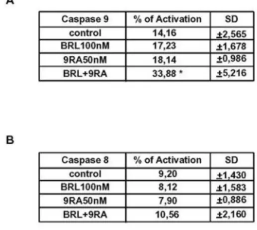
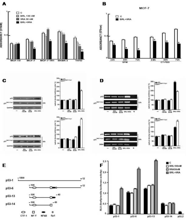
failure (Arakawa et al., 2004; Rangwala and Lazar, 2004; Staels, 2005). On the other hand, the natural ligand for RXR, 9RA (Leblanc and Stunnenberg, 1995) has been effective in vitro against many types of cancer, including breast tumor (Crouch et al,. 1991; Delia et al,. 1993; Rubin et al., 1994; Sun et al.,1997; Wu et al., 1997). Recently, RXR-selective ligands were discovered to inhibit proliferation of ATRA-resistant breast cancer cells in vitro and caused regression of the disease in animal models (Bishoff et al., 1998). The undesirable effects of RXR-specific ligands on hypertriglyceridemia and suppression of the thyroid hormone axis have been also reported (Pinaire and, Reifel-Miller, 2007). The additive antitumoral effects of PPARγ and RXR agonists, both at elevated doses, have been shown in human breast cancer cells (Elstner et al. 2002; Grommes et al., 2004 and references therein). However, high doses of both ligands have remarkable side effects in humans such as, weight gain and plasma volume expansion for PPARγ ligands (Arakawa et al., 2004; Rangwala and Lazar, 2004; Staels, 2005) and hypertriglyceridemia and suppression of the thyroid hormone axis for RXR ligands (Pinaire and Reifel-Miller, 2007). Thus, in the present study it has been demonstrated that nanomolar concentrations of BRL and 9RA in combination do not induce noticeable influences in cell vitality on normal breast epithelial cells, whereas they exert significant antiproliferative effects on breast cancer cells. The molecular mechanism by which combined treatment with BRL and 9RA at nanomolar doses triggers apoptotic events in breast cancer cells, have been elucidated, suggesting potential therapeutical uses for these compounds. To investigate whether low doses of combined agents are able to inhibit cell growth, the capability of 100 nM BRL and 50 nM 9RA to affect normal and malignant breast cell lines was first assessed. We observed that treatment with BRL alone does not elicit any significant effect on cell viability in all breast cell lines tested, while 9RA alone reduces cell vitality only in T47-D cells (Fig. 4A). In the presence of both ligands cell viability is strongly reduced in all breast cancer cells: MCF-7, its variant MCF-7TR1, SKBR-3 and T-47D, while MCF-10 normal breast epithelial cells are completely unaffected (Fig. 4A). To evaluate the effectiveness of both ligands in the
presence of serum, we performed MTT assay in MCF7 cells treated with low doses of BRL and 9RA in SFM as well as in 5% CT-FBS (Fig. 4B). The molecular mechanism underlying these effects has been elucidated in MCF-7 cells in which an upregulation of tumor suppressor gene p53 has been observed. A significant increase in p53 and p21WAF1/Cip1 content was observed by Western Blot only upon combined treatment after 24 and 36 h (Fig. 4C). Furthermore, we showed an upregulation of p53 and p21WAF1/Cip1 mRNA levels induced by BRL plus 9RA after 12 and 24 h (Fig. 4D). To investigate whether low doses of BRL and 9RA are able to transactivate the p53 promoter gene, we transiently transfected MCF-7 cells with a luciferase reporter construct (named p53-1) containing the upstream region of the p53 gene spanning from -1800 to +12 (Fig. 4E). Treatment for 24 h with 100 nM BRL or 50 nM 9RA did not induce luciferase expression, whereas the presence of both ligands increased in the transactivation of p53-1 promoter (Fig. 4F). To identify the region within the p53 promoter responsible for its transactivation, we used constructs with deletions to different binding sites such as CTF-1, nuclear factor-Y (NF-Y), NFkB and GC sites (Fig. 4E). In transfection experiments performed using the mutants p53-6 and p53-13 encoding the regions from -106 to +12 and from -106 to -40, respectively, the responsiveness to BRL plus 9RA was still observed (Fig. 4F). In contrast, a construct with a deletion in the NFkB domain (p53-14) encoding the sequence from -106 to -49, the transactivation of p53 by both ligands was absent (Fig. 4F), suggesting that NFkB
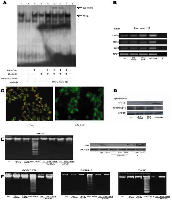
site is required for p53 transcriptional activity. To gain further insight into the involvement of NFkB site in the p53 transcriptional
response to BRL plus 9RA, we performed electrophoretic mobility shift assay experiments using syntethic oligodeoxyribonucleotides corresponding to the NFkB sequence within p53 promoter. We observed the formation of a specific DNA binding complex in nuclear extracts from MCF-7 cells (Fig. 5A, lane 1), where specificity is supported by the abrogation of the complex by 100-fold molar excess of unlabeled probe (Fig. 5A, lane 2). BRL treatment induced a slightly increase in the specific band (Fig. 5A, lane 3), while no changes were observed on 9RA exposure (Fig. 5A,
![Figure 2. (A) Incorporation of [ 3 H]RA (100 nM) into proteins vi Retinoylation reaction on rat adrenal glands mitochondria at 37°C for 90 min in the absence and in the presence of 5 and 10 mM ATP](https://thumb-eu.123doks.com/thumbv2/123dokorg/2877561.10044/24.892.96.790.103.684/figure-incorporation-proteins-retinoylation-reaction-adrenal-mitochondria-presence.webp)
![Table 2. Retinoylation reaction on adrenal glands cellular sub-fractions Incorporation of [ 3 H]RA (100 nM) into proteins by cellular fractions of adrenal glands in duplicate, incubated in a buffer without ATP for 90 min at 37°C, as described in Materia](https://thumb-eu.123doks.com/thumbv2/123dokorg/2877561.10044/25.892.147.751.422.671/retinoylation-fractions-incorporation-fractions-duplicate-incubated-described-materia.webp)