Alma Mater Studiorum – Università di
Alma Mater Studiorum – Università di
Bologna
Bologna
DOTTORATO DI RICERCA
IN BIOINGEGNERIA
Ciclo XXI
Settore scientifico disciplinare di afferenza: ING-IND/34
TITOLO TESI
SVILUPPO DI UNA PROCEDURA SPERIMENTALE
PER CARATTERIZZARE IL CEMENTO OSSEO ACRILICO
DEVELOPMENT OF AN EXPERIMENTAL PROCEDURE
TO TEST ACRYLIC BONE CEMENT
Presentata da: Ing. EWA BIALOBLOCKA-JUSZCZYK
Coordinatore Dottorato:
Relatore:
Prof. Angelo Cappello
Prof. Luca Cristofolini
Co-relatore:
Ing. Massimiliano Baleani
Ing. Marco Viceconti
CONTENTS
RIASSUNTO……….….….5
SUMMARY………..…...…9
I INTRODUCTION………...………...…..…12
1.1 PMMA based bone cement………...…....12
1.2 Biocompatibility………13
1.3 Cemented Total Joint Replacements………...14
1.4 Total Hip Arthroplasty: indication for surgery……….…...14
1.5 Cemented Total Hip Arthroplasty: cementing techniques………….……...16
1.5.1 Bone preparation……….….….17
1.5.2 Lavage………..……...…...17
1.5.3 Cement mixing……….…..…18
1.5.4 Stem centralizers……….….….18
1.5.4 Cement delivery………....….18
1.5.5 Cement pressurization and stem insertion………..…...19
1.6 Long-term stability of cemented THA……….…...20
1.7 Aseptic loosening………...………...21
1.8 Physical and mechanical properties of bone cement………...…21
1.9 Weak points of current standards………...…..….22
1.10 Aim………..…..23
1.11 Bone cement investigated within the project………..…...24
1.12 References……….…25
II CHARACTERIZATION OF STATIC MECHANICAL PROPERTIES: A COMPARATIVE STUDY ON THREE COMMERCIAL BONE CEMENTS ………..………..….29
2.1 Introduction………..…….29
2.3 Results……….……….……30
2.4 Discussion……….…31
2.5 References………...…..…31
III FRACTURE PROPERTIES OF AN ACRYLIC BONE CEMENT….………..32
3.1 Abstract………..……….32
3.2 Introduction……….33
3.3 Materials and methods………34
3.3.1 Materials and specimens preparation……… ……..34
3.3.2 Fatigue testing……….……...34
3.3.3 Fatigue crack propagation testing……….….……...35
3.3.4 Fracture toughness testing………. …….36
3.4 Results………..……….…...36
3.4.2 Fatigue testing……….…..36
3.4.3 Fatigue crack propagation testing………..…...36
3.4.4 Fracture toughness testing……….……….……….…...…37
3.5 Discussion………..………..……....…37
3.6 References………..……….……….…39
IV FRACTURE PROPERTIES COMPARISON OF THREE COMMERCIAL BONE CEMENTS……….….44
4.1 Abstract………44
4.2 Introduction………..45
4.3 Materials and methods……….…………46
4.3.1 Cement type selection………46
4.3.2 .Specimen preparation………..………47
4.3.3 Fatigue testing……….………..….48
4.3.4 Fatigue crack propagation testing……….….…………49
4.3.5 Fracture toughness testing……… …….…….49
4.4 Results……….……….….……...50
4.4.2 Fatigue crack propagation testing………....……50
4.4.3 Fracture toughness testing……….….…....51
4.5 Discussion………..…..51
4.6 Acknowledgment……….…53
4.7 References………...……….…53
V EFFECT OF LONG-TERM PHYSIOLOGICAL ACTIVITY ON THE LONG-TERM STEM STABILITY OF CEMENTED HIP ARTHROPLASTY:IN VITRO COMPARISON OF THREE COMMERCIAL BONE CEMENTS……….…...…..56
5.1 Abstract……….….……..56
5.2 Introduction………....…..57
5.3 Material and methods……….……..59
5.3.1 Bone cement selection……….……….59
5.3.2 Preparation of the specimens………..…….…..60
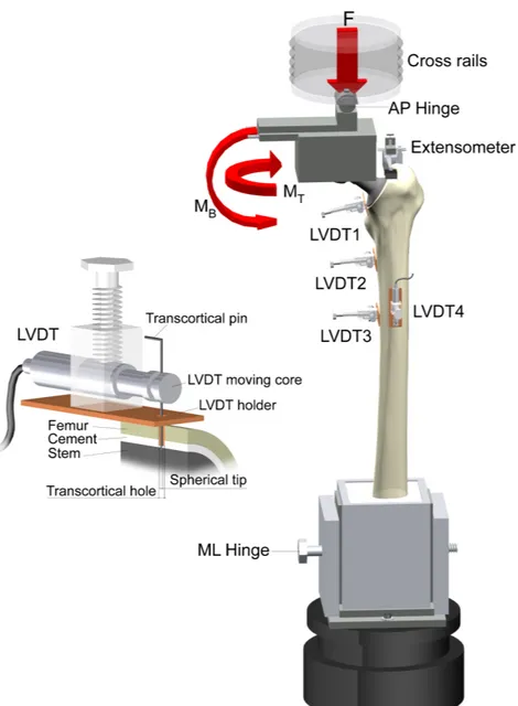
5.3.3 The load history and testing setup……….….…..61
5.3.4 Micromotion measurement……….…..…..63
5.3.5 Cement mantle inspection for fatigue damage………..…..…65
5.3.6 Statistical analysis……….………….….….66
5.4 Results……….….…66
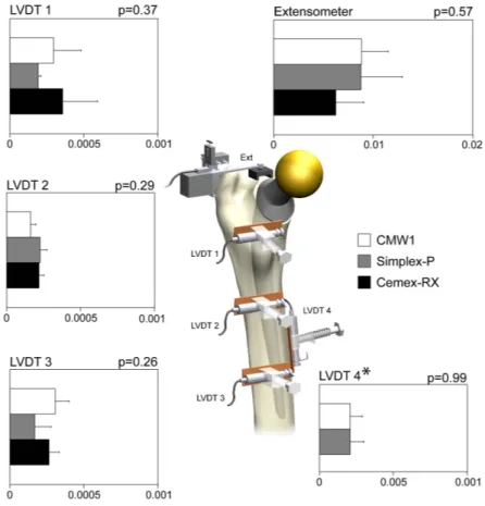
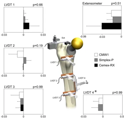
5.4.1 Inducible and permanent micromotions……….….….…66
5.4.2 Fatigue damage in the cement mantle………..……68
5.5 Discussion……….….…..72
5.6 Acknowledgments………..…………..…76
5.7 References………..…………..…76
VI EFFECT OF STEM PREHEATING ON THE FATIGUE BEHAVIOUR OF BONE CEMENT USED FOR CEMENTED PROSTHESES……….…………...79
6.1 Abstract………...………….……….…..79
6.2 Introduction……….………80
6.4 Results……….……….…..……….83
6.5 Discussion……….……….….84
6.6 Acknowledgments………..….……86
6.7 References……….….….86
VII VACUUM MIXING AND STEM PRE-HEATING IMPROVE MECHANICAL PERFORMANCE OF THE BONE CEMENT MANTLE…….……….…..…89
7.1 Abstract………...………89
7.2 Introduction……….………90
7.3 Material and methods………..………91
7.3.1 Materials and bone cement mantle moulding……….………91
7.3.2 Monitoring the curing process………...……..92
7.3.3 Specimens extraction………...…………93
7.3.4 Bending test………..….94
7.3.5 Fatigue test………...……….94
7.3.6 Porosity analysis………..95
7.3.7 Statistical analysis procedure………..………….95
7.4 Results………...…..96 7.4.1 Curing process……….………96 7.4.2 Bending strength……….……….……97 7.4.3 Fatigue strength………..………….97 7.4.4 Porosity………...…..98 7.5 Discussion………..………….99 7.6 References………..100 VIII CONCLUSIONS………..…………106
APPENDIX A THE COMPRESSIVE PROPERTIES AND FRACTURE TOUGHNESS OF PMMA BASED ACRYLIC RESIN REINFORCED WITH MILLED AND MICRONISED GLASS FLAKES ………108
RIASSUNTO
Attraverso il pioneristico esperimento di Charney nel 1960 [1], il cemento osseo polimetilmetacrilato (PMMA) diventò il più diffuso materiale di fissaggio, per protesizzazione totale dei giunti articolari. Assicura un’ottima stabilità delle protesi in poco tempo dopo la polimerizzazione. Tra i vari tipi di protesizzazione cementata (spalla, gomito, anca ginocchio e caviglia) la sostituzione totale d’anca (Total Hip Replacement THR) è l’intervento più frequente [2].
Il cemento osseo crea un legame meccanico tra la componente protesica ed il tessuto osseo circostante. La sopravvivenza a lungo termine della sostituzione articolare dipende dalle proprietà meccaniche del cemento. Per questo motivo, prima di introdurre un nuovo modello di PMMA nella pratica clinica, il materiale deve soddisfare i requisiti descritti nella ISO 58333
Comunque, risultati clinici dimostrano che le prove meccaniche richiesti dalla normativa ISO non predicono le prestazioni a lungo termine del cemento [3]. L’inconsistenza degli attuali test di validazione pre-clinica è legata alla limitazione dei protocolli definiti per le prove meccaniche statiche, i.e. prove a compressione e flessione. Infatti, prove meccaniche dinamiche potrebbero essere maggiormente rappresentative delle condizioni di carico causate dalle attività fisiologiche quotidiane del paziente [4].
Numerosi studi sono stati pubblicati in passato, riportando proprietà dinamiche (fatica, resistenza a frattura, propagazione delle crepe a fatica) di diversi cementi ossei [5]. Comunque la variabilità tra protocolli di prova e risultati rende impossibile identificare un valido metodo di predizione a lungo termine delle proprietà dei cementi. Esempi di questa variabilità sono i seguenti [5]:
• propagazione della cricca a fatica (valori delle costanti di Paris C e n); C varia da 4.03*10-8 a 9.5*10-4, e n varia da 4.71 a 9.77.
Per questa ragione lo scopo del presente progetto è stato quello di identificare una procedura sperimentale per la validazione pre-clinica del cemento osseo acrilico. L’assunzione principale fatta all’interno di questo progetto è stata quella di realizzare una completa caratterizzazione meccanica di tre cementi ossei commerciali e di confrontare i risultati ottenuti con informazioni provenienti da protocolli fisiologici in vitro. In fine, dopo aver confrontato tutte le informazioni ottenute dagli esperimenti con risultati clinici dei tre cementi ossei scelti (Cemex RX, Surgical Simplex P and CMW1), è stato identificato il protocollo di prova il protocollo di prova che ha mostrato le predizioni più accurate.
Il lavoro presentato in questa tesi è stato sviluppato nel Laboratorio di Tecnologia Medica del’Istituto Ortopedico Rizzoli (Bologna, Italia).
La prima parte del progetto (Capitolo 1) è dedicata alle prove condotte secondo lo standard ISO 5833. Il cemento osseo selezionato è stato testato in termini di proprietà a compressione e a flessione e i risultati ottenuti sono stati paragonati con quelli forniti dal produttore e quelli trovati in letteratura. Lo scopo è stato quello di acquisire le informazioni di base relative allo standard utilizzato.
La prima serie di prove meccaniche è stata condotta in accordo con il protocollo interno LTM. Sono state analizzate (Capitolo 2) le proprietà dinamiche i.e., la resistenza a fatica, la resistenza alla propagazione delle crepe e la resistenza a frattura, di un cemento osseo acrilico (Cemex RX). La metodologia presentata è stata applicata in una seconda fase del lavoro, dedicata al confronto dei tre cementi selezionati e all’identificazione di quale tra i protocolli presentati sia maggiormente predittivo delle prestazioni a lungo termine del cemento osseo (Capitolo 3).
Il passo successivo del progetto è stato quello di caratterizzare tre cementi commerciali usando un protocollo fisiologico per simulare le attività più critiche in termini di carico di fatica (salita e discesa delle scale, entrata ed uscita dalla macchina, entrate ed uscita dalla vasca da bagno e inciampo) (Capitolo 4). Per
controllare la stabilità dello stelo protesico sono stati misurati i micromovimenti durante la prova. L’analisi della qualità del mantello di cemento, dopo una simulazione di 24 anni di attività del paziente, ha permessola verifica del protocollo per la predizione delle prestazioni a lungo termine del cemento osseo.
È stato sviluppato un protocollo che simulasse le condizioni di polimerizzazione presenti in sala operatoria, allo scopo di verificare l’influenza del pretrattamento dello stelo femorale e dei metodi di miscela del cemento, sulla resistenza meccanica del mantello di cemento. Durante le prove la temperatura e il corrispondente tempo di polimerizzazione sono stati monitorati in differenti punti. Provini, estratti dal mantello, sono stati sottoposti a prove di flessione o fatica. Il Capitolo 5 riporta i risultati dello studio sul preriscaldamento dello stelo femorale ed i suoi effetti sulla vita del mantello di cemento sottoposto a fatica, mentre nel Capitolo 6 è stato discusso l’effetto combinato del preriscaldamento e del metodo di miscela del cemento sulle prestazioni a fatica ed a flessione.
Una applicazione pratica del progetto di validazione dei metodi di prova, uno studio preliminare sulla formulazione di un nuovo cemento, è stata svolta presso Leeds University (UK). I dettagli di questa applicazione sono riportati nell’Appendica A. Una serie di prove meccaniche sono state eseguite su PMMA addizionato con quattro diverse concentrazione di glass flakes, e con due diverse dimensioni di glass flakes. Anche l’influenza della miscelazione sotto vuoto è stata analizzata. In totale, includendo i gruppi di controllo, 18 gruppi sono stati testati a compressione (ISO 5833) e alla resistenza alla frattura per doppia torsione
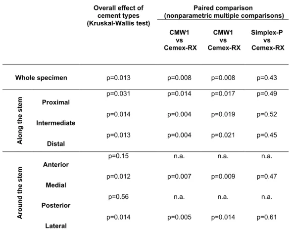
In conclusione, le informazioni ottenute dagli standard ISO indicano il cemento CMW1 come avente migliori proprietà meccaniche. Al contrario, dalla prove di fatica e le simulazioni fisiologiche in vitro, il CMW1 risulta essere il cemento a maggior rischio di fallimento, rispetto alle altre due formulazioni selezionate. Questi risultati sono in accordo con i dati clinici. Conseguentemente, i due test presentati si sono dimostrati capaci di predire la sopravvivenza delle protesi cementate relativamente alla formulazione di cemento osseo usata.
1. Charnley, J., Surgery of the hip-joint: present and future developments. Br Med J, 1960. 1(5176): p. 821-6. 2. Finerman, G.A.M., et al., Total Hip Arhroplasty Outcomes. 1998, New York: Churchill Livingstone Inc. 3. Nilsen, A.R. and M. Wiig, Total hip arthroplasty with Boneloc:Loosening in 102/157 cases after 0.5-3 years.
Acta Orthopaedica, 1996. 67(1): p. 57-59.
4. NIH, Total Hip Joint Replacement. NIH Consens Statement 1994 Sep 12-14; 12(5): 1-31, 1994.
SUMMARY
Through the pioneering experiment of Charnley in 1960 [1], PMMA bone cement became the most widely utilized fixing material in total joint replacements. It assures an optimal stability of the prosthesis in short time after polymerization. Among all types of cemented joint replacements, i.e. shoulder, elbow, hip, knee and ankle, the Total Hip Replacement (THR) is the most frequent intervention [2].
As bone cement creates a mechanical bond between the prosthetics components and surrounding bone tissue, the long-term survival of cemented joint replacement depends on mechanical properties of the cement. In fact, before introducing new PMMA bone cement into clinical use, the formulation must comply with the ISO 58333 standard requirements.
However, clinical outcomes demonstrate that the requested ISO standardized mechanical tests do not predict the long-term performance of the cement [3]. The irrelevance of current pre-clinical validation is due to the limitation of defined protocols to static mechanical test, i.e. compressive and bending test. In fact, dynamic mechanical testing would be more relevant to the loading condition caused by physiological daily activity of the patient [4].
Numerous studies have been published in the past, reporting dynamic (fatigue, fracture toughness, fatigue crack propagation) properties of different bone cements [5]. However, the variability among testing protocols and results makes it impossible to identify a valid method to predict long-term outcome of the cement. Examples of this variability are as fallows [5]:
• fracture toughness; 1.03 to 2.32 MPa*m1/2
• fatigue crack propagation constants (values of Paris constants C and
For this reason, the aim of presented project was to identify an experimental procedure for pre-clinical validation of acrylic bone cement. The main assumption of the project was to perform complete mechanical characterization of three commercially available bone cements and to confront obtained results with data from an in vitro physiological protocol. Finally, after comparing all collected data from the experimental testing with clinical outcome of the three chosen bone cements (Cemex RX, Surgical Simplex P and CMW1), the testing protocol leading to the most accurate predictions has been identified.
The work presented within this thesis was carried out in Laboratorio di Tecnologia Medica of Istituto Ortopedico Rizzoli (Bologna, Italy).
The first part of the project (Chapter 1) is dedicated to the tests conducted in accordance to the ISO 5833 standard. The selected bone cements have been tested in terms of compressive and bending properties and obtained data have been compared with those provided by manufactures and found in the literature. The purpose was to acquire basic information according to the standard.
The first series of mechanical tests in accordance to the internal LTM protocol have been performed. Dynamic properties i.e. the fatigue strength, the resistance to crack propagation and the fracture toughness, of an acrylic bone cement (Cemex-RX) where investigated (Chapter 2). Presented methodology has been applied in later part of this work, dedicated to the comparison of three selected cements and identification, which of three applied protocols is the most predictive for long-term performance of bone cement (Chapter 3).
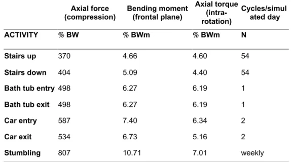
Next step of the project was to characterize three commercially available bone cements using a physiological protocol to simulate the most critical activities in terms of fatigue loading (stair climbing and stair descending, car entry and car exit, bathtub entry and bathtub exit, and stumbling) (Chapter 4). To monitor stem stability, micromotions were measured during the test. Analysis of the quality of the cement mantle after simulation of 24 years of a patient’s activities permitted the verification of a protocol, to predict the long-term performance of bone cement.
To verify the influence of stem pretreatment and cement mixing methods on mechanical strength of the cement mantle, a protocol simulating the surgical curing conditions has been developed. During curing, temperature and corresponding time were monitored at different locations. Specimens, extracted from the mantles, underwent bending or fatigue tests. Chapter 5 reports results of the study on the effect of stem preheating on the fatigue life of cement mantle, while in Chapter 6 an effect of both, stem preheating and mixing method on bending and fatigue performance of cement has been discussed.
As a practical application of the project on validation of testing methods, a preliminary study on developing a new cement formulation was carried out at Leeds University (UK), the details of which are reported in Appendix A. A series of mechanical tests were performed on PMMA with addition of four different glass flakes concentration, and with two different flake sizes. The influence of vacuum mixing has been investigated. In total, including reference groups (or control groups/samples), 18 groups were tested for compression (ISO 5833) and double-torsion fracture toughness.
To conclude, data obtained from standard ISO test shows the best mechanical properties for CMW1 bone cement. While, from complex fracture characterization and physiological in vitro simulation, the CMW1 results as cement with the highest failure risk, respect to two others selected formulations, which is in accordance with the clinical data. Consequently, those two tests are able to predict the survival of a cemented prosthesis in dependence on used bone cement formulation.
1. Charnley, J., Surgery of the hip-joint: present and future developments. Br Med J, 1960. 1(5176): p. 821-6. 2. Finerman, G.A.M., et al., Total Hip Arhroplasty Outcomes. 1998, New York: Churchill Livingstone Inc. 3. Nilsen, A.R. and M. Wiig, Total hip arthroplasty with Boneloc:Loosening in 102/157 cases after 0.5-3 years.
Acta Orthopaedica, 1996. 67 (1): p. 57-59.
4. NIH, Total Hip Joint Replacement. NIH Consens Statement 1994 Sep 12-14; 12(5): 1-31, 1994.
I INTRODUCTION
1.1 PMMA based bone cement
The majority of bone cements currently available on the market are based on poly(methyl methacrylate) (PMMA), where the monomer of methyl mathacrylate (MMA) is an ester of methacrylic acid.
Studies on methacrylic acids began more than 80 years ago in Tübingen (Germany), when Otto Röhm was given the topic of “polymerization products of acrylic acid” for his thesis. Later, based on his research, he founded a company (Röhm and Haas) for the development of acrylates [1]. Following, the establishment of a large number of technical synthesis techniques for MMA (by 1928), a new application was found for it in denture production. In 1935 Bauer patented this technique.
In 1936 the company Kulzer had discovered that mixing the PMMA powder with MMA liquid produces a dough, which became stiff if benzoyl peroxide (BPO) is added and the mixture is heated to 100oC. In this way the first clinical use of PMMA mixture (Paladon 65) was developed for close cranial defects in humans. Prefabricated plates of PMMA, made under laboratory conditions, were adjusting by surgeons during the surgery [1].
Since chemists found that the polymerization process of MMA occurs at room temperature if a co-initiator is added, the companies Degussa and Kulzer established a protocol for the chemical production of PMMA bone cements in 1943. They used tertiary aromatic amines as the co-initiator. Thus, the defined procedure is valid until today, and it should be considered as the birth of PMMA based bone cement.
Some of them have already been withdrawn from the market, others are used only in certain countries or play a minor role in clinical use. They can differ from each other by the chemical composition of the base mixture i.e. polymers molecular weight and concentration of accelerator and/or initiator. Moreover, they differ in the included additives e.g. barium sulfate or zirconium dioxide (as radiopaque medium), Na-fluoride, antibiotics, antiblastics,ethylene-oxide (sterilization), chlorophyllin (dye) or mechanically reinforcing particles (carbon fibers, glass fibers) [2] [3, 4]. Each change in composition of final bone cement mixture will influence its viscosity, working time, setting and in effect mechanical properties of the polymerized bone cement bulk [5-8].
Nowadays, PMMA based bone cements are widely used in surgery for the augmentation of fractured tissue (vertebroplasty and kyphoplasty) and dentistry, but more frequently as a fixing material that provides transmission of mechanical loading from the prosthesis to the bone. Generally, the application of cemented prosthesis is recommended when tissue surrounding an implant is too fragile/weak to support the load generated during physiological activity of the patient, e.g. patients with osteoporosis [9, 10].
1.2 Biocompatibility
The reaction of the tissue to PMMA bone cement, was the essential question just after Charnley developed his implantation technique. The extensive study of Hullinger [11] proved the biocompatibility of hardened PMMA. Also, Lehmann and Jenny [12] in their study with cell cultures have shown that PMMA did not cause any cytotoxic reactions. Further studies have shown, that cell reaction to PMMA is associated with particle size and it is not a specific immune response [10, 13]. However, the investigation of biocompatibility is not part of the scope of this thesis.
1.3 Cemented Total Joint Replacements
The PMMA based bone cement is widely used in Total Joint Replacements (TJR). Annually, over 1 mln of implantation procedures is performed worldwide, considering all types of joint replacements. More than 50% of the total TJR are the procedures of total hip arthroplasty [14]. Second, less numerous but also very common reconstructive procedure is total knee arthroplasty, followed by replacements of the ankle and shoulder.
The survival of joint reconstruction depends on many factors (infections, wearing, aseptic loosening, dislocations, fractures). Critical factors responsible for failure of the prosthesis can differ in dependence on type of the joint, e.g., wearing in knee replacements or implant design in ankle replacements. However, for the most frequently reconstructed joint, i.e. the hip, aseptic loosening results as the most critical factor leading to the revision [15, 16]. For that reason, this study is dedicated to problems related with cemented total hip arthroplasty.
1.4 Total Hip Arthroplasty: indication for surgery
The hip joint is one of the most severely loaded joints in the human body, designed for many different types of movement. It consists of the head of the femur, which is shaped like a ball; and a part of the pelvic bone called the acetabulum, which looks like a hollow or socket (Fig.1).
Fig.1 Section of the hip joint.
In a healthy hip joint, a layer of cartilage lies between the head of the femur and the acetabulum. The cartilage keeps the bony surfaces from grinding against each other, and allows the head of the femur to rotate in different directions inside the socket formed by the acetabulum. This is the natural range of motion, as well as the ability of the hip permit to support the weight of the upper body, but these can be gradually lost when the hip joint deteriorates. The prostheses that are used in hip replacement surgery are intended to restore as much of the functioning of to the hip joint as possible. The level of function in the hip after the surgery depends in part on the reason for the damage to the joint.
Disorders and conditions that may lead to the need for hip replacement surgery include [9]:
• Osteoarthritis (OA). Osteoarthritis is a disorder in which the cartilage in the joints of the body gradually breaks down, allowing the surfaces of the bones to rub directly and wear against each other (FIG). Eventually the patient experiences swelling, pain, inflammation, and an increasing loss of mobility.
OA effects appear most often in adults over the age of 45, and is thought to result from a combination of wear and tear on the joint, lifestyle, and genetic factors.
• Rheumatoid arthritis (RA). Rheumatoid arthritis is a disease that begins earlier in life than OA and affects the whole body. Its symptoms are caused by the immune system's attack on the body's own cells and tissues.
• Trauma. Damage to the hip joint from a fall, automobile accident, or workplace or athletic injury may trigger the process of cartilage breakdown in the hip joint.
• Avascular necrosis. Avascular necrosis, which is also called osteonecrosis, is a disorder caused by the loss of blood supply to bone tissue. Bone starved for blood supply becomes weak and eventually collapses. The most common reasons for loss of blood supply include trauma, the use of steroid medications, certain blood disorders, and alcoholism.
• Ankylosing spondylitis (AS). Ankylosing spondylitis is a less common form of arthritis that primarily affects the bones in the spine and pelvis. These bones gradually fuse together when the body replaces inflamed tendons or ligaments with new bone instead of elastic connective tissue.
1.5 Cemented Total Hip Arthroplasty: cementing techniques
The first surgery to anchor the femoral head prosthesis in the femur with auto-polymerizing PMMA was successfully performed by Charnley in 1958 [17]. In 1962 he introduced a new technique of total hip joint replacement using a stainless steel ball mounted on a stem that was inserted into the bone to replace the femoral head. A high-density polyethylene socket was fitted into the acetabular side of the joint. Both parts of the Charnley prosthesis fixed to their respective sides of the joint with an acrylic polymer cement. Soon, his totally innovative surgical method using modular implants found many enthusiasts among surgeons and researchers. More recent
developments include the use of cobalt chrome or titanium alloys or ceramic materials in place of stainless steel.
1.5.1 Bone preparation
Bone preparation is critical for long-term survivorship of both the cemented stem and the cup [9].The aim is to provide a clean, stable bony bed for cement interdigitation into the remaining cancellous bone and to maintain stable interfaces between the implant and cement, and the cement and the bone. Most investigators would agree that a surgeon should remove all loose cancellous bone but leave the remaining dense bone nearest to the cortex to enhance interdigitation of the cement into the remaining bone. This increases the shear strength of the cement and gives the best contact of the cement mantle to the remaining bone stock. Reaming with cylindrical or tapered reamers in the femur is often performed to remove the loosest bone but should be done by hand to leave a residuum of cancellous bone. It is important not to ream away all cancellous bone, as this will leave a smooth inner cortex and decreases the ability for the cement to bond to the bone [9].
Some implant systems are designed to be reamer-less and all bone preparation is meant to be done by a broach. Broaching, which compacts the bone rather than removes it as a reamer does, is an important step in the femoral preparation. The broaches, which in many systems are also used for sizing and trialing of the femoral implant, create a reproducibly larger envelope of 2 mm to 3 mm circumferentially around the stem. This allows for a uniform thickness of the cement mantle around the stem. Aggressive broaching should be avoided to prevent denuding of the inner cortical bone. Plugging the femoral canal improves the ability to pressurize the cement and limits the size and extent of the cement column. This increases the uniformity of the cement column.
1.5.2 Lavage
Once the bony bed has been broached, the cancellous bone compacted, and the canal plugged, the bone must be cleaned. Pulsative lavage has been shown to be an effective means of removing further loose bone and fat content [18, 19]. This step has
been shown to increase penetration of cement into the bone and has been considered critical in achieving an adequate cement interdigitation.
Brushing the canal has not been shown to have any added value [18]. Once the bone has been cleaned, it should appear white, signifying that most blood and fat have been removed. Several authors believe that the bone should be dried to maintain this clean, white state [20]. This can be achieved by using either hypotensive anesthesia or dilute epinephrine, or hydrogen peroxide mixtures. Frequent and regular drying of the canal with sponges will keep the field clean and dry. The drier the bone, the better the interdigitation and microlock of the cement to the bone.
1.5.3 Cement mixing
The cement can be mixed once the bone preparation has occurred. It has been shown that certain mixing methods can decrease cement porosity and fume exposure and influence mechanical properties of the cement mantle (see chapter 5 and 6) [21-23]. Porosity reduction has been well documented to increase tensile and fatigue strength in the cement, increasing the cement’s longevity (see chapter 5) [24, 25].The newer mixing systems are more user-friendly than the older mixing systems that were uncomfortable to use.
1.5.4 Stem centralizers
Centralization has been shown to increase the likelihood of long-term success on the cemented femur. Both proximal and distal centralizers, which are now considered part of third-generation cement technique, are widely used as they have shown an increased ability to maintain a more uniform circumferential cement mantle around the stem [26].Consistently placing the stem in the center of the cement mantle has been shown to be a basic goal of cement technique; centralizers are important to enable the surgeon to achieve a reliable stem location.
1.5.5 Cement delivery
Although digital packing of the cement has been associated with a good deal of voids and cement mantle defects, the use of a caulking gun and the concept of
retrograde filling of the canal have been instrumental in improving the cement technique [27, 28].A surgeon must ensure that the tip of the cement gun is placed at the tip of the plug so that the cement column begins directly on the tip of the plug and then, with steady pressure on the handle, a surgeon should allow the cement column itself to gently force the gun’s syringe back out of the bone.
It is important to avoid the common mistake of having the cement migrating proximally while it is being introduced around the syringe. Removing the syringe from this type of a cement column leaves a defect in the mantle. The timing of cement introduction depends upon the type of cement being used. The doughier (fast setting) cements must be introduced right away and sometimes cannot be used with a gun because of the resistance generated by the cement.
The lower viscosity cements must be placed later in their setting cycle to avoid the cement running out of the canal and leaving retained cement around the joint. The ideal time for cement introduction is when the cement is just becoming doughy with a dull appearance and not sticky. The cement guns consistently deliver cement at pressures that can decrease the chances of blood mixing with the cement at the bone-cement interfaces and have been shown to decrease the incidence of air voids.
1.5.5 Cement pressurization and stem insertion
Once the cement column has been applied, it should be pressurized to further increase interdigitation and microlock. This can be accomplished by placing the thumb over the top of the canal and manually pressurizing the cement or by applying a cone or disk to the cement gun to improve proximal pressurization. This method has been shown to generate pressures >30 mm Hg in the proximal cement mantle [18, 26, 29]. When the cement has been pressurized, the stem is ready to be inserted. It is essential that the stem be inserted accurately into the envelope, which had been created by the broach. Cement mantle thickness is important. Although the ideal thickness is the subject of debate, it has been shown that stems with a 2 mm to 5 mm medial mantle had the best outcomeand that 3 mm to 4 mm cement mantles appear to have the best stress curves [30-32].The stem should be centrally aligned in the canal. A stem introducer, which can control the version of the stem while it is being
introduced, is a helpful tool. The optimal time for stem insertion is when the cement is in a slightly doughy shape as this has been associated with the best cement penetration [33]. Gentle steady manual pressure should be applied with version controlled by an inserter. It is imperative to hold both the leg and implant in place while the cement cures to avoid the creation of cement voids. All excess cement can be removed at this time.
1.6 Long-term stability of cemented THA
Application of cemented THA improved incredibly the quality of patients’ life upon the last few decades. Only in Sweden, between 1979-2006 for 256520 primary total hip arthroplasty up to 96% were cemented. Due to the developed research on PMMA material properties and studies on bone cement with antibiotic release, long-term survival increased significantly. Yearly revision rates for cemented hip implants decreased to 7% per year, while it is higher for uncemented prostheses (ca. 13%) [15].
Unfortunately, like all surgical intervention on the human body and insertion of artificial materials into it, hip prosthesis implantation brings many risks. There are two groups of complications that can occur after a THA [10, 14]:
• Early complications (within few weeks after primary THA) i.e., primary infections, fractures, nerve injuries, dislocations, deep vein thrombosis and pulmonary embolism and wound complication
• Late complications (more than 2 month after primary THA) i.e., deep/secondary infections, heterotopic ossification, aseptic loosening.
An important cause for failure of cemented THA is biologic loosening due to two factors. First, thermal necrosis of surrounding bone tissue, affecting blood circulation and increase predisposition to formation of fibrous membrane at the cement-bone interface [34]. Rising temperature of polymerizing bone cement registered in vivo is between 67-1240C, depending on the formulation [35].Second, is a chemical necrosis due to the release of an unreacted monomer (MMA) [36].
1.7 Aseptic loosening
Although, the most frequently complication for cemented THA is aseptic loosening. Following The Swedish Hip Registry, for 21 519 cases of first revisions (where 83.1% are cemented THA), reported between 1979-2006 in Sweden the 74% are caused by aseptic loosening [15].
There are many factors involved in aseptic loosening of cemented arthroplasty e.g., interfacial failure, bond failure, bone remodeling and cement failure, porosity and stem-cement fretting [37-39]. The primary failure mechanism of cement mantle in vivo is fatigue, which is derived by the application of dynamic, repeated loads during daily activity [40, 41]. In that way, PMMA debris inducted by mantle fragmentation and/or fretting at stem-cement interface can generate a biological reaction, which accelerates bone destruction [13].
Currently, there is only one consensus among researchers, that mechanical failure of the cement at any of three so called weak-link zones (implant-cement interface, cement mantle, cement-bone interface) is critical for long-term survival of cemented prosthesis [38, 40, 42, 43]. Hence, it is of great importance to test the mechanical properties of bone cements under standardized conditions.
1.8 Physical and mechanical properties of bone cement
To create uniform and reproducible testing bases for PMMA bone cements, in 1976 American Society for Testing and Materials (ASTM) began the establishment of a standard. Based on this, a shortly afterwards, the first version of ISO 5833/1 (1979) was developed. Nowadays, before introducing new bone cement into the market, it must comply with the present ISO 5833 standard, established in 2002.
However, upon the last few decades, some alternative testing protocols have been developed, so material scientists have a greater number of testing methods at theirs disposal. There are several static tests (compressive, bending, tensile) that can be performed in different environments (Ringer solution, buffered phosphate
solution, room or body temperature, etc.), or at different times after polymerization. In addition, dynamic tests are possible (e.g. tensile fatigue test, fatigue crack propagation, etc.), which predict the long-term resistance of the material, these require the application of 5-10 millions alternating loads with a frequency of 3-5Hz [44-46]. Unfortunately, possible variability of testing protocols, samples storage, data presentation amongst different research group makes the comparison of theirs results difficult [46].
Comparative studies of various bone cements that have been published in the past, frequently deal only with a few parameters. The study of new cement compared with well known old bone cement as a reference (e.g. Palacos R), is a very popular method, but often only highlights certain benefits of the chosen cement. For example, Kindt-Larson et al. [47] made a detailed comparison of Boneloc, newly developed bone cement, and four other bone cements available in U.S. market. From mechanical tests performed on Boneloc and other bone cements in accordance to ISO 5883, Boneloc performed best. But, following its clinical introduction , it had to be withdraw from the market after only 2 years of follow-up [48-50].
That example shows the other existing problem. Namely, that case characterization of new bone cement based only on ISO 5833 standards was insufficient and not predictive of clinical performance. To avoid such mistakes in the future, it is of great importance to create a basis for valid tests for all cements by revising the standards.
1.9 Weak points of current standards
Clinical outcome of cemented prostheses shows that existing standard tests established to characterize mechanical properties of PMMA bone cement are not able to predict long-term performance of the cemented implantation. This lack of relevance of pre-clinical validation tests is due to limitations of the standardized test to static loading conditions (ISO 5833), while cement mantle during normal daily activity of patients is subjected to complex cycling loading. In the literature dedicated
to bone cement mechanical characterization, a large-volume of data on dynamic performance, i.e fatigue limit, fatigue crack propagation, fracture toughness, can be found [46, 51]. However, the variability of protocols for determining those properties and treatment of the results does not permit the selection of a valid and relevant procedure for pre-clinical prediction of long-term performance of bone cement.
1.10 Aim
The aim of this project was to develop a testing protocol able to predict the long-term performance of an acrylic bone cement.
Therefore, three commercially available bone cements has been chosen in dependence on theirs clinical outcome. Afterward, a complete characterization of chosen bone cements was performed, in terms of:
• mechanical properties required by ISO 5833
• fracture characteristic: fatigue endurance limit, fatigue crack propagation and fracture toughness tests
• long-term physiological-like performance: an in vitro simulation of loading spectrum that replicates all critical physiological activities.
Finally, data were collected and compared with clinical outcome to identify the most predictive mechanical test for long-term performance of bone cement.
To investigate the influence of stem pretreatment and cement mixing method on mechanical cement mantle properties, an in vitro protocol for cement mantles molding had to be developed. Fatigue and bending properties of specimens retrieved from an in vitro cement mantle were investigated using one of selected bone cements (Surgical Simplex-P).
1.11 Bone cements investigated within the project
Referring to the availability of bone cement formulation and data regarding clinical outcome, three cement formulation (listed in table 1) have been chosen for entire project.
Table 2- Chemical composition of selected bone cements [1], where:
PMMA - poly(methylmethacrylate), MMA - methylmetacrylate DmpT - N,N-dimethyl-p-toluidine, BaSO4 - barium sulphate as opacifier HQ - hydroquinone as stabilizer, BPO - benzoyl peroxide
Trade name (manufacture) Composition of polymer Additives Viscositytype
Cemex RX, (Tecres SpA, Sommacampagna, Italy)
polymer powder (40g): 88.25% PMMA, 3% styrene, 2.75% BPO
monomer liquid (13.30g): 99.1% MMA, 0.9% DmpT and 75 ppm of HQ 9% BaSO4 low Surgical Simplex P[11], (Stryker-Howmedica, Howmedica International, Limerick, Ireland)
polymer powder (40g): 15% PMMA, 75% MMA-styrene copolymer 3% MMA-styrene, 1.5% BPO monomer liquid (18.79g): 97.4% MMA, 2.6%
DmpT and 80 ppm of HQ
10% BaSO4 medium
CMW1,
(DePuy Internationa Ltd., Blackpool,UK)
polymer powder (40g): 88.85% PMMA, 2.05% BPO
monomer liquid (18.37g): 99.18% MMA, 082% DmpT and 25 ppm of HQ
9.1% BaSO4 high
First two cement types (Cemex-RX and Simplex-P) have a documented extremely positive clinical outcome [52, 53]. They were chosen to represent a successful cement types in this study. Including two cement types having comparable clinical outcome enable assessing if the in vitro protocol yielded consistent results.
The third cement type (CMW1) has a negative clinical outcome [52, 54]. It was chosen to represent a cement type with poor performance in this study. Including this cement type in the study enabled assessing, in comparison against the other two
types, the ability of the in vitro protocol to discriminate between cement types having different clinical outcome.
In order to minimize the influence of additional ingredients (e.g. antibiotics or different radio-opacifiers), all bone cements chosen included BaSO4 as the only
additive.
1.12 References
1. Kühn, K.D., Bone cements. Up-to-date comparison of physical and chemical properties of commercial materials. 2000, Berlin: Springer.
2. Vallittu, P.K., A review of methods used to reinforce polymethyl methacrylate resin. J Prosthodont, 1995. 4(3): p. 183-7.
3. Persson, C., et al., Mechanical effects of the use of vancomycin and meropenem in acrylic bone cement. Acta Orthopaedica, 2006. 77(4): p. 617-621.
4. Frutos, P., et al., Release of gentamicin sulphate from a modified commercial bone cement. Effect of (2-hydroxyethyl methacrylate) comonomer and poly(N-vinyl-2-pyrrolidone) additive on release mechanism and kinetics. Biomaterials, 2002. 23(18): p. 3787-97.
5. Lewis, G., et al., Influence of two changes in the composition of an acrylic bone cement on its handling, thermal, physical, and mechanical properties. J Mater Sci Mater Med, 2007. 18(8): p. 1649-58.
6. Lewis, G., et al., Influence of the radiopacifier in an acrylic bone cement on its mechanical, thermal, and physical properties: barium sulfate-containing cement versus iodine-containing cement. Journal of Biomedical Materials Research (Applied Biomaterials), 2005. 73(1): p. 77-87.
7. Lewis, G., S. Janna, and A. Bhattaram, Influence of the method of blending an antibiotic powder with an acrylic bone cement powder on physical, mechanical, and thermal properties of the cured cement. Biomaterials, 2005. 26(20): p. 4317-25.
8. Lewis, G. and S. Mladsi, Effect of sterilization method on properties of Palacos R acrylic bone cement. Biomaterials, 1998. 19(1-3): p. 117-24.
9. Cameron, H.U., The Technique of Total Hip Arthroplasty. 1992, St. Louis: Mosby-Year Book.
10. Petty, W., Total Joint Replacement. 1991: W.B. Saunders Company, Philadelphia.
11. Hullinger, L., Untersuchungen uber die Wirkungvon Kunstarzen in Gewebekulturen. Arch. Orthop. Unf. Chir., 1962. 54: p. 581.
12. Lehmann, R.A. and M. Jenny, Tierexperimentelle un histologishe Ergebnisse bei der Frakturleimung mit dem Polyurethanopolymer Ostamer. Schweiz. Med. Wochenschr., 1961. 91: p. 908-914.
13. Jasty, M., W. Jiranek, and W.H. Harris, Acrylic fragmentation in total hip replacements and its biological consequences. Clinical Orthopaedics and Related Research, 1992. 285: p. 116-28.
14. Finerman, G.A.M., et al., Total Hip Arhroplasty Outcomes. 1998, New York: Churchill Livingstone Inc.
15. Herberts, P., J. Karrholm, and G. Garellick, Annual report 2006 - The Swedish National Hip Arthroplasty Register. 2007, Department of Orthopaedics, Sahlgrenska University Hospital: Gothenburg.
16. Aldinger, G. and J. Gekeler, Aseptic loosening of cement-anchored total hip replacements. Arch Orthop Trauma Surg, 1982. 100(1): p. 19-25.
17. Charnley, J., Surgery of the hip-joint: present and future developments. Br Med J, 1960. 1(5176): p. 821-6.
18. Lee, A.J.C. and R.S.M. Ling, Improved cementing techniques. Instructional Course Lectures, 1981. 30: p. 407-13.
19. Dorr, L.D., et al., Factors influencing the intrusion of methylmethacrylate into human tibiae. Clinical Orthopaedics and Related Research, 1984. 183: p. 147-52. 20. Majkowski, R.S., et al., Bone surface preparation in cemented joint replacement.
The Journal of Bone and Joint Surgery. British Volume, 1993. 75(3): p. 459-462. 21. Lewis, G., J.S. Nyman, and H.H. Trieu, Effect of mixing method on selected
properties of acrylic bone cement. Journal of Biomedical Materials Research (Applied Biomaterials), 1997. 38(3): p. 221-8.
22. Lewis, G., Effect of mixing method and storage temperature of cement constituents on the fatigue and porosity of acrylic bone cement. Journal of Biomedical Materials Research (Applied Biomaterials), 1999. 48(2): p. 143-9. 23. He, S., C. Scott, and P. Higham, Mixing of acrylic bone cement: effect of oxygen
on setting properties. Biomaterials, 2003. 24(27): p. 5045-8.
24. Murphy, B.P. and P.J. Prendergast, The relationship between stress, porosity, and nonlinear damage accumulation in acrylic bone cement. J Biomed Mater Res, 2002. 59(4): p. 646-54.
25. Kenny, P., et al., Vacuum loading of the femoral cement gun: the effect on mantle porosity. Hip International, 1998. 8(4): p. 219-22.
26. Noble, P.C., et al., Pressurization and centralization enhance the quality and reproducibility of cement mantles. Clin Orthop Relat Res., 1998(355): p. 77-89. 27. Mau, H., et al., Comparison of various vacuum mixing systems and bone cements
as regards reliability, porosity and bending strength. Acta Orthopaedica Scandinavica, 2004. 75(2): p. 160-72.
28. Reading, A.D., et al., A comparison of 2 modern femoral cementing techniques: analysis by cement-bone interface pressure measurements, computerized image analysis, and static mechanical testing. The Journal of Arthroplasty, 2000. 15(4): p. 479-87.
29. Cornell, C.N. and C.S. Ranawat, The impact of modern cement techniques on acetabular fixation in cemented total hip replacement. The Journal of Arthroplasty, 1986. 1(3): p. 197-202.
30. Sih, G.C., G.M. Connelly, and A.T. Berman, The effect of thickness and pressure on the curing of PMMA bone cement for the total hip joint replacement. Journal of Biomechanics, 1980. 13(4): p. 347-52.
31. Skinner, J.A., et al. Effect of cement mantle thickness at the proximal femur: comparison of two cementing techniques. in IMechE: Engineers & Surgeons joined at the hip: Refining future strategies in total hip replacement. 2002. London: The Institution of Mechanical Engineers.
32. Ramaniraka, N.R., L.R. Rakotomanana, and P.F. Leyvraz. Total Hip Arthroplasty: Influence of cement thickness and bone-cement rugosity on the cement stress and on the femoral stem stability. in 7th Annual Meeting European Orthopaedic Research Society. 1997. Barcelona.
33. Baleani, M., R. Fognani, and A. Toni, The influence of stem insertion rate on the porosity of the cement mantle of hip joint replacements. Proc Inst Mech Eng [H], 2003. 217(3): p. 199-205.
34. DiPisa, J.A., G.S. Sih, and A.T. Berman, The temperature problem at the bone-acrylic cement interface of the total hip replacement. Clinical Orthopaedics and Related Research, 1976(121): p. 95-8.
35. Wang, J.S., et al., Does Vacuum Mixing of Bone Cement Affect Heat Generation? Analysis of Four Cement Brands. Journal of Applied Biomaterials, 1995. 6: p. 105-108.
36. Linder, L., Reaction of bone to the acute chemical trauma of bone cement. J Bone Joint Surg Am, 1977. 59(1): p. 82-7.
37. Verdonschot, N. and R. Huiskes, Cement debonding process of total hip arthroplasty stems. Clin. Orthop. Rel. Res., 1997. 336: p. 297-307.
38. Verdonschot, N.J., Biomechanical failure scenarios for cemented total hip replacement. 1996, Wageningen: Ponsen & Looijen bv publ.
39. Race, A., et al., Early cement damage around a femoral stem is concentrated at the cement/bone interface. J Biomech, 2003. 36(4): p. 489-96.
40. Verdonschot, N. and R. Huiskes, Dynamic creep behavior of acrylic bone cement. Journal of Biomedical Materials Research, 1995. 29(5): p. 575-81. 41. NIH, Total Hip Joint Replacement. NIH Consens Statement 1994 Sep 12-14;
42. Topoleski, L.D., P. Ducheyne, and J.M. Cuckler, A fractographic analysis of in vivo poly(methyl methacrylate) bone cement failure mechanisms. Journal of Biomedical Materials Research, 1990. 24(2): p. 135-154.
43. Verdonschot, N. and R. Huiskes, The effects of cement-stem debonding in THA on the long-term failure probability of cement. J Biomech, 1997. 30(8): p. 795-802.
44. Morlock, M.M., et al., Duration and frequency of every day activities in total hip patients. J. Biomechanics, 2001. 34(7): p. 873-881.
45. Morlock, M.M., et al. Quantification of duration and frequency of every day activities in total hip patients with a mini computer based system. in 17th I.S.B. Congress. 1999: ISB99.
46. Lewis, G., Fatigue testing and performance of acrylic bone-cement materials: state-of-the-art review. J Biomed Mater Res B Appl Biomater, 2003. 66(1): p. 457-86.
47. Kindt-Larsen, T., D.B. Smith, and J.S. Jensen, Innovations in acrylic bone cements and application equipment. Journal of Applied Biomaterials, 1995. 6: p. 75-83.
48. Suominen, S., Early failure with Boneloc bone cement. 4/8 femoral stems loose within 3 years. Acta Orthopaedica, 1995. 66(1): p. 13.
49. Nilsen, A.R. and M. Wiig, Total hip arthroplasty with Boneloc:Loosening in 102/157 cases after 0.5-3 years. Acta Orthopaedica, 1996. 67(1): p. 57-59. 50. Havelin, L.I., et al., The effect of the type of cement on early revision of Charnley
total hip prostheses. A review of eight thousand five hundred and seventy-nine primary arthroplasties from the Norwegian Arthroplasty Register. The Journal of Bone and Joint Surgery. American Volume, 1995. 77(10): p. 1543-50.
51. Lewis, G., Properties of acrylic bone cement: State of the art review. J. Appl. Biomater., 1997. 38: p. 155-182.
52. Herberts, P. and H. Malchau, How outcome studies have changed total hip arthroplasty practices in Sweden. Clin Orthop Relat Res, 1997(344): p. 44-60. 53. Nivbrant, B., et al., Bone cement with reduced proportion of monomer in total
hip arthroplasty: preclinical evaluation and randomized study of 47 cases with 5 years' follow-up. Acta Orthopaedica Scandinavica, 2001. 72(6): p. 572-84.
54. Havelin, L.I., et al. Prospective studies of hip prostheses and cements. A presentation of the Norwegian Arthroplasty Register 1987-1999. in 67th Annual meeting of the academy of orthopaedic surgeons. 2000. Orlando.
II
CHARACTERIZATION OF STATIC
MECHANICAL PROPERTIES (ISO 5833):
A COMPARATIVE STUDY ON THREE
COMMERCIAL BONE CEMENTS
2.1 Introduction
The bone cements, before being introduced to the clinical use must comply within present ISO 5833 (2002) standards. Several studies comparing mechanical properties obtained in accordance to ISO standards for various bone cements have been published in the past [1]. However, even though ISO 5833 obliges, users often have great difficulty in reproducing the specifications on the material properties given by differ researchers and/or manufacturer. The variability of obtained results can be due to the factors like storage temperature of cement formulation prior to mixing and mixing method and manual skills of the user.
The purpose of this study was to acquire necessary skills to guarantee reproducibility of specimens’ fabrication in later steps of the whole project. For this reason a series of mechanical tests based on ISO 5833 was performed. Three bone cements selected for this project have been compared in terms of compressive and bending properties.
2.2 Material and methods
Three commercial bone cements were investigated: Cemex RX, Surgical Simplex P and CMW1. All bone cements were storied at least for 24h in 230C prior to mixing process. Two testing group were performed, first for compressive test, and second four-point bending test. For both compressive and bending cement formulation were mixed in air.
Specimens preparation, aging and test environment condition were in accordance with ISO 5833 (2002). In total for each cement formulation 12 specimens for compression and 12 for bending test were exanimate. The Chauvenet’s criterion has been applied to exclude the outliers.
2.3 Results
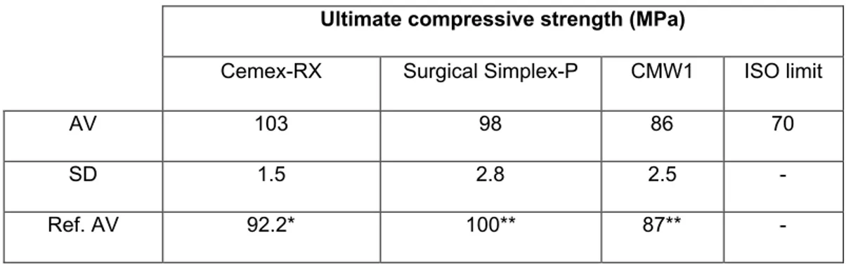
Obtained results from compressive test and four-point bending have been presented adequately in Table1 and Table2.
Table 1- Compressive mechanical properties of three commercial bone cements. Relevance with the
literature: * Kühn K.D. [1], **Hansen, D.; Jensen, J. S., [2]
Ultimate compressive strength (MPa)
Cemex-RX Surgical Simplex-P CMW1 ISO limit
AV 103 98 86 70
SD 1.5 2.8 2.5
-Ref. AV 92.2* 100** 87**
-Table 2- Results of four-point bending test and correspondence with data from literature. Relevance
with the literature: *Baleani et al., [3], **Weber et al., [4], *** Hansen, D.; Jensen, J. S., [2], **** Kühn K.D., [1]
Ultimate bending strength (MPa) Elastic modulus (MPa)
Cemex-RX Simplex-PSurgical CMW1 ISOlimit Cemex-RX Simplex-PSurgical CMW-1 ISOlimit
AV 58 53 64 50 2503 2460 3027 1800
SD 3.6 5.1 7.2 - 111 135 19
-Ref.
-2.4 Discussion
The purpose of this study was to acquire necessary skills to guarantee reproducibility of specimen fabrication in later steps of the whole project. For this reason a series of mechanical tests based on ISO 5833 was performed. Three bone cements selected for this project have been compared in terms of compressive and bending properties.
Obtained results are in correspondence with data found in literature. Existing small differences between our results and previously published data can be due to differences in bone cement storage, mixing time or environment (polymerization of acrylic cements is extremely sensitive even to negligible variation of mentioned factors). The results obtained through testing protocol defined in ISO 5833 standards, indicated CMW1 as the cement type with the best bending strength, comparing with two other cement types. This observation is opposite to clinical outcomes [5]. That experiment confirms the hypothesis that additional testing methods need to be defined in ISO standards to validate pre-clinically new cement brands.
2.5 References
1. Kühn, K.D., Bone cements. Up-to-date comparison of physical and chemical properties of commercial materials. 2000, Berlin: Springer.
2. Hansen, D. and J.S. Jensen, Mixing does not improve mechanical properties of all bone cements. Manual and centrifugation-vacuum mixing compared for 10 cement brands. Acta Orthopaedica, 1992. 63(1): p. 13-8.
3. Baleani, M., R. Fognani, and A. Toni, The influence of stem insertion rate on the porosity of the cement mantle of hip joint replacements. Proc Inst Mech Eng [H], 2003. 217(3): p. 199-205.
4. Weber, S.C. and W.L. Bargar, A comparison of the mechanical properties of Simplex, Zimmer and Zimmer lowviscosity bone cements. Biomater Med Devices Artif Organs, 1983. 11: p. 3-12.
5. Herberts, P. and H. Malchau, How outcome studies have changed total hip arthroplasty practices in Sweden. Clin Orthop Relat Res, 1997(344): p. 44-60.
III FRACTURE PROPERTIES
OF AN ACRYLIC BONE CEMENT
Published on Acta of Bioengineering and Biomechanics 2008; 10(1):21-6
E. Bialoblocka-Juszczyk 1,2, M. Baleani1, L. Cristofolini1,2 and M. Viceconti1
1Medical Technology Laboratory, Rizzoli Orthopaedic Institute, Bologna, Italy 2Engineering Faculty, University of Bologna, Bologna, Italy
3.1 Abstract
This study investigated experimentally the fracture properties, i.e., the fatigue strength, the resistance to crack propagation and the fracture toughness, of an acrylic bone cement (Cemex RX). The mean endurance limit was determined following the staircase method. The endurance limit was estimated at 9.2 MPa. The fatigue crack propagation rate was measured according to the ASTM E647 standard. The equation of the line fitting the crack growth per cycle (da/dN) versus the stress-intensity factor range (delta K), in a log-log graph, was used to calculate the empirical constants of Paris' law for the selected bone cement: a/dN (m/cycle) = 3.56 x 10 (-7) x delta K (MPa x m1/2) 5.79. This power-law relationship described well (R2 = 0.96) the growth rate in the stable crack growth region, i.e., in the mid delta K range. The fracture toughness K(IC) of the bone cement was determined according to the ASTM E399 standard. The K(IC) mean value was 1.38 MPa x m1/2. These experimental results provide the set of necessary inputs for numerical studies aimed to investigate the damage accumulation process in the mantle fixing cemented prostheses.
Keywords
3.2 Introduction
Polymethylmethacrylate (PMMA) based bone cement is the most common, commercially available material used in the orthopaedic field to fix cemented prostheses to the hosting bone. The use of PMMA assures an optimal implant stability after the surgical session which should be guaranteed for the entire implant life.
Clinical data from Swedish Total Hip Replacement Register show that aseptic loosening has caused nearly 60% of the failures in cemented implants during the last 26 years [1]. Many causes may contribute to the complex phenomenon which causes implant loosening. Among others, one of the potential causes for aseptic loosening is the long-term mechanical performance of the cement mantle. It has to transfers loads, generated during daily activities, from the implant to the periprosthetic tissues. Therefore, in vivo the cement mantle undergoes complex cyclic loadings. These loads cause cyclical stresses which may crumble the cement mantle [2, 3], promoting loosening of the prosthetic component [4-12].
The long-term behaviour of the cement mantle depends on the mechanical properties of the bone cement and how it is stressed in vivo [5, 13-16]. The former are characteristics of the materials itself, generally referred to as fracture properties [17]. However, the stress levels within the cement mantle are affected by prosthesis design, mantle thickness and quality, and support of the bone tissue surrounding the implant [9, 18-22]. The prosthesis-cement mantle-bone system can be investigated by means of Finite Element Models (FEMs). FEMs are used to calculate the stress within the mantle and to predict the fatigue damage under simulated physiological conditions [6, 10, 23-25]. These studies require the knowledge of the fracture properties of the bone cement.
Although there are many reports about the mechanical characteristics of bone cement [14, 26-31], a complete characterisation of the fracture properties of a commercial bone cement is missing. The aim of this study is to determine all the fracture properties, that are the fatigue strength, the resistance to crack propagation and the fracture toughness, of a PMMA based radiopaque bone cement.
3.3 Materials and methods
3.3.1 Materials and specimens preparation
Cemex RX (Tecres, Verona, Italy) was selected for this study. It is a PMMA based bone cement. A percentage of 9% barium sulphate was present in this formulation to assure the required radiopacity, as in most of the commercially available formulations of bone cements. The bone cement was mixed at a temperature of 23±1°C and at a relative humidity falling in the range of 40-60%, in agreement to ISO 5833 recommendations. The bone cement was mixed in air and, once reached the doughing time, the dough was poured into the moulds to cast the specimens of defined dimensions (see Figure 1). After 1h of polymerization specimens were stored in saline solution at 37°C for 14 days before testing. Prior to testing both sides of each specimen were polished using 800-grid sand paper to adjust the thickness to the desired value, with an accuracy of 0.1 mm. Before testing, specimens were X-ray checked, rejecting all ones with macro-porosity (pore diameter>1 mm) in the working region [17, 32]. Since this inspection was not possible for the 10 mm thick specimens, the fracture surface was examined after testing: if a macro-porosity was found on the crack surface, the specimen was discarded [31]. All the specimens were tested in air at 23±1°C on a material testing machine (MTS Mini Bionix 858, MTS System Corp., Minneapolis, MN). The frequency of cyclic loading was set to 4 Hz.
3.3.2 Fatigue testing
Fatigue tests were performed on dog-bone like specimens. The dimensions and geometry of the specimen were chosen in agreement to ISO 527-2. Working part dimensions were: length (l) 80mm, width (w) 10 mm, and thickness (t) 4 mm (figure 1).
Fig.1 The dimensions of the dog-bone like specimen and the C(T) specimen. t indicates the
thickness of the specimen.
Fatigue testing were performed at selected load levels until specimen fracture or runout (test completed). Runout was fixed at 10 million cycles. Preliminary testing were performed above the rough estimated endurance limit. This series continued decreasing the load level until a specimen did not fail during the test, i.e. reached 10 million cycles. At this point the up-and down scheme of the straircase method started and continued until 15 specimens were tested in the failure-not failure region. The collected data were used to determine the median endurance limit [33].
3.3.3 Fatigue crack propagation testing
The crack propagation rate was measured according to a method based on ASTM E647. Standard compact-type (C(T)) specimens were moulded. Specimen dimensions were: width (w) 40 mm, and thickness (t) 4 mm (figure 1).
A razorblade was used to produce a pre-crack in the specimen notch before subjecting the specimen to cyclic loading. A sinusoidal tensile load between 0 and 60 N was applied. Before testing, Krak Gages (Mod. B20CE, Rumul, Switzerland) were attached to both sides of the specimen to allow the measurement of the crack length (a) during the test. The number of load cycles
(N) was recorded at each crack length increment of 0.4±0.1mm. Five test repetitions were performed. A linear regression was used to fit the data, plotted in a log-log graph of the crack growth per cycle (da/dN) versus the stress-intensity factor range (∆K). The regression equation was used to calculate the empirical constants C and n of the Paris’ law (da/dN = C*(∆K)n). This power-law relationship describes the growth rate in the stable crack growth region of the log-log graph, referred to as region II [33].
3.3.4 Fracture toughness testing
The fracture toughness was determined according to a method based on ASTM E399. Specimen dimensions were: width (w) 10 mm, and thickness (t) 10 mm (figure 1). Preliminary, the specimens were pre-cracked by applying a cyclic load. To maintain the pre-crack growth rate in the order of 10-3 mm/cycle, four decreasing load levels were chosen. The crack length was monitored by means of an extensometer attached to the specimen mouth. Pre-cracking was stopped when the a/W ratio fell in the range of 0.45-0.55. Then the specimen was preloaded with 100 N and subjected to a monotonic tensile test at a crosshead rate of 10 mm/min. The load and the corresponding crack opening were recorded throughout the test to calculate the critical stress intensity value, KIC, according to the ASTM
standard. Experimental series continued until five valid specimens were tested.
3.4 Results
3.4.1 Fatigue testing
15 specimens were tested in the failure-not failure region. Six specimens completed the test. As runout was the less frequent event, its occurrence was used to estimate the mean endurance limit. The specimen fraction not-failed at 360N was 60%, at 370N was 40%, while all specimens failed at 380N. On the basis of these experimental data, the mean endurance limit was estimated to 9.2MPa. 3.4.2 Fatigue crack propagation testing
A set of fatigue crack growth data versus stress-intensity range was collected for each specimen. All the five sets are plotted in Figure 2 together with the regression line.
Fig.2 A linear line fitting the fatigue crack growth data for the investigated bone cement.
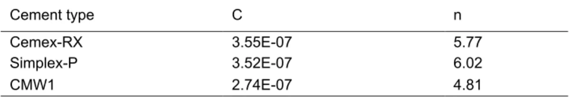
The coefficient of determination was R2=0.96. From the equation of the regression line the constants C and n of the Paris’ law were calculate: C=3.56*10 -7 (m/cyclex(MPa * m_)-n); n=5.79.
3.4.3 Fracture toughness testing
Seven C(T) specimens were tested. Two of these were excluded since a macro-porosity was found on the fracture surface, and therefore the KIC value was
not calculated from the experimental data. The KIC mean value calculated for the
five valid specimens was 1.38 MPa x m1/2. The coefficient of variation for KIC of
these five specimens was 3.6%.
3.5 Discussion
This study aimed to assess the fracture properties of a commercial, PMMA base, bone cement. The fracture properties characterise the material behaviour
![Table 2- Chemical composition of selected bone cements [1], where:](https://thumb-eu.123doks.com/thumbv2/123dokorg/8217082.128347/25.918.141.783.349.735/table-chemical-composition-selected-bone-cements.webp)
![Table 1 – Chemical composition of selected bone cements [22], where: PMMA - -poly(methylmethacrylate), MMA - methylmetacrylate DmpT - N,N-dimethyl-p-toluidine, BaSO 4 -barium sulphate as opacifier HQ - hydroquinone as stabilizer, BPO - benzoy](https://thumb-eu.123doks.com/thumbv2/123dokorg/8217082.128347/61.892.137.758.210.685/chemical-composition-methylmethacrylate-methylmetacrylate-toluidine-opacifier-hydroquinone-stabilizer.webp)