Research Article
Epoetin Alpha and Epoetin Zeta: A Comparative Study on
Stimulation of Angiogenesis and Wound Repair in an
Experimental Model of Burn Injury
Natasha Irrera,
1Alessandra Bitto,
1Gabriele Pizzino,
1Mario Vaccaro,
1Francesco Squadrito,
1Mariarosaria Galeano,
2Francesco Stagno d’Alcontres,
2Ferdinando Stagno d’Alcontres,
2Michele Buemi,
1Letteria Minutoli,
1Michele Rosario Colonna,
2and Domenica Altavilla
31Department of Clinical and Experimental Medicine, University of Messina, 98125 Messina, Italy
2Department of Dentistry and Medical and Surgical Experimental Sciences, University of Messina, 98125 Messina, Italy
3Department of Paediatric, Gynaecological, Microbiological and Biomedical Sciences, University of Messina, 98125 Messina, Italy
Correspondence should be addressed to Francesco Squadrito; [email protected] Received 19 December 2014; Revised 2 April 2015; Accepted 15 April 2015
Academic Editor: Themis R. Kyriakides
Copyright © 2015 Natasha Irrera et al. This is an open access article distributed under the Creative Commons Attribution License, which permits unrestricted use, distribution, and reproduction in any medium, provided the original work is properly cited. Deep second-degree burns are characterized by delayed formation of granulation tissue and impaired angiogenesis. Erythropoietin (EPO) is able to stimulate angiogenesis and mitosis, activating vascularization and cell cycle. The aim of our study was to investigate whether two biosimilar recombinant human erythropoietins, EPO-𝛼 and EPO-Ζ, may promote these processes in an experimental model of burn injury. A total of 84 mice were used and a scald burn was produced on the back after shaving, in 80∘C water for 10 seconds. Mice were then randomized to receive EPO-𝛼 (400 units/kg/day/sc) or EPO-Ζ (400 units/kg/day/sc) or their vehicle (100𝜇L/day/sc 0.9% NaCl solution). After 12 days, both EPO-𝛼 and EPO-Ζ increased VEGF protein expression. EPO-𝛼 caused an increased cyclin D1/CDK6 and cyclin E/CDK2 expression compared with vehicle and EPO-Z (𝑝 < 0.001). Our study showed that EPO-𝛼 and EPO-Ζ accelerated wound closure and angiogenesis; however EPO-𝛼 resulted more effectively in achieving complete skin regeneration. Our data suggest that EPO-𝛼 and EPO-Ζ are not biosimilars for the wound healing effects. The higher efficacy of EPO-𝛼 might be likely due to its different conformational structure leading to a more efficient cell proliferation and skin remodelling.
1. Introduction
Erythropoietin (EPO) is a renal glycoprotein hormone that regulates red blood cell production by inhibiting apoptosis of erythroid precursor cells, but it is also involved in the mech-anisms of proliferation and differentiation in hematopoietic tissues [1,2]. EPO acts through its specific cytokine receptor, the erythropoietin receptor (EPOR), allocated on cell surface [3,4] of many different haematopoietic and nonhaematopoi-etic tissues and cells [5]. EPOR has been identified on neu-rons, astrocytes, microglia, endothelial cells, macrophages, fibroblasts, keratinocytes, mast cells, melanocytes, liver, and uterus. When activated, the receptor promotes the Janus
Kinase (JAK)/Signal Transducer and Activator of Transcrip-tion (STAT) pathway, stimulating mitosis and differentiaTranscrip-tion in all these cell lines [6,7].
Several studies have suggested that EPO may be involved in the regulation of physiological wound healing process. It has been demonstrated that locally erythropoietin admin-istration into subcutaneous fibrin chambers was able to promote the formation of wound granulation tissue in a rat model of wound healing [8]. This effect was ascribed to the stimulation of physiological angiogenesis and upregulation of iNOS expression in skin.
Furthermore, additional studies have shown that EPO may interact with vascular endothelial cells and vascular
Volume 2015, Article ID 968927, 9 pages http://dx.doi.org/10.1155/2015/968927
endothelial growth factor (VEGF), involved in the angiogenic process, thus stimulating cellular motility and mitosis in endothelium and leading to the formation of new capillaries [9, 10]. These experimental evidences prompted the use of EPO in improving impaired wound healing; as a matter of fact our group already demonstrated the efficacy of recombinant EPO in ameliorating skin repair during diabetes and burn injury [11,12].
Recombinant human erythropoietin (rHuEPO) has been largely used in the clinical setting and new formulations have been synthesized, such as Epoetin alpha (EPO-𝛼) and Epoetin Z (EPO-Z) [13–15]. EPO-𝛼 was the first genera-tion recombinant erythropoietin with the same amino acid sequence as human erythropoietin and it exerts the maxi-mum therapeutic effect when administered subcutaneously [16]. EPO-Z is a new generation biosimilar with the same amino acid sequence of EPO-𝛼 and overlapping carbohydrate composition; it may be used as an alternative choice for the treatment of patients with end-stage renal diseases and anemia. EPO-Z was developed and registered in agreement with the European Medicines Agency (EMEA) guidelines. The regulatory approval of EPO-Z is based both on extensive physicochemical/molecular, preclinical, and clinical charac-terization studies and on a comprehensive postmarketing surveillance program, as required for all European “biosimi-lars.”
However, biosimilars may have some differences in the materials used for their preparation and may show variability in manufacturing processes, and, in addition, the active form may differ with regard to the size and the complexity of the structure [17]. The biotechnological techniques used for the preparation of the compounds make the molecules more heterogeneous, and this is the result of changes in structure such as glycosylation and/or alterations in amino acid sequence. These variations are often responsible for immune effects [18–20].
Since the new EPO-Z has never been tested for its skin repair effects, the purpose of our study was to investigate whether EPO-𝛼 and EPO-Z may display overlapping specific action in an experimental model of burn injury.
2. Materials and Methods
2.1. Animals and Experimental Burn Model. The protocol
was evaluated and accepted by the Ethics Committee of the University of Messina and all animal procedures were carried out according to Guide for the Care and Use of Laboratory Animals. A total of 84 male C57BL/6 mice weighing 20–22 g were used in this study and were purchased by Charles River Italy (Calco, Italy). Animals were housed and maintained under controlled environmental conditions (12 h light/dark cycle, temperature approximately 23∘C) and provided with standard laboratory food and water ad libitum. Skin injury was performed on the shaved back of mice: after general anaesthesia with sodium pentobarbital (80 mg/kg/i.p.), hair on the back was shaved using a depilatory cream to reduce any possible injury due to hair removal. Burn injury was produced with 80∘C water, and mice were immersed for 10 seconds with a burn template so that the dorsum was
exposed to hot water through a 2× 3 cm window. In this way, we produced a deep-dermal second-degree burn; post-burn sedation and analgesia were provided with diazepam (50 mg/L added to drinking water) for 7 days to alleviate pain. Following thermal injury, animals were divided into 3 groups and randomized to receive EPO-𝛼 (400 I.U./kg/day in 100𝜇L 0.9% NaCl subcutaneously), EPO-Z (400 I.U./kg/day in 100𝜇L 0.9% NaCl subcutaneously), or vehicle (100 𝜇L/day 0.9% NaCl subcutaneously). The dose chosen in this study has been previously shown effective for EPO-𝛼 in previous studies for the treatment of wound healing [11, 12]. Nei-ther lower dose regimens nor different schedule treatments showed beneficial effects in preliminary experiments (results not shown). At 3, 6, and 12 days 7 animals per group were sacrificed and the remaining 7 animals from each group were observed and treated to assess the time to final wound closure. At the time of sacrifice blood was collected by cardiac puncture, and burn areas were excised and used for histological and molecular analysis.
2.2. Erythrocyte Count and Haemoglobin Level. Number of
erythrocytes and haemoglobin (Hb) level were evaluated from blood samples at 12 days after burn injury. Red blood cell count was carried out on stained smears according to the standard morphologic criteria for mouse [11,12].
2.3. Western Blot Determination of VEGF, Cyclin D1, CDK6, Cyclin E, CDK2, p15, and p27. Skin samples were
homog-enized in lysis buffer (1% Triton X100, 20 mM Tris/HCl, pH 8.0, 137 mM NaCl, 10% glycerol, 5 mM EDTA, 1 mM phenylmethylsulfonyl fluoride, 1% aprotinin, and 15𝜇g/mL leupeptin). Protein samples (30𝜇g) were denatured in a reducing buffer (62 mmol/L Tris pH 6.8, 10% glycerol, 2% SDS, 5%𝛽-mercaptoethanol, and 0.003% bromophenol blue) and separated by electrophoresis on a sodium dodecyl sul-phate (SDS) polyacrylamide gel (10% or 12%). The separated proteins were transferred onto a PVDF membrane using a transfer buffer (39 mmol/L glycine, 48 mmol/L Tris, and pH 8.3, 20% methanol) at 100 V for 1 hour. The membranes were stained with Ponceau S (0.005% in 1% acetic acid) to confirm blotting, blocked with 5% nonfat dry milk in TBS-0.1% Tween for 1 hour at room temperature, washed 3 times for 10 minutes each in TBS-0.1% Tween, and then incubated with a primary antibody for cyclin D1, CDK6, cyclin E, CDK2, p15, or p27 (Cell Signaling, Beverly, MA) and VEGF (Abcam, Cambridge, UK) in TBS-0.1% Tween overnight at 4∘C. The day after, antibody was removed by washing the membranes 3 times for 10 minutes each in TBS-0.1% Tween. Membranes were incubated with a secondary antibody peroxidase-conjugated (Pierce, Rockford, IL) for 1 hour at room temperature. After washing, the membranes were analyzed by the enhanced chemiluminescence system according to the manufacturer’s protocol (Amersham, Little Chalfont, UK). The VEGF, cyclin D1, CDK6, cyclin E, CDK2, p15, and p27 protein signals were quantified by scanning densitometry using a bioimage analysis system (Bio-Profil Celbio, Milan, Italy). Equal loading of protein was assessed on stripped blots by𝛽-actin staining with a rabbit monoclonal antibody (Cell Signaling, Beverly, MA).
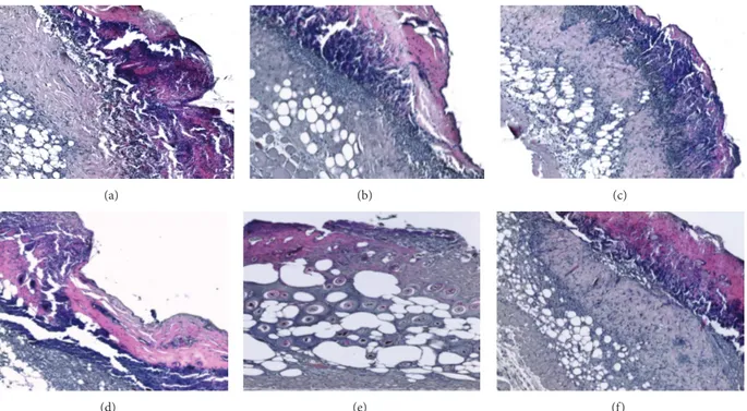
(a) (b) (c)
(d) (e) (f)
Figure 1: H&E stained sections of burned skin at days 3 and 6 examined under light microscopy. The untreated group (a) as well as groups treated with EPO-𝛼 (b) and EPO-Z (c) show extensive skin defects, necrosis and inflammatory cells in the subcutaneous layer, and absence of hair follicles and skin glands. At day 6 the untreated group (d) shows the persistence of the crust and of necrotic and inflammatory areas; EPO-𝛼 treatment (e) promotes the reduction of the inflammatory infiltrate, partial restoration of the dermal structure with the presence of hair follicles and sweat glands; EPO-Z treated skin (f) shows the presence of a partial regeneration behind the crust, with some areas of necrosis and inflammatory cells infiltration.
2.4. Histologic Evaluation. Skin samples were removed from
the animals of each experimental group and fixed in 10% buffered formalin at room temperature for at least 24 hours. Perpendicular sections to the anterior-posterior axis of the burn area were dehydrated in graded ethanol, cleared in xylene, and embedded in paraffin according to rou-tine procedures. Five-micrometer-thick sections of paraffin-embedded tissues were mounted on glass slides, hydrated to distilled water, and then stained with hematoxylin and eosin. In particular, histological slides were examined at×10 to×20 magnification to observe morphologic alterations or improvements (i.e., presence or absence of reepithelialization, crusting, blistering, granulation tissue and collagen matrix organization, inflammation, congestion, and edema), as pre-viously reported [11, 12]. Evaluations were obtained from a pathologist without knowledge of the treatments.
2.5. Immunohistochemistry for CD31. Paraffin-embedded
tis-sues were sectioned (5𝜇m) and rehydrated, and antigen unmasking was performed by using 0.05 M sodium citrate buffer (pH 6.0). Specimens were treated with 3% hydrogen peroxide to block endogenous peroxides and with horse normal serum (Vector Laboratories, Burlingame, CA, USA) to avoid nonspecific staining. Slides were then incubated overnight at 4∘C in a humid box with primary antibody to detect CD31 (Abcam, Cambridge, UK). The day after, sections were washed with PBS, a secondary antibody was used
(Vector Laboratories), and the reaction was revealed adding a DAB solution (diaminobenzidine tetrahydrochloride, Sigma, Milan, Italy) for 1–3 minutes. Slides were counterstained with haematoxylin, dehydrated, mounted, and examined by a pathologist without knowledge of the treatments, by using masked slides from×10 to ×40 magnification with a Leica (Leica Microsystems, Milan, Italy) microscope.
2.6. Statistical Analysis. All data are expressed as means±
S.D. Comparisons between different treatments were anal-ysed by one-way ANOVA followed by Bonferroni’s multiple-comparison test. The histological score analysis was carried out by two-way ANOVA followed by Bonferroni’s multiple-comparison test. The possibility of error was set at𝑝 < 0.05 and it was considered statistically significant. Graphs were drawn using GraphPad Prism (version 5.0 for Windows).
3. Results and Discussion
3.1. Erythrocyte Count and Blood Hb. Since EPO is effective
in stimulating the erythroid lineage, we evaluated the number of red cells and Hb in our experimental groups, to confirm that EPO-𝛼 and EPO-Z may promote the production of erythrocytes and haemoglobin. We have already observed in our previous studies that rHuEPO stimulates erythroid lineage [11,12]. As expected, the administration of EPO-𝛼 and EPO-Z increased both the number of circulating erythrocytes
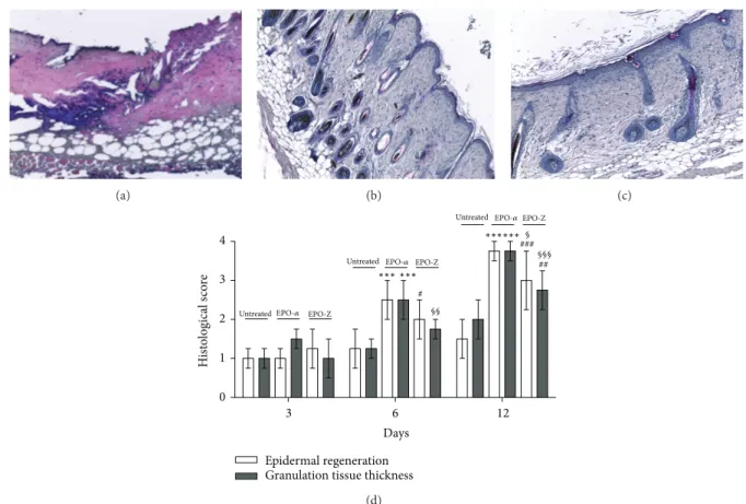
(a) (b) (c) 3 6 12 0 1 2 3 4 Epidermal regeneration Granulation tissue thickness
### ## # Days H ist o logical s co re ∗∗∗∗∗∗ ∗∗∗ ∗∗∗ EPO-𝛼 EPO-Z EPO-𝛼 EPO-Z §§ § §§§ Untreated Untreated EPO-𝛼 EPO-Z Untreated (d)
Figure 2: H&E stained sections of burned skin at day 12 observed under light microscopy. Skin of untreated animals (a) shows necrosis surrounded by a contraction of the panniculus carnosus as well as disorganized dermal-epidermal layers. Animals treated with EPO-𝛼 (b) demonstrate a well regenerated tissue with a keratin layer and reorganized subdermal structures. Animals treated with EPO-Z (c) show a still incomplete healing process characterized by a linear external layer with a low staining of hair keratin compared with the EPO-𝛼 treated group. The graph (d) represents the mean of epidermal regeneration and granulation tissue thickness at several time points in the studied animals.∗∗∗𝑝 < 0.001 versus untreated;#𝑝 < 0.05 versus untreated;#𝑝 < 0.05 versus untreated;###𝑝 < 0.001 versus untreated;§𝑝 < 0.05 versus EPO-Z;§§𝑝 < 0.01 versus EPO-Z;§§§𝑝 < 0.001 versus EPO-Z. Significance between treatments is considered within the same day of observation.
and Hb at day 12 (data not shown). No difference was observed between the two compounds. Our results confirm that these two molecules equally stimulate erythroid lineage, as reported in clinical studies, and act as biosimilars [15].
3.2. Skin Repair. Wound healing is characterized not only by
the stimulation of angiogenesis, but also by the formation and remodelling of matrix components and, in this setting, macrophages are responsible for the production of growth factors. Since EPOR is located on the surface of these cells, EPO may stimulate macrophages, and this mechanism of action explains the role of erythropoietin in the skin repair process [21].
Additionally, an in vitro study, evaluating the effects of EPO and EPO receptors in human hair follicles (HFs) [22], showed that also hair follicle cells synthesize EPO protein in situ and express EPO receptor. These structures are well-known not only for producing hair but also for their impor-tant contribution to tissue repair through reepithelialization, and both investigators and clinicians have noted for years that hair-bearing areas tend to heal more quickly than areas lacking follicles [23, 24]. Langton et al. [25] demonstrated
that acute wound healing is delayed in absence of HFs. HFs-derived keratinocytes are responsible for reepithelization during wound healing [26], while HF dermal sheath cells are a key source of fibroblasts in response to wounding and have a role in replacing skin dermis [27].
In our study, at day 3, control group as well as groups treated with EPO-𝛼 and EPO-Z showed extensive skin defects, necrosis and inflammatory cells in the subcutaneous layer, and absence of hair follicles and skin glands. At day 6, the control group showed the persistence of the crust and of necrotic and inflammatory areas. The treatment with EPO-𝛼 promoted the reduction of the inflammatory infiltrate and determined partial restoration of the dermal structure with the presence of hair follicles and sweat glands; instead EPO-Z treated skin showed only the presence of a partial regeneration behind the crust, with some areas of necrosis and inflammatory cells infiltration (Figure 1).
At the end of experimental period (day 12), skin of control animals showed necrosis surrounded by a contrac-tion of the panniculus carnosus as well as disorganized dermal-epidermal layers. We observed that animals treated with EPO-𝛼 demonstrated a well regenerated tissue with
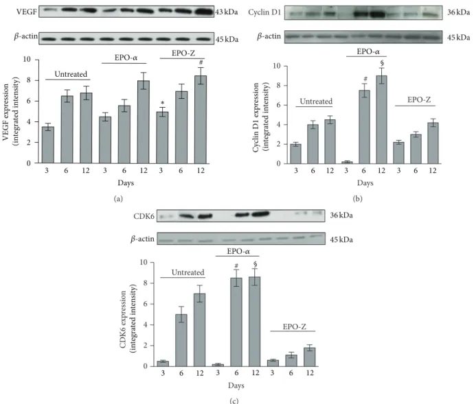
VEGF 0 2 4 6 8 10 Untreated EPO-Z # Days 3 6 12 3 6 12 3 6 12 EPO-𝛼 43 kDa 45 kDa 𝛽-actin VEGF exp re ss io n (in tegra te d in te n si ty) ∗ (a) 0 2 4 6 8 10 Untreated EPO-Z Days # EPO-𝛼 3 6 12 3 6 12 3 6 12 C yc lin D 1 exp re ssio n (in te gra ted in te n si ty) 𝛽-actin 36 kDa 45 kDa § Cyclin D1 (b) 0 2 4 6 8 10 Untreated EPO-Z # Days CDK6 3 6 12 3 6 12 3 6 12 EPO-𝛼 CD K6 exp ressio n (in tegra te d in te n si ty) 𝛽-actin 36 kDa 45 kDa § (c)
Figure 3: Effects of EPO-𝛼 and EPO-Z on VEGF (a) protein expression in skin tissue samples. EPO-𝛼 increases the expression of VEGF at all time points even if not significantly. The administration of EPO-Z significantly increases the expression of VEGF at both 3 and 12 days. ∗𝑝 < 0.01 versus untreated;#𝑝 < 0.01 versus untreated. Effects of EPO-𝛼 and EPO-Z treatment on cyclin D1 (b) and CDK6 (c) proteins expression in skin tissue samples.#𝑝 < 0.001 versus untreated and EPO-Z,§𝑝 < 0.001 versus untreated and EPO-Z. Values are expressed as mean and SD for each group. Significance between treatments is considered within the same day of observation.
a keratin layer and reorganized subdermal structures. Ani-mals treated with EPO-Z showed a still incomplete healing process compared with the EPO-𝛼 treated group (Figure 2).
Therefore, the use of EPO-𝛼 caused reduced inflamma-tion, restored dermal layers, and accelerated complete healing with a “restitutio ad integrum” of skin structures, such as hair follicles. By contrast, EPO-Z did not determine a complete healing compared with EPO-𝛼: tissue structures were orga-nized, inflammation was reduced, and matrix components were visible but incompletely formed.
3.3. Angiogenesis: Expression of VEGF. A deep second-degree
burn is characterized by the disruption and alteration of the whole epidermis; furthermore, the burned area shows a non-regenerating, mesenchyma-mediated wound healing, with delayed formation of granulation tissue, decreased deposition of collagen, and impaired angiogenesis. EPO can protect
epidermis, dermis, and its vital structures, in particular capillaries and blood vessels, from further damage following burn injury [28]. In fact, it has been demonstrated that erythropoietin may also act on nonhaematopoietic tissues via the EPOR, stimulating tissue healing, particularly following injury [29,30].
The angiogenic process is the result of the interaction of several factors and VEGF has a pivotal role in orchestrating wound healing, promoting endothelial cell proliferation from the very first moments. In a previous study we demonstrated that the synergism between VEGF and EPO promotes the angiogenic process, confirming the role of rHuEPO as a growth factor in an experimental model of wound healing [11].
In the present experiment we compared EPO-𝛼 and EPO-Z in the stimulation of angiogenesis. Physiologically, the healing of burn wounds causes an increase in VEGF
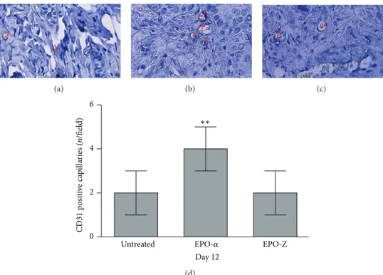
(a) (b) (c) 0 2 4 6 Untreated EPO-Z Day 12 ∗∗ EPO-𝛼 CD 31 p osi ti ve ca p illa ries (n /field) (d)
Figure 4: CD31 immunostaining of burned skin from mice untreated (a) or treated with EPO-𝛼 (b) or EPO-Z (c) at day 12. Arrows indicate staining positivity in small capillaries; administration of EPO-𝛼 stimulates neovessel formation, as shown in the graph, (d) more than EPO-Z (∗∗𝑝 = 0.0017 versus untreated and EPO-Z).
compared to normal conditions. For this reason, we studied VEGF expression in wounds at days 3, 6, and 12 after treatment. As shown in Figure 3, EPO-𝛼 and of EPO-Z stimulated VEGF expression at all time points, and EPO-Z stimulation was significant at days 3 and 12. The reason for this apparent discrepancy could be due to the fact that VEGF is not the only and one factor involved in new vessel formation, and indeed during impaired wound healing conditions the receptor of VEGF (VEGF-R2) and other growth mediators are downregulated. As a matter of fact, despite a lower increase in VEGF (as compared to EPO-Z), EPO-𝛼 caused a faster and more organized skin healing underlying that VEGF increase alone is not sufficient. To prove the efficacy of angiogenesis stimulation, besides VEGF synthesis, we investigated the newly formed capillaries through CD31 immunostaining which was investigated in skin samples at days 6 and 12. At day 6 no CD31 staining was observed in skin tissue (data not shown). As demonstrated
inFigure 4, at day 12 untreated animals presented a positive
staining (Figure 4(a)) and the administration of EPO-𝛼 further increased the staining in small capillaries, indicating a sustained angiogenesis (Figure 4(b)). EPO-Z did not improve neovascularisation at day 12, confirming the delayed healing observed with H&E staining (Figure 4(c)).
3.4. Regulation of Cell Cycle Machinery. The processes of
(i) angiogenesis, (ii) matrix remodelling, and (iii) wound healing are promoted by activation of cell cycle and cell proliferation executed in a timely and orderly manner. The phases of cell cycle in mammalian cells are controlled by
different types of cyclins. Cyclins, cyclins-dependent kinases (CDKs), and negative regulators, such as cyclin-dependent kinase inhibitors belonging to the p16 and p21 family, work together to regulate cell cycle progression. Cyclin D1 and cyclin E are fundamental in the activation of the G1 phase. Cyclin D1 and cyclin E increase is followed by a concomitant upregulation of CDK6 and CDK2 activity. Both complexes trigger progression of the G1 phase and entry into the S phase. Cell cycle negative regulators are divided into 2 families, the p21 family (p21, p27, and p57) and the p16 family (p15, p16, p18, and p19). Any alteration in the complex equilibrium between cyclin/CDK complexes and their negative regulators could be responsible for an impaired cell growth and, in turn, for a delayed tissue repair [31,32].
It has been demonstrated that EPO gene expression is related to the upregulation of cyclins/CDK and the admin-istration of EPO may promote DNA synthesis, as well as cell proliferation, and upregulates the expression of cyclins [33]. In agreement with this finding, it has also been shown that improvement of neovascularisation induced by EPO may be attributed to a direct effect mediated not only by VEGF, but also by endothelial cell proliferation [34].
In our experiment we tested not only 𝛼 and EPO-Z for the same efficacy in promoting wound healing in experimental burn wounds but also whether these effects were dependent upon the activation of cyclins and cyclin-dependent kinases (CDKs). The cell cycle positive regulators were evaluated at days 3, 6, and 12 following burn injury. The burn wounds of mice treated with vehicle showed a moderate expression of cyclin D1 and CDK6 at days 3, 6, and 12
0 2 4 6 8 10 # Untreated EPO-Z Days EPO-𝛼 ∗ 3 6 12 3 6 12 3 6 12 𝛽-actin 45 kDa 48 kDa C yc lin E exp re ssio n (in tegra te d in te n si ty) § §§ Cyclin E (a) 0 2 4 6 8 10 Untreated EPO-Z # CDK2 EPO-𝛼 Days 3 6 12 3 6 12 3 6 12 𝛽-actin 45 kDa 33 kDa CD K 2 exp re ssio n (in tegra te d in te n si ty) (b) 0 2 4 6 8 10 Untreated EPO-Z # Days p15 EPO-𝛼 ∗ 3 6 12 3 6 12 3 6 12 𝛽-actin 45 kDa 15 kDa p15 exp res sio n (in tegra te d in te n si ty) § (c) 0 2 4 6 8 10 Untreated EPO-Z # p27 EPO-𝛼 Days 3 6 12 3 6 12 3 6 12 𝛽-actin 45 kDa 27 kDa p27 exp res sio n (in tegra te d in te n si ty) § (d)
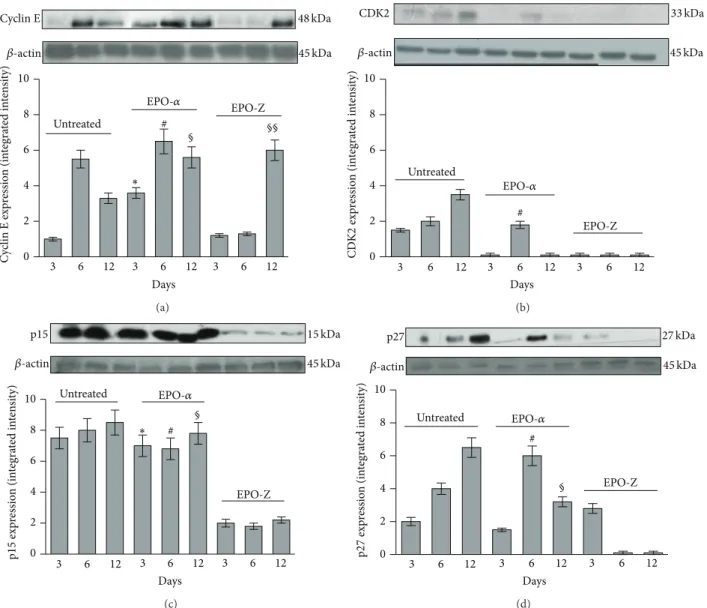
Figure 5: Effects of EPO-𝛼 and EPO-Z treatment on cyclin E (a) and CDK2 (b) protein expression in skin tissue samples.∗𝑝 < 0.001 versus untreated;#𝑝 < 0.001 versus EPO-Z;§𝑝 < 0.001 versus untreated and EPO-Z. Effects of EPO-𝛼 and EPO-Z treatment on p15 (c) and p27 (d) protein expression in skin tissue samples.∗𝑝 < 0.001 versus EPO-Z;#𝑝 < 0.001 versus EPO-Z;§𝑝 < 0.001 versus EPO-Z. Values are expressed as mean and SD for each group. Significance between treatments is considered within the same day of observation.
(Figures3(b)and3(c)). The administration of EPO-𝛼 signif-icantly upregulated the expression of cyclin D1 at days 6 and 12 (𝑝 < 0.001) compared to the treatment with vehicle and EPO-Z (Figure 3;𝑝 < 0.001).
CDK6, the kinase responsible for the activation of cyclin D, showed a trend similar to its cyclin in wounds treated with vehicle (Figures3(b)and3(c)). The administration of EPO-𝛼 determined a significant increase in the expression of CDK6 at days 6 and 12 (𝑝 < 0.001). EPO-Z did not cause significant differences in the expression of CDK6.
The expression of cyclin E in skin of mice treated with vehicle increased only at day 6. The treatment with EPO-𝛼 significantly enhanced the expression of cyclin E at all time points; the administration of EPO-Z increased cyclin E
expression only at day 12 (Figure 5(a)). CDK2 expression did not show any significant upregulation following the treatment with both EPO-𝛼 and EPO-Z compared to the animals treated only with vehicle (Figure 5(b)).
Interestingly, we observed an opposite situation for the expression of p15 protein: in fact, in this case, the treatment with EPO-Z reduced only the expression of the inhibitor p15 in a significant manner and at all time points, while EPO-Z did not modify the expression of the cyclins (Figure 5(c)). However, the only reduction in the expression of the inhibitor p15 is not sufficient to activate the cell cycle and accelerate the wound healing process following burn injury.
On the other hand, the treatment with EPO-𝛼 did not modify the expression of this inhibitor in a significant way.
The expression of p27 was increased at days 6 and 12 in burn mice treated with vehicle and EPO-𝛼 and only at day 3 in mice treated with EPO-Z (Figure 5(d)).
Under normal conditions, the progression of granulation tissue and cell proliferation are controlled by cyclin D1/CDK6 and cyclin E/CDK2 complexes. Our data demonstrate that EPO-𝛼 increased more efficiently the expression of cyclins. In this way, cell cycle is certainly promoted and this event contributes to the regeneration of the burned tissue and its damaged structures. Therefore it is tempting to speculate that the better wound healing effect of EPO-𝛼 is due to a greater ability to prime the cell cycle machinery.
4. Conclusions
It has been demonstrated that EPO represents particularly promising approach for the treatment of burn wounds [35– 37]. EPO-𝛼 and EPO-Z are biosimilars, and they should have overlapping therapeutic efficacy. In fact both compounds did not show significant differences in Hb levels, in ery-throcytes number, and in expression of VEGF. EPO-𝛼 and EPO-Z also accelerate wound closure and angiogenesis, but EPO-𝛼 resulted more effectively in achieving complete skin regeneration. We hypothesize that the superiority in this specific therapeutic effect of EPO-𝛼 with respect to EPO-Z might be related to a more robust activation of the cell cycle machinery. At this moment, we do not have a clear-cut explanation for this “dissimilarity.” It could be hypothesized that molecular differences in manufacturing processes used for the preparation of these two formulations or changes in structure, for example, in glycosylation of some residues, may lead to a different activation of cell cycle and may account for the different wound healing effect. In addition, these possible conformational changes might be responsible for a lower receptor affinity that determines an alteration of receptor bond and therefore of the correlated signalling of response.
However further experiments are needed to fully eluci-date the molecular mechanisms underlying this difference between the two biosimilars.
All these data propose that the abnormalities of wound healing in burn injury might be a result of significant alterations in the cell cycle machinery of the repair tissue, and the administration of EPO might be suggested as a potential strategy to restore the impaired wound healing, stimulating both angiogenesis and cell cycle of the damaged burned tissue.
Conflict of Interests
The authors declare that there is no conflict of interests regarding the publication of this paper.
Authors’ Contribution
Natasha Irrera and Alessandra Bitto equally contributed to this work.
References
[1] W. Jelkmann, “Biology of erythropoietin,” Clinical Investigator, vol. 72, no. 6, pp. S3–S10, 1994.
[2] M. Piagnerelli and J.-L. Vincent, “The use of erythropoiesis-stimulating agents in the intensive care unit,” Critical Care
Clinics, vol. 28, no. 3, pp. 345–362, 2012.
[3] H. Youssoufian, G. Longmore, D. Neumann, A. Yoshimura, and H. F. Lodish, “Structure, function, and activation of the erythropoietin receptor,” Blood, vol. 81, no. 9, pp. 2223–2236, 1993.
[4] J. Rossert and K.-U. Eckardt, “Erythropoietin receptors: their role beyond erythropoiesis,” Nephrology Dialysis
Transplanta-tion, vol. 20, pp. 1025–1028, 2005.
[5] M. Brines and A. Cerami, “Discovering erythropoietin’s extra-hematopoietic functions: biology and clinical promise,” Kidney
International, vol. 70, no. 2, pp. 246–250, 2006.
[6] F. P. Barbone, D. L. Johnson, F. X. Farrell et al., “New epoetin molecules and novel therapeutic approaches,” Nephrology
Dial-ysis Transplantation, vol. 14, no. 2, pp. 80–84, 1999.
[7] P. J. Connolly, S. K. Wetter, W. V. Murray et al., “Synthesis and erythropoietin receptor binding affinities of N, N-disubstituted amino acids,” Bioorganic and Medicinal Chemistry Letters, vol. 10, no. 17, pp. 1995–1999, 2000.
[8] Z. A. Haroon, K. Amin, X. Jiang, and M. O. Arcasoy, “A novel role for erythropoietin during fibrin-induced wound-healing response,” American Journal of Pathology, vol. 163, no. 3, pp. 993–1000, 2003.
[9] D. Ribatti, M. Presta, A. Vacca et al., “Human erythropoietin induces a pro-angiogenic phenotype in cultured endothelial cells and stimulates neovascularization in vivo,” Blood, vol. 93, no. 8, pp. 2627–2636, 1999.
[10] M. Buemi, A. Lacquaniti, D. Bolignano et al., “The erythropoi-etin and regenerative medicine: a lesson from fish,” European
Journal of Clinical Investigation, vol. 39, no. 11, pp. 993–999,
2009.
[11] M. Galeano, D. Altavilla, D. Cucinotta et al., “Recombinant human erythropoietin stimulates angiogenesis and wound heal-ing in the genetically diabetic mouse,” Diabetes, vol. 53, no. 9, pp. 2509–2517, 2004.
[12] M. Galeano, D. Altavilla, A. Bitto et al., “Recombinant human erythropoietin improves angiogenesis and wound healing in experimental burn wounds,” Critical Care Medicine, vol. 34, no. 4, pp. 1139–1146, 2006.
[13] C. Baldamus, S. Krivoshiev, M. Wolf-Pflugmann, M. Siebert-Weigel, R. Koytchev, and A. Bronn, “Long-term safety and tolerability of epoetin zeta, administered intravenously, for maintenance treatment of renal anemia,” Advances in Therapy, vol. 25, no. 11, pp. 1215–1228, 2008.
[14] V. Wizemann, B. Rutkowski, C. Baldamus, P. Scigalla, and R. Koytchev, “Comparison of the therapeutic effects of epoetin zeta to epoetin alfa in the maintenance phase of renal anemia treatment,” Current Medical Research & Opinion, vol. 24, no. 3, pp. 625–637, 2008.
[15] S. Krivoshiev, V. V. Todorov, J. Manitius, S. Czekalski, P. Scigalla, and R. Koytchev, “Comparison of the therapeutic effects of epoetin zeta and epoetin alfa in the correction of renal anaemia,”
Current Medical Research and Opinion, vol. 24, no. 5, pp. 1407–
1415, 2008.
[16] T. McGowan, N. M. Vaccaro, J. S. Beaver, J. Massarella, and M. Wolfson, “Pharmacokinetic and pharmacodynamic profiles of
extended dosing of epoetin alfa in anemic patients who have chronic kidney disease and are not on dialysis,” Clinical Journal
of the American Society of Nephrology, vol. 3, no. 4, pp. 1006–
1014, 2008.
[17] S. D. Roger, “Biosimilars: how similar or dissimilar are they?”
Nephrology, vol. 11, no. 4, pp. 341–346, 2006.
[18] G. Barosi, A. Bosi, M. P. Abbracchio et al., “Key concepts and critical issues on epoetin and filgrastim biosimilars: a position paper from the Italian Society of Hematology, Italian Society of Experimental Hematology, and Italian Group for Bone Marrow Transplantation,” Haematologica, vol. 96, no. 7, pp. 937–942, 2011.
[19] A. S. Tsiftsoglou, S. Ruiz, and C. K. Schneider, “Development and regulation of biosimilars: current status and future chal-lenges,” BioDrugs, vol. 27, no. 3, pp. 203–211, 2013.
[20] S. Tamilvanan, N. L. Raja, B. Sa, and S. K. Basu, “Clinical concerns of immunogenicity produced at cellular levels by biopharmaceuticals following their parenteral administration into human body,” Journal of Drug Targeting, vol. 18, no. 7, pp. 489–498, 2010.
[21] Z. A. Haroon, K. Amin, X. Jiang, and M. O. Arcasoy, “A novel role for erythropoietin during fibrin-induced wound-healing response,” American Journal of Pathology, vol. 163, no. 3, pp. 993–1000, 2003.
[22] B. M. Kang, S. H. Shin, M. H. Kwack et al., “Erythropoietin promotes hair shaft growth in cultured human hair follicles and modulates hair growth in mice,” Journal of Dermatological
Science, vol. 59, no. 2, pp. 86–90, 2010.
[23] G. H. Bishop, “Regeneration after experimental removal of skin in man,” American Journal of Anatomy, vol. 76, no. 2, pp. 153– 181, 1945.
[24] V. Martinot, V. Mitchell, P. Fevrier, A. Duhamel, and P. Pellerin, “Comparative study of split thickness skin grafts taken from the scalp and thigh in children,” Burns, vol. 20, no. 2, pp. 146–150, 1994.
[25] A. K. Langton, S. E. Herrick, and D. J. Headon, “An extended epidermal response heals cutaneous wounds in the absence of a hair follicle stem cell contribution,” Journal of Investigative
Dermatology, vol. 128, no. 5, pp. 1311–1318, 2008.
[26] M. Ito, Y. Liu, Z. Yang et al., “Stem cells in the hair follicle bulge contribute to wound repair but not to homeostasis of the epidermis,” Nature Medicine, vol. 11, no. 12, pp. 1351–1354, 2005. [27] C. A. B. Jahoda and A. J. Reynolds, “Hair follicle dermal sheath cells: unsung participants in wound healing,” The Lancet, vol. 358, no. 9291, pp. 1445–1448, 2001.
[28] C. I. G¨unter, A. Bader, U. Dornseifer et al., “A multi-center study on the regenerative effects of erythropoietin in burn and scalding injuries: study protocol for a randomized controlled trial,” Trials, vol. 14, no. 1, article 124, 2013.
[29] E. Bod´o, A. Kromminga, W. Funk et al., “Human hair follicles are an extrarenal source and a nonhematopoietic target of erythropoietin,” The FASEB Journal, vol. 21, no. 12, pp. 3346– 3354, 2007.
[30] M. V. A. Arroyo, M. A. Castilla, F. R. G. Pacheco et al., “Role of vascular endothelial growth factor on erythropoietin-related endothelial cell proliferation,” Journal of the American Society of
Nephrology, vol. 9, no. 11, pp. 1998–2004, 1998.
[31] P. Nurse, “A long twentieth century of the cell cycle and beyond,”
Cell, vol. 100, no. 1, pp. 71–78, 2000.
[32] J. Bartkova, B. Grøn, E. Dabelsteen, and J. Bartek, “Cell-cycle regulatory proteins in human wound healing,” Archives of Oral
Biology, vol. 48, no. 2, pp. 125–132, 2003.
[33] H.-S. Peng, X.-H. Xu, R. Zhang et al., “Multiple low doses of erythropoietin delay the proliferation of hepatocytes but promote liver function in a rat model of subtotal hepatectomy,”
Surgery Today, vol. 44, no. 6, pp. 1109–1115, 2014.
[34] H. Sorg, C. Krueger, T. Schulz, M. D. Menger, F. Schmitz, and B. Vollmar, “Effects of erythropoietin in skin wound healing are dose related,” The FASEB Journal, vol. 23, no. 9, pp. 3049–3058, 2009.
[35] D. Schmauss, F. Rezaeian, T. Finck, H. G. Machens, R. Wettstein, and Y. Harder, “Treatment of secondary burn wound progres-sion in contact burns—a systematic review of experimental approaches,” Journal of Burn Care & Research, 2015.
[36] M. Tobalem, Y. Harder, F. Rezaeian, and R. Wettstein, “Sec-ondary burn progression decreased by erythropoietin,” Critical
Care Medicine, vol. 41, no. 4, pp. 963–971, 2013.
[37] A. Bader, S. Ebert, S. Giri et al., “Skin regeneration with conical and hair follicle structure of deep second-degree scalding injuries via combined expression of the EPO receptor and beta common receptor by local subcutaneous injection of nanosized rhEPO,” International Journal of Nanomedicine, vol. 7, pp. 1227– 1237, 2012.
Submit your manuscripts at
http://www.hindawi.com
Hindawi Publishing Corporation
http://www.hindawi.com Volume 2014
Anatomy
Research International
Peptides
Hindawi Publishing Corporation
http://www.hindawi.com Volume 2014
Hindawi Publishing Corporation http://www.hindawi.com
International Journal of
Volume 2014
Zoology
Hindawi Publishing Corporation
http://www.hindawi.com Volume 2014
Molecular Biology International
Genomics
International Journal of
Hindawi Publishing Corporation
http://www.hindawi.com Volume 2014
The Scientific
World Journal
Hindawi Publishing Corporation
http://www.hindawi.com Volume 2014
Hindawi Publishing Corporation
http://www.hindawi.com Volume 2014
Bioinformatics
Advances inMarine Biology
Journal ofHindawi Publishing Corporation
http://www.hindawi.com Volume 2014 Hindawi Publishing Corporation
http://www.hindawi.com Volume 2014
Signal Transduction
Journal ofHindawi Publishing Corporation
http://www.hindawi.com Volume 2014
BioMed
Research International
Evolutionary Biology
International Journal of
Hindawi Publishing Corporation
http://www.hindawi.com Volume 2014
Hindawi Publishing Corporation
http://www.hindawi.com Volume 2014
Biochemistry Research International
Archaea
Hindawi Publishing Corporation
http://www.hindawi.com Volume 2014 Hindawi Publishing Corporation
http://www.hindawi.com Volume 2014 Genetics
Research International
Hindawi Publishing Corporation
http://www.hindawi.com Volume 2014
Advances in
Virology
Hindawi Publishing Corporation http://www.hindawi.com
Nucleic Acids
Journal ofVolume 2014
Stem Cells
International
Hindawi Publishing Corporation
http://www.hindawi.com Volume 2014
Hindawi Publishing Corporation
http://www.hindawi.com Volume 2014
Enzyme
Research
Hindawi Publishing Corporation
http://www.hindawi.com Volume 2014
International Journal of