UNIVERSITY OF CATANIA
International PhD program in “Neurobiology”
XXVIII Cycle
PhD dissertation
Effect of alpha lipoic acid and choline-containing compounds on the
expression of some astroglial biomarkers during proliferation and
differentiation of astrocytes and neuroblastoma cultures
Dr. Vincenzo Bramanti
COORDINATOR: Prof. Roberto Avola
TUTOR: Prof. Roberto Avola
CO-TUTOR: Prof. Maria Viola
____________________________________________________________
XXVIII Cycle 2012-2016
INDEX
Chapter 1
………..………pag. 6INTRODUCTION
………pag. 61.1 Growth factors ….……….……....pag. 7 1.1.1 Fibroblast growth factors (FGFs) ………pag. 9 1.1.2 Epidermal growth factor (EGF) ………..pag. 10 1.1.3 Insulin (INS)……….pag. 10 1.1.4 Insulin like growth factors (IGF-I and IGF-II)……….pag. 11 1.2 Cholinergic precursors ……….. pag. 12 1.3 Steroid hormones ………. pag. 14 1.4 Alpha lipoic acid ……….. pag. 17 1.4.1 Lipoic acid and cancer……….. pag. 19 1.5 Lipoic acid in the mechanisms of resistance to bortezomib in SH-SY5Y
neuroblastoma cells……… pag. 20
1.6 Alpha glycerylphosphorylcholine (GPC) .……… pag. 22
1.7 Intermediate filaments cytoskeletal proteins ………. pag. 26 1.7.1 Vimentin…….………... pag. 27 1.7.2 Glial fibrillary acidic protein (GFAP) ………... pag. 29 1.8 Tissue transglutaminase (TG-2) ……….... pag. 30 1.9 Cyclin D …….………... pag. 30 1.10 Ornithine decarboxylase……….... pag. 32 1.11 Mitogen-activated protein (MAP) kinases ……….... pag. 32
Chapter 2
……….. ..pag. 35AIM OF INVESTIGATION
……….…….. pag. 35Chapter 3
……….…….………...….. pag. 383.1
EXPERIMENTAL PROCEDURES
………. pag. 383.1 Astroglial cell cultures ………...………. pag. 38 3.2 Treatment with Cholinergic Precursors ………. pag. 38 3.3 Monodansyl-Cadaverine Labelling of Astrocytes……….………. pag. 39 3.4 Drug Treatment for lipoic acid on astroglial cell cultures ……...…….………. pag. 39 3.5 Lipoic acid and GPC treatments on astroglial cell cultures ...…….………. pag. 40 3.6 Immunocytochemical Analysis ...………....………. pag. 40 3.7 Comet assay……...…….………...………. pag. 40 3.8 Drug treatment in astroglial cell cultures at 24 DIV for DNA labeling assay……….pag. 41 3.9 Drug treatment in astroglial cell cultures at 22 DIV for DNA labeling assay and protein
expression detection……… pag. 42 3.10 For DNA labeling assay……….……… pag. 43 3.11 Determination of Astroglial cell Viability ……….………… pag. 43 3.12 [Methyl-3H]Thymidine incorporation into DNA ……….……… pag. 43 3.13 NB cell cultures and treatments……….……… pag. 43 3.14 NB Cell viability assay………..……… pag. 44 3.15 HO-1 Gene expression analysis by real-time PCR (qRT-PCR) ………...… pag. 44 3.16 Western blot analysis …………..………..……… pag. 45 3.17 Immunofluorescence …………..………..……… pag. 45 3.18 Statistical Analysis …………..………..……… pag. 46
Chapter 4
………pag. 474.1
RESULTS
…….………pag. 474.2 Treatment with Choline containing compound on astrocyte cultures ……….………pag. 47 4.3 LA-GPC…….………...………pag. 48
4.4 Pretreatment bFGF-E2 ………..………...………pag. 49
4.5 Pretreatment with competence GF and DEX and subsequent treatment
with progression GF……..………..………...….……pag. 51 4.6 Effects of LA on BTZ induced-cytotoxicity in neuroblastoma cell lines ……...….……pag. 53 4.7 LA reduces HMOX1 expression induced by BTZ and increase its nuclear
traslocation………....………..………...….……pag. 53 4.8 LA modulates both ER-stress and autophagy signaling …………..………...….……pag. 53
Chapter 5
………....………..………...….……pag. 555.1
DISCUSSION
………..………..………...….……pag. 556.1 Treatment with Choline containing compound on astrocyte cultures……..……...….……pag. 55 7.1 Lipoic acid-GPC treatment………pag. 57 8.1 Pretreatment with competence growth factors and E2 and treatment with
progression GF……….………pag. 59 9.1 Pretreatment with competence growth factors and DEX and subsequent treatment with
progression GF……….………pag. 64 10.1 Effect of lipoic acid in the mechanisms of resistance to bortezomib in
Chapter 6
………pag. 716.1
CONCLUSION
…….………pag. 717.1 Concluding Remarks ……….………pag. 71 8.1 Future Perspectives ….………pag. 71
9.1
REFERENCES
……….…………pag. 7310.1
FIGURES
………...……….pag. 88CHAPTER 1
INTRODUCTION
The central nervous system of vertebrates (CNS) is formed by neurons and glia, two heterogeneous classes of cells playing main and complex functions. During the neurogenesis, the proliferation and differentiation processes and the consequent formation in different types of neural cells play a key role in the development of the nervous system.
The interactions between neurons and astrocytes play a crucial role during the process of development and in the adult brain. During development, in fact, glial cells promote the growth of the precursors.
Astrocytes (star-shaped cells) are involved in the physical structuring of the brain and they are the most abundant glial cells in the brain that are closely associated with neuronal synapses and regulate the transmission of electrical impulses within the brain.
Astrocytes function to maintain the homeostatic environment of the CNS and also play an important role in immune regulation, acting as a source of chemokines, cytokines, and effector molecules. The idea that astrocytes have active roles in the modulation of neuronal activity and synaptic neurotransmission is now widely accepted.
In addition, it is well known that glial cells are also involved in providing neurotrophic signals to neurons required for their survival, proliferation and differentiation.
Glial cells contribute to control neuronal activities and are influenced by nerve cells through cell-to-cell interactions or through the release of soluble molecules such as neurotransmitters.
In the interactive dialogue between neurons and glia, the growth factors, coordinate some complex processes, such as the development and maturation of astrocytes and neurons. Growth factors also influence the activity of the target cells by interaction with specific receptors located on the plasma membrane.
Furthermore, it is known in the literature that the astroglia proliferation is closely related to cell growth (Wang et al. 1995).
In addition, steroid hormones play a key role on the function and development of the brain in mammals. In particular, it has been observed that estrogens act as protective factors in neurodegenerative diseases, such as Parkinson's disease and Alzheimer's diseases (Xu et al. 1998). In contrast to this regulatory function and protective, estrogens play a different role during neuronal development. Alterations in the levels of estrogen in the central nervous system (CNS) in development, affect critical aspects of cell differentiation including the extension of neurites, synapse formation, myelination, the expression of neurotransmitters and neuropeptides, death and survival cell (Miranda et al. 1994). Estrogens are crucial hormones for differentiation of the CNS. In addition, estrogens affect the astroglial compartment and can promote the effects of neurotrophins at the level of the genome through interactions with the regulatory pathways of neurotrophic factors (Toran-Allerand 1996).
GROWTH FACTORS
The growth factors (GFs) are protein molecules with low molecular weight that play an important role in cellular communication, through interaction with specific receptors located in the plasma membrane of target cells. The growth factor interaction greatly resembles that of some hormones. At the level of the S.N.C. astrocytes express receptors for various GFs, neurotransmitters and neuromodulators. Nerve cells can respond to growth factors of astrocytic origin and can control the functions astrocyte through different signaling molecules and pathways of intracellular signal transduction. The growth factors, neurotransmitters, and peptides represent the principal agents of cell signaling. The GFs include both the components that stimulate proliferation, as mitogens, and the components that depress it (for example, TGF and interferons).
However, these types of response are attributable to distinct GFs, as a single factor may stimulate or inhibit the proliferation in different cell populations, or in the same cell type, but in different conditions (concentration and time of application of the factor growth, functional status of the target cells, etc.). The stimulation of the cells with GFs induce a proliferative effect after about 15-20 hours after the receipt of the stimulus. Mitogenic activity exercised by growth factors on quiescent cells induces G0-G1 transition phase of the cell cycle, resulting a progression versus the S phase of DNA replication.
This process is characterized at least by two subphases: an initial "competence" phase, which in the case of quiescent cells coincides with their transition from G0 to G1 phase, followed by a "progression" phase in which the cell synthesizes components necessary for DNA synthesis, such as DNA polymerase, the "Proliferating cell nuclear antigen" (PCNA), ribonucleotide reductase, thymidine kinase.
In some cases, the stimulation of proliferation requires the presence of the growth factor during all the time that elapses between the receipt of the stimulus and the stage of progression. In other cases, instead, it is sufficient that the growth factor is present only during the phase of competence.
The epidermal growth factor (EGF), basic fibroblast growth factor (bFGF), the insulin-like growth factor I (IGF-I), and insulin (INS) are neurotrophic agents inducing neuronal and astroglial cells proliferation and differentiation in culture (Avola et al. 1988a; Avola et al. 1988b).
Stiles C.D. and coll. (Stiles et al. 1979) have classified the growth factors such as factors of
"competence" or "progression". These two classes of growth factors cooperate for a full mitogenic
response of the cell.
The factors of "competence" are not able to initiate DNA synthesis, but are able to induce the "competence" to respond to other growth factors ("progression" factors) that stimulate the "progression" through the cell cycle. The competent cells do not respond to the factors of "progression" and stop growth. The factors of "competence" include: the platelet-derived growth
factor (PDGF) and fibroblast growth factor (FGF), while the factors of "progression" include the epidermal growth factor (EGF), and the insulin growth factor family (IGFs).
In addition to growth factors mentioned above, there is a wide range of molecules that participate in various development processes and neuronal growth. Among the most important are the class of neurotrophins that includes the "nerve growth factor" (NGF), the "brain derived neurotrophic factor" (BDNF) and neurotrophin 3 and 5.
The biological action of NGF is mediated by its binding to two classes of receptors localized on the plasma membrane: p75, known as low-affinity receptor, which binds also to other neurotrophins, and Trk-A, a glycoprotein of 140 kDa with activity of tyrosine kinases. NGF has multiple functions and in the near future could be used in the treatment of certain diseases such as neurotrophic corneal ulcer, Parkinson's disease, Alzheimer's disease, multiple sclerosis and some peripheral neuropathies.
Fibroblast growth factors (FGFs)
The family of fibroblast growth factors (FGFs) includes the aFGF and bFGF, two mitogenic proteins that were originally purified basing on their ability to bind to heparin, the FGF-5, FGF-6, the factor growth of keratinocytes, and the products of oncogenes int-2 and hst (Hefti and Knusel 1988).
The fibroblast growth factors are able to elicit strong morphological effects on the astroblasto, characterized by a narrowing of its cell body, and by an increase of the extension of cellular processes.
The FGFs are present in many peripheral tissues and are potent mitogens agents for various types of cells (Baird et al. 1986). The brain and the pituitary gland are particularly rich in bFGF. The basic fibroblast growth factor (bFGF) is a protein from the PM of about 17,000 D, can stimulate the proliferation of endothelial, mesenchymal and neuronal cells.
The bFGF promotes the survival and differentiation of cholinergic neurons in rat mesencephalic culture, of mesencephalic dopaminergic neurons (Mayer et al. 1993) new-striatal GABAergic neurons (Zhou and DiFiglia 1993) and immortalized hypothalamic neurons (Gallo et al. 1996).
Epidermal growth factor (EGF)
The epidermal growth factor (EGF) is an acidic protein, that belongs to a large family of proteins which includes some viral proteins.
It is a "progression" polypeptide factor (molecular weight of about 6000 Da), and it is known in the literature its stimulatory effect on the proliferation of the epidermis, its inhibitory effect on gastric secretion, as well as the proliferation and differentiation of various cell types (Avola et al. 1988a). A lot of studies concerning EGF were addressed both to the role as a mitogen, both to its ability to affect the differentiation of not-neuronal cells (Carpenter G. and Cohen S. 1979), such as astroglial cells where the EGF stimulates both the synthesis of DNA and RNA, that the activity of fosfoinositil 3-kinase (Avola R. et al. 1988a and 1988b). The EGF is able, moreover, to modify the morphology and to induce astrocyte "up-regulation" in the levels of glutamine synthetase and S-100 (Avola R. et al. 1988a and 1988b).
In recent years it has been demonstrated that EGF induces numerous biological responses: • increased or decreased cell adhesion
• morphological changes
• induction of ornithine decarboxylase • phosphorylation reactions.
I
nsulin
Insulin and insulin like growth factors of type I and II are a family of proteins involved in the regulation of metabolism and cell growth of various tissues.
Insulin is a peptide hormone secreted by the cells of the pancreatic island of Langerhans , has a PM of 5807 Da and consists of two polypeptide chains of 21 (A-chain) and 30 (B-chain) amino acids.
Insulin is able to promote some metabolic pathways, and in particular it induces some key enzymes of glycolysis and causes a lowering of the levels of activity of fructose 1,6-bisphosphatase and pyruvic carboxykinase in the liver, causing a slowdown in gluconeogenesis.
The central nervous system has been regarded, for a long time, as a court Insulin-dependent, because the insulin produced by the pancreas crosses the blood-brain barrier to a limited extent. Currently there is increasing evidence that insulin found in the brain is synthesized on site probably by astrocytes and that insulin can function in nerve cells, more like that neurotransmitter neuromodulator (Wei et al. 1990).
Insulin, synthesized and released by nerve cells, has also been associated with the growth of the brain. In fact, it is well known that the extent of protein synthesis in the brain is positively correlated with the number of insulin receptors since it promotes an active uptake of amino acids through specific transporters and that insulin stimulates the synthesis of macromolecules in mixed cultures brain and in cultured astrocytes from neonatal rat (Avola et al. 1988a; Avola et al. 1988b). This action may be specific for astrocytes, which regulate the local availability of glucose in the central nervous system. In this way the Insulin could affect the availability of extra glucose in case of ischemic events, due to its ability to promote the "uptake" of glucose through its specific carrier (GLUT4).
Insulin like growth factors (IGF-I and IGF-II)
Insulin like growth factors (IGFs) (IGF-I and IGF-II) are two peptides (MW about 7500) that promote cell growth and are structurally related to insulin.
The IGF-I is a potent mitogen and acts through interaction with its specific receptor type I, and it belongs to the family of tyrosine kinase receptors. This receptor for IGF-I has a higher affinity than IGF-II and insulin. The receptors for IGF-I are widely distributed in the central nervous system of mammals. E 'likely that, through the link with its high affinity receptor, the IGF-I is able to increase the synthesis of RNA in neurons and to stimulate DNA synthesis in embryonic brain cells in vitro. In the brain, IGF-I and IGF-II expression is remarkable during early development, but their expression decreases in the adult brain.
The IGF-I stimulates glial and neuronal growth and in the CNS increases the production of myelin stimulates DNA synthesis and induces a phenotype catecholaminergic in neural cells of chicken crest (Nataf and Monier 1992). In addition, in astrocytes, IGF-I stimulates cell proliferation and glucose "uptake".
Insulin, IGF-I and IGF-II promotes the survival and stimulate neurite outgrowth in central neurons and peripheral culture, including mesencephalic dopaminergic neurons (Engele and Bohn 1991). The fact that IGF-I is expressed in neurons during synaptogenesis in the projection, made me think for a functional role of IGF-I in the synaptic formation or stabilization (Hefti and Knusel 1988). IGF-I has also an important role in programmed cell death mechanisms by exerting a antiapoptotic role (LeRoith et al. 1995).
Cholinergic precursors
Phospholipids are important components of mammalian cells, including neurons and glial cells, and exert different biological functions. They form lipid bilayers that provide structural integrity necessary for protein function, act as energy reservoirs, and serve as precursors for various second messengers. A better knowledge of various aspects of phospholipid metabolism will contribute to understand the role of lipids in maintaining cell physiology and how alterations in their metabolism can contribute to various nervous system disorders.
Choline is a quaternary amine obtained primarily from the diet and synthesized in brain and liver as well. Choline is also a precursor in the biosynthesis of the neurotransmitter acetylcholine (ACh) and of phosphatidylcholine (PC), a major membrane constituent (Wurtman 1992). Choline plays a crucial role as a key structural and functional component of cell membranes (Wurtman 1992). The neurotransmitter acetylcholine may have an important role in controlling glial activation. Glial cells release the acetylcholine degrading enzyme acetylcholinesterase (AChE) (Anderson et al. 2008; Bond et al. 2006) and express cholinergic receptors (Xiu et al. 2006). Glial response to cholinergic activation results from the balance between the direct hyperpolarizing action of acetylcholine and the depolarizing modulation of glutamate from the neighbouring neurons (Amenta and Tayebati 2008; Seigneur et al. 2006).
Degeneration of basal forebrain cholinergic system is thought to play an important role in the pathophysiology of Alzheimer’s disease and dementia with Lewy bodies, which represent common forms of dementia in the elderly.
The neuropathology of Alzheimer’s disease and dementia with Lewy bodies is related to cholinergic dysfunctions and the main therapeutic strategies to counter these disorders are represented by enhancement of cholinergic neurotransmission primarily by slowing-down AChE/cholinesterase degradation with cholinesterase inhibitors (for a review see (Amenta and Tayebati 2008; Parnetti et al. 2007). Cholinergic precursor loading therapy was the first approach tried to relief cognitive impairment in dementia disorders, but controlled clinical trials failed to show significant improvements with choline or phosphatidylcholine (lecithin), a choline-containing phospholipids, alone or in association with cholinesterase inhibitors (tacrine plus choline, or physostigmine plus choline) (Parnetti et al. 2007). The reasons for the lack of effect of this precursor strategy are unclear, but some recent studies are suggesting that negative effects with choline or phosphatidylcholine cannot be generalized for all cholinergic precursors (Parnetti et al.
2007). This is true for the cholinergic precursors cytidine 50-diphosphocholine (CDP-choline) and choline alphoscerate (alpha-glyceryl-phosphorylcholine), which increase acetylcholine content and release. Among these two precursors, choline alphoscerate is more effective than CDP-choline in rising plasma choline levels (Amenta and Tayebati 2008).
Astroglial cells during proliferation and differentiation in primary cultures represent a valuable tool to study biochemical mechanisms involved during development and maturation of these cultured nerve cells. Neuronal nicotinic acetylcholine receptors are expressed by hippocampal astrocytes and their activation produces rapid currents and calcium transients (Sharma and Vijayaraghavan 2001).
STEROID HORMONES
The organization of neural circuits is controlled by a broad spectrum of neuroendocrine responses. In particular, the cognitive and behavioral functions in adult mammals are constantly affected by specific sex hormones and the different exposure of the central nervous system (CNS) to steroid hormones produced by the gonads, especially estrogens and androgens.
The biosynthesis of steroid hormones is made from cholesterol, which is the precursor of the five most important classes of steroids, hormones, such as progestins and in particular progesterone, androgens, including testosterone, estrogens, which include estrone and estradiol (E2), glucocorticoids, such as cortisol and mineralocorticoids such as aldosterone. The most important sites of synthesis of these classes of hormones are:progestins in the corpus luteum, estrogen in the ovaries, androgens in the testes, glucocorticoids and mineralocorticoids in the adrenal cortex.
Steroid hormones once in the bloodstream, bind in large part to carrier proteins albumin and globulins.
As part of the nervous system estrogen, together with glucocorticoids and androgen hormones are key to many brain activity.
The presence of estrogen appears to be crucial during brain development for perinatal sexual differentiation, masculinization and development of structures and functions of the CNS (AP
Arnold and RA Gorski 1984). Numerous studies have confirmed that perinatal exposure to estrogen in the CNS changes permanently glial and neuronal morphology.
Estrogens also modulate functional and neuromorphological properties such as cell size, the formation of synapses, axonal growth and dendritic arborization and are involved in the control of neuronal development and, in particular, in the formation of brain sex-specific circuits. The formation of brain estrogens is catalyzed by the enzyme aromatase (also known by the name of estrogen synthase or P450arom). The enzyme aromatase is known for its ability to catalyze the conversion of androgens such as testosterone and androstenedione into estrogens such as 17- E2 and estrone.
Many studies, in which were used monolayer cell cultures of rodent brain, have shown that the expression of aromatase and its activity are mainly localized in neurons with very low levels in glial cells (Beyer and Hutchison 1997). Some researchers (Zwain et al. 1997), found that the production of E2 and the expression of aromatase in cultured astrocytes derived from the cerebral cortex of neonatal rats, suggesting that under specific culture conditions, aromatase can be expressed by astrocytes. Astrocytes are potentially able to express the aromatase in response to injury and the distribution of aromatase immunoreactive astrocytes after a lesion and the distribution of reactive astrocytes after brain damage. Estrogens that are formed in astrocytes would be issued and would act as a trophic factor for damaged neurons in the processes of cell growth (Garcia-Segura et al. 2001). Glial cells do not express the aromatase under normal conditions, the induction of the enzyme may be part of the program of glial activation and may cooperate with the new conditions in which it comes to finding the nerve tissue after damage.
The brain aromatase activity appears to be mainly regulated by the steroid hormones.
The receptors for steroid hormones are also phosphoproteins and their functions are regulated by phosphorylation. This post-translational modification may play a role in the nuclear translocation, in DNA binding, and in interactions with other proteins in trans-activation. The primary site of phosphorylation is induced dall'E2 serine 118 (Ser118).
The action of estrogens on neurite outgrowth includes the stimulation of the release of Ca++ from intracellular stores and the consequent activation of the trasductional cascade cAMP/PKA/pCREB (Beyer 1999). The mobilization of cytosolic calcium and the subsequent activation of intracellular Ca++ cascades seems to be a prerequisite for neuronal survival and dendritic growth.
It has been shown that estrogen inducing an increase of Ca++ a adenilate cyclase stimulate, through the activation of a kinase Ca++/Calmodulin dependent (CaMK) (DMF Cooper et al. 1995). The rapid increase of Ca2+ ions is specific for 17-E
2, in fact, testosterone and E2 at high concentrations do not seem to affect the levels of intracellular Ca2+.
In the nerve cells have been described interactions between the signaling pathways dell'E2 and growth factors (Singh et al. 1999; Toran-Allerand et al. 1988). In particular, has been demonstrated co-localization of estrogen receptor with the ligand of neurotrophins and their receptor systems (p75 and the receptor tyrosine kinase) in neurons of the CNS in development (Toran-Allerand et al. 1992). The IGF-I is a growth factor with prominent neurotrophic effects, which stimulates the differentiation and survival of specific neuronal populations.
Estrogens may have a neuroprotective effect by preventing programmed cell death, the effect of many stress agents, reducing the risk and improving the symptoms of neurodegenerative diseases. One of the mechanisms by which estrogen may exert their neuroprotective effects, involving molecules important to apoptosis. The family of proteins related to the Bcl-2 is involved in the regulation of cell death of many types of cells, including neurons. Some members of this family such as Bcl-2 and Bcl-XL are negative regulators of apoptosis, other as Bax, Bad and Bid, act as positive regulators of apoptosis (Martinou et al. 1994).
Depletion of estrogen after menopause is thought to increase the susceptibility of women to Alzheimer's. In fact, in women there is an increased prevalence of Alzheimer's disease (Bachman et al. 1992) than men. The estrogen replacement therapy may have therapeutic effects against Alzheimer's disease and improve cognitive function and stopping the progression of the disease. However, the mechanism by which estrogens induce neuroprotection is unclear. As well as in
neurodegeneration is apoptosis involved, in neuroprotection by estrogen seems involved the modulation of apoptosis.
ALPHA LIPOIC ACID
Lipoic acid (1,2-dithiolane-3-pentanoic acid; LA) as well as its reduced form dihydrolipoic acid (DHLA) are compounds having a chiral center (Fig. 1). LA contains two thiol groups, which may be oxidized or reduced. It is part of a redox pair, being the oxidized member of the reduced form dihydrolipoic acid (DHLA) and both the oxidized and reduced forms of LA are antioxidants. In addition, the asymmetrical carbon atom, provides special optical properties.
LA has two enantiomers: the R-enantiomer [R-LA or (+)LA] and the S-enantiomer [S-LA or (-)LA]. LA is present in nature as R-enantiomer (Fig. 1), but synthetic LA is a racemic mixture of (+)LA and (-)LA [(+/-)LA]. Furthermore, both forms have different functions. LA is also an pivotal component of mitochondrial complex of four important proteins partecipating on the synthesis and degradation of glycine molecule.
In view of its important role in biochemical processes, LA was enclosed into vitamin B complex, although, now researchers do not consider it a vitamin. The chemical activity of LA and DHLA is mainly based in its dithiolane ring (Fig. 1), in addition, the position of the two sulfur atoms in the ring creates an exceptionally high electron density, which confers special properties to LA. These structural features provide to LA a potential reactive under physiological conditions. It is well known that in biological systems only the NAD(P)H/NAD(P)+ redox couple has a higher reduction potential. DHLA, the reduced form of LA, exercises an antioxidant effect directly by donating electrons to a pro-oxidant or an oxidized molecule. It can regenerate reduced vitamin C from dehydroascorbic acid and it can indirectly regenerate vitamin E back from its oxidized state. Moreover, LA metabolites have been shown to have antiinflammatory and antioxidant effects (Kwiecien et al. 2013). However, it has been shown that LA is able to exert a significant antioxidant effect through a scavenger activity on free radicals (Packer et al. 2001), as well as its capability of
LA to chelate metals (Ghibu et al. 2009). Thus, the chemical nature of LA and DHLA make them capable of taking part to a variety of biochemical reactions where redox state is meaningful.
A significant activity of LA is the capability to chelate toxic metals and also to increase glutathione levels inside the cells. Glutathione plays important role in the capability of the system to chelate and discharge a wide variety of toxins and toxic metals. Several metals known to form these complexes are manganese, zinc, cadmium, lead, cobalt, nickel, iron, copper, cadmium, arsenic and mercury.
LA is a naturally occurring compound that is synthesized in small amounts by plants and animals, including humans (Smith et al. 2004). Endogenously synthesized LA is covalently bound to specific proteins, which function as cofactors for mitochondrial dehydrogenase enzyme complexes.
In addition, to the physiological functions of protein-bound LA, there is an increasing scientific and medical interest in potential therapeutic uses of pharmacological doses of free LA. Considering its role in biochemical processes, lipoic acid was initially included in the vitamin B complex.
LA is unique among natural antioxidants in its ability to fulfil all of these requirements, making it a potentially highly effective therapeutic agent for a number of conditions in which oxidative damage has been implicated.
Furthermore, since oxidative stress cooperates to develop the disease pathogenesis, it has been investigated to use LA as a treatment alternative for Alzheimer’s disease and diabetic polyneuropathy. There is evidence that LA performs therapeutic activity in diabetic condition because it is able to induce a lowering glucose levels. In fact, LA is administered to the patients suffering of diabetic polyneuropathy, a pathologic condition associated with increased oxidative stress. (Golbidi et al. 2011; Vallianou et al. 2009; Ziegler et al. 2004).
LA supplementation has multiple beneficial effects on the regression of the mitochondrial function and on oxidative stress associated with several diseases and aging. However, appropriate plasma levels need to be obtained in order to warrant maximum therapeutic benefit. The use of the LA as drug or food supplement is interfered by its rapid metabolism (man plasma half live of 30 min and bioavailability after oral administration of 30%) and its stability problems since it is known that LA can polymerize. Its degradation in the presence of light was characterized by a physical change in the compound and a shift in the ultraviolet spectrum.
Lipoic acid and cancer
Oxidative stress possesses also a main role in tumorigenesis (Durand and Mach 2013). LA has been administered as an anticancer agent mainly in experimental studies of different tumorigenesis cells type with encouraging results (Al Abdan 2012; Feuerecker et al. 2012; Guais et al. 2012; Kim et al. 2012; Mantovani et al. 2003; Michikoshi et al. 2013). Until now the molecular mechanisms implicated in this process are still unknown. In addition to its antioxidant power, another possibility could be its capability to provoke cellular apoptosis as recently demonstrated in lung cells (Michikoshi et al. 2013). This effects may originated by activation of caspase proteins induced by endoplasmic reticulum stress (Mantovani et al. 2003).
Another possible hypothesis could be linked to the cancer cells metabolism, which converts and transforms preferentially the glucose to lactate, a mechanism known as the Warburg effect (Kim et al. 2012). In fact, LA is the cofactor of pyruvate deydrogenase which converts and transforms the pyruvate molecules to acetil CoA, resulting in a decrement in the formation of lactate molecules (Feuerecker et al. 2012). The clear and immediate consequence of this process is the block of the glycolytic way. In addition, the block of mTOR (target of rapamycina), a well known signaling pathway involved on cell growth and correlated to insulin receptor phosphorylation- PI3K-AKT activation, has been showed in several researches using insulinoma cells (Targonsky et al. 2006). This action provoked an inhibition of insulin secretion as well as a decrement beta-cells growth (Targonsky et al. 2006).
Combinations of LA with well-known drugs as well as the synthesis of LA conjugates with other bioactive scaffolds are two strategies toward the development of effective agents for the prevention or treatment of various disorders and diseases. In agreement to the reviewed patents, LA can be used in combination therapy, with drugs (anticancer, antidiabetic, antimicrobial) having synergistic effects and reduced toxicity (Kates et al. 2014). In fact, in some research studies, LA was administered associated with other antioxidant agents or with other anticancer drugs (Diesel et al. 2007).
Lipoic acid in the mechanisms of resistance to bortezomib in SH-SY5Y
neuroblastoma cells
Neuroblastoma (NB), the well known extracranial solid cancer in childhood, is the most common cancer in infancy (Park et al. 2010). It is a neuroendocrine tumor, arising from any neural crest element of the sympathetic nervous system (SNS). Neuroblastoma is one of the few human malignancies able to vary from the highly aggressive chemoresistant disease to the spontaneous regression (Ohira and Nakagawara 2010). Although the standard treatment for neuroblastoma is based on combination chemotherapy with drugs such as doxorubicin, vincristine, cyclophosphamide
and cisplatin, the disease becomes chemoresistant over time and is eventually fatal. Therefore, novel therapeutic approaches to treat this disease are needed.
Bortezomib (BTZ), a reversible inhibitor of the 26S proteasome, is used as first line drug for treating multiple myeloma (MM) (Romano et al. 2013). Recent experimental evidence has shown that BTZ treatment is able to overcome cancer cell resistance in different solid tumors, including NB (Mujtaba and Dou 2011). A synergistic effect of its combination with doxorubicin in vitro has been reported (Du et al. 2012). BTZ treatment in NB cell lines induces over-expression of heme oxygeanse 1 (HO-1) which characterizes the increase of resistance to proteosome-inhibition (Furfaro et al. 2014). Recently, some researches showed that BTZ-induced HO-1 is involved in resistance to proteosome inhibitor and is involved in genomic instability of MM (Tibullo et al. 2016). HO‑1, also known as heat shock protein 32, is an enzyme that catalyze the heme degradation to generate biliverdin, free heme iron and carbon monoxide. The classical physiological functions of HO-1 is to decrease oxidative stress and inflammatory responses and to protect against apoptosis by the removal of heme, a potent pro-oxidant and pro-inflammatory mediator. In neoplastic cells HO-1 is considered to play a major role as an essential survival factor, protecting against chemotherapy-induced increase in ROS (Abe 2011; Goswami et al. 2008; Meister et al. 2007; Richardson et al. 2014; Teicher et al. 1999).
In addition, it is well known that bortezomib induces endoplasmic reticulum (ER) stress and its adaptive response pathway, known as the unfolded protein response (UPR). The UPR enables the cell to survive reversible environmental stresses.
However, if the stress is severe or prolonged, UPR activation eventually leads to cell-cycle arrest (Brewer and Diehl 2000; Brewer et al. 1999) and the induction of apoptosis (Zinszner et al. 1998). Emerging data indicate that ER stress is also a potent inducer of macroautophagy, a process whereby eukaryotic cells recycle their macromolecules and organelles. Depending on the context, autophagy counterbalances ER stress-induced ER expansion, enhances cell survival or leads the cell to non-apoptotic death. Recent studies have been revealed that autophagy plays a critical role in cell
death decisions, and autophagy induction is correlated to apoptosis resistance. Effective autophagy may reduce cell death by inhibiting apoptosis (Degenhardt et al. 2006; Zinszner et al. 1998), whereas inhibition of autophagy may promote cell death by potentiating apoptosis (Liu et al. 2011; Zhou et al. 2014). Autophagy refers to an evolutionarily conserved process in which cytoplasmic components are sequestered in double-membraned autophagosomes. Auto-phagosomes are eventually fused with lysosomes in order to form autolysosomes for the purpose of recycling cellular components to maintain cytoplasmic homeostasis (Lum et al. 2005). Concerning the cell death/survival decisions, the role of autophagy is highly contextual, autophagy can act as an alternative cell death pathway or it can play a cytoprotective role (Mani et al. 2015).
The antioxidant role played by LA and its particular ability in restoring glutathione content may be correlated to proliferative and differentiative state of astrocyte cells (Bramanti et al. 2010b), as demonstrated by up and down modulation of several astroglial biomarkers expression.
In neurological fields, accumulating evidence suggests that LA has neuroprotective effects in the models of both brain ischemia (Clark et al. 2001) and neurodegeneration such as Alzheimer’s disease (AD) (Siedlak et al. 2009) and Parkinson’s disease (PD) (Karunakaran et al. 2007). Recently, it has been demonstrated that LA prevented cell death induced by drugs which deplete glutathione in NB cells (Yamada et al. 2011).
Alpha glycerylphosphorylcholine (Choline alphoscerate, αGPC)
Changes in cholinergic function are implicated in the pathogenesis of learning and memory alterations occurring in adult-onset cholinergic dysfunction including dementia disorders (Gottfries et al., 1994). The cholinergic system is not the only neurotransmitter system affected in cognitive dysfunction common of Alzheimer’s disease or vascular dementia, but analysis of its involvement in cognitive functions has shown that central cholinergic receptors might be involved in learning and memory through complex mechanisms (Gottfries et al., 1994).
Cholinergic strategies were therefore developed for restoring deficient cholinergic neurotransmission which occurs primarily in basal forebrain (Terry et al., 2003). Cholinergic precursors have represented an old approach to treat cholinergic dysfunction and cognitive decline in adult-onset dementia disorders (Amenta et al., 2001; Parnetti et al., 2007). Many of these precursors were early leaved because their efficacy was not clearly demonstrated. This is not true for choline alphoscerate, a cholinergic precursor available in the pharmaceutical market of several countries, which has been studied both in preclinical paradigms and in clinical trials.
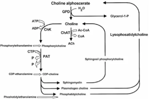
Figure A: Chemical structure of choline alphoscerate
Choline alphoscerate or alpha-glycerylphosphorylcholine (ATC code N07AX02) (GPC) (Figure A) is a semi-synthetic derivative of lecithin.Following oral administration, it is converted to phosphorylcholine, a metabolically active form of choline able to reach cholinergic nerve terminals where it increases acetylcholine synthesis, levels and release (Amenta et al., 2008).
Figure B summarizes acetylcholine anabolic pathways (Amenta et al., 2008).
As shown, the enzyme glyceryl phosphorylcholine diesterase transforms alpha glyceryl-phosphorylcholine into a molecule of choline and another of glycerol-1-phosphate.
Figure B: Acetylcholine synthetic pathways . Interference of choline-containing compounds.
This figure shows the steps in which choline alphoscerate can influence neurotransmitter biosynthesis. Cythidin diphosphate (CDP); Cythidin triphosphate (CTP); Glyceryl-phosphorylcholine diesterase (GPD); Choline acetyltransferase (ChAT); Choline kinase (ChK); Phosphocholine cytidyl transferase (PCT)
Mechanisms of action of choline alphoscerate are mainly two. In fact the compound interferes with brain phospholipid metabolism and increases brain choline and acetylcholine levels and release (Amenta et al., 2008; Amenta et al., 2001).
Pharmacodynamic studies on choline alphoscerate during phases of development of the compound were focused primarily on its role in potentiating brain cholinergic neurotransmission and in interfering with brain phospholipid metabolism. Pre-clinical studies have demonstrated that choline alphoscerate increases the release of acetylcholine in rat hippocampus, facilitates learning and memory in experimental animals (Sigala et al., 1992), improves brain transduction mechanisms (Lopez et al., 1991; Schettini et al., 1990) and decreases the age-dependent structural changes occurring in the rat frontal cortex and hippocampus (Amenta et al., 1993). Moreover, the compound contributes to anabolic processes responsible for membrane phospholipid and glycerolipid synthesis, positively influencing membrane fluidity (Aleppo et al., 1994). In several animal
paradigms of impaired cognitive function, choline alphoscerate was demonstrated to improve cognitive deficit in experimental models of aging brain (Canonico et al., 1990; Drago et al., 1990) and to reverse mnemonic deficits induced by scopolamine administration (Parnetti et al., 2007; Sigala et al., 1992). Based on the above evidence, the central parasympathomimetic activity of choline alphoscerate was defined, suggesting its clinical use in patients affected by cognitive decline. Consistently with the activity profile, choline alphoscerate was classified as a centrally acting parasympathomimetic drugs both in international pharmacopoeias (Reynolds et al., 1996) and in the Chemical Therapeutical Anatomical Classification.
A restorative role of choline alphoscerate on central cholinergic system was also documented by studies performed in old rodents. In these investigations the compound was able to counter age-related changes in brain acetylcholine synthesizing (choline acetyltransferase) and degrading (acetylcholinesterase) enzymes (Amenta et al., 1994) and some subtypes of muscarinic cholinergic receptors (Amenta et al., 1994; Muccioli et al., 1996).
Neuroprotective effects of choline alphoscerate were also documented in a rodent model of altered cholinergic neurotransmission caused by lesioning of the Nucleus Basalis Magnocellularis which represents the main source of cholinergic innervation of cerebral neocortex (Amenta et al., 1995; Bronzetti et al., 1993). A neuroprotective effect of treatment with choline alphoscerate on hippocampus microanatomy and glial reaction which represents an early sign of brain damage was documented in spontaneously hypertensive rats, used as an animal model of brain vascular injury (Tomassoni et al., 2006). A model used to mimic to some extent neuropathological changes occurring in vascular dementia (Tomassoni et al., 2006). Among cholinergic precursors tested, choline alphoscerate elicited the most relevant stimulation on vesicular acetylcholine transporter, and choline transporter in the same model of brain vascular injury suggesting that it represents a strong enhancer of central cholinergic neurotransmission (Tayebati et al., 2011).
Effects of choline alphoscerate were limited not only in rodent models of aging of lesioning of brain cholinergic nuclei, but also in Rhesus monkeys. In this species the compound revealed general
facilitatory properties on retinal neurotransmission as well as specific spatial frequency tuning effects on retinal information processing (Antal et al., 1999).
Another series of more recent studies has shown that association of choline alphoscerate with (acetyl)cholinesterase inhibitors potentiates effects on cholinergic neurotransmission. In fact, administration of choline alphoscerate plus the acetylcholinesterase inhibitor rivastigmine induced an increase of brain acetylcholine levels and of high affinity choline uptake binding sites more pronounced than single drugs (Amenta et al., 2006). This investigation has suggested that combination of a suitable precursor of brain acetylcholine such as choline alphoscerate and of an acetylcholinesterase inhibitor may represent an association worthwhile of being further investigated as a cholinergic replacement therapy in pathologies characterized by impaired cholinergic neurotransmission (Amenta et al., 2006). This working hypothesis was supported by the demonstration of a more sustained neuroprotective action by choline alphoscerate plus the acetylcholinesterase inhibitor galantamine than the two drugs administered alone (Tayebati et al., 2009).
Intermediate filaments cytoskeletal proteins
One of the main hallmarks of developmental neurobiology is to understand the molecular mechanisms by which such cellular diversity is generated. Such diversification occurs at an early stage of development, especially by activation of sets of cell type-specific genes, which gives cells distinctive functions and morphological characteristics. Some of these cell-specific genes are the intermediate filaments (IF) protein genes, which are regulated during cell development. The two major IF proteins of astrocytes are vimentin and GFAP. In the course of astrocyte development, a transition in the expression of IF protein genes is observed. Early during development, radial glia and immature astrocytes express mainly vimentin. Towards the end of gestation, a switch occurs whereby vimentin is progressively replaced by GFAP in differentiated astroglial cells. At present, there is no consensus on the functional role of these IF proteins. The application of molecular
genetic approaches to IF function has been providing some significant insights as well as raising new questions about the functional role of individual IF proteins.
Vimentin
Vimentin IFs are the only IF type found in a variety of cells including astrocytes, fibroblasts, endothelial cells, macrophages, neutrophils and lymphocytes (Evans 1998). Functional analysis of the vimentin gene promoter has already been carried out and several negative and positive elements were identified within this region (Gomes et al. 1999). Data obtained from vimentin knockout mice (-/-) demonstrated that those animals developed and reproduced without presenting an obvious new phenotype, thus heavily calling into question the biological function of vimentin (Colucci-Guyon et al. 1994). Several data, however, argue in favor of a relevant function for vimentin. Using the same vimentin (-/-) lineage as Colucci-Guyon and collaborators found that GFAP filaments were also absent in certain glial cells that normally co-express vimentin and GFAP such as the Bergmann glia and an astrocyte subpopulation of the corpus callosum (Colucci-Guyon et al. 1994). This was not due to the inability to express GFAP. Transfection of cultured vimentin-/- astrocytes with a vimentin cDNA restores the vimentin-GFAP filament network, suggesting that in these cells vimentin might be required for coassembly with GFAP filaments (Galou et al. 1996).
Reactive gliosis is a prominent result of many types of insult to the central nervous system (CNS) and leads to the formation of glial scar that impedes the regeneration of axons. The intermediate filament protein vimentin is found in pathology of the CNS, mainly in the vicinity of injuries to the CNS. In the present study some authors investigated the role of vimentin in the formation of glial scars in vitro and in vivo by using immunohistochemistry, Western blot analysis, and in situ hybridization. In vitro experiments showed that the intensity of immunofluorescent labeling for vimentin and glial fibrillary acidic protein (GFAP) was consistently decreased in astrocytes after transfection with a retrovirus carrying antisense complementary DNA (cDNA) for vimentin. Transfection also inhibited the growth of astrocytes and decreased the expression of vimentin
mRNA. In vivo studies demonstrated that transfection with the retrovirus carrying the antisense cDNA vimentin inhibited the upregulation of vimentin and GFAP in stab wounds in rat cerebrum. These results suggest that vimentin may play a key role in the formation of glial scars in the CNS. Moreover, vimentin appears to accompany the formation of glial scars. Vimentin may stabilize the formation of GFAP-type IF in some reactive astrocytes, and its expression may be required for the formation of GFAP in these cells (Galou et al. 1996). In normal adult CNS, vimentin is not expressed in astrocytes, but only in some specialized glial cells such as those of Bergmann glia and radial glia, and ependymal cells. These findings suggest that vimentin may take part in the formation of glial scar, and that there may be a relationship between the expression of GFAP and that of vimentin. During the formation of GFAP networks in some reactive astrocytes, vimentin may act as a cytoskeleton associated protein (Fuchs and Cleveland 1998). At early stages of CNS development, IF in radial glia and immature astrocytes are composed of vimentin. Subsequently, at about the time of birth, a transition from vimentin to GFAP takes place; vimentin disappears and is progressively replaced by GFAP in differentiated astroglial cells, which transiently coexpress these two proteins (Galou et al. 1996). The transient expression of vimentin observed in the present study has also been observed immunocytochemically in most models of gliosis (Stringer 1996). In the normal adult rodent brain, vimentin expression is restricted to specialized glia such as ependymal cells, Bergmann glia of the cerebellum, and Schwann cells, which has led to the suggestion that vimentin may be a more specific marker of gliosis than is GFAP (Lenz et al. 1997). Galou founds that the astrocytes in the immediate vicinity of stab wounds expressed considerable GFAP (Galou et al. 1996). However, these cells did not express GFAP after the vimentin gene had been knocked out, whereas in wild-type mice the cells not only expressed vimentin but also expressed GFAP. These results suggest that the expression of GFAP in these astrocytes depends on the expression of vimentin, and that changes in the expression of vimentin affect the expression of GFAP. In summary, the authors Lin and Kai found that the expression of vimentin and GFAP increased markedly after injury to CNS, and that restricting vimentin decreased the expression both of
vimentin and GFAP, as well as formation of glial scar. In addition, the authors therefore believe that vimentin may play an important role in reactive gliosis and the formation of glial scar. Accordingly, we suggest that manipulating the expression of vimentin may control reactive gliosis and provide an environment that favours the regeneration of injured axons.
Glial fibrillary acidic protein (GFAP)
Glial fibrillary acidic protein is an intermediate filament (IF) protein that is expressed by numerous cell types of the central nervous system (CNS) including astrocytes and ependymal cells. GFAP is a type III IF protein that maps, in humans, to 17q21. It is closely related to its non-epithelial family members, vimentin, desmin, and peripherin, which are all involved in the structure and function of the cell’s cytoskeleton. GFAP is thought to help to maintain astrocyte mechanical strength, as well as the shape of cells but its exact function remains poorly understood, despite the number of studies using it as a cell marker.
GFAP is expressed in the central nervous system in astrocyte cells. It is involved in many important CNS processes, including cell communication and the functioning of the blood brain barrier. GFAP is proposed to play a role in astrocyte-neuron interactions as well as cell-cell communication. In vitro, using antisense RNA, astrocytes lacking GFAP do not form the extensions usually present with neurons.
In addition, GFAP has been widely recognized as an astrocyte differentiation marker, constituting the major intermediate filament (IF) protein of mature astrocyte (Bramanti et al. 2010a). GFAP synthesis is considered an important element of the developmental program of astrocyte differentiation and is part of the reactive response to almost any CNS injury (Bramanti et al. 2010a). It is involved in many important CNS processes, including cell communication and functioning of the blood–brain barrier (Grasso et al. 2014). During GFAP network formation in some reactive astrocytes, vimentin may act as a cytoskeleton associated protein. At early stages of CNS development, IF in radial glia and immature astrocytes are composed of vimentin (Bramanti et al.
2010a). The gene GFAP encodes an intermediate filament protein (50kDa) of mature astrocytes, which may be used as a marker for distinguishing astrocytes from other glial cells during development of the central nervous system. Defects in this gene causes Alexander disease. It is a rare disorder of astrocytes in the CNS
Tissue transglutaminase (TG-2)
Tissue transglutaminase (TG-2) is an important calcium dependent protein, which represents a normal constituent of central and peripheral nervous systems during fetal stages of development. This enzyme is a member of transglutaminases family that catalyzes the formation of isopeptide bridges by calcium Ca2+-dependent-cross linking of the carboxamide moiety of a peptide-bound glutamine either to the e-amino group of a peptide-bound lysine, or to polyamines, with liberation of ammonia. Type-2 transglutaminase (TG-2), which is the most ubiquitous TG isoform, is a multifunctional enzyme involved in the regulation of cell differentiation and survival (Milakovic et al. 2004). TG-2 is the only member of TGs playing a role in cell signaling transduction, differentiation and apoptosis. TG-2 is induced during apoptotic death and is implicated in a variety of human disorders including central nervous system (CNS) disorders (Fesus and Piacentini 2002; Mastroberardino et al. 2002). Ca2+ ions are key regulators of TG-2 activity. When intracellular Ca21 is low, TG-2 behaves like a G protein, coupling different receptors to phospholipase C (Nakaoka et al. 1994). The multiplicity of TG-2 function also depends on its intracellular location.
Cyclin D
Cyclin D is a member of the cyclin protein family that is involved in regulating cell cycle progression. The synthesis of cyclin D is initiated during G1 and drives the G1/S phase transition. Once the cells reach a critical cell size (and if no mating partner is present in yeast) and if growth factors and mitogens (for multicellular organism) or nutrients (for unicellular organism) are present, cells enter the cell cycle. In general, all stages of the cell cycle are chronologically separated in
humans and are triggered by cyclin-Cdk complexes which are periodically expressed and partially redundant in function. Cyclins are eukaryotic proteins that form holoenzymes with cyclin-dependent protein kinases (Cdk), which they activate. The abundance of cyclins is generally regulated by protein synthesis and degradation through an APC/c dependent pathway.
Cyclin D is one of the major cyclins produced in terms of its functional importance. It interacts with four Cdks: Cdk2, 4, 5, and 6. In proliferating cells, cyclin D-Cdk4/6 complex accumulation is of great importance for cell cycle progression. Namely, cyclin D-Cdk4/6 complex partially phosphorylates Rb, which is able to induce expression of some genes (for example:cyclin E) important for S phase progression.
Growth factors stimulate the Ras/Raf/ERK that induce cyclin D production. One of the members of the pathways, MAPK activates a transcription factor Myc, which alters transcription of genes important in cell cycle, among which is cyclin D. In this way, cyclin D is synthesized as long as the growth factor is present.
Even though cyclin D levels in proliferating cells are sustained as long as the growth factors are present, a key player for G1/S transition is active cyclin D-Cdk4/6 complexes. Despite this, cyclin D has no effect on G1/S transition unless it forms a complex with Cdk 4 or 6.
One of the best known substrates of cyclin D/Cdk4 and -6 is the retinoblastoma tumor suppressor protein (Rb). Rb is an important regulator of genes responsible for progression through the cell cycle, in particular through G1/S phase.
In its un-phosphorylated form, Rb binds a member of E2F family of transcription factors which controls expression of several genes involved in cell cycle progression (example, cyclin E). Rb acts as a repressor, so in complex with E2F it prevents expression of E2F regulates genes, and this inhibits cells from progressing through G1. Active cyclin D/Cdk4 and 6 inhibit Rb by partial phosphorylation, reducing its binding to E2F and thereby allowing E2F-mediated activation of the transcription of the cyclin E gene and the cell progresses towards S-phase. Subsequently, cyclin E fully phosphorylates Rb and completes its inactivation.
Cyclin D is regulated by the downstream pathway of mitogen receptors via the Ras/MAP kinase and the β-catenin-Tcf/LEF pathways and PI3K. The MAP kinase ERK activates the downstream transcription factors Myc and AP-1 which in turn activate the transcription of the Cdk4, Cdk6 and Cyclin D genes, and increase ribosome biogenesis. Rho family GTPases and Focal Adhesion Kinase (FAK) activate Cyclin D gene in response to integrin.
Ornithine decarboxylase
The Ornithine decarboxylase (ODC) is an enzyme involved in polyamines metabolism: by decarboxylation of ornithine, reaction catalyzed by the enzyme ornithine decarboxylase (ODC), is formed putrescine.
The expression of this enzyme is regulated by different stages, by transcriptional and post-transductional levels.
The first reaction consists in the production of putrescine by the ODC enzyme. It requires pyridoxal phosphate as a cofactor and also reducing agents containing thiol groups.
The polyamines are divalent regulators of cell function, promote the growth or cell death depending on the environmental signals and recently it has been shown that the polyamines are also involved in cell cycle regulation.
Recent "in vivo" studies shown that during the cell cycle also occur changes in the activity of ODC and in the concentration of polyamines.
Supporting the correlation between polyamines and cell growth, some reports indicate that high levels of polyamines, resulting in an increase of their synthesis, are present in the cells that make up many solid tumors, where the ODC results to be overexpressed, as well as in different precancerous manifestations and consequently exposure to chemical carcinogens.
Mitogen-activated protein (MAP) kinases are serine/threonine-specific protein kinases belonging to the CMGC (CDK/MAPK/GSK3/CLK) kinase group. The closest relatives of MAPKs are the cyclin-dependent kinases (CDKs). MAPKs are involved in directing cellular responses to a diverse array of stimuli, such as mitogens, osmotic stress, heat shock and proinflammatory cytokines. They regulate proliferation, gene expression, differentiation, mitosis, cell survival, and apoptosis-among many others. The first mitogen-activated protein kinase to be discovered was ERK1 (MAPK3) in mammals. Since ERK1 and its close relative ERK2 (MAPK1) are both involved in growth factor signaling, the family was termed "mitogen-activated".
MAP kinases are found in eukaryotes only, but they are fairly diverse and encountered in all animals, fungi and plants, and even in an array of unicellular eukaryotes. Most MAPKs have a number of shared characteristics, such as the activation dependent on two phosphorylation events, a three-tiered pathway architecture and similar substrate recognition sites. These are the "classical" MAP kinases. But there are also some ancient outliers from the group as sketched above, that do not have dual phosphorylation sites, only form two-tiered pathways, and lack the features required by other MAPKs for substrate binding. These are usually referred to as "atypical" MAPKs. It is yet unclear if the atypical MAPKs form a single group as opposed to the classical ones.
Mitogen-activated protein kinases are catalytically inactive in their base form. In order to become active, they require (potentially multiple) phosphorylation events in their activation loops. This is conducted by specialized enzymes of the STE protein kinase group.
In the case of classical MAP kinases, the activation loop contains a characteristic TxY (threonine-x-tyrosine) motif (TEY in mammalian ERK1 and ERK2, TDY in ERK5, TPY in JNKs, TGY in p38 kinases) that needs to be phosphorylated on both the threonine and the tyrosine residues in order to lock the kinase domain in a catalytically competent conformation. In vivo and in vitro, phosphorylation of tyrosine precedes phosphorylation of threonine, although phosphorylation of either residue can occur in the absence of the other.
The ERK1/2 pathway of mammals is probably the best characterized MAPK system. The most important upstream activators of this pathway are the Raf proteins (A-Raf, B-Raf or c-Raf), the key mediators of response to growth factors (EGF, FGF, PDGF, etc.); but other MAP3 kinases such as c-Mos and Tpl2/Cot can also play the same role. All these enzymes phosphorylate and thus activate MKK1 and/or MKK2 kinases, that are highly specific for ERK1 and ERK2. The latter phosphorylate a number of substrates important for cell proliferation and cell cycle progression (RSK kinases, Elk-1 transcription factor, etc.)
Extracellular Regulated Kinases (ERKs)
Extracellular-signal-regulated kinases (ERKs), a well known member of the MAP kinase family, act as an integration point for multiple biochemical signals, and in addition, they are involved in a wide variety of cellular processes such as proliferation, differentiation, transcription regulation and development. The activation of this kinase requires its phosphorylation by upstream kinases. Many different stimuli, including growth factors, cytokines, virus infection, ligands for heterotrimeric G protein-coupled receptors, transforming agents, and carcinogens, activate the ERK pathway. Some studies demonstrated an alternative estrogen signaling pathway that displayed similarities to the one used by growth factor and specifically the MAP-kinase cascade (Singh et al. 1999).
CHAPTER 2
AIMS OF INVESTIGATION
In view of the possible interest of selected cholinergic precursors to counter cholinergic deficits typical of several neurological diseases, the first session of present study we will design to evaluate the effects of treatment for 24h with acetylcholine and the cholinergic precursors choline, CDP-choline and CDP-choline alphoscerate on TG-2 expression and pattern in primary cultures of rat astrocytes at 14, 21 and 35 days in vitro (DIV).
In addition, as mentioned before, because α-lipoic acid plays a pivotal role as antioxidant and metabolic component of some enzymatic complexes involved in glucose metabolism of different cell types, the second session of our research we will focus to evaluate the effect of (+)lipoic acid or (+/-)lipoic acid and /or 10 mM -GPC for 24h treatment on astroglial cell cultures.
In particular, we will evaluate the expression of some proliferation and differentiation markers in 15 or 21 DIV astrocyte cultures treated with 50 µM (+) lipoic acid or (+/-)lipoic acid and /or 10 mM -GPC for 24h. In particular, we will evaluate by Western blot analysis the expression of GFAP, vimentin, cyclin D1 Ornithyne decarboxilase and MAP-kinase, a signalling transduction pathway biomarker, in 15 or 21 DIV astrocyte cultures. In addition, it will interesting to evaluate the possible genoprotective effect by analysis of DNA status detected by Alkaline Comet assay.
Furthermore, in the third session of the investigation, we will study the estrogens and growth factors activities as mitogens promoting cellular proliferation. Although it was considered originally that these agents manifested their mitogenic action trough separate pathways, more recent data suggest that EGF and estrogen-mediated signaling pathways are intertwined. For this reason, the aim of the third session of our investigation will be particularly devoted to evidence the interactions between the “competence” growth factor bFGF and/or estrogen 17--estradiol and the “progression” growth
factors (EGF or IGF-I or INS) on DNA labeling and on proliferation and differentiation activity of primary astroglial cell cultures under different experimental conditions.
In particular, our attention will be focused to study first the labeling of [methyl-3H]thymidine in the DNA of primary astrocyte cultures at 24 days in vitro (DIV), pretreated with estradiol (17--E2) and/or “competence” growth factor bFGF and subsequent treated with “progression” GFs by two or by three in the last 12h or 24h.
Subsequently, we will study Cyclin D1, ERK1/2, GFAP and vimentin expression in our astroglial cultures at 24 DIV pretreated for 36h with estradiol and treated with “competence and progression” growth factors by two or by three for 24h.
In addition, we will hypothesize that the joint pretreatment with “competence” growth factor bFGF and DEX and subsequent treatment with “progression” GFs can stimulated an upward modulation of cellular proliferation and differentiation.
This last evidence seeks to evaluate the eventual synergistic effect played by trophic factors added in the presence of glucocorticoids and to elucidate the different behaviors of competence and progression GFs interacting with DEX under our experimental conditions. Additionally, this last one seeks to elucidate the relationships among these classes of neuroactive molecules, steroids, and GFs in the regulation of astrocyte proliferation and differentiation in culture that might have significant implications for future therapeutic approaches to neurologic disorders associated with astrogliosis.
Finally, the last session of the research project will be focused in order to evaluate the antioxidant effects of LA combined with BTZ on NB cell lines. In particular, we will focus our attention on HO-1 modulation, on gene HMOX-1, as well as on the study of several of ER-stress proteins expression markers, such as the chaperons Binding Immunoglobulin Protein (BiP1) and Inositol-requiring enzyme 1 (IRE1α), ER oxidoreductin 1 (ERO1α) and Protein disulfide isomerase (PDI), attivated by cell in order to counteract ER stress response.
Finally, we will study some protein related to the autophagy such as the protein of Autophagy protein 5 (ATG5), Microtubule-associated protein 1 (MAP1) and Beclin-1 protein (BECN1).