1
Department of Health Sciences
PHD PROGRAM IN MEDICAL SCIENCE AND
BIOTECHNOLOGIES
XXIX cycle
Academic years 2013-2016
PhD THESIS
Endothelial MMP-9 Drives the Inflammatory Response in
Abdominal Aortic Aneurysm (AAA)
SSD (Settore Scientifico Disciplinare) BIO16 Anatomia Umana
Supervisor:
Prof.ssa Francesca Boccafoschi
PhD Coordinator:
PhD Student:
2
Table of contents
Pag.
Abstract
41 Introduction
61.1 Macro- and micro-anatomy of human aorta 7
1.1.1 Human aorta 7
1.1.2 Structural features of human arterial wall 8
1.1.3 Extracellular matrix in vascular tissues 13
1.1.4 Biomechanical properties of human arterial wall 17
1.2 Abdominal aortic aneurysm 21
1.2.1 Definition, epidemiology and risk factors 21
1.2.2 Symptoms, diagnosis and treatment 24
1.2.3 Histological features of AAA 29
1.3 The MMP family 32
1.3.1 Focus on human MMP-9 34
1.4 Molecular pathway included in abdominal aortic aneurysm 38 1.5 Future perspective: Epigenetic in AAA pathogenesis 40
2 Aim of the project
443 Material and Methods
463.1 Study of protocol and sample collection 47
3.2 Immuno-histological analyses 47
3.3 Gelatin zymography for detection of MMP-2 and MMP-9 48
3.4 Cell culture 48
3.5 Human peripheral blood mononuclear cells (PBMCs) isolation 49 3.6 Cytofluorimetric analysis for macrophages characterization 49
3.7 Lentiviral vector transduction 49
3.8 Cytokines antibody arrays and immunofluorescent microscopy on
EA.hy926 cells
50
3
3.10 NF-kB activation 50
3.11 Tube formation assay 50
3.12 Western blot 51
3.13 Preparation of conditioned media (CM) and silver staining 51
3.14 Statistical analysis 52
4 Results
534.1 Study subjects: AAA patients and control group. 54 4.2 Histological features of aneurysm-affected abdominal aorta 55 4.3 MMP-9 expression within healthy and AAA wall 59
4.4 In vitro model of ECs silenced for MMP-9 60
4.5 Tube formation assay 61
4.6 NF-kB activation 61
4.7 MMP-9 regulates cytokines production by ECs and inflammatory cells adhesion
62
4.8 Effect of conditioned media (CM) on vascular smooth muscle cells (vSMC) and macrophages
64
5 Discussion
676 References
724
Abstract
Abdominal aortic aneurysm (AAA) is a complex multi-factorial disease leading to life- threatening complications. Chronic inflammation and extracellular matrix degradation are the major pathological features of AAA. Vascular inflammation involves complex interaction among inflammatory cells (i.e. neutrophils, lymphocytes, monocytes, macrophages), endothelial cells (ECs), vascular smooth muscle cells (VSMCs), and extracellular matrix (ECM). Although vascular endothelium plays a key role in aneurysm disease, the molecular mechanisms underlying its involvement is only partially understood. In this study, we have characterized the role of matrix metalloproteinase-9 (MMP-9) as potential trigger of the inflammatory response during the reciprocal interaction between ECs and VSMCs. Briefly, in biopsies of human AAA we found increased level of MMP-9, IL-6 and monocyte chemoattractant protein-1 (MCP-1), which correlated with a massive medial neo-angiogenesis. In particular, in vitro silencing of MMP-9 in ECs, using specific shRNA delivered by lentiviral vectors, inhibited TNF-alpha mediated activation of NF-kB. In addition, ECs void of MMP-9 failed to migrate in 3D matrix and affected VSMC behavior in terms of matrix remodeling. Overall our findings indicate that silencing of MMP-9 may represent a therapeutic target to restore vascular extracellular matrix remodeling.
5
Riassunto
L’aneurisma addominale aortico (AAA) è una malattia multi-fattoriale complessa che porta a complicanze rischiose per la vita. Le principali caratteristiche patologiche dell’AAA sono l'infiammazione cronica e la degradazione della matrice extracellulare.
L’infiammazione coinvolge una complessa interazione tra cellule infiammatorie (in particolare neutrofili, linfociti, monociti e macrofagi), cellule endoteliali (ECs), cellule vascolari muscolari lisce (VSMC) e la matrice extracellulare (ECM). Anche se l’endotelio vascolare svolge un ruolo chiave nello sviluppo dell’aneurisma, i meccanismi molecolari alla base del suo coinvolgimento sono solo parzialmente delineati. In questo studio abbiamo caratterizzato il ruolo delle metalloproteinasi-9 (MMP-9) endoteliale come potenziale agente promuovente della risposta infiammatoria durante l'interazione tra EC e VSMC. In particolare, abbiamo rilevato in biopsie di AAA una maggiore espressione di MMP-9, IL-6 e monocyte chemoattractant protein-1 (MCP-1), che correla con una massiccia neo-angiogenesi presente anche nello strato medio della parete vascolare. È stato valutato il silenziamento in vitro di MMP-9 in EC, utilizzando specifici shRNA veicolati da vettori lentivirali, ed è stata osservata un’inibizione nell’attivazione di NF-kB a seguito di stimolazione con TNF-alfa.
Inoltre, le cellule endoteliali private di MMP-9 non sono in grado di migrare in una matrice 3D, e influenzano il comportamento VSMC in termini di rimodellamento della matrice. Nel complesso i nostri risultati indicano che il silenziamento dell’MMP-9 endoteliale potrebbe rappresentare un bersaglio terapeutico per ripristinare il rimodellamento fisiologico vascolare della matrice extracellulare.
6
7
1.1 Macro- and micro-anatomy of human aorta
1.1.1 Human aorta
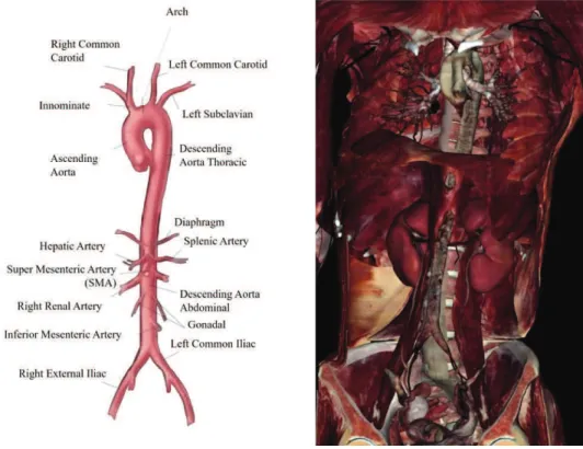
The aorta is the largest artery of the human vascular system and it is designated to deliver blood from heart to the systemic circulation. The human aorta arises from the left ventricle and descends to the abdomen; it can be distinguished into distinct segments: ascending aorta (between the left ventricle and the aortic arch), aortic arch, descending thoracic aorta (above the diaphragm) and abdominal aorta (below the diaphragm). The aorta belongs to the elastic arteries, which are large-diameters vessels (larger than 2.5 cm) with close localization to the heart. Healthy aorta of an adult normally measured roughly 3 cm diameter at the origin (ascending aorta), 2.5 cm along the descending thoracic aorta and 1.8-2 cm along the abdominal aortic segment. Elastic artery structure is kept by a network of connective fibres, elastin and collagen, that contributes to the arterial distension and recoil during left ventricle contraction (systole) and relaxation (diastole) respectively; moreover, the elastic artery recoil allows the blood entering into the small arteries. Conversely, the muscular arteries are located at periphery and provide blood to the whole tissue (skeletal muscle and inner organs). Muscular arteries are medium-size vessels (about 0.4 cm) and they are mainly composed of smooth muscle. This structure is functional to the muscular artery ability to vary the laminal diameter depending blood pressure, and regulate blood flow to each organ.
8
Figure 1. Human aorta anatomy. Diaphragm divides aorta in two portions: thoracic and abdominal aorta. Virtual dissection
technology from Anatomage Table (Anatomage, CA, USA)
1.1.2 Structural features of human arterial wall
The healthy wall of aorta is composed by three layers called tunicae: intima, media and adventitia (Gasser et al., 2006)
Figure 2. Microanatomy of a vessel wall.
Three layers compose arterial wall. The intima and the media are separated by the internal elastic membrane. The external elastic membrane separates the media and the adventitia.
9
Tunica intima is the innermost layer of the artery wall; it consists of a single layer of
endothelial cells (ECs) placed on a thin basal lamina and a sub-endothelial layer of various thickness depending on localization, age and pathological changes. ECs compose the luminal side of the vessel wall and they are in contact with the blood flow; ECs are characterized by an elongated flat shape, that can undergo changes depending on the direction of the blood flow. ECs communicate to each other through a network of three types of junctions: I) tight junctions, or zonulae occludens, regulating the substances delivery across the endothelium; II) adherens
junctions, whose main component is given by cadherins; they are designated to control the
endothelium permeability to circulating cells; III) gap junctions, structures designated to allow ions and metabolites exchange between cells (Bazzoni and Dejana, 2004). ECs possess several structural and regulatory roles, participating to the vascular homeostasis and vessel wall integrity. They are responsible for the maintaining of a selectively permeable and non-thrombogenic barrier, that regulates the smooth muscle cells tone through the secretion of vasoconstrictors and vasodilatators, they modulate the immune response through the expression of chemotactic and adhesion molecules. ECs are located on the basal lamina, composed of collagen type IV, proteoglycans, laminin and fibronectin. Regarding more specifically the abdominal aorta, the intima layer is mainly composed by type I collagen (16%), while type III and type IV collagen are present in a lower percentage. The sub-endothelial layer mainly contains smooth muscle cells and bundles of collagen fibrils. Collagen fibres are not arranged according to the same orientation through the whole layer, but many layer of collagen with a distinct pattern of orientation are present. (Gasser et al., 2006). The tunica intima terminates with an internal elastic lamina, which creates the limit with the medium layer of the artery wall. The internal elastic lamina changes according to the arterial type: it is very thin and hard to be distinguished in elastic arteries, whereas it is very prominent in muscular arteries.
The media is the middle and, in terms of mechanical properties, the most important layer
of the aortic wall. The internal elastic lamina and the external elastic lamina separate the tunica media from the intima and adventitia, respectively. The media is composed by smooth muscle cells (vSMCs), organized as complex network of elastin and collagen, mainly type I, III, IV and V. An extracellular matrix (ECM), containing proteoglycans, surrounds the cellular and elastic components. vSMCs are spindle shaped, with an elongated nucleus and assume a specific orientation in elastic and muscular arteries. In elastic arteries, vSMCs are arranged into 5-15µm thick concentric structures, separated by elastin. At physiological distending pressures, the aortic smooth muscle, elastin and collagen are precisely organized into distinct layers, termed lamellar
10
mammals proposed by Wolinsky and Glagov, elastin fibres are organized into fenestrated sheets, or lamellae, forming concentric layers, with finer elastin fibres that interconnect adjacent lamellae; bundles of collagen are interposed between elastin sheets, circumferentially oriented (Wolinsky and Glagov, 1964). In a later work, Clarke and Glagov defined the lamellar unit as the
“musculo-elastic fascicle”, that constitutes both the structural and functional unit of the aortic
media; the number as well as the orientation and the composition of these units are functional to the homogeneous distribution and magnitude of the tensile stress along the vessel wall (Clark and Glagov, 2015). Medial vSMCs are essential to the correct functionality of the vasculature; indeed, their contraction or relaxation modulate the arterial structural and diameter changes, adapting to the blood dynamics and keeping blood pressure. In addition, SMCs are responsible of the synthesis of ECM components, vSMCs are susceptible to the phenotype changes, shifting from contractile to synthetic, characterized on the basis of different cell shape and structure, marker expression, proliferative and migration rates. Contractile vSMCs show an elongated, spindle-shape morphology, substituted by a less elongated, epitheloid-like spindle-shape in synthetic vSMCs. The synthetic phenotype is characteristic for the presence of a high number of organelles appointed to protein synthesis, as well as for a high proliferative rate. In contractile vSMCs, biosynthetic organelles are replaced by contractile filaments; in addition, the contractile SMC phenotype presents increased contractile markers, such as alpha-smooth muscle actin, smoothelin, smooth muscle heavy chain, that are not represented in synthetic vSMC (Rensen et al., 2007). vSMC heterogeneity exists not only between different tissues and vascular districts, but also in the same vessel, suggesting a genetic basis. Moreover, vSMC phenotype changes in dearly development: during blood vessel formation, vSMCs assume a contractile phenotype, corresponding to a reduced ECM production and an increased myofilament formation, allowing the vessel contraction and dilation under the effects of blood flow and pressure. A reversal of vSMC phenotype, from contractile into synthetic, induces cell proliferation and invasion of the intima tunica, characteristic of vascular disorder. Some differences concerning the number of lamellar units, the elastic fibres composition and the nutrition mechanism have been observed between the thoracic and abdominal region of human aorta, thus, implying a different response to injuries and pathological processes. The number of lamellar units increases with arterial diameter and mechanical force; in addition, it defines the thickness of the aortic media and influences the presence of vasa vasorum, small vessels that arise from the adventitia and supply nutrients to the media. When the number of lamellar units is about 28-30 (0.5 mm thickness), as in smaller elastic arteries, the media receives nutrients through a simple diffusion mechanism from blood vessel lumen, traversing endothelial layer. Arteries with more than 28 units receive nutrient from vasa
11
vasorum. Human thoracic aorta contains 55-60 lamellar units, that increase in number through the synthesis of new additional lamellar units; human abdominal aorta, regardless its diameter, has 20-32 units, that undergo an expansion process without a numerical increase. This difference may explain the major elastin content in thoracic aorta than abdominal aorta; in addition, the elastin content was shown to decrease with age. A reduced number of lamellar units, a decreased elastin content and an inadequate nutrient transport create the structural conditions (loss of stiffness and recoil capacity of elastin) that predispose the abdominal aorta to an increased risk of aneurysm formation. (Wolinsky, 1970) (Ruddy et al., 2008). The media of muscular arteries has a less-defined structure, predominantly made of thick smooth muscle layer and an internal elastic lamina. Smooth muscle is arranged into concentric layers, whose number can range between 25 and 35 in larger arteries; these smooth muscle structures are surrounded by connective matrix that works as a substrate for the cell components and, meanwhile, contributing to the artery strength and force.
The adventia (tunica adventitia) is the outermost layer of the arterial wall, representing the 10% of elastic arteries and the 50% of muscular arteries. It is mainly composed of: fibroblast, macrophages, collagen and groudmatrix. The adventitia shows a various level of thickness, depending on the localization and function of the vessel wall. Type I collagen is the predominant component of the connective tissue and polarized light microscopy showed an organization into helical structures. The collagen is the main constituent and it contributes to the adventitial providing support and strengthen to the arterial wall, preventing the excessive distension of the vessels. Nerves and vasa vasorum are also characteristic of adventitial structures. Nerves contribute to the regulation of medial smooth muscle activity, through the release of neurotransmettitors, like norepinephrine and acetylcholine. Vasa vasorum represent an intra-vascular network of small vessels, including arterioles, venules and capillars, that can reach the media, as a nutritive support to the arteries whose thickness does not allow the nutrient delivery through the diffusion from the intima. The structural changes that affect the arterial wall is named
vascular remodeling, which interests larger elastic arteries more than smaller and muscular ones.
Ageing represents the main decisive factor for arterial remodeling, mainly characterized by an increased thickness of media and, most frequently, intima. In a young healthy subject, the intima is very thin; ageing can be responsible of connective tissue alterations, followed by an increase of collagen content. Moreover, endothelial cells can be influenced by ageing process both on the structural and functional hand. First, can change their morphology, acquiring a bigger size and an irregular shape; they can also loose most of their regulatory roles. Endothelial layer can become
12
more permeable, allowing the transit of many substances and vSMC infiltration. These alterations characterized the atherosclerotic process.
13
Figure 3. Molecular and cellular events occurring during vascular remodeling.
In aged arteries, all these events contribute to decrease compliance, resilience, and increase stiffness. ECM, extracellular matrix; MMP, matrix-metalloproteinases; NO, nitric oxide; AGE, advanced glycation end-products. (Duca et al., 2016)
14
1.1.3 Extracellular matrix in vascular tissues
Extracellular matrix (ECM) is synthesized by vSMCs and adventitial fibroblasts, and constitutes the scaffold that supports the structure and the function of the vessel wall, driving its biomechanical properties. ECM does not act only as a structural support, but also regulates many cell functions, including proliferation, migration, differentiation and morphological changes (Daley et al., 2008). The main ECM components are fibrous proteins that confer tensile strength and visco-elasticity to the wall, as well as proteins that contribute to ECM network building.
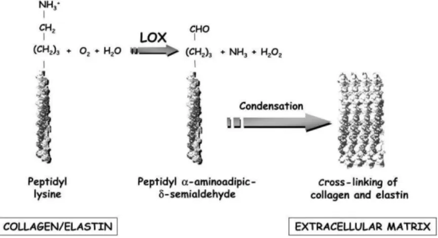
Collagen is a tense protein that prevents vessel wall excessive distension. About 26 members of this family have been identified and their expression and distribution depend on the cell type. Vascular cells, type I and type III collagen are the most common types, representing 30% and 60% of the vascular wall; the remaining 10% is composed type V, XII and XIV collagen (Wagenseil and Mecham, 2009). Structurally, the basic unit of collagen is given by a 300 nm triple helix. Each chain, called α-chain, contains 338 (type I) and 340 (type II) triplets of the aminosequence Gly-X-Y (generally, X is a proline and Y is an hydroxyproline). Post-trascriptional modifications are required before collagen maturation into a functional protein. Crosslinking Lysil-oxidase increases collagen insolubility and tensile strength by crosslinking. Lysyl oxidase (LOX) is a copper-dependent amine oxidase that initiates the covalent cross-linking of collagen and elastin. LOX catalyses an oxidative de-amination of lysine and hydoxylysine residues to peptidyl a-aminoadipicd-semialdehydes. These highly reactive semialdehydes can spontaneously condense to form intra-and intermolecular covalent cross-linkages that assure ECM stability. Strong evidence about the involvement of a reduction of LOX activity in the pathogenesis of AAA is available. (Rodríguez et al., 2008)
Figure 4. Reaction catalyse by LOX. LOX oxidizes primary amines on collagen and elastin substrates to reactive
15
Blood vessels are endowed with the property of viscoelasticity, which allows to buffer pressure variations during the cardiac cycle, permitting an adequately constant blood flow and correct organ perfusion. This mechanical phenomenon is due to the presence of highly resilient elastic fibres. They are arranged in concentric fenestrated elastic lamellae. Each elastic lamella alternates and it is physically connected with a concentric ring of vSMC forming the lamellar unit. Mature elastic fibres and lamellae are comprised of a homogenous core containing elastin that is assembled along parallel microfibrils. Microfibrils are composed of numerous heterogeneous glycoproteins, such as fibrillin-1 and fibrillin-2, microfibrin-associated glycoproteins (MAGP1 and MAGP2) and the latent transforming growth factor β (TGF- β)-binding proteins (Kielty et al., 2002). Microfibrils interact with other proteins localised to the elastin-microfibril interface or to the cell surface-elastic fibre interface called elastin microfibril interface-located proteins (EMILINs) and fibulins. However, elastin is the most prominent component of the large arteries wall.
Elastin gene is located on chromosome 7 and its expression is high during pre- and neonatal development (Arribas et al., 2006). In humans, its activity is restricted to the early life, and the mature form of elastin has a half-life of 40 years. Elastin degradation is related to aging and diseases. Elastin gene encodes for a precursor form of the functional protein, named tropoelastin. This is a monomeric protein (64-72 kDa), whose structure is characterized by the alternans of hydrophobic domains and lysine residues with cross-linking motifs. When tropoelastin is released in the extracellular space, lysine residues undergo modifications to form covalent binds with elastin molecules. As collagen, crosslinking is mediated by lysil-oxidase and takes to sequential reactions leading to the functional and insoluble form of elastin. Unlike elastin that has 15-20 cross-links per unit, collagen only contains 1-4 cross-links. The high number is critical for elastin recoil property, as well as for insolubility and high half-life (Eyre et al., 1984). As reported before, under native conditions elastic fibres have short period of active synthesis, restricted to fetal life and neonatal period. During this period, tropoelastin is continuously produced and assembled into extracellular elastic fibres. Afterwards, tropoelastin is not actively synthesized in blood vessels. The first prerequisite step in elastogenesis involves assembly of microfibrils close to the cell surface, probably modulated by the cell surface integrins and heparin sulfate proteoglycans. It requires proteolytic modifications of the secreted fibrillins before the association with MAGPs and other microfibrillar components. The hydrophobic and non-glycosylated tropoelastin requires to be chaperoned through the intracellular secretory pathways. In the endosomal and Golgi compartments, tropoelastin is escorted by a 67 kDa elastin binding protein (EBP) that protects from premature intracellular self-aggregation and degradation by
16
serine proteinases. This interaction triggers the release of newly secreted tropoelastin from the chaperone and their subsequent orderly binding to the microfibrillar scaffold. This systematic binding secures a proper mutual orientation of the adjacent tropoelastin molecules and allow for the process of ordered self-assembly (coarcevation). Coarcevation allows for the proximity of lysine residues (located in “cross-linking domains”) that can be covalently cross-linked together after their oxidative deamination by a LOX (Kagan and Li, 2003) to form linkages called desmosines and isodesmosine. The fully cross-linked elastin turns into an “insoluble” polymer. Vascular elastin is produced by SMCs in the media and fibroblasts in the adventitia, but also endothelial cells display some limited elastogenic abilities. Elastin confers elasticity to the vessel wall, the most essential property in large diameter arteries which are subjected to high pressures generated by heart beating and blood flow, defining their ability to contract and distend according to the cardiac cycle. In addition, elastin and elastin-derived peptides (EDP) have been shown to regulate cell proliferation and phenotype.
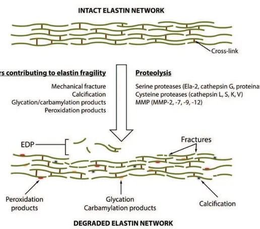
Figure 5. Elastin remodeling.
During ageing, elastin is subject to remodeling and fragmentation. Different factor such as mechanical fracture, calcification, glycation, carbamylation and peroxidation can contribute to elastin fragility and rupture. In addition, these factors enhance elastin
17
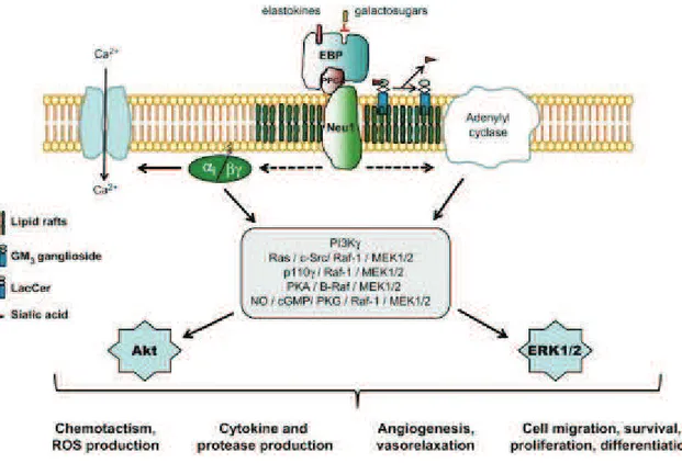
Elastokines are the bioactive EDP generated by the action of elastases and, all elastokines act by binding to a singular cell surface receptor named the elastin receptor complex (ERC). ERC is derived from the lysosomal β-galactosidase (β-Gal) complex and is expressed at the plasma membrane of numerous cell types (Duca et al., 2004). It is a heterotrimer composed of a peripheral 67 kDa subunit noted elastin-binding protein (EBP), which binds elastin peptides, a 55 kDa protective protein or cathepsin A (PPCA), and a 61 kDa membrane-bound neuraminidase, Neu-1 (Hinek et al., 2006). From a mechanistic point of view, when an elastin peptide binds to EBP, it is recruited to the cell membrane where it interacts with PPCA and Neu-1 to form the functional ERC enhancing diverse signaling pathways. Depending on the cell type, various signaling modules are triggered, converging to the activation of ERK1/2, which appear as crucial actors in elastin peptide signaling. In smooth muscle cells, EBP-induced proliferation involves activation of Gi proteins concomitantly with opening of Ca2+ L-type channels. Finally, in endothelial cells, a PI3Kγ/Akt/eNOS/NO/PKG1 module is involved in ERK1/2 activation (Fahem et al., 2008).
Figure 6. Elastokines signaling.
Elastin receptor complex (ERC)-induced signaling pathways and associated cellular response. cGMP, cyclic guanosine monophosphate; EBP, elastin-binding protein; ERK1/2, extracellular signal-regulated kinase 1/2; MEK1/2, mitogen-activated
protein kinase/extracellular signal-regulated kinase 1/2; NO, nitric oxyde; PI3Kγ, phosphoinositide-3 kinase γ; PKG, protein kinase G. (Maurice et al., 2013)
18
1.1.4 Biomechanical properties of human arterial wall
Vessel wall are continuously exposed to a wide range of hemodynamic forces. Because of the pulsatile nature of blood and the complexity of the arterial system, being composed of various size vessels, branches and bifurcations, the fluid mechanic laws cannot be simply applied to explain these mechanics (Resnick et al., 2003). Blood vessels are constantly subjected to various types of hemodynamic forces, including hydrostatic pressure, cyclic stretch, and fluid shear stress. Shear stress (τ) is parallel to the vessel wall and represents the frictional force that blood flow exerts mainly on the endothelial layer. Instead, cyclic stretch (ρ) is the stress perpendicular to the vessel wall and represents the circumferential deformation of the blood vessel wall during distension and relaxation of the recurring cardiac cycle.
Figure 7. Mechanical and hemodynamic forces associated with blood flow.
Shear stress (τ) is parallel to the vessel wall and represents the frictional force that blood flow exerts mainly on the endothelial surface of the vessel wall. Instead, cyclic stretch (ρ) is the stress perpendicular to the vessel wall and represents the circumferential deformation of the blood vessel wall during distension and relaxation of the recurring cardiac cycle.
Hemodynamic forces associated with blood flow play a central role in the homeostasis of the circulatory system. These forces act on the vascular wall, contributing to the regulation of myogenic tone, responses to vasoactive molecules, gene regulation, vascular permeability and remodeling. Hemodynamic loading breakdown, can be responsible for the development of vascular diseases, including hypertension, thrombosis, aneurysms and atherosclerosis. While physiological cyclic stretch induces cell cycle arrest in VSMC, chronically increased blood pressure and vascular transmural stress activate vascular cell proliferation (leading to luminal stenosis), collagen and fibronectin synthesis which results in thickening of the vascular wall as a feature of hypertension-induced vascular remodeling eventually altering arteries compliance. (Hipper and Isenberg, 2000)(O’Callaghan and Williams, 2000)
Although vascular endothelial cells and vascular smooth muscle cells are exposed to both types of mechanical forces, shear stress resulting from blood flow is sensed mainly by ECs
19
(Davies, 1995), whereas both ECs and SMCs are subjected to cyclic stretch resulting from pulsatile pressure. Vascular cells have with numerous receptors that sense external stimuli such as strain, pressure and fluid shear stress. These receptors transduce the mechanical signals into a biological response through a process named mechanotransduction. The cytoskeleton and other structural components have an established role in mechanotransduction, being able to transmit and modulate tension within the cell. SMCs and ECs are able to perceive stretch and shear stress alterations through a system of receptors, that include integrins, G-protein receptor, ion channels, adhesion molecules and activate a signal transduction, also involving extracellular matrix (Resnick et al., 2003) (Lehoux et al., 2006).
As previously described, endothelium works as a selectively permeable barrier that regulates the macromolecules entrance from the blood into the vessel wall (i.e. low-density lipoprotein, LDL). In this view, endothelium has a protective function, together with an anti-thrombogenic property; moreover, it guides the inflammatory process and drives the SMC contraction through the release of vasocostrictors and vasodilatators. Any impairment in these activities is crucial for the development of pathological processes, especially related to proatherogenic and pro-thrombotic events. Each shear stress variation is sensed by ECs, that undergo several mechanisms of adaptation to acute as well as chronic changes. One of EC characteristics consists in the alignment according to the blood flow. Under physiologic condition, the mean intensity of shear stress is typically of 10-15 dynes/cm2 in large arteries. (Tsou et al., 2008) ECs with an elongated shape and aligned with blood flow reflect a laminar and unidirectional flow; closely to side branches, bifurcations and curvatures, the flow becomes turbulent, creating secondary flows with different directions as well as recirculation sites (vortices); in this case, ECs assume a less oriented configuration, a polygonal shape and this morphological alteration is mediated by the cytoskeleton and microtubules rearrangement.
The introduction of DNA microarray technique allowed the identification of a great quantity of genes regulated by shear stress, highlighting many molecular mechanisms involved in vascular wall pathophysiology. Interestingly, genes encoding for anti-oxidant factors, ECM proteins, growth arrest and gap junction proteins were shown to be induced under chronic laminar shear stress, revealing an atheroprotective role; conversely, proatherogenic and pro-inflammatory genes were stimulated in response to an altered shear stress (Malek et al., 1999) (Li et al., 2005). Although shear stress mainly acts on ECs, it can influence SMC phenotype and alignment, in mechanism mediated by ECs. According to SMC culture in an in vitro two-dimensional level, a laminar shear stress stimulates the acquisition of a contractile phenotype, increases SMC
20
apoptosis mediated by EC release of NO and reduces the cell proliferation (Sterpetti et al., 1993), inducing the cell-cycle arrest. Conversely, an oscillatory flow takes to SMC proliferation and migration, in a MMP-dependent mechanism, characteristics of the synthetic phenotype. Interestingly, SMC proliferation was observed closely to atherosclerotic sites, with high shear stress (Yoshida et al., 1990). Differently from shear stress, mechanical stretch directly affects SMCs, both at the structural and molecular level. At physiological strain (10%, 1 Hz), SMC proliferation resulted inhibited through different mechanism, one of which involves the G1/S arrest during cell cycle. All these data are concurrent with the belief that SMC under physiological stretch are differentiated and reveal a contractile phenotype. Under hypertensive condition, SMCs are exposed to high level of stretch, due to the increased blood pressure and, according to Laplace’s equations (T=Pr/h), increased proliferation and enhanced synthesis of collagen and elastin (Lehoux et al., 2006).
From a molecular point of view, SMCs express many different receptors and ion channels, which can be activated by mechanical stress leading to the activation of multiple classic signaling pathways, such as G protein, kinases, calcium, cAMP, NO, eNOS, MAPK and MKP-1. Moreover, mechanical stresses appeared simultaneously to initiate multiple signaling pathways. Mechanical stresses stimulate conformational activation of cell integrins and increase cell binding to extracellular matrix. The dynamic formation of new ligand-integrin connections is required for stretch induced mechanotransduction. During the stimulation of vascular cells by mechanical factors such stretch, several signaling events are associated with the formation of focal adhesion, which comprise integrin cluster and cytoskeletal protein. The proteins present at focal adhesion become phosphorylated on tyrosine when the cells are stimulated, a FAK activation is an indicator in focal adhesion formation (Boccafoschi et al., 2011)(Lehoux et al., 2006).
Once cells adhere to ECM proteins, integrins became activated and form cluster at the cell surface thus initiating the formation of adhesion complexes which will enhance FAK’s catalytic activity and increase FAK tyrosine phosphorylation. FAK phosphorylation at tyrosine 397 (Y397) leads to the recruitment of Src and Src-family kinases as well as to an increased phosphorylation of other proteins present in the adhesion complex such as paxilin and p130Cos. (Arias-Salgado et al., 2003) (Lehoux et al., 2005) Subsequent phosphorylation of specific tyrosine residues leads to the recruitment of additional SH2-domain-containing signaling proteins such as PI3K and Grb2.43 Among integrin receptors, integrin α5β3 has been identified as having particularly interesting expression pattern among the vascular cells during angiogenesis and vascular remodeling. In particular, integrin α5β3 binds numerous ECM ligands with exposed RGD peptide
21
as fibronectin, vitronectin, fibrinogen and it is involved in the response to mechanical strain. In the ‘90s, Wilson and colleagues established a link between the mechanical stimuli and the action of growth factors. Their studies demonstrated that cyclic stretch increased VSMC proliferation when plated on collagen, fibronectin or vitronectin, but not on laminin or elastin; further, these effects required the production of PDGF suggesting a tight interaction between integrins and growth factors downstream signaling pathways related to mechanical stimuli. Generally, growth factors receptors belong to two classes of membrane proteins: tyrosine kinase receptors and G-coupled receptors and G proteins. Another membrane structurse involved in mechanosensing are ion channels. VSMCs stimulated by mechanical stress result in a transient increase in intracellular calcium and divalent cations and depolarization which maintain smooth muscle tension. Opening these channels causes Ca2+ and Na+ influx and membrane depolarization, which contributes to the myogenic response to mechanical stretch. (Marrero et al., 1996)
The altered stretch-activated channels in arterial SMCs may contribute to the enhanced myogenetic responses as well as the generation of hypertrophy and remodeling of arterial tissue in hypertension. All the cited membrane mechanosensors have a common intracellular pathway converging to MAP kinase signaling. MAP kinase comprises a ubiquitous family of threonine/tyrosine kinases, and includes extracellular signal-regulated kinases (ERK), stress activated protein kinases (SAPK) or c-Jun NH2-terminal kinases (JNK) and p38/ERK kinases. (Karin, 1998) This cascade is an important pathway whereby signal originating from mechanical forces can lead to gene expression and protein synthesis in vSMC.
22
1.2 Abdominal Aortic Aneurysm
1.2.1 Definition, epidemiology and risk factors
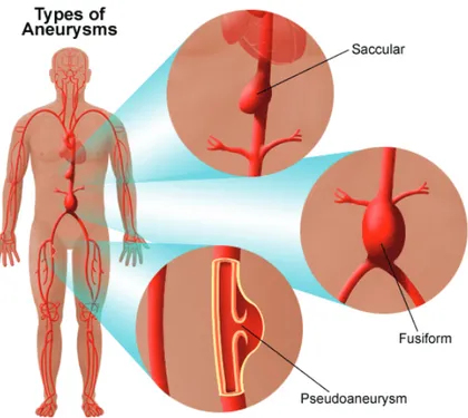
The term “aneurysm” is used to define a permanent focal dilatation of the vessel wall. Mc Gregor et al. defined abdominal aortic aneurysm (AAA) as an aortic segment with a diameter larger than 3cm (McGregor et al., 1975); following this definition, aneurysms of the infrarenal, intrarenal and suprarenal aorta are included (Sakalihasan et al., 2005). Aneurysms can be distinguished into fusiform and saccular, according as the whole aortic circumference, or only a small segment of the aortic circumference, is interested.
Figure 8. Type of aneurysms (stanfordhealthcare.org)
AAAs comprise the 15th leading cause of death among men over 65 and result in more than 15,000 surgical procedures annually in the United States. Although prevalence of aneurysms is increasing the mechanisms that regulate aortic aneurysm development remain almost undefined (Lederle et al., 2000) (Salmon et al., 2013). Approximately 80% of all aortic aneurysms occurs below the renal arteries, and the majority has an atherosclerotic cause. Other less common causes include inflammatory aneurysm, tuberculous aneurysm and syphilitic or infectious aneurysm.
It usually remains asymptomatic until the rupture occurs. Because of the ageing of the population, the increasing of smokers and the improved screening and diagnostic tools AAA incidence is recently increased. Major risk factors for the occurrence of AAA are male gender,
23
age, smoking, race, pre-existent cardiovascular affections, such as hypertension and atherosclerosis. The risk of AAAs increases dramatically after 60 years of age (Singh, 2001). Clinically relevant aneurysms (more than 4 cm in diameter) are present in approximately 1% of men between 55 and 64 years of age, and the prevalence increases by 2% to 4% per decade thereafter (Singh, 2001). AAAs are four to six times more common in men than in women (Scott et al., 1995) (Lederle et al., 2001). Moreover, AAAs develop in women approximately 10 years later than in men (McFarlane, 1991). The related mechanisms of sex differences are not fully understood, although women seem to be protected by female sex hormones (Makrygiannis et al., 2014).
AAAs were found to occur more frequently in Caucasian population than in Afro-American ethnicity. Due to its promoting effects on inflammation, proteolysis, and smooth muscle cell (SMC) apoptosis, smoking is another strong risk factor for the development of AAAs. (Li and Dai, 2012). In addition to these environmental components, genetic aspects play an important role. A positive familiar history for AAA, especially in male first-degree relatives, is associated with an increased risk. Rupture of an AAA and its associated catastrophic physiological insult carry the overall mortality more than 80%, and 2% of all deaths are AAA-relate (Nordon et al., 2011).
Most aneurysms are small in size and they do not need the surgical repair, but the increase in the aortic diameter can lead to rupture, which can be fatal. Medical therapy may be helpful in patients with small- to medium-sized aneurysms not elective for surgery (thoracic and abdominal aortic aneurysms). Appropriate and immediate therapy is essential with the aim of stabilizing the patient and improving the clinical outcome. Therapy’s targets are blood pressure control, decrease of shear stress, optimization of anticoagulation, volume management, and pain control. Different pharmacological treatments are available: beta-blockers, tetracycline, statins and angiotensin-converting enzyme Inhibitors/Angiotensin Receptor Blockers. β-blockers may be beneficial for reducing the rate of aortic dilatation. This is thought to be due to the effect of β-blockers in reducing left ventricular dP/dt and reducing shear stress. In addition, β-blockers reduce dP/dt in the aorta and might be beneficial via this mechanism and the resultant effect on shear stress in the aorta (Danyi et al., 2011). Although data regarding therapeutic benefit of beta-blockers in management of AAA are limited, beta-blockers have been shown to significantly reduce the expansion rate of AAA. The 2005 ACC/AHA guidelines recommended beta-blocker therapy in patients with an AAA not elective for surgery. Doxycycline is a nonspecific MMPs inhibitor (Petrinec et al., 1996). This antibiotic has been used in conditions with MMPs overexpression
24
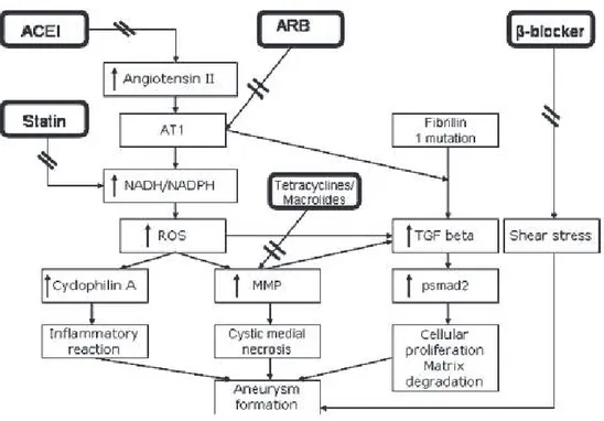
(e.g., periodontal disease, rheumatoid arthritis) (Hanemaaijer et al., 1997). In animal models, doxycycline slowed elastin degradation and aneurysm development (Xiong et al., 2008). In human, doxycycline decreased MMP-9 levels (Baxter et al., 2002) and slowed the rate of progression of AAA. Statin treatment is one of the elective therapies in cardiovascular diseases. Statins reduce the progression of atherosclerosis and improve clinical outcomes. In addition to their lipoprotein-reducing properties, statins have pleiotropic effects. For instance, they reduce oxidative stress by blocking the effects of reactive oxygen species on aneurysms. This effect is independent of their lipid-lowering properties. Statins achieve these results through suppressing the NADH/NADPH oxidase system (Ejiri et al., 2003). Angiotensin II has been shown to have several biological effects on the cardiovascular system. It promotes vascular hypertrophy, cell proliferation, production of extracellular matrix, and activation of macrophages, and it activates NADH/NADPH oxidase of vascular smooth muscle cells. Angiotensin-converting enzyme inhibitors (ACEIs) have been shown to both stimulate and inhibit MMPs and the degradation of extracellular matrix in aortic aneurysms (Rizzoni et al., 2003).
Figure 9. Molecular mechanisms of aneurysm formation and the effects of different medications. Angiotensin promotes
aneurysm formation through angiotensin 1 (AT1) receptors, increasing ROS via NADH/NADPH system. This promotes inflammatory reaction and subsequent medial degeneration, leading to AAA. Fibrillin gene mutations cause enhanced transforming growth factor (TGF)-β signaling, causing cell proliferation and MMP synthesis. Angiotensin-converting enzyme
inhibitors (ACEIs) block angiotensin II. Statins block the NADH/NADPH system; tetracyclines and macrolides reduce MMP activity. β-Blockers reduce shear stress on the vessel. (Danyi et al., 2011)
25
1.2.2 Symptoms, diagnosis and treatment
Differently from inflammatory AAAs, non-inflammatory aneurysms are generally asymptomatic: only in 8-18% of AAA cases, patients reported symptoms (i.e. pain that radiates to back or legs or low blood pressure). For this reason, AAA are accidentally diagnosed, after clinical investigation in presence of other cardiovascular disease or during screening test. In order to confirm a diagnosis of AAA, a complete medical history and physical examination is performed in combination with instrumental diagnostic procedures such as: X-ray, computed tomography scan (CT-scan) and magnetic resonance imaging scan (MRI).
Conversely, rupture of aneurysm is accompanied by abdominal pain, hemorrhagic and a pulsatile abdominal mass. Aneurysm evolution and possible complications are generally evaluated by diameter measures, which represent the major index of aortic rupture risk. The use of non-invasive techniques detecting circulating levels of degradation peptides or proteins that actively participate to AAA molecular pathogenesis would be more useful and accurate to decide drug therapy or surgical intervention. About 30% of asymptomatic aneurysms are recognized by physical palpation as a pulsatile abdominal mass, but this type of inspection bears on inter-operator variability. Preferential diagnostic is represented by ultrasonography (UT), also applied to follow-up, surveillance of asymptomatic aneurysms and screening. UT has a high sensitivity and sensibility, without high costs. When surgical procedures are needed, CT is performed. It can also be helpful to define the best surgical approach. CT gives more detailed information about the aortic diameter, the shape of the aneurysm, the thickness of intraluminal thrombus, together with the presence of entrapped blood. In presence of an inflammatory AAA (IAAA), CT also indicates the extent of the inflammation. MRI is an alternative and more accurate diagnostic procedure, but it requires quite expensive cost.
The treatment choice depends on the aortic size and diameter (Sakalihasan et al., 2005): · aortic diameter less than 5 cm: follow-up and surveillance through UT;
· aortic diameter between 5 and 5.5 cm: follow-up or surgery (the latter in case of female patient, familial history, high serum markers such as, plasma fibrinogen, D-dimer and IL-6)
· aortic diameter larger than 5.5 cm: surgery.
The goal for abdominal aortic aneurysm is to prevent rupture by controlling wall dilatation and weakening. Two different treatments for AAA are reported: open repair or endovascular repair.
26
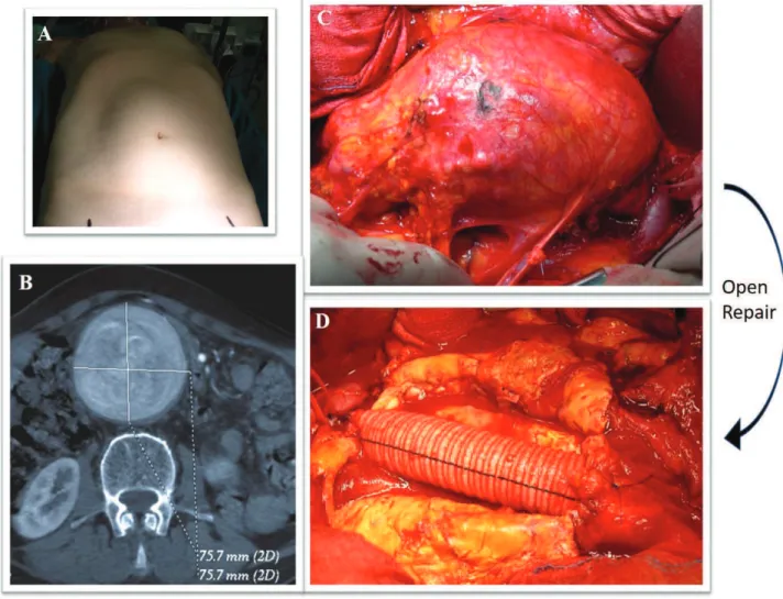
Open surgical repair (OSR) was first reported in 1962 and still remains the treatment with the best long-term results. It is a major surgical procedure done under general anaesthesia, usually consisting of a midline laparotomy and cross-clamping of the aorta and iliac vessels. OSR is followed by the removal of the dysfunctional aortic segment and the insertion of a graft as substitute (DuBost et al., 1952). The mortality rate for OSR is between 3% and 7% and increases significantly in patients with comorbidities, particularly with coronary and carotid artery disease.
Figure 10. Open surgical repair (A) Abdomen of AAA patient (B) Abdominal CT showing aorta dilatation C) Aneurysm sac during surgical open repair D) Abdominal aortic graft in Dacron. Courtesy of Dr. Casella
The development of Endovascular Aneurysm Repair (EVAR) opens the way for a safer management of AAA. The long-term results of EVAR are comparable to those of OSR. EVAR was first described by Parodi in 1991. (Parodi et al., 1991) Juan Parodi implanted the first endograft in a human body and EVAR has become a milestone in the treatment of AAA, especially in presence of significant comorbidities. EVAR consists of the placement of a tubular graft within the aneurysm sac, using a metallic stent to fix it to the normal aortic and iliac wall.
27
The graft acts like an artificial blood vessel, excluding the damaged aortic segment from the normal blood flow, and thus, preventing the aneurysm progression and rupture. This reparative alternative does not require the surgical incision of the abdomen, thus avoiding complications consequent to the open surgery; in addition, it allows a shorter recovery time, reduced hospital stay and implies a lesser blood loss improving the patient quality of life. (Paravastu et al., 2014) Several devices are available to treat AAA, differing with respect to design, modularity, metallic composition and structure of the stent, thickness, porosity and possible presence of an active method of fixing the device to the aortic wall. Appropriately-sized aortic endograft should be selected in relation to patient anatomy. The ideal characteristics are:
ü Stent-graft size ranging ü Long durability
ü Good biocompatibility ü Device delivery flexibility ü Radial force stability ü Low overall costs
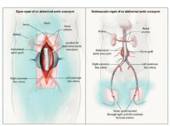
Figure 11. Open surgical repair (A) and Endovascular repair (B). (Schermerhorn et al., 2008)
One of the most frequent cause of EVAR failure is the “endoleak”, which happens when the blood flows into the aneurysm sac, external to the graft, and this condition can lead to
28
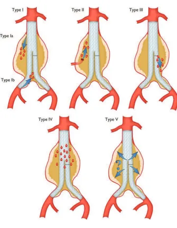
aneurysm expansion and rupture. Endoleak can be detected by angiography, CT scan or duplex ultrasound imaging and can be classified according to the timing of development. Primary endoleak (early endoleak) includes all cases that occur during the 30 days’ perioperative period; secondary endoleak (late endoleak) represents a late complication of the correct seal (White et al., 1997). Endoleaks are further distinguished into five different types: type I, occurs when there is persistent blood flow around the proximal end of the graft or around the distal aspect of the graft;
type II is caused from back-bleeding from side branches (inferior mesenteric artery and lumbar
arteries) that retrograde fill the aneurysm sac; type III, derived from a mechanical failure or a structural defect of the graft; type IV, consequent to a graft wall porosity; type V, often referred to as “endotension,” it happens when the aneurysm sac becomes pressurized without an identifiable source. (White et al., 1998)(Baum et al., 2003)
Figure 12. Classification of endoleaks.
(https://academic.oup.com/eurheartj/article/35/41/2873/407693/2014-ESC-Guidelines-on-the-diagnosis-and-treatment)
Although many researchers suggest that aneurysm formation is due to a combination of genetic, environmental and immune factors (Sakalihasan et al., 2005), the most common form of this dilatative affection rises consequently to atherosclerotic plaque disease, except in specific
29
cases of aneurysms associated with inflammatory diseases, connective tissue affections or traumatic event. The theory on the atherosclerotic origin of AAA has been based on the presence of atherosclerosis pathogenesis features in patients affected by aortic aneurysm. Current investigations and data about the specific nature of this relationship are still controversial; in addition, the aetiology and the key mechanisms involved in aneurysm initiation and expansion have not been completely elucidated. However, common features of aortic aneurysms are inflammation, matrix degradation and smooth muscle cells depletion. According to the most established theory, the “atherosclerotic aneurysm” develops as a complication to the pathological processes deriving from the atherosclerotic plaque development: matrix remodeling, thrombus formation and release of pro-inflammatory cytokines. An alternative theory considers aneurysm and atherosclerosis as independent events, that result from the same environmental and genetic conditions, but different dynamics are involved. In any case, according to current data describing the type of association between the two arterial affections, the majority of AAA are associated with atherosclerosis and elucidating this process would be crucial to develop more specific and targeted therapies. In the following table 1, major features of both atherosclerosis and AAA are listed.
30
1.2.3 Histological features of AAA
The destruction of the lamellar architecture of the aortic media is the predominant histopathological feature of aneurysm disease. As previously described, ECM represents the dynamic structure that supports the aortic wall, driving its main functions. ECM remodeling is strictly regulated under physiological conditions and abnormal expression and activity of the main ECM remodeling mediators are characteristic of many vascular disorders. Elastin fragmentation represents an early event in AAA formation, consequently causing wall weakening; degradation of fibrillar collagen type I and III occurs in a later phase of the AAA expansion, leading to the loss of aortic tensile strength and causing a decisive step in the aortic rupture. Many experimental data, obtained from human aneurysm tissues, as well as from in vitro and in vivo animal models, highlighted the MMPs involvement in ECM abnormal degradation, evidenced by an increased expression of these proteolytic enzymes during aneurysm initiation and/or progression, correlating with aneurysm size. MMPs are secreted by vascular and inflammatory cells; MMP over-expression in terms of mRNA, protein, as well as enzymatic activity, is not only associated with an increased production and secretion, but it is also the result of the unbalanced MMPs/TIMPs ratio. MMP-2 and MMP-9 are the most commonly enzymes associated with aneurysm disease. They both degrade elastin and collagen. MMP-2 is constitutively expressed in small aneurysm, suggesting a role in aneurysm initiation and formation, whereas MMP-9 is more prevalent in large diameter aorta, indicating a main participation in aneurysm progression and expansion. In addition, MMP-8 is a type I collagenases and, together with MMP-9, was shown to be increased in ruptured aneurysms. In addition to MMPs, other proteases localize at aneurysm wall: u-PA (urokinasePlasminogen activator) and t-PA (tissue-Plasminogen activator) convert plasminogen into plasmin, which in turns activates MMPs; cathepsin S and K, with elastolytic action, and cathepsin L, which degrade type IV and V collagen, laminin, elastin and proteoglycans. Medial SMCs constitute the main vascular cell population that participate to aortic structure, through the synthesis of ECM components as well as the secretion of ECM remodeling mediators, like MMPs and their tissue inhibitors TIMPs. The importance of SMC behaviour in the aneurysm development was explored by Lopez-Candales et al, underlining an increased SMC apoptosis, marked by an increased expression of p53, which is indicative of cell cycle arrest, in aneurysmal tissues versus non-aneurysmal and atherosclerotic aortic tissues (Lopez-Candales et al., 1997)(Holmes et al., 1996). The elastin degradation is tightly related to the abnormal hemodynamic shear stress that influences aneurysm growth. The SMCs respond to elastin degradation through an increased tropoelastin synthesis; this is confirmed by increased mRNA levels of tropoelastin in aneurysm tissue. This adaptation is not followed by the correct elastin
31
organization into the mature effective structures, resulting in the aortic wall inability to respond to the increased shear stress.
Moreover, another feature of AAA is the intraluminal thrombus (ILT). In 70–80% of AAA patients, the vessel wall is covered by an intraluminal thrombus which generally does not preclude blood flow and shows little compression throughout the cardiac cycle (O’Leary et al., 2014)(Vorp et al., 1996). While mural thrombosis is frequently observed in aneurysmal disease, the complete vessel occlusion is a comparably rare event associated with a high rate of mortality (Cervantes et al., 1985). In recent studies, more attention has been dedicated to the mechanical properties of the ILT. Indeed, the mechanical role of ILT on the wall stress distribution and the underlying local wall strength is yet unclear.
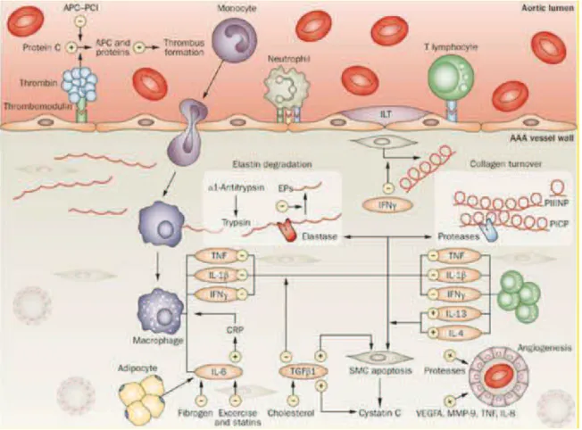
In addition to the loss of structural integrity, aortic aneurysm is characterized by an intensive transmural inflammation. The presence of the inflammatory infiltrate may be due to elastin fragmentation. In fact, this process releases soluble peptides exerting a chemotactic effect that recruits inflammatory cells, which further secrete proteolytic enzymes, thus amplifying the ECM degradation. Studies on the inflammatory infiltrate invading the AAA wall revealed the presence of aggregating clusters of CD3+ T and CD19+ B lymphocytes, together with macrophages, and localized at the adventitial vasa vasorum (Koch et al., 1990). In addition, an imbalance of the ratio between CD3+ T helper and CD8+ T suppressor cells has been observed, with the predominance of T-helper cells (Koch et al., 1990) (Tang et al., 2005). Inflammatory cells activate a cascade of reactions increasing inflammation environment and exacerbating the proteolysis process. CD3+ T lymphocytes produce IL-4, -5, -8, -10 that attract other inflammatory cells, stimulate the cytokine release by T cells and the neoangiogenesis; in addition, CD3+ stimulate the IFN-γ release, which, in turn, induces MMP expression. The presence of antigen presenting cells in aneurysm wall suggests an antigen driven T cell response as leading cause to aneurysm development. Matrix proteins, such as elastin, collagen, aortic aneurysm antigenic protein-40 (AAAP-40) (Xia et al., 1996) and exogenous agent (i.e. herpes simplex virus, cytomegalovirus) have been proposed as potential antigen capable to induce T cells activation. Interestingly, the molecular mimicry is mechanism of T cells activation, and it is supposed to be involved in aneurysm pathogenesis. The molecular mimicry is based on sharing of common sequences and epitops between a foreign antigen (a microorganism) and self (host) antigen: the immune reaction against a virus or bacteria, through a mechanism of cross reaction (Ozsvath et al., 1996)(Oleszak et al., 2004). As-consequence, the immune response inside the aortic wall is exacerbated, thus complicating the proteolytic process. According to these findings, an
32
autoimmune origin of AAA has been proposed (Hirose and Tilson, 2001). Macrophages actively participate to parietal remodeling and inflammation: immunohistochemical and in-situ hybridization data revealed an intensive MMP-9 expression by aneurysm-infiltrating macrophages (Thomson et al., 1995); later works demonstrated the macrophage-mediated release of cytokines, such as Il-1β, TNF-α, IL-8, thus stimulating B-cell and cytotoxic T-cell differentiation, cytokine and protease synthesis. Neo-angiogenesis consists in the formation of new blood vessels from pre-existing ones and for this purpose it requires the ECM degradation by proteolytic enzymes to promote the ECs migration from mature vessels; these steps are driven by specific growth factors, such as VEGF, PDGF, EGF, TNF-α. The occurrence of neovascularisation in AAA wall has been demonstrated by Thomson et al (1995), evidenced by an increased number of neo-vessels in aneurysmal tissues in comparison to normal aortic specimens; moreover, the degree of neovascularisation was shown to be correlated with the inflammatory infiltrate amount (Holmes et al., 1995). These findings, together with immunolocalization studies showing MMPs expression in correspondence of neo-vessels (Herron et al., 1991), support a crucial role for the medial neo-vessels formation to the aneurysm pathogenesis. Moreover, increased neovascularisation and expression of angiogenic cytokines were detected at the site of rupture, indicating an involvement of the angiogenic response into aneurysm rupture.
33
Figure 13. Schematic diagram of the mechanisms involved in abdominal aortic aneurysm, which primarily affect two main processes: inflammation and extracellular matrix turnover (F. a M. V. I. Hellenthal et al., 2009).
1.3 The MMP family
The mammalian MMPs are a group of 23 structurally related enzymes that have a catalytic Zn2+ ion site (Nagase et al., 2006; Visse and Nagase, 2003). MMPs belong to the family of proteolytic enzymes that degrade several components of ECM. MMPs generally consist of a prodomain, a catalytic domain, a hinge region, and a hemopexin domain. The cysteine switch motif PRCGXPD in the propeptide that maintains MMPs in their zymogen form (proMMP), and the zinc-binding motif HEXGHXXGXXH in the catalytic domain is used to distinguish the proteinases family. They are either secreted from the cell or anchored to the plasma membrane. On the basis of substrate specificity, sequence similarity, and domain organization, vertebrate MMPs can be divided into six groups (Figure 14). The key feature of collagenases (MMP-1, MMP-8, MMP-13 and MMP-18) is the ability to cleave interstitial collagens I, II, and III at a specific site. MMP-2 and MMP-9 also belong to collagenases group. They digest denaturated collagens, gelatin. These enzymes have three repeats of type II fibronectin domain inserted in the catalytic domain, which bind gelatin, collagen and laminin.
34
Stromelysin’s group are: stromelysin 1 (MMP-3) and stromelysin 2 (MMP-10) with similar substrate specificities, while MMP-3 has a proteolytic efficiency higher than MMP-10. MMP-11 is called stromelysin 3, but it is usually grouped with “other MMPs” because the sequence and substrate specificity diverge from those of MMP-3.
Matrilysins are characterized by the lack of hemopexin domain. Matrilysin 1 (MMP-7) and matrilysin 2 (MMP-26) belong to this group. MMP-7 processes cell surface molecules such as pro-α- defensin, Fas-ligand, pro-tumor necrosis factor (TNF-α), and E-cadherin, while MMP-26 digests several ECM components. There are six membrane-type MMPs (MT-MMPs): four are type I transmembrane proteins (MMP-14, MMP-15, MMP-16 and MMP-24) and two are glycosylphosphatidylinositol (GPI) anchored proteins (MMP-17 and MMP-25). Seven MMPs are not classified in the above categories. MMP-12 (mainly expressed by macrophages) digests elastin and other ECM molecules. MMP-19, MMP-20, MMP-22, MMP-23 and MMP-28 are part of this group.
Figure 14. Classification of MMP family. The main features of matrix metalloproteinases (MMPs) are illustrated, showing the
minimal domain structures. One clear division is between MMPs that are secreted and those that are anchored to the cell surface by an intrinsic motif namely, a transmembrane (TM) domain (MMP-14, -15, -16 and -24), a glycosylphosphatidylinositol (GPI)
anchor (MMP-17 and MMP-25) or an amino (N)-terminal signal anchor (SA) (MMP-23). Nine MMPs, including all of the membrane-anchored enzymes, have a furin-recognition domain. C5, type-V-collagen-like domain; Col; collagenase-like protein;
Cs, cytosolic; Cys, cysteine array; Fn, fibronectin repeat; Fr, furin-cleavage site; Pro, pro-domain; SH, thiol group; SP, signal peptide; Zn, zinc. (Parks et al., 2004)
35
All MMPs are synthetized as pre-pro-enzymes and secreted as inactive pro-MMPs. Many of the MMPs have the ability to cleave and activate the pro-forms of other MMPs, thereby acting in protease cascades that amplify their effectiveness (Nagase et al., 2006) (Visse and Nagase, 2003). One of the features of MMP is that many of those genes are “inducible” by different effectors such as growth factors, cytokines, chemical agents, physical stress, and oncogenic cellular transformation. Moreover, MMPs are regulated at different levels: gene expression, localization (that is, the pericellular accumulation of enzyme), pro-enzyme (or zymogen) activation and enzyme inactivation (Ra and Parks, 2007). Also, endogenous tissue inhibitors of MMPs (TIMPs) provide a balancing mechanism to prevent excessive ECM degradation. An imbalance between MMPs and TIMPs could lead to MMPs activity increasing and, thus, to pathological changes in the vessel wall structure associated with vascular disease (Raffetto and Khalil, 2008)
1.3.1 Focus on human MMP-9
Human MMP-9 consists of an NH2- terminal pro-domain, a catalytic domain, a linker domain, and a COOH-terminal hemopexin-like domain that combines to form a 92 kDa pro and 88 kDa active enzyme. (Papazafiropoulou and Tentolouris, 2009) The catalytic domain of MMP-9 contains two zinc ions, five calcium ions, and three repeats homologous to the type II module of fibronectin. The catalytic zinc ion is essential for the proteolytic activity. MMP-9 has a unique domain termed “fibronectin-like domain”, which consists of three repeats of fibronectin type II of about 58 amino acids. This domain is heavily O-glycosylated and contains elongated linker between catalytic and hemopexin-like domain (Opdenakker et al., 2001). The fibronectin-like domain is essential for binding to denatured collagen or gelatin (O’Farrell and Pourmotabbed, 1998). Hemopexin-like domain shares a sequence similarity to plasma hemopexin and it is present in MMP-9. In pro-MMP-9, the hemopexin-like domain forms a tight complex with TIMP-1 and TIMP-3 though their COOH-terminal domains (Nagase et al., 2006), Pro-MMP-9 is complexed with TIMP-1 in the Golgi apparatus of the cell before secretion (Roderfeld et al., 2007). TIMP-1 is bound to the pro-MMP-9 via COOH-terminal domain, leaving the NH2 terminus capable of inhibiting other MMPs.
The expression of MMP-9 is regulated on multiple levels: transcription, post-translation (also involving non-proteolytic activation); local translation (Dziembowska et al., 2012); sequestration on the cell membrane (e.g. binding to cell adhesion molecules, such as hyaluronian receptor CD44 (Bourguignon et al., 1998), integrins (Wang et al., 2003), lipoprotein
receptor-36
related protein-1 (LRP-1), and megalin/LRP-2 (Van den Steen et al., 2006)); internalization (Hahn-Dantona et al., 2001); and delayed activation that involves cleavage of the propeptide and co-secretion with TIMP-1, its endogenous inhibitor (Sbai et al., 2010).
At the transcriptional level, MMP-9 is positively regulated by multiple factors, including E-26 (Ets) transcription factors, NF-kB, polyomavirys enhancer A-binding proteins-3 (PEA3), activator protein-1 (AP-1), specificity protein 1 (Sp-1), and serum amyloid A-activating factor (SAF)-1 (16). Ets are a family of transcription factors associated with a variety of biological functions including cellular differentiation, cell migration, proliferation, apoptosis and angiogenesis. NF-kB is capable of binding to kB DNA on the promoters or enhancers of genes to regulate expression. Bond et al. reported an increase in MMP-9 levels in vascular smooth muscle cells via NF-kB mechanisms (Bond et al., 2001a). Inhibition of transcription factor NF-κB reduces MMP-9 production in vascular smooth muscle cells and macrophages (Bond et al., 2001) (Grimm et al., 2006). Reactive oxygen species can activate MMP-9, both directly and indirectly, by activating transcription factors such as NF-κB (Okamoto et al., 2001). Ang II has direct and indirect effects on MMP-9 expression. In ventricular myocytes, Ang II directly stimulates NF-κB to induce MMP-9 expression (Rouet-Benzineb et al., 2000). Ang II activates epidermal growth factor receptor and the mitogen-activated protein kinase pathway to induce MMP-9 expression (Shah and Catt, 2003). Aldosterone, which is produced locally in the myocardium, triggers MMP-9 production through NF-κB signaling (Li et al., 2000). PEA3 and AP-1 binding sites are present in the MMP-9 promoter (Wu et al., 2009). AP-1 has two binding sites on MMP-9, and the activation of MMP-9 is preceded by a rapid transient increase in AP-1 protein levels (Woessner, 1991). Thrombospondins stimulate production of MMP-9 by activating AP-1 (Greenwood et al., 1998). Donnini and colleagues showed that fragments of thrombospondin promote MMP-9 production in bovine capillary endothelial cells (Donnini et al., 2004). In order to increase transcription, Sp-1 binds to the MMP-9 promoter to induce transcription. Sp-1 undergoes several post-transcriptional modifications such as phosphorylation and glycosylation (Murthy et al., 2012) (Taheri and Bazan, 2007). Conversely, inhibition of Sp-1 leads to decreased MMP-9 expression (Murthy et al., 2012). In addition, the transcription factor described above cross-interact to regulate MMP-9 expression. In fact in vascular smooth muscle cells, upregulation of MMP-9 is mainly attributed to expression and activation of NF-κB and AP-1 transcription factors (Valen et al., 2001). AP-1 alone, however, is not sufficient for maximal MMP-9 transcription, and the cooperation of either NF-κB or Sp-1 binding proteins upstream of the AP-1 site is required for full MMP-9 transcription induction (Benbow and Brinckerhoff, 1997). SAF-1 is an inflammatory responsive transcription factor that induces MMP-9 transcription via cooperation with AP-1 (Ray