RESEARCH ARTICLE
Gene expression regulated by abatacept
associated with methotrexate and correlation
with disease activity in rheumatoid arthritis
Ce´line DerambureID1, Gaelle Dzangue-TchoupouID2, Maria Antonietta D’Agostino3, Thierry Lequerre´2,4*, Olivier Vittecoq2,41 Normandie Univ, UNIROUEN, Inserm U 1245, Rouen, France, 2 Normandie Univ, UNIROUEN, Inserm U
1234, Rouen, France, 3 Department of Rheumatology, AP-HP Ambroise Pare´ Hospital, University of Versailles Saint Quentin en Yvelines, Boulogne-Billancourt, France, 4 Department of Rheumatology Inserm CIC/CRB1404, Rouen University Hospital, Rouen Cedex, France
Abstract
Objectives
Abatacept acts as a competitive inhibitor of the CD28/(CD80/86) costimulation signal required for T cell activation. Mechanisms of action of abatacept have not been fully investi-gated. The objective of this study was to provide detailed insight into the mode of action of Abatacept based on gene expression data.
Methods
In this ancillary study from the APPRAISE trial, we investigated the global molecular effects of Abatacept in whole blood samples collected prospectively in biologic naive rheumatoid arthritis patients (n = 19) at baseline and 6 months after the initiation of Abatacept therapy concomitant with methotrexate. Whole human genome microarrays (4x44K) were per-formed on both baseline and 6-month samples from responders and non-responders patients categorized according to EULAR criteria. T-test with Benjamini-Hochberg correc-tion was performed to identify significant gene expression changes. Gene Ontology and Single Experiment Analysis tools allowed us to highlight specific biological mechanisms involved in methotrexate/Abatacept.
Results
In methotrexate/Abatacept responders, 672 genes were significantly (q<0.05) dysregulated at 6 months compared to baseline. Correlation analysis highlighted 19 genes whose dysre-gulations were significantly associated with disease activity variation (p<0.05) and whose functions were associated with proliferation, apoptosis of cells and mitochondrial metabo-lism, suggesting a restoration of oxidative signaling. The other 653 gene expression changes were relative to direct or indirect effects of methotrexate/Abatacept treatment and were significantly (p<0.005) involved in pathways relative to mRNA processing, protea-some, angiogenesis, apoptosis and TCR signaling. This study highlights new mechanisms
a1111111111 a1111111111 a1111111111 a1111111111 a1111111111 OPEN ACCESS
Citation: Derambure C, Dzangue-Tchoupou G, D’Agostino MA, Lequerre´ T, Vittecoq O (2020) Gene expression regulated by abatacept associated with methotrexate and correlation with disease activity in rheumatoid arthritis. PLoS ONE 15(8): e0237143.https://doi.org/10.1371/journal. pone.0237143
Editor: Masataka Kuwana, Nippon Medical School, JAPAN
Received: September 11, 2019 Accepted: July 21, 2020 Published: August 6, 2020
Copyright:© 2020 Derambure et al. This is an open access article distributed under the terms of the Creative Commons Attribution License, which permits unrestricted use, distribution, and reproduction in any medium, provided the original author and source are credited.
Data Availability Statement: All relevant data are uploaded to the National Center for Biotechnology Information’s Gene Expression Omnibus (GEO) database and publicly accessible via accession numbers GSE91079 and GSE68215.
Funding: The APPRAISE trial was funded by Bristol-Myers Squibb and this ancillary study was funded by Bristol-Myers Squibb which was involved in the design of the study as well as in the collection of clinical data and biological samples, in
of action of methotrexate/Abatacept and may provide new therapeutic targets to prevent autoimmunity in rheumatoid arthritis.
1. Introduction
Abatacept (ABA) is indicated for the treatment of adult patients with moderately to severely active RA. ABA may be used as monotherapy or concomitantly with disease-modifying anti-rheumatic drugs (DMARDs) such as methotrexate (MTX). ABA is a soluble human recombi-nant fusion protein for which the extracellular domain of human cytotoxic T lymphocyte-associated molecule 4 (CTLA-4Ig) is bound to the Fc portion of human IgG1. ABA decreases the T cell responses involved in RA pathophysiology. In fact, T cells are activated by the engagement of the T cell receptor (TCR) and the interaction of costimulatory molecules, such as CD80/CD86 on antigen presenting cells (APC), with CD28 on T cells. Activated T cells express T-lymphocyte antigen-4 (CTLA-4), which binds both costimulatory molecules CD80 and CD86, but in contrast to CD28 binding, delivers anti-proliferative signals that downregu-late T cell activation [1–3]. Thus, CD80 and CD86 appear to present dual functions within the immune system: activation signals by their interaction with CD28 as well as inhibition signals by their interaction with CTLA-4. Therefore, we sought to understand how ABA can act on both APC and T cells when ABA interacts with CD80/CD86.
Overall, CTLA-4Ig limits T cell proliferation, promotes T cell tolerance, induces reverse sig-naling in antigen-presenting cells, reduces the migratory capacity of monocytes decreasing the expression of several adhesion molecules, induces the 2,3-dioxygenase indolamine pathway in dendritic cells and heme oxygenase in Tregs, restores T cell small GTPase Ras-related protein 1 (Rap1) function and inhibits osteoclast precursor cell differentiation [4–7]. Despite some data available on synovium, the mechanisms of action of ABA have not been fully investigated at the molecular level [8,9]. Gene expression signatures can provide an unbiased view of the molecular changes underlying biologically and medically interesting phenotypes.
In this ancillary study from the APPRAISE trial, we investigated the global molecular effects of ABA therapy concomitant with MTX in whole blood samples collected prospectively in bio-logic naive RA patients at baseline and 6 months after the initiation of therapy. The objective of this study was to provide detailed insight into the mode of action of ABA based on gene expression data on whole blood.
2. Materials and methods
Patients from the APPRAISE trial
A total of 19 RA patients from the APPRAISE trial (NCT00767325) were enrolled in this ancil-lary study [10,11]. Eligible patients were �18 years of age, had American College of Rheuma-tology (ACR)-defined RA for at least 6 months according to the 1987 classification criteria [12], and were receiving MTX (�15 mg/week) for at least 3 months prior to baseline. Patients were required to have active disease, defined by a baseline Disease Activity Score 28 (DAS28-CRP) of >3.2 or tender and swollen joint counts of �6 and a C reactive protein ((DAS28-CRP) level greater than the normal upper limit. All patients received intravenous infusions of ABA (10 mg/kg) at baseline, and at weeks 2, 4, 8, 12, 16, 20 and 24, in addition to stable doses of con-comitant MTX (�15 mg/week). Oral corticosteroid use (stable dose of �10 mg prednisone/ day) was permitted during the study. For this study, 5 ml of whole blood were collected in the interpretation of data; in the writing of this
manuscript and in the decision to submit the manuscript for publication. The statistical analyses concerning this ancillary study were performed by CD independently of Bristol-Myers Squibb. This does not alter our adherence to PLOS ONE policies on sharing data and materials.
Competing interests: M-AD’A has received fees from Bristol-Myers Squibb, AbbVie, UCB, MSD, Novartis and Roche Pharma, as well as research grants from Pfizer. TL and OV reports consultancy fees from Bristol-Myers Squibb outside the submitted work; TL and OV reports consultancy and personal fees from AbbVie, Chugai, Merck Sharp & Dohme, Novartis, Sanofi, Pfizer, UCB and grant from BMS. CD and GDT have nothing to disclose. CD, TL, OV, Bristol-Myers Squibb and Inserm Transfert have a patent application in conjunction with the University of Rouen for the use of predictive biomarkers able to predict abatacept responsiveness (METHODS OF PREDICTING DRUG RESPONSIVENESS OF PATIENTS SUFFERING FROM AUTOIMMUNE INFLAMMATORY DISEASES; PCT/EP2017/ 050928). This does not alter our adherence to PLOS ONE policies on sharing data and materials.
PAXgene RNA tube (Qiagen, PreAnalytiX, GmbH, Courtaboeuf, France) just before the first infusion and 6 months later and stored at -80˚C until use.
The study was approved by the Institutional Review Board/Independent Ethics Committee (IRB/IEC) and local ethics committees (Hopital Ambroise Pare, Comite´ de Protections des Personnes Idf Vlll Lab D’Anatomopathologie, 9 Ave Charles De Gaulle, Boulogne-Billancourt 92100, France. Comitato Etico Per La Sperimentazione Del Farmaco, Asur, Territoriale 5 Di Jesi Via Gallodoro 68, Jesi (An) 60035, Italy. Azienda Ospedaliera Universitaria Policlinico G. Martino, Comitato Etico Scientifico Via Consolare Valeria, 1, Messina 98124, Italy. Leeds East Research Ethics Committee, Yorkshire and Humber Rec Cntr Off. First Floor, Millside Mill Pond Lane, Leeds, England LS6 4RA, United Kingdom. Ceic Fundacio Unio Catalana D’Hos-pitals, Area De Serveis C/Bruc 72–74 1a, Barcelona 08009, Spain. Azienda Universitaria Senese, Comitato Etico Locale La Sperimentazione Clinica Dei Medicinali Farmacia Aous— Viale Bracci, Siena 53100, Italy. De Videnskabsetiske Komiteer For Region Hovedstaden, Kon-gens Vaenge 2, Hillerod 3400, Denmark. Com Sperimentazione Clin Dei Med Azienda Osp-Univ Pisana, Via Roma 67, Pisa 56126, Italy. Comitato Etico Dell’Osp-Universita Cattolica Del Sacro Cuore, Policlinico Universitario Agostino Gemelli Di Roma Largo A Gemelli 8, Roma 00168, Italy. Rek Sorost, Frederik Holsts Hus Ulleval Terrasse Ulleval Sykehus Kirkeveien 166, Oslo 0450, Norway. Ceic—Fundacion Jimenez Diaz-Ute, Avda. Reyes Catolicos, 2-2a, Madrid 28040, Spain. Hospital Mostoles, Ceic Area 8 C/Rio Jucar S/N, Madrid 28935, Spain. Univer-sita Di Roma La Sapienza, Comitato Etico Dell’Azienda Policlinico Umberto I, Roma 00161, Italy. Ceic Area 9-Hosp Severo Ochoa De Leganes Y Hosp De Fuenlabrad, Avenida Orellana S/N Leganes, Madrid 28911, Spain. Hospital Universitario La Princesa, C/Diego De Leon, 62, Madrid 28006, Spain. Farmakologiai Etk Bizottsaga, Arany J.U. 6–8., Budapest 1051, Hungary. Comitato Etico Per La Sperimentazione Dell’Azienda, Ospedaliera Istituti Ospitalieri Di Verona Piazzale Stefani 1, Verona 37126, Italy. Ethikkommission Der Ludwig-Maximilians Universitaet, Marchioninistr. 15, Muenchen 81377, Germany) and was conducted in accor-dance with the ethical principles underlying the European Union Directive 2001/20/EC and the United States Code of Federal Regulations on Good Clinical Practice as defined by the International Conference on Harmonisation. All patients provided written informed consent.
Clinical evaluation and response to MTX/ABA
Clinical characteristics were collected at baseline and 6 months later: age, gender, disease dura-tion, MTX and corticosteroid doses. All the patients were treated with MTX concomitant with ABA whereas 52.6% of the patients (10/19) received corticosteroids. Anti-citrulinated peptide antibody (ACPA) and rheumatoid factors titers were not collected in this study. The response to MTX/ABA was evaluated at 6 months with EULAR response criteria based on DAS28-CRP [13]. These 19 RA patients were selected among the patients from the APPRAISE trial on the basis of their highest amplitude of response for good responders (change in DAS28-CRP �2.3) after 6 months of treatment (n = 14). Only five non responders were at our disposal (S1 Fig).
RNA preparation
Total RNAs from whole blood were extracted with PAXgene blood RNA kit according to the manufacturer’s recommendations (Qiagen PreAnalytiX GmbH, Courtaboeuf, France) and stored at -80˚C until use. The quality and quantity of isolated RNAs were assessed using the 2100 Bioanalyzer (Agilent Technologies, Santa Clara,California, USA) and the Nanodrop
Microarrays
Whole human genomic DNA microarrays were used to analyze two-colored gene expression profiling (4x44K Whole Human Genome, Agilent Technologies, Les Ulis, France). To identify dysregulated gene expression under treatment, baseline samples (100ng) were Cyanine-3 labeled and co-hybridized with their cyanine-5 paired samples after treatment (6 months) according to the manufacturer’s instructions (Low Input QuickAmp Labeling Kit, Agilent Technologies, Les Ulis, France). The microarrays were scanned with a 5μM pixel size using the DNA Microarray Scanner GB (Agilent Technologies, Les Ulis, France). Image analysis and extraction of raw and normalized signal intensities by Lowess method were performed with Feature Extraction Soft-ware 10.5.1.1 (Agilent Technologies, Les Ulis, France). The data were in agreement with the Minimum Information About a Microarray Experiment guidelines and were deposited in the National Center for Biotechnology Information’s Gene Expression Omnibus (GEO) database (https://www.ncbi.nlm.nih.gov/geo/). The data are accessible using the accession number GSE91079 for gene expression changes between baseline and 6 months under MTX/ABA. Data from a previous study were used to recover the ratio of gene expression from RA patients at baseline just before treatment compared to 10 healthy donors (GSE68215) [14].
Statistical and functional analysis
Comparisons of clinical and biological data using GraphPad Prism software version 5.0 were performed using unpaired or paired Student’s t-test; unpaired t-test for age, methotrexate and corticosteroid doses, CRP, DAS28, TJC, SJC and VAS between R and NR at onset (0 month); paired t-test for differences between 0 and 6 months in both R and NR subsets; and Fisher’s exact test for frequency comparisons (sex-ratio).
Data from the transcriptomic analysis were analyzed with GeneSpring GX V.14.0 (Agilent Technologies, Les Ulis, France). A first step of filtration was applied to remove the spike-in probes, the flagged probes (not uniform, saturated or outlier) and the probes whose intensities were close to the background (<100 fluorescence intensity) both at baseline and at 6 months in R and NR separetely. Student’s t-test with Benjamini Hochberg correction to check the False Discovery Rate ((FDR), FDR = 0.05) was used to determine the statistical significance (q-value < 0.05) in gene expression levels between 6-month samples and paired-baseline samples. The Gene Ontology (GO) analysis was used to investigate the biological processes, molecular function or cellular localization enriched in the transcript list showing significant changes between 6 month and baseline.P value was computed by standard hypergeometric
distribu-tion. The GeneSpring Single Experiment Analysis (SEA) bio-informatics tool was used for computational analysis to identify potential curated canonical pathways which are enriched in the transcript list showing a significant gene expression difference between 6 month and base-line, using WikiPathways database (http://www.wikipathways.org). 760 pathways were evalu-ated and gene enrichment was measured by Fisher’s exact test.
For genes with a statistically significant change in expression between 6 month and base-line, correlation between change in gene expression and change in DAS28 was computed. Nominal significance of the Pearson’s correlation test was used to determine if the change in gene expression was related to ABA/MTX response.
3. Results
Characteristics of RA patients and their response to MTX/ABA
From the APPRAISE trial, 19 RA patients were selected among R (n = 14) and NR (n = 5) treated by MTX/ABA according to EULAR response criteria.Table 1provides demographic
and clinical data for these 19 RA patients at baseline and after 6 months of treatment. The characteristics of RA patients from R and NR at baseline were comparable for all parameters suggesting absence of bias in patient selection (p>0.05). In the R subset, tender joint count,
swollen joint count, global assessment of disease measured by the patient with visual analog scale, disease activity score (DAS28-CRP) (p<0.001) and CRP (p<0.05) improved significantly
after 6 months of treatment, while in the NR subset no parameters improved significantly (Table 1).
Gene expression changes under MTX/ABA in R
After filtration steps, 21,529 transcripts in R were retained for subsequent differential gene expression analyses between baseline and 6 months. By t-test adjusted with Benjamini-Hoch-berg correction (5% FDR withq< 0.05) for multiple testing, 672 genes were significantly
dys-regulated in R between baseline and 6 months of treatment (Fig 1A). The size of NR subset of patients (n = 5) was too low and the analysis statistically underpowered to identify significant gene expression changes in this subset. Among the dysregulated genes in R, 481 genes were up-regulated while 191 genes were down-regulated under treatment (Fig 1B). In order to dif-ferentiate gene expression changes (log2(6-month expression/ baseline expression)) relative
to both disease activity extent of improvement (Δ DAS28-CRP = DAS28-CRP at baseline— DAS28-CRP at 6 months) and/or treatment effects in R, correlation analysis was performed between gene expression changes andΔ DAS28-CRP between baseline and 6 months. Among the 672 genes dysregulated in R, 19 transcripts were correlated with DAS28 extent of improve-ment (p <0.05) (Fig 2,S2 Fig). The 653 other transcripts were not linked to disease activity extent of improvement suggesting maybe a gene expression change relative to direct or indi-rect treatment effects.
Gene expression change relative to disease activity variation
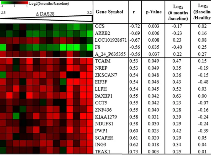
Among the 19 genes dysregulated relative to disease activity between baseline and 6 months in R, 5 were negatively (-0.72 < r < -0.56) correlated with change in disease activity (increase in gene expression was correlated with worse response) and 14 were positively (0.53 < r < 0.73) correlated with change in disease activity (increase in gene expression was correlated with bet-ter response) (Fig 2,S2 Fig). Based on the findings of our previous study, half of these 14 Table 1. Clinical and biological parameters of RA patients.
R (n = 14) NR (n = 5)
0 6 months 0 6 months
Age 58.79 +/- 4.87 58.60 +/- 4.2
Gender (F/M) 11/3 3/2
Duration of disease (years) 4.5 +/- 1.75 8.80 +/- 4.13
Methotrexate dose (mg/week) 15.36 +/- 1.11 18 +/- 1.23
Corticosteroid dose (mg/day) 6 +/- 0.75 6.5 +/- 1.5
Tender joint count/28 15.07 +/- 1.46��� 0.43 +/- 0.17 9.60 +/- 4.35 10.80 +/- 2.96
Swollen joint count /28 12.78 +/- 1.66��� 1.5 +/- 0.66 9.40 +/- 1.78 8.40 +/- 1.83
Patient assessment of disease (VAS scale/100 mm) 62.07 +/- 6.81��� 17.14 +/- 4.9 49.4 +/- 12.29 49.0 +/- 12.77
DAS28-CRP 5.77 +/- 0.23��� 2.09 +/- 0.12 4.95 +/- 0.76 4.97 +/- 0.68
CRP (mg/L) 13.83 +/- 4.83� 2.91 +/- 0.53 20.47 +/- 8.50 16.55 +/- 9.03
Mean +/- Standard error of the mean.P values were determined by Student’s t-test or Fisher exact test (�: p<0.05;���: p<0.0001). CRP: C reactive protein; DAS28:
disease activity score 28; NR: non-responders; R: responders; VAS: visual analog scale. Data available inS3 Fig.
genes, were down-regulated at baseline in RA responders to MTX/ABA compared to 10 healthy subjects (Fig 2). Conversely, for the expression of the 5 genes negatively correlated with disease activity change between baseline and 6 months, gene expression ratios between baseline in RA responders to MTX/ABA and 10 healthy subjects were all positive (Fig 2) Fig 1. Gene expression changes between baseline and 6 months after MTX/ABA in responders. Baseline samples were co-hybridized with 6 months samples on 4x44K Agilent whole human genome microarrays. Data are expressed by a log2ratio (6 months / baseline). A: Volcano plots in responders
(n = 14) was built with at-test with Benjamini-Hochberg correction for false discovery rate estimation. 672 genes were significantly dysregulated in R
(q<0.05). Fold change (FC) is the ratio of relative abundance of transcripts in RA patients after treatment (6 months)vs before treatment (baseline). B:
Hierarchical clustering (Pearson’s correlation coefficient and complete linkage metrics) showing the 481 up-regulated genes and the 191 down-regulated genes in responders between baseline and 6 months. C: single experiment analysis of the 653 genes whose dysregulations between baseline and 6 months were associated with MTX/ABA in responders. We identified 6 signaling pathways involved in MTX/ABA response from the WikiPathways database (enrichment p-value < 0.005).
suggesting that these 5 genes were up-regulated at baseline for RA responders compared to healthy subjects. The up or down regulation of these 19 genes compared to regulations between healthy subjects and responders suggest a trend toward the normalization of these gene expressions at 6 months compared to baseline. The analysis of these 19 transcripts with GO tool showed no biological process or molecular function enriched in this transcript set. Moreover, when these 19 mRNA were submitted to WikiPathways via SEA tool from Gene-Spring software, we found a significant enrichment of 7 signaling pathways (p<0.05) but including just one gene out of 19 for each pathway. Therefore, these 19 transcripts cannot be mapped to specific biological function. These results allowed us to consider that the gene expression of these 14 genes increased and that of the 5 genes decreased to trend toward a nor-mal gene expression level, when disease activity variation improved. These results are specific Fig 2. Disease activity correlates with gene expression changes between baseline and 6 months in MTX/ABA responders. Pearson’s correlation coefficients (r) were calculated for all 672 transcripts which were significantly dysregulated between baseline and 6 months after treatment in RA responders (n = 14). Nineteen transcripts were correlated (p<0.05) with change in disease activity score 28 (Δ DAS). For each transcript mean of Log2(6
months / baseline) and mean of log2(baseline / healthy controls) are indicated. A_24_P635355: Agilent probe ID; ARRB2: arrestin beta-2; CCS: copper
chaperone for superoxide dismutase; CCT5: Chaperonin containing TCP1 subunit 5; EIF3F: eukaryotic translation initiation factor 3 subunit; F8: Coagulation factor VIII; ING3: inhibitor of growth family member 3; KIAA1279: KIF1 binding protein; LLPH: LLP homolog. long-term synaptic facilitation; NDUFS1: NADH-ubiquinone oxidoreductase 75 kDa subunit; NREP: neuronal regeneration related protein; PAXBP1: PAX3 and PAX7 binding protein 1; PWP1: Periodic tryptophan protein 1 homolog; SCAPER: S-phase cyclin A-associated protein in the endoplasmatic reticulum; TCAIM: T-cell activation inhibitor; TRAK1: Trafficking protein Kinesin binding 1; ZKSCAN7: Zinc finger with KRAB and SCAN domains 7; ZNF436: zinc finger protein 436.
to R patients since the gene expression levels of these 19 genes did not change significantly in 5 NR between baseline and 6 months.
Gene expression changes relative to ABA in responders
Among the 672 genes whose gene expression differed under MTX/ABA in R between baseline and 6 months, 653 were relative to ABA since their gene expression levels were dysregulated with no correlation with disease activity improvement (DAS28-CRP). These 653 transcripts were submitted to GO analysis showing a significant enrichment of 20 GO classes relative to nuclear localization and mRNA processes.
The WikiPathways analysis by SEA tool of these 653 genes led us to identify a significant enrichment of 6 signaling pathways involved in ABA response: 2 mRNA processes, protea-some, angiogenesis, apoptosis and TCR signaling (Fig 1C) (p<0.005). mRNA processing path-ways were in accordance with GO analysis results. All these genes dysregulated under ABA and involved in these 6 pathways are indicated inTable 2.
4. Discussion
The main objective of this study was to investigate the molecular effects of ABA on gene expression in whole blood from RA patients. We compared the molecular changes under ABA by means of transcriptomic analysis of whole blood. From the APPRAISE trial, we selected 14 R and 5 NR to the MTX/ABA concomitant treatment. At baseline, all the 19 patients were comparable and had high disease activity (Table 1). After 6 months of treatments, DAS28 decreased from 5.8± 0.2 to 2.1 ± 0.1 in R, while it was unmodified in NR (from 4.9 ± 0.8 to 5± 0.7). We did not consider corticosteroids as a potential bias since both the proportion of patients treated in concomitance with corticosteroids and corticosteroids doses are not signifi-catively different between R and NR subsets (p = 0.63 and 0.77 respectively).
In R, ABA significantly changed the gene expression level of 672 genes including 481 up-regulated and 191 down-up-regulated genes. The question arises as to whether the changes of the genes expressions results from the decrease of disease activity or from the direct or indirect effects of ABA. Only 19 genes had a gene expression change nominally correlated with extent of improvement of disease activity variation measured with DAS28-CRP. Since the absolute correlation coefficients range between 0.53 and 0.73, these gene expression changes are not fully explained by modification of the disease activity. Gene enrichment analysis did not iden-tify any significant pathways for these 19 genes. Nevertheless, one relevant biological process was found from data in the literature.
A decrease of disease activity at 6 months compared to baseline was correlated with an increase of expression level for TCAIM, TRAK1, KIA1279 (KIF1 binding protein), NDUFS1 and CCT5, which are involved in mitochondria processes [15–17].NDUFS1 encodes a
mito-chondrial protein NADH-ubiquinone oxidoreductase subunit, regulating reactive oxygen spe-cies (ROS) production [18]. These results are in accordance with the dysregulation ofCCT5
which encodes a molecular chaperone that is a member of the chaperonin containing TCP1 complex involved in autophagy, clearance of dysfunctional mitochondria and production of ROS [19]. Interestingly, in a previous study, we found a pre-silencing of the electron transport chain (ETC) pathway in R compared to NR before initiation of MTX/ABA [14]. The down reg-ulation of genes expressed in mitochondria reflected the redox imbalance observed in R [14]. After 6 months of treatment, ABA significantly upregulated some ETC genes, including
NDUFS1, in R probably leading to a slight increase in ROS, restoring the redox balance and
improving T cell response as recently demonstrated by Yang Z [20]. This study has shown that MTX/ABA treatment modulates the gene expression involved in mitochondria processes and
Table 2. Transcripts relative to significantly enriched pathways in MTX/ABA responders.
ProbeName Log2(FC) Gene Symbol Gene Name RefSeqAcc UniGeneID
Hs_mRNA_processing_WP411_45374 (p = 2,1.10−6)
A_23_P61551 -0.26 CD2BP2 CD2 (cytoplasmic tail) binding protein 2 NM_006110 Hs.202677 A_23_P28688 0.28 CPSF3 cleavage and polyadenylation specific factor 3. 73kDa NM_016207 Hs.515972 A_23_P37137 0.36 DNAJC8 DnaJ (Hsp40) homolog. subfamily C. member 8 NM_014280 Hs.433540 A_24_P517901 0.34 HNRNPA1 heterogeneous nuclear ribonucleoprotein A1 NM_002136 Hs.546261 A_32_P142028 0.25 HNRNPC heterogeneous nuclear ribonucleoprotein C (C1/C2) NM_031314 Hs.508848 A_24_P724984 0.62 HNRNPR heterogeneous nuclear ribonucleoprotein R NM_001102398 Hs.373763 A_24_P157424 0.52 NCBP2 nuclear cap binding protein subunit 2. 20kDa NM_007362 Hs.591671 A_24_P151727 0.23 NONO non-POU domain containing. octamer-binding NM_007363 Hs.533282 A_23_P129614 0.24 NUDT21 nudix (nucleoside diphosphate linked moiety X)-type motif 21 NM_007006 Hs.528834
A_32_P193646 0.27 RBMX RNA binding motif protein. X-linked NM_002139 Hs.380118
A_23_P162945 0.25 SRP54 signal recognition particle 54kDa NM_003136 Hs.167535
Hs_Processing_of_Capped_Intron-Containing_Pre-mRNA_WP1889_42105 (p = 7.2.10−6)
A_23_P61551 -0.26 CD2BP2 CD2 binding protein 2 NM_006110 Hs.202677
A_23_P37137 0.36 DNAJC8 DnaJ (Hsp40) homolog. subfamily C. member 8 NM_014280 Hs.433540 A_24_P517901 0.34 HNRNPA1 heterogeneous nuclear ribonucleoprotein A1 NM_002136 Hs.546261 A_32_P77502 0.38 HNRNPA3 heterogeneous nuclear ribonucleoprotein A3 NM_194247 Hs.516539 A_32_P142028 0.25 HNRNPC heterogeneous nuclear ribonucleoprotein C (C1/C2) NM_031314 Hs.508848 A_24_P724984 0.62 HNRNPR heterogeneous nuclear ribonucleoprotein R NM_001102398 Hs.373763 A_24_P157424 0.52 NCBP2 nuclear cap binding protein subunit 2. 20kDa NM_007362 Hs.591671
A_32_P193646 0.27 RBMX RNA binding motif protein. X-linked NM_002139 Hs.380118
Hs_Proteasome_Degradation_WP183_45274 (p = 9.88.10−5)
A_24_P113674 0.49 HLA-B major histocompatibility complex. class I. B NM_005514 Hs.77961 A_23_P113716 0.49 HLA-C major histocompatibility complex. class I. C ND Hs.743218 A_24_P311926 0.48 HLA-G major histocompatibility complex. class I. G NM_002127 Hs.512152 A_24_P418044 0.35 HLA-J major histocompatibility complex. class I. J (pseudogene) NR_024240 Hs.720762 A_32_P57870 0.20 PSMC1 proteasome (prosome. macropain) 26S subunit. ATPase. 1 NM_002802 Hs.356654 A_23_P109928 0.24 PSMD6 proteasome (prosome. macropain) 26S subunit. non-ATPase. 6 NM_014814 Hs.152536
A_24_P681301 0.26 UBC ubiquitin C NM_021009 Hs.520348
Hs_TCR_Signaling_Pathway_WP69_45093 (p = 0.004)
A_24_P128145 0.32 ATF2 activating transcription factor 2 NM_001880 Hs.592510
A_24_P945262 0.35 CARD11 caspase recruitment domain family. member 11 ND Hs.665701
A_23_P34676 0.36 CD247 CD247 molecule NM_198053 Hs.156445
A_24_P407717 -0.17 GRB2 growth factor receptor-bound protein 2 NM_002086 Hs.444356 A_23_P159920 -0.24 IKBKG inhibitor of kappa light polypeptide gene enhancer in B-cells NM_003639 Hs.43505
A_23_P44112 0.24 LAT linker for activation of T cells NM_014387 Hs.632179
Hs_Apoptosis_Modulation_by_HSP70_WP384_41236 (p = 0.003)
A_23_P72537 0.36 AIFM1/PDCD8 apoptosis-inducing factor. mitochondrion-associated. 1 NM_004208 Hs.424932
A_24_P29665 0.34 CYCS cytochrome c. somatic NM_018947 Hs.437060
A_23_P86917 -0.20 FADD Fas (TNFRSF6)-associated via death domain NM_003824 Hs.86131 Hs_angiogenesis_overview_WP1993_44954 (p = 6.62.10–4)
A_24_P128145 0.32 ATF2 activating transcription factor 2 NM_001880 Hs.592510
A_24_P407717 -0.17 GRB2 growth factor receptor-bound protein 2 NM_002086 Hs.444356
A_23_P40174 -0.66 MMP9 matrix metallopeptidase 9 NM_004994 Hs.297413
A_23_P13969 0.41 PXN paxillin ND Hs.446336
A_23_P107401 -0.32 TIMP2 TIMP metallopeptidase inhibitor 2 NM_003255 Hs.633514
proliferation or apoptosis of cells. When we compared the gene expression of these 19 genes in healthy subjects and in patients with disease activity improvement, the gene expression of half of these genes dysregulated in RA patients fluctuated toward the normal gene expression level found in healthy subjects. The normalization of gene expression level under MTX/ABA treat-ment and the correlation between gene expression level and disease activity represent two arguments to consider the involvement of these genes in RA pathophysiology.
The gene expression level of the majority of genes with a significant variation of expression under MTX/ABA in R was not correlated with disease activity. This may be due to little varia-tion (2.3–5.2) in the response amongst the R group. The submission of these genes to GO and Wikipathway databases found 20 significant biological processes and 6 signaling pathways.
ABA increased mRNA processes through two pathways, that seems relevant since Naka-mura Set al. also found that dysregulation of genes was involved in RNA elongation arrest and
recovery, regulation of apoptosis and formation of RNA polymerase II elongation complex [21].
Concerning TCR signaling pathway, the expression level ofGRB2 and IKBKγ decreased
whileATF2, CD247, LAT and CARD11 increased at 6 months compared to baseline under
MTX/ABA. The growth factor receptor-bound protein 2 (GRB2) modulates and integrates sig-nals from receptors on cellular surfaces in inner signaling pathways and is crucial for amplifi-cation of TCR signalling [22]. Moreover, IKBKγ, also called NEMO, exercises a negative action on TCR-induced NF-κB activation [22,23]. On the contrary,LAT, CD247, ATF2 and CARD11 were overexpressed under MTX/ABA in R. Linker for activation of T cells (LAT)
binds Grb2 to mediate T cell activation, proliferation and cytokine production [24]. The T-cell receptor T3 zeta chain (CD3z) or CD247 is essential for assembly, surface expression and sig-naling cascade of the T-cell receptor-CD3 (TCR/CD3) complex. A reduced density of synovial T-cell surface CD3z has been observed in RA patients, suggesting a decrease in TCR signaling, thus encouraging positive selection of autoreactive T effector cells in the thymus [25,26]. While ABA is supposed to block T cell activation, our analysis of the TCR signaling pathway and the literature suggest a surprising activation of T cells. Normally, ABA binds CD80/86 to prevent the costimulatory signal relative to the interaction between CD28 and CD80/86, lead-ing to T cell inactivation. Since T cells were activated in RA, the level of CTLA4 expressed on T cell membranes was increased. CTLA4 is also a ligand to CD80/86 to induce anti-proliferative effects on T cells [27]. Therefore, since ABA binds CD80/86, the interaction between CTLA4 and CD80/86 is blocked, inducing the suppressive effects of inhibition on T cell, leading finally to T cell activation. This presumed mechanism could explain the T cell activation observed in our study. Further functional analyses are necessary to confirm this new mechanism of action of ABA.
Protein degradation by the proteasome is one of the major regulatory mechanisms in the cell. In our study, 7 genes from this pathway (UBC, PSMC1, PSMD6, HLA-B, HLA-C, HLA-G
andHLA-J) were mainly up-regulated in MTX/ABA responders at 6 months compared to
baseline. These results are in agreement with the increase in mRNA processes. Indeed, protea-some activation may reflect the activation of mRNA processes since the ubiquitin-proteaprotea-some Table 2. (Continued)
ProbeName Log2(FC) Gene Symbol Gene Name RefSeqAcc UniGeneID
A_23_P132611 0.56 VHL von Hippel-Lindau tumor suppressor. E3 ubiquitin protein ligase NM_000551 Hs.517792
FC: fold change (6months/baseline). RefSeqAcc: accession number in RefSeq database. Probe name: Agilent probe ID. ND: not determined.
system has both proteolytic and non-proteolytic functions in multiple aspects of the transcrip-tion process, including initiatranscrip-tion, elongatranscrip-tion and mRNA processes [28].
AIFM1, ATF2 and CYCS up-regulated in our study, are released from the mitochondrion
intermembrane space into the cytosol and the nucleus and are pro-apoptotic factors. CYCS induces apoptosome formation involved in the cell death by caspase-dependent pathway, while AIFM1 contributes to apoptotic nuclear DNA damage in a caspase-independent way [29]. ATF2 and AIFM1 also induce mitochondria to release the apoptogenic proteins cyto-chrome c (CYCS). In addition,CARD11 and IKBKG (also called NEMO) involved in TCR
pathway, were dysregulated under ABA in favor of apoptosis since CARD11 is an upstream activator of BCL10 and NF-κB signaling. On the contrary, the gene expression of Fas-associ-ated protein with Death Domain (FADD) was decreased under ABA enhancing cell survival.
To explain the opposite effects of MTX/ABA in apoptosis, further studies are necessary in order to better understand the effect of ABA on different blood cells.
Finally,GRB2, MMP9, TIMP2 were down-regulated under MTX/ABA at 6 months
com-pared to baseline in RA responders, whereasATF2, PXN and VHL were up-regulated. All
these genes are involved in the angiogenesis process [30].GRB2 and MMP9 are known to
pro-mote angiogenesis. Indeed, MMP9 is thought to play an important role in the degradation of extracellular matrix (ECM) and to facilitate the process which is useful for angiogenesis. The dysregulation ofVHL, GRB2 and MMP9 suggests a decrease of angiogenesis at 6 months
com-pared to baseline under MTX/ABA in R [31].
The limitation of this study was the low number of NR to MTX/ABA, the concomitant treatment of MTX making the interpretation of the own effects of ABA difficult. Moreover, the study was performed from the whole blood and not from cell subsets such as monocytes, T or B cells. RNA sequencing on each cell population would be more appropriated to understand the specific cellular effects of ABA.
5. Conclusions
The MTX/ABA concomitant treatment regulates few genes whose expression is correlated with extent of improvement of disease activity in RA responders. These genes are involved in inflammation and in mitochondrial processing suggesting the restoration of oxidant signaling. Most dysregulated genes at 6 months compared to baseline under MTX/ABA treatment were relative to drugs without disease activity correlation and were involved in RNA processes, TCR signaling, apoptosis, angiogenesis or proteasome pathways. Genes relative to TCR signal-ing were dysregulated in favor of T cell activation in responders to ABA. ABA, targetsignal-ing CD80/86, blocks the T cell activation in RA which is usually mediated by the interaction of CD28 and CD80/86. ABA also blocks the T cell inhibition mediated by the dominant interac-tion of CTLA4 and CD80/86, resulting in T cell activainterac-tion. Moreover, ABA might activate mRNA processing and proteasome with pro-apoptotic and anti-angiogenesis effects. Although further investigations are required to confirm these effects, this study already highlights new mechanisms of action of ABA associated with MTX in RA responders.
Supporting information
S1 Fig. Flow chart of rheumatoid arthritis patients selected from APPRAISE trial. (PDF)
S2 Fig. Correlation analysis between variation of disease activity and changes of gene expression associated with MTX/ABA in responders. Pearson correlation coefficients (r) were calculated for each 672 transcripts wich are significantly dysregulated between baseline
and 6 months. Fold change (FC; 6 months/baseline) of 19 transcripts were significantly (p<0.05) correlated to variation of disease activity (D DAS). A_24_P635355: Agilent probe ID; ARRB2: arrestin beta-2; CCS: copper chaperone for superoxide dismutase; CCT5: Chaperonin containing TCP1 subunit 5; EIF3F: eukaryotic translation initiation factor 3 subunit; F8: Coag-ulation factor VIII; ING3: inhibitor of growth family member 3; KIAA1279: KIF1 binding protein; LLPH: LLP homolog, long-term synaptic facilitation; NDUFS1: NADH-ubiquinone oxidoreductase 75 kDa subunit; NREP: neuronal regeneration related protein; PAXBP1: PAX3 and PAX7 binding protein 1; PWP1: Periodic tryptophan protein 1 omolog; SCAPER: S-phase cyclin A-associated protein in the endoplasmatic reticulum; TCAIM: T-cell activation inhibi-tor; TRAK1: Trafficking protein Kinesin binding 1; ZKSCAN7: Zinc finger with KRAB and SCAN domains 7; ZNF436: zinc finger protein 436.
(PDF)
S3 Fig. Clinical and biological data from responders and no-responders rheumatoid arthritis patients. CRP: C reactive protein; DAS28: disease activity score 28; NR: no-respond-ers; R: respondno-respond-ers; VAS: visual analog scale.
(PDF)
Acknowledgments
The authors would like to thank all principal investigators and patients who participated in the APPRAISE study. The study Design and logistics were initiated and funded by Bristol-Myers Squibb. Editorial assistance was provided by Nikki Sabourin-Gibbs Rouen University Hospital.
Author Contributions
Conceptualization: Ce´line Derambure, Thierry Lequerre´. Data curation: Ce´line Derambure, Thierry Lequerre´.
Formal analysis: Ce´line Derambure, Gaelle Dzangue-Tchoupou, Thierry Lequerre´. Funding acquisition: Thierry Lequerre´, Olivier Vittecoq.
Investigation: Ce´line Derambure, Gaelle Dzangue-Tchoupou, Thierry Lequerre´. Methodology: Ce´line Derambure, Gaelle Dzangue-Tchoupou, Thierry Lequerre´.
Project administration: Maria Antonietta D’Agostino, Thierry Lequerre´, Olivier Vittecoq. Resources: Maria Antonietta D’Agostino, Thierry Lequerre´, Olivier Vittecoq.
Software: Ce´line Derambure.
Supervision: Ce´line Derambure, Thierry Lequerre´, Olivier Vittecoq. Validation: Ce´line Derambure.
Visualization: Ce´line Derambure.
Writing – original draft: Ce´line Derambure, Thierry Lequerre´.
Writing – review & editing: Ce´line Derambure, Thierry Lequerre´, Olivier Vittecoq.
References
1. Alegre ML, Frauwirth KA, Thompson CB: T-cell regulation by CD28 and CTLA-4. Nat Rev Immunol 2001, 1(3):220–228.https://doi.org/10.1038/35105024PMID:11905831
2. Leibson PJ: The regulation of lymphocyte activation by inhibitory receptors. Curr Opin Immunol 2004, 16(3):328–336.https://doi.org/10.1016/j.coi.2004.03.006PMID:15134782
3. Walunas TL, Lenschow DJ, Bakker CY, Linsley PS, Freeman GJ, Green JM, et al.: CTLA-4 can function as a negative regulator of T cell activation. Immunity 1994, 1(5):405–413. https://doi.org/10.1016/1074-7613(94)90071-xPMID:7882171
4. Bonelli M, Ferner E, Goschl L, Bluml S, Hladik A, Karonitsch T, et al.: Abatacept (CTLA-4IG) treatment reduces the migratory capacity of monocytes in patients with rheumatoid arthritis. Arthritis Rheum 2012, 65(3):599–607.
5. Bonelli M, Goschl L, Bluml S, Karonitsch T, Hirahara K, Ferner E, et al.: Abatacept (CTLA-4Ig) treat-ment reduces T cell apoptosis and regulatory T cell suppression in patients with rheumatoid arthritis.
Rheumatology (Oxford), 55(4):710–720.
6. Fiocco U, Sfriso P, Oliviero F, Pagnin E, Scagliori E, Campana C, et al.: Co-stimulatory modulation in rheumatoid arthritis: the role of (CTLA4-Ig) abatacept. Autoimmun Rev 2008, 8(1):76–82.https://doi. org/10.1016/j.autrev.2008.07.035PMID:18718877
7. Patakas A, Ji RR, Weir W, Connolly SE, Benson RA, Nadler SG, et al.: Abatacept Inhibition of T Cell Priming in Mice by Induction of a Unique Transcriptional Profile That Reduces Their Ability to Activate Antigen-Presenting Cells. Arthritis Rheumatol 2015, 68(3):627–638.
8. Buch MH, Boyle DL, Rosengren S, Saleem B, Reece RJ, Rhodes LA, et al.: Mode of action of abatacept in rheumatoid arthritis patients having failed tumour necrosis factor blockade: a histological, gene expression and dynamic magnetic resonance imaging pilot study. Ann Rheum Dis 2009, 68(7):1220– 1227.https://doi.org/10.1136/ard.2008.091876PMID:18772191
9. Remans PH, Wijbrandts CA, Sanders ME, Toes RE, Breedveld FC, Tak PP, et al.: CTLA-4IG sup-presses reactive oxygen species by preventing synovial adherent cell-induced inactivation of Rap1, a Ras family GTPASE mediator of oxidative stress in rheumatoid arthritis T cells. Arthritis Rheum 2006, 54(10):3135–3143.https://doi.org/10.1002/art.22139PMID:17009234
10. D’Agostino MA, Boers M, Wakefield RJ, Berner Hammer H, Vittecoq O, Filippou G, et al.: Exploring a new ultrasound score as a clinical predictive tool in patients with rheumatoid arthritis starting abatacept: results from the APPRAISE study. RMD Open 2016, 2(1):e000237. https://doi.org/10.1136/rmdopen-2015-000237PMID:27175297
11. D’Agostino MA, Wakefield RJ, Berner-Hammer H, Vittecoq O, Filippou G, Balint P, et al.: Value of ultra-sonography as a marker of early response to abatacept in patients with rheumatoid arthritis and an inad-equate response to methotrexate: results from the APPRAISE study. Ann Rheum Dis 2016, 75 (10):1763–1769.https://doi.org/10.1136/annrheumdis-2015-207709PMID:26590174
12. Arnett FC, Edworthy SM, Bloch DA, McShane DJ, Fries JF, Cooper NS, et al.: The American Rheuma-tism Association 1987 revised criteria for the classification of rheumatoid arthritis. Arthritis Rheum 1988, 31(3):315–324.https://doi.org/10.1002/art.1780310302PMID:3358796
13. van der Heijde DM, van’t Hof MA, van Riel PL, Theunisse LA, Lubberts EW, van Leeuwen MA, et al.: Judging disease activity in clinical practice in rheumatoid arthritis: first step in the development of a dis-ease activity score. Ann Rheum Dis 1990, 49(11):916–920.https://doi.org/10.1136/ard.49.11.916 PMID:2256738
14. Derambure C, Dzangue-Tchoupou G, Berard C, Vergne N, Hiron M, D’Agostino MA, et al.: Pre-silenc-ing of genes involved in the electron transport chain (ETC) pathway is associated with responsiveness to abatacept in rheumatoid arthritis. Arthritis Res Ther 2017, 19(1):109.https://doi.org/10.1186/ s13075-017-1319-8PMID:28545499
15. Keeren K, Friedrich M, Gebuhr I, Philipp S, Sabat R, Sterry W, et al.: Expression of tolerance associated gene-1, a mitochondrial protein inhibiting T cell activation, can be used to predict response to immune modulating therapies. J Immunol 2009, 183(6):4077–4087.https://doi.org/10.4049/jimmunol.0804351 PMID:19684086
16. Randall TS, Moores C, Stephenson FA: Delineation of the TRAK binding regions of the kinesin-1 motor pro-teins. FEBS Lett 2013, 587(23):3763–3769.https://doi.org/10.1016/j.febslet.2013.09.049PMID:24161670
17. Wozniak MJ, Melzer M, Dorner C, Haring HU, Lammers R: The novel protein KBP regulates mitochon-dria localization by interaction with a kinesin-like protein. BMC Cell Biol 2005, 6:35.https://doi.org/10. 1186/1471-2121-6-35PMID:16225668
18. Lopez-Fabuel I, Le Douce J, Logan A, James AM, Bonvento G, Murphy MP, et al.: Complex I assembly into supercomplexes determines differential mitochondrial ROS production in neurons and astrocytes.
Proc Natl Acad Sci U S A 2016, 113(46):13063–13068.https://doi.org/10.1073/pnas.1613701113 PMID:27799543
19. Pavel M, Imarisio S, Menzies FM, Jimenez-Sanchez M, Siddiqi FH, Wu X, et al.: CCT complex restricts neuropathogenic protein aggregation via autophagy. Nat Commun 2016, 7:13821.https://doi.org/10. 1038/ncomms13821PMID:27929117
20. Yang Z, Shen Y, Oishi H, Matteson EL, Tian L, Goronzy JJ, et al.: Restoring oxidant signaling sup-presses proarthritogenic T cell effector functions in rheumatoid arthritis. Sci Transl Med 2016, 8 (331):331ra338.
21. Nakamura S, Suzuki K, Iijima H, Hata Y, Lim CR, Ishizawa Y, et al.: Identification of baseline gene expression signatures predicting therapeutic responses to three biologic agents in rheumatoid arthritis: a retrospective observational study. Arthritis Res Ther 2016, 18:159. https://doi.org/10.1186/s13075-016-1052-8PMID:27435242
22. Radtke D, Lacher SM, Szumilas N, Sandrock L, Ackermann J, Nitschke L, et al.: Grb2 Is Important for T Cell Development, Th Cell Differentiation, and Induction of Experimental Autoimmune Encephalomyeli-tis. J Immunol 2016, 196(7):2995–3005.https://doi.org/10.4049/jimmunol.1501764PMID:26921310
23. Wang K, Diao LH, Gong Y, Liu X, Li Y: NEMO differentially regulates TCR and Talpha induced NF-kappaB pathways and has an inhibitory role in TCR-induced NF-NF-kappaB activation. Cell Signal, 24 (8):1556–1564.https://doi.org/10.1016/j.cellsig.2012.03.022PMID:22513115
24. O’Brien SA, Zhu M, Zhang W: The Importance of IL-6 in the Development of LAT-Mediated Autoimmu-nity. J Immunol 2015, 195(2):695–705.https://doi.org/10.4049/jimmunol.1403187PMID:26034173
25. Maurice MM, Lankester AC, Bezemer AC, Geertsma MF, Tak PP, Breedveld FC, et al.: Defective TCR-mediated signaling in synovial T cells in rheumatoid arthritis. J Immunol 1997, 159(6):2973–2978. PMID:9300721
26. Sakaguchi N, Takahashi T, Hata H, Nomura T, Tagami T, Yamazaki S, et al.: Altered thymic T-cell selection due to a mutation of the ZAP-70 gene causes autoimmune arthritis in mice. Nature 2003, 426 (6965):454–460.https://doi.org/10.1038/nature02119PMID:14647385
27. Egen JG, Kuhns MS, Allison JP: CTLA-4: new insights into its biological function and use in tumor immunotherapy. Nat Immunol. 2002, 3(7):611–618.https://doi.org/10.1038/ni0702-611PMID: 12087419
28. Yao T, Ndoja A: Regulation of gene expression by the ubiquitin-proteasome system. Semin Cell Dev
Biol 2012, 23(5):523–529.https://doi.org/10.1016/j.semcdb.2012.02.006PMID:22430757
29. Saelens X, Festjens N, Vande Walle L, van Gurp M, van Loo G, Vandenabeele P: Toxic proteins released from mitochondria in cell death. Oncogene 2004, 23(16):2861–2874.https://doi.org/10.1038/ sj.onc.1207523PMID:15077149
30. Mazaki Y, Hashimoto S, Sabe H: Monocyte cells and cancer cells express novel paxillin isoforms with different binding properties to focal adhesion proteins. J Biol Chem 1997, 272(11):7437–7444.https:// doi.org/10.1074/jbc.272.11.7437PMID:9054445
31. Ding C, Luo J, Fan X, Li L, Li S, Wen K, et al.: Elevated Gab2 induces tumor growth and angiogenesis in colorectal cancer through upregulating VEGF levels. J Exp Clin Cancer Res 2017, 36(1):56.https://doi. org/10.1186/s13046-017-0524-2PMID:28420432