Alma Mater Studiorum – Università di Bologna
DOTTORATO DI RICERCA INScienze Farmacologiche e Tossicologiche dello Sviluppo e del Movimento Umano
_______________________
Ciclo XXVIII
Settore Concorsuale di afferenza: 05/A1 BotanicaSettore Scientifico disciplinare: BIO/15 Biologia Farmaceutica
TITOLO TESI
MEDICINAL PLANTS FROM ANCIENT TRADITION AS A SOURCE FOR MATRIX PROTEASES INHIBITORS
STUDY OF CORRELATION BETWEEN BIOLOGICAL ACTIVITY AND PHYTOCHEMICAL PROFILE
Mandrone Manuela
Presentata da: ___________________________________________
Coordinatore Dottorato
Relatore
Prof. Patrizia Hrelia
Prof. Ferruccio Poli
INDEX
1.
Introduction
1.1 The Extracellular Matrix: functions and homeostasis
1.2 Matrix Metalloproteases (MMPs): a valuable target for the drug discovery
1.2.1
Structural features1.2.2
Biological role and regulation of MMPs1.2.3
MMPs as a factor of virulence of human pathogens infections1.2.4
Synthetic inhibitors of MMPs1.3 Medicinal plants and natural products as MMPs inhibitors
1.4 Ethnobotany and ‘reverse pharmacology’
1.5 Metabolomics: a new tool to analyze complex matrices
1.5.1
NMR-based metabolomics1.5.2
UV-Vis spectroscopy based metabolomics1.5.3
Chemometrix analysis2. Aim of the thesis
3. Methods and Material
3.1 Biological activity screening and 1H NMR metabolomic analysis of 49 plants
3.1.1 Plant Material
3.1.2 Extraction for the biological activity test 3.1.3 Procedure of tannins removal
3.1.4 Extraction for the 1H NMR-Matabolomic analysis 3.1.5 NMR measurement
3.1.6 LC-MS analysis
3.1.7 Multivariate data analysis 3.1.7 Collagenase inhibitory assay
3.2. Alchemilla vulgaris bio-guided assay
3.2.1 Extraction and liquid/liquid partition 3.2.2 Column Chromatography
3.3 Biological activity screening and 1H NMR metabolomic analysis of 39 Tea varieties
3.3.1 Plant material, extraction and 1H NMR-based metabolomic analysis 3.3.2 UV-Vis-based metabolomic analysis
3.3.3 Isolation of the active fractions
4. Results and discussion
5. Conclusion
1. Introduction
1.1 The Extracellular Matrix: functions and homeostasis
The extracellular matrix (ECM) is defined as an organized and dynamic network of insoluble macromolecules surrounding cells. It is composed of biochemically distinct constituents, mainly proteins (e.g. collagens and elastin), proteoglycans, matricellular proteins (dynamically expressed extracellular non-structural proteins), polysaccharides and hyaluronic acid, which is an anionic non sulphureted glycosaminoglycan (without protein core).1
Structurally these components make up different ECM types, namely: the basement membrane (surrounding epithelia, endothelia, muscle, adipose tissue and nerves) the stromal or interstitial matrix, the elastic fibers (surrounding the connective tissue: skin, lung, large blood vessels), the bone, tooth and cartilage ECMs and the tendon and ligament ECMs.2
The overall ECM forms an insoluble scaffold, which is essential to maintain organs positional homeostasis and to support tissue architecture, integrity and compartmentalization (e.g. epidermis vs. dermis). As well as, the ECM constitutes the cells microenvironment and provides structural support and hardness for cell organization and integrity.
Despite its evident structural role, the ECM serves many other functions and in recent years it has been increasingly recognized as a highly dynamic and interactive network, which represents one of the most important regulators of both individual and collective cellular behavior. As a matter of fact, cells continually adapt to their environment by perceiving its diverse physical, biochemical and biomechanical properties, which are directly modulated by ECM composition, structure and organization. In this sense, the ECM, far from being an inert scaffold, is rather than a dynamic structure accomplishing several and versatile functions in the organisms.
First of all, the physical properties of the ECM play a key role in the maintenance of tissue polarity, which is essential for correct cells behavior. Moreover, modifications in ECM physical properties, depending on the contexts, are able to block or facilitate cell migration serving as anchorage site, barrier or movement track (fig.1 stages 1-2-3), influencing, in this way, development, regenerative and healing processes.
Another ECM clue, involved in cell-ECM communication, is represented by its biochemical
properties, which pertain to its indirect and direct signaling capabilities. Indirectly, through its
for growth factors and cytokines, in fact, by preventing their otherwise free diffusion, it helps to shape a concentration gradient of this molecules (fig. 1 stage 4).3 Moreover, certain ECM components, including heparan sulfate proteoglycans and the hyaluronic acid receptor CD44, can influence the direction of cell-cell communication, functioning as a signal coreceptor and/or as ‘signal presenter’, which selectively bind different growth factors (fig. 1 stages 5 and 6).4
Fig.1 ECM-cell interactions. Stages 1-2-3 show the importance of the physical property of the ECM in regulating cells behavior in particular during the migration processes. Stage 4 shows the ability of ECM to acts as a sink for growth factor signaling molecules helping to shape a concentration gradient, by binding to these signals and preventing their otherwise free diffusion. Stage 5 shows the possibility for certain ECM components to selectively bind different growth factors and function as a signal coreceptor or a presenter (stage 6) and help determine the direction of cell-cell communication. The ECM also directs signals to the cell by using its endogenous growth factor domains (not depicted) or functional fragment derivatives after being processed by proteases such as MMPs (stage 7). Finally, cells directly sense the biomechanical properties of the ECM, including its stiffness, and change a wide variety of behaviors accordingly (stage 8).
(The figure is adapted form Pengfei Lu J. Cell Biol. 196, 395-406)
The ECM is also able to direct signals to the cell by its endogenous growth factor domains (not depicted in fig.1) or functional fragment generated after being processed by proteases (fig. 1 stage 7).
Lastly, cells directly sense the biomechanical properties of the ECM, for instance perturbations in ECM stiffness induces a wide variety of cells behaviors accordingly.
Considering all the possible ways for cell-ECM interaction, it results clear that ECM is involved in a huge number of physiological processes such as: development, morphogenesis, tissue repair and remodeling, cells adhesion, migration and proliferation, differentiation.
However, it’s important to underline that cell-ECM interactions are definitely reciprocal, thus the ECM properties are, in turn, constantly influenced by intrinsic and extrinsic stimuli. This concept lead to figure out the ECM as a structure subjects to a continuous dynamic remodeling in its microenvironment, even when the macroscopic topology remains mostly unchanged (the latter changes only in response to growth or repair stimuli).
The dynamic nature of the ECM microenvironment is related to the concept of ECM hemostasis, which referred to the correct balance between the synthesis and degradation of ECM components. Considering all the crucial physiological functions of the ECM, it results clear that perturbations in its homeostasis lead to the development and progression of several pathological conditions. For instance, the ECM remodeling is essential for normal wound healing processes but its excessive deposition, forming an aberrant ECM, determines fibrotic and degenerative diseases including, among the others, tumor cell invasion and metastasis. 5
Several different factors are responsible for the tightly control of ECM homeostasis, most prominent among them are enzymes such as the matrix metalloproteases (MMPs). As a consequence, the prolonged misregulation of these proteases is frequently observed in many multifactorial diseases.
1.2 Matrix Metalloproteases (MMPs): a valuable target for the drug discovery
1.2.1 Structural features
MMPs belong to the metzincins family and are multidomain calcium-dependent zinc (II)-containing endopeptidases.6
The majority of the MMPs are consisted of four distinct domains, namely: N-terminal pro-domain, catalytic domain, hinge region, and C-terminal hemopexin-like domain (fig. 2).
Regarding the membrane-type MMPs (MT-MMPs), they additionally contain a transmembrane domain, which anchors them in the cell surface.7
Fig.2 Structure of the MMP family basic domain.
The catalytic domain of this proteases superfamily is characterizes by two distinct regions:a groove centered on the catalytic zinc ion and an S1′ site, the latter plays a significant role in determining the substrate specificity.
Thus, characteristic of the active site grove is the presence of Zn (II) binding motif (HEXXHXXGXXH) in which the zinc atom is coordinated by three histidine residues (fig. 3); as well as a glutamic acid residue which facilitates catalysis and a strictly conserved methionine-containing tight 1,4 β-turn, forming a hydrophobic cleft for the catalytic zinc ion.8
Fig.3 Characteristic catalytic domain of MMPs, the gray ball represents the Zn (II) ion.
Furthermore, the catalytic domain is characterized by the presence of a second ‘structural’ zinc (II) ion and two or three calcium ions, which stabilize the structure. The catalytic mechanism of MMPs (most probably) involves the carboxylate group of the highly conserved glutamate residue (in the
catalytic pocket), which draws a proton from a zinc-bound water molecule allowing its nucleophilic attack on the substrate’s scissile amide bond.9
This process is facilitate by the positively charged zinc (II) catalytic ion, which allows to stabilize a negative charge at the carbon of the scissile amide and a conserved alanine residue, which helps to stabilize positive charge at the nitrogen of the scissile amide.
Since the catalytic domain is strictly conserved in this proteases family, the substrate specificity is often determine by non-catalytic ancillary domains.10
The C-terminal domain is present in almost all of the MMPs except MMP-7 and MMP-23, and seems to regulate the enzyme activity.11
1.2.2 biological role and regulation of MMPs
The earliest descriptions of MMPs were proposed in 1949 and they were defined as depolymerizing enzymes, which could be involved in tumor growth. After about 13 years, the first MMP was identified in 1962 as the protease responsible for the digestion of fibrillar collagen and was thus primarily named collagenase and subsequently MMP-1.12 Nowadays, 23 different MMPs (17 secreted and 6 membrane bound) have been identified in humans and although heir catalytic domain results highly conserved, they differ for many other characteristics, in particular with respect to substrate specificity, cellular and tissue localization, membrane binding and regulation. Regarding MMPs role in the organisms, they have been historically perceived as proteases responsible for the modulation and regulation of the ECM turnover by direct hydrolytic degradation of its components.13 Nowadays, the foregoing appears not sufficient to provide a complete picture of MMPs biology, in fact the knowledge about substrate repertoires and physiological pathways under MMPs control result still inadequate. In fact, the dogma that MMPs are just matrix degraders has been resoundingly overturned, and more comprehensive degradomics approaches have reveled the possibility of MMPs to process ‘non-matrix’ substrates and therefore to play a ‘multifunctional’ role in the organisms.
Moreover, in order to better frame the MMPs biological relevance, it is crucial to focus on the consequences of their cleavage activity. The latter can, in fact, determine the liberation of biologically active proteins such as cytokines, growth factors and chemokines from their membrane-anchored proforms. Although degradomics is still in its ‘infancy’, this approach reveled that MMPs are effectors able to generate protein species with vastly differing activities from a single, original gene product. 14 These findings make the MMPs a highly versatile class of enzymes
responsible for a multitude of physiological roles, many of which, as above discussed, are still not fully understood.15
Some examples of physiological and pathological roles involving MMPs are reported in tab. 1; among them is noteworthy the intriguing activity of the MMPs in cellular apoptotic mechanisms, in fact, they can affect cell survival and proliferation both positively and negatively. These opposing roles played by the proteolytic activity of MMPs involve different mechanisms of actions. For instance, the interaction with cell surface receptors stimulates apoptosis and on the other hands the cleavage of other cell surface receptors, including proteinase-activated receptors (e.g. MMP-12), is involved in shedding of ICAM and CD44 during apoptosis of endothelial and epithelial cells (thus impeding apoptosis). 16
Moreover, the MMPs can also promote a type of apoptosis induced by inadequate/altered cell-ECM interaction, the so called: anoikis (term coined in 1994).17 The physiological role of the anoikis is related to processes such as: tissue remodeling and neoangiogenesis and it becomes a pathological event when determines decreased cancer-cell apoptosis influencing both tumor growth and angiogenesis.18
The excretion of the MMPs is due to different connective tissue and pro-inflammatory cells including: fibroblasts, osteoblasts, endothelial cells, macrophages, neutrophils, and lymphocytes.17 Under normal physiological conditions, the activity of these enzymes is tightly regulated at multiple levels, first of all transcriptional controlled by: inflammatory cytokines, growth factors, hormones, cell-cell and cell-matrix interaction. Moreover, the MMPs are expressed as inactive zymogens, which are subsequently processed by other proteolytic enzymes (such as serine proteases, furin, plasmin, and others) in order to remove the prodomain, which limits entrance and catalysis of the substrate by blocking the catalytic zinc (II) ion via the so called ‘cysteine switch’ mechanism. The ‘cysteine switch’ motif PRCGXPD is a polypeptide segment, which lies in the substrate binding pocket, the -SH group of the cysteine in this propetide coordinate with the catalytic zinc ion keeping pro-MMPs inactive by preventing the binding to the zinc atom to the water molecule essential for catalysis. Thus, the MMPs are activated when the Cys-Zn (II) bond is disrupted and the active site is made available for the water molecule and the substrate.
Several agents, other than proteases, can also readily activate MMPs, including mercurial compounds, -SH reagents and chaotropic agents (this property is often used to activate pro-MMPs in the laboratorial assays). Moreover, oxidants such as HOCl and ONOO- activate pro-MMPs by ‘cysteine switch’ mechanism, this activation process might take place in the organism under inflammatory conditions. 19-20
In addition to the activation of the zymogens, the MMPs activity is also regulated by inhibition of the active forms by two major types of endogenous inhibitors: α2-macroglobulin (and related proteins) and various tissue inhibitors of MMPs named TIMPs, which are a group of four proteins of 21-30 kDa.
Human α2-macroglobulin is a plasma glycoprotein (725 kDa) constituted of four identical subunits of 180 kDa. It inhibits especially MMPs in fluid phase through the formation of a complex, which is rapidly cleared by a receptor-mediated endocytosis.21 The crystallographic structure of TIMP-MMP showed that the mechanism of inhibition involved the TIMPs N-terminal amino group and the carbonyl group of Cys1, which expels the water molecule bound to the zinc atom, bidentately chelate the MMPs catalytic zinc atom.11
In pathological conditions this controlling mechanisms are deregulated and the equilibrium is shifted toward increased MMP activity and consequent tissue degradation and the developing of a huge number of diseases (Tab. 1).
In addition to the genetic, several environmental, dietary and life-style related factors, including smoking, 22 lead to an altered level of MMPs in the tissues, serum and saliva, affecting the ECM turnover.
PHYSIOLOGICA PROCESSES PATHOLOGICAL PROCESSES
Angiogenesis Apoptosis Blastocyst implantation Bone remodeling Cervical dilation Embryonic development Endometrial cycling Hair follicle cycling Immune response Inflammation Nerve growth Organ morphogenesis Ovulation
Postpartum uterine involution Wound healing
Arthritis
Alzheimer’s disease Atherosclerosis
Breakdown of blood-brain barrier Cancer
Cardiovascular disease
Central nervous system disorders Corneal ulceration
Emphysema
Fibrotic lung disease Gastric ulcer Guillian-Barre disease Liver cirrhosis Liver fibrosis Metastasis Multiple sclerosis Nephritis Neurological disease
Osteoarthritis (OA) and Rheumatoid arthritis Periodontal disease
Photoaging
Skin ulceration and non healing wounds Sorby’s fundus disease
Tab.1. MMPs involvement in some physiological and pathological processes (table is extracted form Verma P., 2007 17).
1.2.3. MMPs as a factor of virulence of human pathogens infections
For various human pathogens, including virus, bacteria, fungal and protozoa, the first step to invade and colonize the host organism is the attachment to host tissues, thus the interaction with ECM is of prime importance in this regard. Among other factors, it was reported that the invasive capacity of various microorganisms is linked to their ability to secrete serine proteases, collagenases and others MMPs that eventually degrade laminins and collagens. For instance, secreted proteases produced by invasive pathogens can breach tissue barriers such as the blood-brain and blood-cerebrospinal fluid (CSF) barriers and cause infections in the central nervous system. Moreover, during the infection, an increase in the ECM degradation is also due to the host MMPs activation as a consequence of the inflammatory responses. On the other hands, some pathogens can also manipulate this mechanism, inducing host MMPs expression during invasion in order to degrade ECM.23-24 In addition, the partially degraded and exposed ECM components, generated during the ECM degradation process, are defiantly other attractive targets for adherence of pathogens.25
For this reasons, in recent years, MMPs inhibitors have been also considered as valuable antimicrobial therapeutics both to inhibit the invasive capacity (virulence) of pathogens and to control host ECM degradation.2627 In fact, controlling the microbial-dependent laminin/collagen interactions might be an attractive target for future antimicrobial therapeutics.
1.2.4. Synthetic inhibitors of MMPs
Considering their crucial role in several pathological events, the MMPs have been a highly active set of targets for the design of new therapeutic agents.
Some common features among the molecules endowed with MMPs inhibitory activity has been reported: i) a zinc-binding group (ZBG) as functional group capable to chelate the catalytic zinc (II) ion [e.g. hydroxamate (CONH-O−), carboxylate (COO−), thiolate (S−), phosphinyl (PO2-), etc.]; ii); at least one functional group that provides a hydrogen bond interaction with the enzyme backbone in order to recognize the target enzyme and to achieve improved binding potency iii) one or more
subsite by van der Waals interactions.28 On this base several synthetic MMPIs have been produced but although the first MMPs inhibitor (MMPI) was synthetized in the late 1970’s, until now the only FDA medically approved MMPI (in the United States) still remains Periostat® (doxycycline hydrate, an analog of antibiotic tetracycline), which is prescribed (as MMPI) for the therapy of periodontal disease.29 The scant success of the synthetics MMPI has largely been due to the disappointing results obtained in the clinical and preclinical studies. In particular they showed poor bioavailability and selectivity and several undesirable side effects, such as: tissue toxicity, promotion of liver metastasis and frequent occurring musculoskeletal syndrome.30 For this reason plants and natural products can be considered an alternative valuable source in order to find active compounds able to modulate the activity of MMPs with an improved side-effect profile.
A large number of natural products and vegetal extracts have been investigated, as MMPI and some of them were found active.
1.3 Medicinal plants and natural products as MMPs inhibitors
A considerable amount of preliminarily investigations have been carried out on crude and refined extracts derived from plants or pure natural compounds in order to investigate their ability to modulate MMPs activity. The promising results of these studies have lead to define plants a ‘gold mine’ and ‘green ally’ for the maintenance of ECM homeostasis and to prevent and treat MMPs related diseases. Despite these increasing evidences, the potential of natural products in this field remains largely under-exploited. Objective of this paragraph is to provide an overview of some class of natural product found active in regulating MMPs activity. According to a research accrued out by www.scopus.com on December 2015, the most investigated and promising class of secondary metabolites are the flavonoids followed by the alkaloids. Concerning, terpenes and quinons the results obtained using the name of the general class are not really representative and it results more interesting to search directly using the name of their subclass (e.g. saponins, anthraquinons). The results obtained are depicted in fig. 4.
Fig.4 Number of articles (reported in brackets) found using www.scopus.com and searching in: Article Title, Abstract, Keywords: MMP and each reported class of secondary metabolites. In the case of terpenes and quinones a more specific research for subclass showed a highest number of articles.
Considering the highest number of publications found for the flavonoids, a more deep research targeted on the subclass of flavonoids and MMPs was carried on. As showed in fig. 5 the highest number of publication is focused on catechins and gallochatechins, which are particularly abundant in the Camellia sinensis (Tea).
The research was also extended to flavonoids and the specific MMPs targeted (fig. 6) and the highest number of publication involves MMP-2 (collagenase) and MMP-9 (gelatinase) followed by MMP-1 (fibroblast collagenase), while few results are reported for the modulation of other MMPs and no results were found for MMP-4-5-11-15 and MMPs from -17 to -23.
Fig. 5 Number of articles (reported in brackets) found using www.scopus.com and searching in Article Title, Abstract, Keywords for MMP and each reported class of flavonoids.
Fig. 6 Number of articles (reported in brackets) found using www.scopus.com and searching in Article Title, Abstract, Keywords: flavonoids and different MMP type. Searching for MMP-4-5-6-11-15-17-18-19-20-21-22-23 no results were found (not shown in the graphic).
ANTHOCYANIDINS (1) ANTHOCYANINS (47) AURONES (1) CATECHIN (204) CHALCONES (42) EGCG (161) EPIGALLOCHATECHIN (227) FLAVANONES (74) FLAVONES (76) FLAVONOLS (27) THEAFLAVIN (16) MMP-1 (87) MMP-2 (207) MMP-3 (29) MMP-7 (9) MMP-8 (5) MMP-9 (277) MMP-10 (1) MMP-12 (4) MMP-13 (22) MMP-14 (4) MMP-16 (1)
1.4 Ethnobotany and ‘reverse farmacology’
The use of medicinal plants for the prevention and treatment of diseases represents the cultural heritage handed down by traditional medicines, from which is possible to draw in order to identify plant sources as starting point for drug discovery studies.31 The reevaluation of ancient traditions and the upgraded knowledge in this area is not only finalized to support the ethnobotanical use of these plants but also to discover new therapeutic potentials especially for the treatment of diseases of current interest such as complex multifactiorial diseases. In this context, the combination of traditional knowledge and modern science constitutes a significant innovation giving rise to the so called ‘reverse pharmacology’, which translates the canonical ‘laboratory-to-clinic’ approach to the ‘clinics-to-laboratories’ one, in which safety is awaited to be a starting point.32-33
The concept of “reverse pharmacology” was coined in India studying Ayurvedic medicines, and was also championed by the Chinese in the 1950s. The saving in time and cost coming from this approach is due to the substantial experience of traditional use, which increases the chances for a remedy to be found effective and safe. Certainly, before proceeding to clinical studies, it is important to establish that the remedy is unequivocally considered safe. In this context, WHO guidelines state that: “If the product has been traditionally used without demonstrated harm, no specific restrictive regulatory action should be undertaken unless new evidence demands a revised risk-benefit assessment.” 34 WHO maintains the position that there is no requirement for pre-clinical toxicity testing and evidence of traditional use or recent clinical experience is sufficient. 35 Moreover, same plants are often traditionally used both as a food and medicine and clearly no toxicological tests are required for foods. Pre-clinical toxicity testing is only required for new medicinal herbal products which contain herbs with no traditional history of use. Therefore, if preliminary field studies have shown that the preparation is of common and ancient use and no known important side effects were reported, toxicological studies are unnecessary.36
1.5 Metabolomics: a new tool to analyze complex matrices
Metabolomic is defined as: ’the area of research which strives to obtain complete metabolic fingerprints (the metabolome), to detect differences between them and to provide hypothesis to explain those differences’.36 Metabolites are small molecules (molecular weight of 30-3000 Da), thus this category doesn’t include polymeric biomolecules such as polysaccharides, lignin, peptides, proteins, DNA and RNA. The most suitable analytical strategy for metabolomics results, indeed, a
non-targeted approach, which exclude an a priori knowledge of biologically interesting
metabolites to detect. This approach allows to carry on an inductive (hypothesis generating) experiment instead of a hypothesis testing one.
Following this strategy, metabolomics can be figured out as a snapshot of an organism, showing the quali-quantitative composition of the metabolites at a given time point. It provides a platform from which it would be possible to answer various biological questions starting from an observation-based systems biology hypothesis, in which a broad spectrum of observations are possible (e.g. metadata). Thus, in order to extract information from a metabolomics analysis, it’s required a large number of samples to compare in order to identify and underline the possible correlations between the observations.
In addiction to the other ‘omics’ (genomics, transcriptomics, proteomics and others), the study of the metabolome and the consequent database obtainment recently emerges as a tool for diagnosis of disease and evaluation of drug toxicity. Moreover, non-targeted metabolomic represents a valuable and innovative tool to analyze the complex mixture represented by plant extracts. However, it is important to remember that metabolomic is a tool useful to reveal slow changes related to lengthier processes (such as development and aging) but to measure and define faster metabolic processes it is necessary to study the fluxes (e.g. fluxomics) rather than the concentrations of metabolites, using labeled isotopes such as 13C.37
1.5.1 NMR-based metabolomics
Among the different analytical technics used to perform a metabolomic analysis, nuclear magnetic resonance (NMR) spectroscopy has been used as a major analytical tool and NMR-based methabolomic protocols are validated for humans body fluids, microorganisms and plants
samples.38-39 In particular, 1H NMR has advantages over other techniques because it facilitates high-throughput analysis, in fact it is fast, simple, highly reproducible and avoids samples derivatization treatments. Moreover, NMR analysis provides information on the absolute quantity of metabolites because the signal intensity is only determined by the molar concentration and thus the ratio and amount of components in a mixture can be determined. Furthermore, in order to obtain comprehensive structural information, including stereochemical details, NMR is also the most suitable analytical tool.40
On the other hand, the weakness of NMR lies in its low sensitivity compared to other techniques (especially MS). However, recent technological developments including improvements in hardware (e.g. high-resolution NMR, cryoprobe, microprobe), pulse sequence and spectral acquisition protocols are going to ameliorate this aspect.41 In some cases, another problem related to the 1H-NMR-metabolomics, is represented by signals overlapping (especially in the complex spectra obtained from plant extracts). This limitation can be solved in many cases using multidimensional-NMR spectroscopy, which allows the spread of the resonances in a second dimension, even if these methodologies are more time-consuming because of the long acquisition time required.
To date, numerous applications of NMR-based metabolomic have been reported. In particular, referring to plants, it has been used for: the quality control of botanicals or foods,42 - 43 chemotaxonomy (classification44 and characterization45-46), analysis of genetically modified plants 47-48 and interactions between plants and other organisms,49 post-genomic study of plant models with respect to variations induced by perturbations including environmental changes and physical, biotic, abiotic or nutritional stress50 or to identify biomarkers involved in plant resistance to pests and diseases.51
Moreover, recently, it was introduced the possibility to link the chemical profile of plant extracts to their bioactivity data using NMR-metabolomics combined with projection-based multivariate data analysis (PLS-DA, PLS).52-53-54 This is a system biology approach in which bioassays data are combined with metabolomics and this represents an important innovation and a valid alternative to solve the major challenge in the studies of medicinal plants: identify the active compounds into a
complex biological matrix (that includes also synergism and prodrugs). This new approach is now
rapidly developing for studies on traditional medicine and is sometimes useful for studies of reversed drug discovery or reversed pharmacology, which start from an ethnobotany-based rational.
1.5.2 UV-Vis Spectroscopy based metabolomics
The main drawbacks of the most common used techniques for the metabolomics (NMR, MS) are the expensive equipment, involving large operational or maintenance cost. For this reasons this kind of protocols may result prohibitive for some research group or not suitable for the industry to be applied in routine works. A valuable alternative can, sometimes, be provided by the use of ultraviolet-visible (UV-Vis) and near infrared (NIR) spectroscopy, which are based on inexpensive equipment and very common procedures saving economic resources. UV-Vis spectroscopy is one of the most common techniques used in routine analysis and therefore the possibility to develop metabolomics protocols using these instruments is always interesting.
In tab. 2 are reported some values of maximum absorbance for different class of molecules and natural products even if it is important to consider that absorption wave length other than the maximum may be crucial for the discrimination of the analyzed metabolites and the samples.
CATEGORY SUBSTANCE ABSORPTION (nm)*
ORGANIC COMPOUNDS Ethylene (CH2=CH2) 1.3-butadiene Benzene Naphthalene Anthracene Naphthacene 180 217 255 286 375 477 Betacyanins 530-554; 250-270 Anthraquinones (yellow) 420-460; 220-290 FLAVONOIDS Anthocyanins (mauve or red) 475-550; 275
Chalcones and aurones (yellow) 365-430; 240-260 flavonols (yellow) 365-390; 250-270 Cathechins 380; 460 ALKALOIDS Berberine 348
FAT-SOLUBLE NATURAL COMPOUNDS Retinol β-carotene 328 450 *All the values are approximate, they can vary according to the pH and the solvent used
Tab. 2 Maximum of absorbition (nm) of some natural and organic compounds
Until now few results are published on the use of this technique for metabolomics and the majority of the protocols developed are proposed for food quality control. For instance, UV-based metabolomic analysis coupled with chemometrix techniques have been applied to the geographical and varietal classification of tea samples 55-56 as well as to determine simultaneously the content of several metabolite in a group of samples such as caffeine and theobromine in the tea,57 beta-carboline derivatives in Peganum harmala seeds58 and for the classification of coffees samples (type and conservation state),59 to discriminate between species and subspecies of kiwi and pomelo fruit.60 There are no works developing study of correlation between biological activity and UV-based metabolomics.
The limitations of this technique are due to its applicability to few class of chemical compound and the low selectivity, restrictions clearly connected with the UV-Vis spectroscopy itself.
1.5.3 Chemometrix analysis
In order to extract relevant information from the large metabolomics datasets is important to develop the appropriate automated data processing. Pre-processing and pre-treatment steps are required, followed by variable selection and modeling of the data and statistical validation. Dealing with 1H NMR spectra, the provided data are mono-dimensional and the intensity of the signals can be directly correlated to the abundance of the related metabolites.
For 1H NMR spectra pre-processing procedures usually involves: baseline correction, alignment, binning, normalization and scaling.
Baseline correction (fig. 7) must be applied to the spectra (usually by automatized procedure) in order to avoid problems in the statistical analysis and in the quantification of the metabolites. Moreover, it can happen that a peak shifts between different spectra for different reasons including: instrumental factors, changes of the pH or temperature, changes in salt concentration and so on,
are usually first aligned by spectral referencing, setting the internal reference signal of each spectrum to 0 ppm. Anyway this type of alignment removes only global shifts and may be not sufficient, because in NMR mostly local shifts are observable.
Fig. 7 Example of NMR spectra before (upper part of the figure) and after (bottom of the figure) baseline and alignment.
Fig. 8 Example of NMR binning, the spectrum after the binning procedure is represented in the down part of the figure
After the described first steps of pre-processing, it is required a procedure allowing the digitalization of NMR spectral data to numeric values for further statistical analysis. The objective is reached by binning procedure, by which multiple individuals can be summarized in a conventional data table and consequently classical chemometric tools such as PCA and PLS-based algorithms can be applied. This procedure operates dividing the NMR spectrum into a series of small bins (usually of 0.02 or 0.04 ppm) in which the sum of the signal intensities is then calculated (fig. 8). However, this process determines a decrease in the initial resolution of the spectra and the disadvantage of equal size binning is mainly the lack of flexibility of the boundaries that can determine peak splitting between two bins or peak collapsing within a bin. Therefore, if a peak is split between two bins, this peak frequency may significantly influence the data analysis and in this case more sophisticated methods are required in order to obtain segments of adjustable width sequentially aligned.
Another problem to afford, in order to obtain significant results, is that compounds present at lower concentrations are more difficult to measure and consequently more altered by the analytical noise than high abundance molecules. This makes the high amount metabolites have the highest influence on results of, for example, Principal Component Analysis (PCA) or Partial Least Squares (PLS). Thus, it is important to scale metabolites intensities before analysis to avoid selection of the most abundant metabolites as significant. For this reasons, usually, during the binning step, it is operated a scale to the sum of the total spectrum intensity (scale for total intensity). Anyway this first scaling procedure is not sufficient if in the data there are sub-populations with different variability (heteroscedastic data). Than, in order to make all the samples comparable with each other, a number of scaling methods are commonly applied to the overall set of results, namely: Unit Variance (UV) scaling, Meancentering, Autoscaling, Pareto scaling, Range scaling, Vast scaling, Level scaling. Each kind of scaling has its own limitation and should be carried out carefully. Pareto is a softer scaling and is one of the most suitable procedures for NMR and MS data, it uses weighting each variable with the inverse of the square root of its standard deviation (1/sqrt(SD)). This scaling method stays closer to real measurement but it is sensitive to large changes in the data.
After the pre-processing operations, the multivariate data model must be performed in order to extract the data mining. The first contact with the dataset can be provided by unsupervised model to facilitate the first understanding of the relationship between the samples and eventually the variables responsible for these relationships.Some of the most common unsupervised methods are Principal Component Analysis (PCA), Hierarchical Cluster Analysis (HCA) and the K-means
information can be provided by supervised models like the PLS discriminant analysis (PLS-DA). Moreover the orthogonal PLS algorithm (OPLS) and O2-PLS have also recently been proposed in order to allow an easier interpretation of the models by separating the Y-predictive variability from the orthogonal one. Certainly, this methods need to be validated by using classic statistical additional methods like ANOVA or t-test or permutation test.
2. Aim of the thesis
Considering the crucial involvement of MMPs misregulated activity in the pathogenesis of several degenerative diseases, this class of proteases has been considered a highly active set of targets for the design of new therapeutic agents. However, the scant success of the synthetic MMPs inhibitors, largely due to the disappointing results obtained in the clinical and preclinical studies, makes medicinal plants a valuable source in order to find active principles able to modulate the MMPs activity. In this context, drawing from the knowledge inherited by the traditional medicine systems results an interesting starting point in order to increase the chances to select effective and safe plants.
This work is focused in particular on collagenase, which is the enzyme responsible for the cleavage of native fibrillar collagen and is considered the founding member of the MMPs family. Thus, on the base of Ayurveda, African Traditional Medicine (ATM) and Mediterranean traditional medicine, 49 plants were selected in order to find natural collagenase inhibitors.
In particular, the plants were selected on the basis of their ethnobotanical use to treat conditions possibly linked with MMPs related diseases, such as: wounds and burns healing, rheumatism, arthritis, skin diseases. Moreover, plants used for cosmetic purposes were included considering the involvement of collagenase in the wrinkles formation.
The selected plant material was extracted and the collagenase inhibitory activity of the obtained extracts was tested.
Thus, considering the complex mixture represented by a plant extract, a non-targeted 1H-NMR metabolomics approach was performed in order to obtain most comprehensive information. In particular, the metabolomics analysis was developed in order to investigate the possible correlation between the measured biological activity and the presence of common phytochemicals in the extracts. The NMR data were treated by a multivariate analysis (PLS and OPLS models) using the biological activity data as Y variable.
This procedure allowed to recognize tannins as high active collagenase inhibitors, and it allowed to select Alchemilla vulgaris as the most promising plant to study more in deep.
In fact, performing a tannin-removal procedure, the collagenase inhibitory activity of Alchemilla vulgaris extract resulted more or less unaltered. Thus, in order to identify active compounds other than tannins, it was performed a bio-assay guided fractionation of this plant and the procedure led to identify quercetin-3-O-β-glucuronide as the main active principle.
is one of the most widely consumed beverages worldwide. Moreover, since the ancient time, the tea plant has been also used for medicinal purposes and nowadays several scientific evidences support its healthy-promoting effects. In particular, tea consumption is considered associated with the prevention of several illnesses as: cancer, metabolic and cardiovascular diseases, particularly of atherosclerosis and coronary heart disease. In addition, its anti-aging properties are also reported. The majority of the healthy effects of tea, including the potential anti-aging activity, can be associated with a possible effect on the maintenance of ECM homeostasis and green tea catechins were proved to be MMPs inhibitors.
Considering the existence of several variety of tea, aim of this work was to provide information on eventual significant differences in collagenase inhibitory activity between different tea samples. Moreover, since tea extracts are often used to develop cosmetics or food supplements endowed with a potential modulatory activity on collagenase, the work was focused on developing an UV-Vis based metabolomic protocol able to provide a simple and low-cost technique to select the most active tea on collagenase enzyme.
3. Methods and Materials
3.1 Biological activity screening and 1H NMR metabolomic analysis of 49 plants 3.1.1 Plant Material
The Ayurvedic plants, dried and powdered, were kindly supplied by Maharishi Ayurveda Product Italy (Verona, Italy). They were collected during balsamic period in Ram Bagh (Rajasthan, India) and authenticated by Dr. MR Uniyal, Maharishi Ayurveda Product Ltd., Noida, India.
The African plants were collected from May to September 2007 and 2008, in six villages of Baskoure and Songretenga communes (Burkina Faso). Prof. Joseph Issaka Boussim did their identification, and voucher specimens were deposited in the herbarium of the Botanical Laboratory of the University of Ouagadougou.
The Mediterranean plants were kindly supplied by Byochima S.r.l Italy Località Mocaia, 44B 52031 Anghiari (AR).
All the details of the abovementioned plants are reported in Tab. 3.
TRADITIONAL MEDICIN
USED ACRONYM
PLANT NAME FAMILY PLANT
PART USED AYURVEDA
Ae Aconitum heterophyllum
Wall. Ex Royle Apocynaceae roots As Alstonia scholaris R. Br. Ranuncolaceae bark Ar Asparagus racemosus Willd. Asparagaceae tuberous root Ai Azadirachta indica A. Juss. Melicaceae leaves Bd Bhoeravia diffusa L. Nictagynaceae whole plant Cb Chlorophytum borivillanum Asparagaceae (subf. Agavoideae) tuberous roots Cp Convolvulus pluricaulis Choisy Convulvolaceae whole plant Cn Crataeva nurvala Buch.
Ham.
Capparidaceae bark Co Curculigo orchioides Epoxidaceae ex tuberous
Eo Emblica officnalis
Gaertn.
Phyllantaceae pericarp Hi Hemidesmus indicus L. Apocynaceae roots Mo Moringa oleifera Lam. Moringaceae seeds Rc Rubia cordifolia L. Rubiaceae roots Sc Swertia chirata (Wall.)
C.B. Clarke Gentianaceae whole plants Tb Terminalia bellerica Roxb. Combretaceae fruits Tc Terminalia chebula Retz. Combretaceae pericarp Tco Tinospora cordifolia
Willd. Menispermaceae stem Ws Withania somnifera (L.) Dunal Solanaceae roots AFRICAN (from Burkina Faso) BpL Butyrospermum paradoxum var. pkii C.F.
Gaertn.
Sapotaceace leaves BpR Butyrospermum
paradoxum var. pkii C.F.
Gaertn
Sapotaceace roots bark Csi Cassia sieberiana D.C. Fabaceae roots
bark Ci Chrysanthellum indicum subsp. afroamericanum B.L. Turner. Compositae whole plant Cp Cochlospermum
planchonii Hook. Ef.
Cochlospermacea e tuber Ct Cochlospermum tinctorium A. Rich. Cochlospermacea e leaves Ep Euphorbia paganorum A. Chev. Euphorbiaceae leaves and branches GsL Gardenia sokotensis Hutch Rubiaceae leaves GsS Gardenia sokotensis Hutch Rubiaceae stem bark Ks Khaya senegalensis (Desr.) A. Juss. Meliaceae fruit Ps Panicum subalbidum Kunth Poaceae roots MEDITERRANEAN TRADITION
Ae Agrimonia eupatoria L. Rosaceae aerial parts Av Alchemilla vulgaris L. Rosaceae aerial parts Ao Althaea officinalis L. Malvaceae roots Aso Asparagus officinalis L. Asparagaceae ex
liliaceae
roots Bp Betula pendula Roth. Betulaceae leaves Cao Calendula officinalis L. Asteraceae petals Cs Camellia sinensis L. Teaceae leaves Ce Centaurium erythraea
Rafn.
Gentianaceae aerial parts Ea Equisetum arvense L. Equisetaceae stem Gv Galium verum L. Rubiaceae aerial
parts Gl Gentiana lutea L. Gentianaceae roots
Mb Marrubium vulgare L. Lamiaceae aerial parts Ms Medicago sativa L. Fabaceae aerial parts Po Parietaria officinalis L. Urticaceae aerial parts Ps Pinus sylvestris L. Pinaceae gems Pv Primula veris L. Primulaceae roots Ts Thymus serpyllum L. Lamiaceae aerial parts Tv Thymus vulgaris L. Lamiaceae leaves Vo Verbena officinalis L. Verbenaceae aerial parts
Tab. 3 Details of the plants objects of the study
3.1.2 Extraction for the biological activity test
Fifty mg of dried and powdered plant material was extracted using 2 mL MeOH/H2O (1:1) and sonication for 30 minutes. After this procedure the samples were centrifuged for 20 min and the surnatant was separated from the pellet and dried in to yield the extract.
3.1.3 Procedure of tannins removal
Extracts were solubilized in the minim amount of MeOH/H2O and loaded on a polyamide column (10 x 1 cm) using a ratio of 0.5 g : 100 mg between resin and extract respectively. The elution was realized using 6 mL of EtOH. The collected EtOH eluent was dried in rotary evaporator to obtain the tannin-free extracts.
3.1.4 Extraction for the 1H NMR-Matabolomic analysis
Fifty mg of dried and powdered plant material was extracted using a 1mL of mixture (1:1) of phosphate buffer (Fluka Chemika; 90 mM; pH 6.0) in D2O containing 0.1% trimethylsilylpropionic-2,2,3,3-d4 acid sodium salt (TMSP) and methanol-d4 by ultrasonication for 30 minutes. After this procedure, samples were centrifuged for 20 min and 350 µL of extract were transferred in NMR microtubes.
3.1.5 NMR measurement
1D-1H NMR spectra, 2D J-resolved,1H-1H homonuclear and inverse detected 1H-13C correlation experiments were recorded at 25 °C on a Bruker 600 MHz AVANCE II NMR spectrometer (600.13
used for internal lock purposes. 3.1.6 LC-MS analysis
LC-MS analysis was performed using Bruker microOTOF-Q ESI MS in negative mode with scan begin 150 m/z and scan end 800 m/z, capillary seted at 3800 V, End Plate offset -500 V, collision Cell RF 140.0 Vpp, Nebulized 2.0 Bar, Dry Heater 250 °C, Dry Gas 10.0 l/min.
3.1.7 Multivariate data analysis
The 1H NMR spectra were automatically reduced to ASCII files. Spectral intensities were reduced to integrated regions of equal width (δ 0.04) corresponding to the region of d 0.0-10.0. The regions of δ 5-4.5 and d 3.34-3.30 were excluded from the analysis because of the residual signal of D2O and CD3OD, respectively. Bucketing was performed by AMIX software (Bruker) with scaling on total intensity. Principal component analysis (PCA), Projections to latent structures (PLS) and orthogonal PLS (OPLS) were performed with the SIMCA-P+ software (v. 12.0, Umetrics, Umeå Sweden). Scaling was based on UV for PLS and Pareto for PCA and OPLSDA.
3.1.7 Collagenase inhibitory assay
A concentration of 50 µg/mL of extract was tested. The collagenase assay was performed according to Van Wart and Steinbrink (1981)61 with slight modifications. Collagenase (E.C. 3.4.24.3) from Clostridium histolyticum (type IA, ChC; specific activity 11.72U/mg) was purchased from Sigma– Aldrich Co. (Saint Louis, MO). 20 mU enzyme, prepared in Tris/HCl buffer (0.05 M, pH 7.5), containing 0.4 M NaCl and 0.01 M CaCl2, were incubated for 10 min with test samples at different concentrations (from 10 to 300 µg/mL). The synthetic substrate N-(3-[2-Furyl]-acryloyl)-Leu- Gly-Pro-Ala (FALGPA), prepared in the same buffer solution, was added to start the reaction (final concentration 0.8 mM) in a final volume of 125 µL. The change in absorbance was monitored over a time interval of 3 min at 340 nm under the microplate reader at a constant temperature of 30 °C. The IC50 value was calculated by constructing a linear regression curve showing sample concentrations on the x-axis and percentage inhibition on the y-axis.
The percentage of inhibition of enzyme activity was calculated by the following formula:
dAbs values were calculated in the time interval of 3 minutes.
A mixture of enzyme, substrate and water, instead of samples, was used as negative control. The positive control was represented by antibiotic doxycycline.
3.2. Alchemilla vulgaris bio-guided assay
3.2.1 Extraction and liquid/liquid partition
One Kg of dried and powdered plant material was extracted using 10 L of MeOH/H2O (1:1). The extract was filtered in Bunker funnel and dried in a rotary evaporator.
The extract was than solubilized in H2O and a liquid/liquid partition was performed using in order: hexane, CHCl3 and n-butanol. The obtained extracts were anhydrified using Na2SO4 anhydrous, filtered, dried and tested for the collagenase inhibitory activity.
3.2.2 Column Chromatography
The first three steps of fractionation were performed by BUCHI MPLC equipped with Pump Module C-605, UV Photometer C-640 and Fraction collector C-660.
The n-butanolic extract (13.6 g) was fractionated using MPLC apparatus endowed with HP-20 resin column with flow rate of 50 mL/min and a maximum pressure of 40 bar and detection at λ 220, 250, 280 and 365 nm. Considering that the mobile phase was made by H2O (solvent A) and EtOH (solvent B), the elution system performed is reported in Tab. 5. The performed column yielded 11 fractions (A1-A11).
Fraction A8 (930 mg) showed the highest activity value in the collagenase inhibitory assay, thus was fractionated in MPLC using a Buchi Sepacore Siliga-80g with flow rate of 20 mL/min and a maximum pressure of 50 bar and the abovementioned detection system. Considering that the mobile phase was made by CHCl3 (solvent A) and MeOH/CH3COOH 2% (solvent B), the elution system performed is reported in Tab. 6.
The performed column chromatography yielded 4 fractions (B1-B4).
Fraction B1 and B4 (the most active among the four) were in turn fractionated using MPLC and Buchi Sepacore C18-4 g column with flow rate of 10 mL/min and a maximum pressure of 16 bar and the abovementioned detection system. Considering that the mobile phase was made by MeOH/HCOOH 1% (solvent A) and H2O/HCOOH 1% (solvent B), the elution system performed is reported in Tab. 7. Using this procedure fraction B1 yielded 8 fractions (from C1 to C8) and
Fraction D4 (26.55 mg) was purified on Sephadex LH-20 (GE healthcare) column, using MeOH as eluent and yielding 5 fractions.
The fraction after each column performed where previously analyzed using both H1-NMR and TLC (mobile phase EFAW and visualized by UV-Vis lamp and anisaldehyde spry and heated).
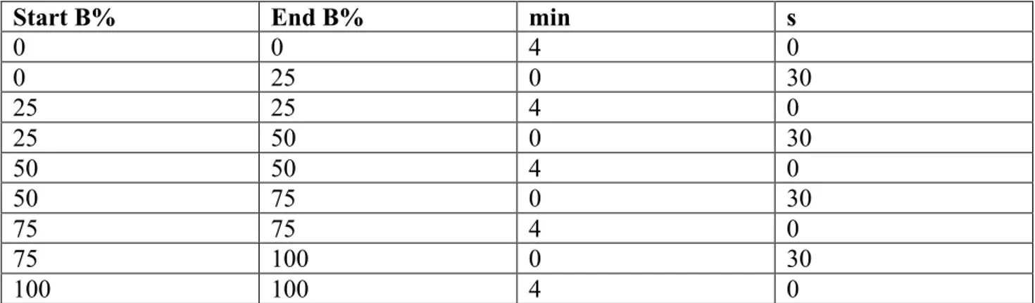
Start B% End B% min s
0 0 4 0 0 25 0 30 25 25 4 0 25 50 0 30 50 50 4 0 50 75 0 30 75 75 4 0 75 100 0 30 100 100 4 0
Tab. 5 Elution system used for the HP-20 column, solvent B is EtOH solvent A is H2O
Start B% End B% min s
90 90 15 0
90 70 5 0
70 70 20 0
50 50 5 0
50 50 10 0
Tab.6 Elution system used for the Silica-gel column, solvent B is MeOH/CH3COOH 2% solvent A is
CHCl3
Tab.7 Elution system used for the C18 column, solvent B is H2O/HCOOH 1% and solvent A is
MeOH/HCOOH 1%
Start B% End B% min
90 90 10 90 80 1 80 80 20 80 70 1 70 70 10 70 50 1 50 50 10 50 30 1 30 30 5
3.3 Biological activity screening and 1H NMR metabolomic analysis of 39 Tea varieties
3.3.1 Plant material, extraction and 1H NMR-based metabolomic analysis
The tea samples were purchased from Simon Lévelt Koffiebranderij en Theehandel A. (Hofmanweg 3 2031 BH Haarlem NL) and the details are reported in Tab. 8.
The extraction and the 1H NMR metabolomics analysis were performed as above described in chapter 3.1. PCA, PLS and OPLS models were also developed as described in chapter 3.1.
Tea Type Used
acronym
Finest Oolong Oolong 1
Grand Pouchong Oolong 2
Puerh Oolong 3
Jangwon Oolong Oolong 4
Pai Mu Tan White 5
White Moonlight White 6
Ambootia Wit White 7
Sencha Classic Green 8
Gyokoru Green 9
Ambootia Groen Green 10
Blackwood Groen Green 11
Lentedauw Mao Feng Green 12
Chunmee Young Hyson Green 13
Nilgiri Groen Green 14
Dragonwell Xi HU Lung Ching Green 15
Snow Bud Lu Xue Ya Green 16
Yunnan Groen Green 17
Nepal Groen Green 18
Pi Lo Chun Groen Green 19
Gunpowder Green 20
Jangwon Groen Green 21
Tarry Melange Black 24
Mokalbari Zomerpluk Black 25
Jangwon Black 26
Dunsandle Jungle Black 27
Ceylon Superieur Black 28
Java Kertasari Black 29
Sumatra Sidamanik Black 30
Darjeeling Zomerpluk Black 31
Ambootia Lentepluk Black 32
Ambootia Regenpluk Black 33
Ambootia summer Black 34
Margareth’s Hope Lentepluk Black 35 Kenya Flowery Orange Pekoe Black 36
Kukicha Black 37
Huang Da Cha Yellow 38
Kukicha Green 39
Tab.8 The table reports all the details of the different tea variety subjected to the study
3.3.2 UV-Vis-based metabolomic analysis
The Tea extracts were solubilized in phosphate buffer (0.05M pH 7.5) at a concentration of 0.4% v/v. The UV-Vis spectra of the extracts were recorded in the spectrophotometer from 700 to 200 nm. The obtained data were exported to Microsoft excel and analyzed by SIMPCA-P+
3.3.3 Isolation of the active fractions
One hundred g of Tea 25 (Black tea Mokalbari Zomerpluk) was extracted in MeOH/H2O (2 L) and sonicated for 1 hour. The procedure was repeated 2 times. After the extraction the extract was filtered and the pellet was discarded. The supernatant was dried in rotary evaporator.
The extract was purified by MPLC and Buchi Sepacore C18-4 g column with flow rete of 10 mL/min and a maximum pressure of 16 bar and the abovementioned detection system. Considering that the mobile phase was made by MeOH/HCOOH 1% (solvent A) and H2O/HCOOH 1% (solvent B), the elution system performed is reported in Tab. 7 (see above).
4. Results and discussion
4.1 Correlation between collagenase inhibitory activity and chemical profile of 49 medicinal plants
In order to identify natural collagenase inhibitors, 49 medicinal plants were selected based on their ethnobotanical uses. In particular, it was given attention to plants traditionally used to promote wounds and burns healing and generally to treat skin related diseases as well as rheumatism, arthritis and bones diseases. In fact, since these illnesses have been associated to a misregulated collagenase activity, it was supposed that an inhibition of this enzyme might be involved in the beneficial effect attributed to the traditional remedy. Moreover, considering the involvement of collagenase in the wrinkles formation, plants used traditionally as anti-aging agents were also considered in the selection stage.
Then, the selected plants (see tab.3) were subjected to a proper extraction procedure and the obtained extracts were tested in the collagenase inhibitory assay at a concentration of 50 µg/mL. According to the obtained percentage of inhibition, the plants were divided in high activity (inhibition >60%), medium (inhibition between 60% and 30%) and low activity (inhibition <30%) (fig.9). Thus, in order to obtain a more comprehensive overview of the relations between the measured biological activity and the chemical profile of the extracts, it was performed a 1H-NMR metabolomic analysis. In particular, this procedure was followed to detect the eventual presence of a common class of metabolites in the most active plants. In order to easily extract this kind of information, the obtained 1H-NMR spectra were subjected to a multivariate data treatment (MVDA). In particular, two supervised models were used, namely PLS and OPLS considering as the y variable the percentage of collagenase inhibitory activity.
Fig. 9 Percentage of collagenase inhibitory activity measured for the 49 crude plant extracts at a concentration of 50 µg/mL. The plants were divided in high (activity>60%), medium (activity between 30% and 60%) and low activity (activity<30%).
The OPLS model (fig.10) showed the general importance of aromatic compounds in the collagenase inhibitory activity. In fact, the presence of several signals in the 1H-NMR spectral region from δ 7.4 to 6.4 was indicated as positively correlated to the biological activity. More in particular, the model identified a signal at δ 7.08 as the most correlated with the collagenase inhibitory activity. Dealing with plant material, spectral signals around δ 7.0 can be attributed to tannin-related compounds, especially gallotannins. The result is consistent with the data reported in literature, in fact, tannins were found capable to inhibit collagenase and other MMPs both in vitro and in vivo models.62-63-64 However, to confirm the hypothesis of tannins involvement in the activity of the selected plants, a tannin-removal procedure was required. Moreover, the possibility to test tannin-free extracts was considered useful in order to investigate the eventual presence of other class of active metabolites taking also into account that some highly polymerized tannins might determine false positive results in the assay (especially in an in vitro test). In fact, this kind of compounds might precipitate proteins leading to an apparent enzyme inhibition.
0 25 50 75 100
Er Ks Ai BpR Ct Eo Cs Ah BpL Tc Ci Gl GsL Ms Vo Cp As Cn Aso Mo Mb Po Bd Ea Ts
% c ol la ge n ase in h ib it io n
A
Fig. 10 OPLS model results. A. Scatter plot colored according to the collagenase inhibitory activity B. VIP plot in which is evident the importance for the activity of the signal at δ 7.08.
Several methods have been reported for tannins-removal but none of them is exempt from disadvantages. However, we elected filtration on polyamide columns as the best method, in fact, it is the most efficient and reproducible procedure, which shows also the advantage to avoid sample contamination with other substances.
Thus, the 14 most interesting extracts (high and medium activity) were eluted on polyamide columns (the yield after column is reported in fig. 11), then the biological activity of the obtained tannin-free extracts was tested.
After the tannins removal, only three out of 14 plants were found still able to inhibit the enzyme with an activity comparable with the one previously showed by the crude extract (fig. 12), namely: Alchemilla vulgaris (Av), Tinospora cordifolia (Tco) and Emblica officinalis (Eo).
Fig. 11 Yield expressed in w/w% obtained after tannin removal procedure for the 14 active plants. 0
20 40 60 80
Tco Hi Av Ae Cs Eo Tb Csi Ai Ct Cpl BpR Ks Er
yi eld after po lyami de (w /w %)
Fig. 12. Comparison in activity, expressed as percentage of collagenase inhibition, between the crude extracts and the respective tannin-free extracts of the 14 active plants. Only the activity of Av, Tco and Eo is almost unaltered after the tannin-removal procedure.
In order to check if the tannin-removal procedure was working efficiently, the extracts obtained after polyamide filtration were compared with the crude extracts by 1H NMR and HPLC-PDA. In most cases, only tannins were irreversibly retained on the polyamide resin, leaving the extracts composition almost unaltered in its other constituents. However, four cases represented an exception, in fact these four extracts were consistently modified in their composition using the polyamide column, namely: Hemidesmus indicus (Hi), Khaya senegalensis (Ks), Butyrospermum paradoxum (roots bark) (BpR) and Embelia ribes (Er).
Although unwanted, in the case of Hi, this inconvenient lead us to obtain useful information on the compounds responsible for its extract activity. In fact, two vanillin positional isomers are present in Hi, namely: 2-hydroxy-4-methoxy-benzaldheyde and 3-hydroxy-4-methoxy-benzaldheyde, and whereas both molecules are detectable in the 1H NMR spectrum of crude Hi extract, the corresponding tannin-free extract completely lost one of them: 2-hydroxy-4-methoxy-benzaldheyde (fig. 13).
The high amount of 2-hydroxy-4-methoxy-benzaldheyde found in the Hi extract and the lost in activity registered after its undesired removal after polyamide column. It suggest the crucial involvement of this compound in the collagenase inhibitory activity of Hi.
In the case of Ks, BPr and Er it was not possible to detect which compounds, other than tannins,
0 20 40 60 80 100 120
Ae Ai Av BpR Cpl Cs Csi Ct Eo Er Hi Ks Tb Tco
% o f c ol la ge n ase in h ib it io n crude extract tannin-free
were missing after the polyamide filtration but the yield was noteworthy low compared to the other tannin-free extracts (fig. 11) and the characteristic red color of this three extracts was totally lost after column. In this case, it is important to consider that, as specified by the suppliers, the polyamide resin irreversibly retains quinones, therefore plants enriched in this class of compound (e.g. Er)65 are not suitable to be treated with this procedure. Accordingly, it is not possible to completely excluded Ks, Bpr and Er from the group of interesting plants to investigate more in deep to found collagenase inhibitors other than tannins.
Fig. 13 Hemidesmus indicus (Hi) 1H-NMR. On the top part is reported the spectrum of the Hi extract and
on the bottom the spectrum of the tannin-free Hi extract. The signals of 2-hydroxy-4-methoxy-benzaldheyde completely disappeared in the tannin-free extract. The blue arrows indicate the signals of the aldehyde and aromatic protons of this compound.
In the second stage of this work it was decided to select the plant less affected by the tannin removal procedure in term of activity, namely Alchemilla vulgaris (Av) and analyze it deeply by a bioassay-guided procedure in order to identify the compounds responsible for the biological activity.
First of all, it was performed a liquid-liquid partition using, in the following order: hexane, CHCl3, n-butanol and H2O to obtain four different fractions enriched in different polarity compounds. The activity of the four fractions was tested and since the n-butanolic one showed the highest activity it was subjected to further purification procedures.
Each fraction obtained from the n-butanolic one was tested for the collagenase inhibitory activity and the highest activity fractions were subjected to further purification steps (fig. 14).
Fig. 14 Scheme of the bioassay-guided procedure performed on Alchemilla vulgaris and yielding miquelianin as the most active compound. The most active fractions are inserted in the red squares.
BIO-ASSAY GUIDED ALCHEMILLA VULGARIS L.
! HP20! Silica!!!!! gel! 1 Kg plant MeOH/H2O 1:1 ! HP20! Silica!!!!! gel!
Alchemilla vulgaris L. Extraction
Liquid/liquid partition MPLC HP-20 SILICA gel Sephadex LH-20 n-butanolic fraction C18 miquelinin
This procedure allowed to purify and identify: quercetin-3-O-β-glucuronide (miquelianin), which resulted the most active compound present in the plant extract (NMR details are reported in fig. 16). The molecule was identified by COSY, J-res, HMBC and HSQC experiments, the sugar configuration was confirmed by the coupling constants obtained by the J-res spectrum and the presence of glucuronic acid in the structure was evidenced by the HMBC correlation between the signal of the proton at δ 3.58 in the sugar moiety and the signal of the carbonylic carbon at δ 173.23. The position 3 of the glycoside was confirmed by HMBC experiment (fig. 15), which showed correlation between the anomeric proton at δ 5.36 and the carbon 3 of the aglycon at δ 135.84 (fig. 15). Miquelianin purity was confirmed by LC-MS, which allowed also to further confirm the structure by the given molecular weigh of 477.08 m/z.
Fig. 15 HMBC spectrum of fraction D4 (containing miquelianin). The correlation between the anomeric proton (H1’’δ) and C3 (135 δ) is evidenced as well as the correlation between proton H4’’ and H5’’ with the carboxylic carbon C6’’ (173.23 δ).
Fig. 16 NMR table of miquelianin
The purified miquelianin was tested in the collagenase inhibition assay and compared with other two flavonoids glycosides with the same aglycone moiety (purchased form Sigma Aldrich) and the positive control doxycycline, the only drug approved by FDA as collagenase inhibitor.
2 3 4 5 6 7 8 9 10 1’ 2’ 3’ 4’ 5’ 6’ 1’’ 2’’ 3’’ 4’’ 5’’ 6’’ (querce3n-3-O-β-glucuronid or miquelian) 1’ 6’ 5’ 4’ 3’ 2’ (querce'n-3-O-β-glucuronid or miquelian)