A
A
l
l
m
m
a
a
M
M
a
a
t
t
e
e
r
r
S
S
t
t
u
u
d
d
i
i
o
o
r
r
u
u
m
m
–
–
U
U
n
n
i
i
v
v
e
e
r
r
s
s
i
i
t
t
à
à
d
d
i
i
B
B
o
o
l
l
o
o
g
g
n
n
a
a
DOTTORATO DI RICERCA IN
Oncologia e Patologia Sperimentale
Progetto Nr.1 : Oncologia
Ciclo XXV
Settore Concorsuale di afferenza: 06/A2 Settore Scientifico disciplinare: MED/05
TITOLO TESI
Ribosome Biogenesis and cell cycle regulation:
Effect of RNA Polymerase III inhibition
Presentata da: Carmine Onofrillo
Coordinatore Dottorato Relatore
Chiar.mo Chiar.mo
Prof. Sandro Grilli
Prof. Massimo Derenzini
Esame finale anno 2013
2
Index:
Relations hip between cell growth and
Cell cycle progression... 5
1 Aim of the thesis
...
6
2 Introductions... 7
2.1 Cell Cycle... 7
2.1.1 Cell cycle control by
CDK/Cyclin system
...
7
2.1.2 Cell Cycle checkpoints
...
12
2.2 The tumor suppressor P53 ... 15
2.2.1 P53 protein structure
...
15
2.2.2 P53 transcriptional target genes
... 18
2.2.3 MDM2 mediated P53 regulation
...
21
2.3 Ribosome Biogenesis... 23
2.3.1 The Nucleolus
...
24
2.3.2 Ribosomal RNA maturation
and Ribosome subunit production
...
26
2.4 Modulation of cell cycle regulators and
control of ribosome biogenesis... 31
2.5 Ribosome biogenesis alterations effects
on nucleolar morphology
and cell cycle progression... 34
2.6 RNA Polymerase III and Non coding RNAs... 37
2.6.1 Enzime structure
...37
3
3 Results. ... 43
3.1 POLR3A interference led to inhibition
of RNA Polymerase III activity ... 43
3.2 RNA Polymerase III inhibition
caused nucleolar fragmentation
and affects rRNA maturation... 45
3.3 The ribosomal stress and nucleolar disruption
induced by RNA Polymerase III inhibition
did not activate the p53 pathway... ...47
3.4 RNA Polymerase III inhibition led to a p53
independent cell cycle arrest... 49
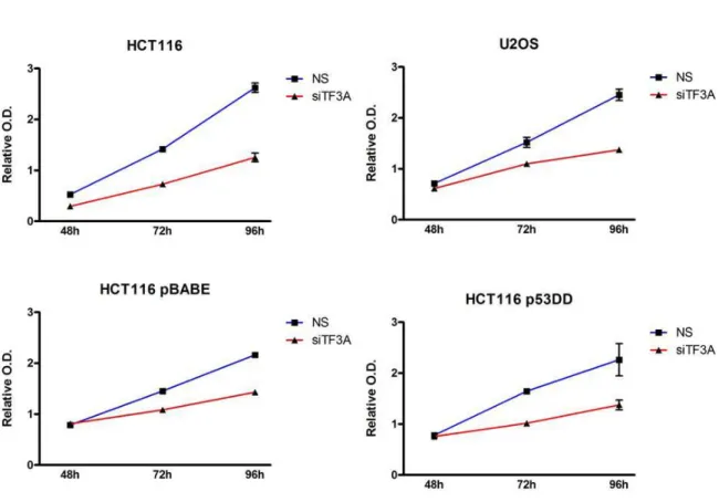
3.5 TF3A interference led to a specific
inhibition of 5S rRNA transcription ... 50
3.6 The lack of 5S rRNA is linked with
an altered RNA processing ... 52
3.7 5S rRNA depletion did
not activate p53 pathway ... 53
3.8 TFIIIA inhibition caused
a P53 independent cell cycle arrest ... 53
4
5 Materials and Methods... 60
5.1 Cell lines, culture conditions
and drug treatments... 60
5.2 RNAi, plasmids and
transfection reagents... 60
5.3 AgNOR staining ... 61
5.4 RNA extraction, reverse
transcription and real-time qPCR... 61
5.5 Analysis of newly synthesized rRNA
with
3H-Uridine labeling... 63
5.6 Polysome profile... 64
5.7 Western blotting... 64
5.8 Immunocytochemistry... 65
5.9 Cristal Violet Staining... 66
5
Ribosome Biogenesis and cell cycle regulation:
Effect of RNA Polymerase III inhibition
In cycling cells positive stimuli like nutrient, growth factors and mitogens increase ribosome biogenesis rate and protein synthesis to ensure both growth and proliferation. In contrast, under stress situation, proliferating cells negatively modulate ribosome production to reduce protein synthesis and block cell cycle progression. The main strategy used by cycling cell to coordinate cell proliferation and ribosome biogenesis is to share regulatory elements, which participate directly in ribosome production and in cell cycle regulation. In fact, there is evidence that stimulation or inhibition of cell proliferation exerts direct effect on activity of the RNA polymerases controlling the ribosome biogenesis, while several alterations in normal ribosome biogenesis cause changes of the expression and the activity of the tumor suppressor p53, the main effector of cell cycle progression inhibition (1. Montanaro L. et al., 2012). The available data on the cross-talk between ribosome biogenesis and cell proliferation have been until now obtained in experimental model in which changes in ribosome biogenesis were obtained either by reducing the activity of the RNA polymerase I (Pol I) or by down-regulating the expression of the ribosomal proteins. The molecular pathways involved in the relationship between the effect of the inhibition of RNA polymerase III (Pol III) activity and cell cycle progression have been not yet investigated. In eukaryotes, RNA Polymerase III is responsible for transcription of factors involved both in ribosome assembly (5S rRNA) and rRNA processing (RNAse P and MRP). Thus, the down-regulation of RNA Polymerase III activity, resulting in ribosome biogenesis stress, should also be responsible for modulation of cell cycle progression.
6
Aim of the thesis
The main purposes of this study are:
To characterize the effect of RNA Polymerase III depletion on ribosome biogenesis by the analysis of nucleolar morphology, rRNA processing status and ribosome subunit production
To verify the effect of RNA Polymerase III depletion on cell cycle progression
To define the molecular pathways linking RNA Polymerase III inhibition to changes in the cell cycle progression
The results that will be obtained might lead to a deeper understanding of the molecular pathway that controls the coordination between ribosome biogenesis and cell cycle, and might give useful information about the possibility to target RNA Polymerase III for cancer treatment.
Before describing the results of the present thesis, a description of the molecular mechanisms controlling the cell cycle progression, the ribosome biogenesis and the established relationship between ribosome biogenesis and cell proliferation will be given.
7
Introduction
2.1 Cell Cycle
Tumorigenesis is the result of deregulation of cell cycle control elements which leads to aberrant cell proliferation. Cell cycle progression is regulated and monitored by a series of coordinated mechanism that ensure the passage through different cell cycle phases only after transition of cell status monitoring checkpoints. The cell cycle divided in two main phases: Mitosis (phase M) and Interphase. While Interphase provides the passage through G1, S and G2 phases, the stage of Mitosis includes prophase, metaphase, anaphase and telophase, replication of DNA occurs in a specific part of the interphase called S phase (2. Norbury and Nurse, 1992). S phase is preceded by a gap called G1 during which the cell is preparing for DNA synthesis and followed by a gap called G2, when the cell prepares for mitosis. G1, S, G2 and M phases are the standard subdivisions of cell cycle. Cells in G1 can, before commitment to DNA replication, enter a resting state called G0. Cells in G0 phase account for the major part of the non-growing, non-proliferating cells in the human body.
2.1.1 Cell cycle control by CDK/Cyclin system
The passage from a cell cycle phase to another one is finely regulated by different cellular factors. The principal elements implicated in cell cycle regulation are Cyclin Dependent Kinases (CDK), a family of serine/threonine kinases that exert theirs activity during the different phases of cell cycle. Nine CDKs are identified in mammalian cells and five of these are involved in cell cycle regulation.
8
In particular G1 phase is characterized by the activity of CDK4, CDK6 and CDK2; S phase is linked to CDK2 activity, while G2 and M phase are CDK1 dependent. The activity of CDK is regulated by the presence of their binding partners known as cyclins. While the protein levels of CDKs are constant during cell cycle progression, cyclins protein levels rise and fall in a selective manner during the different cell cycle phases and periodically activate specific CDKs. (FIG. 2.1). The three D type cyclins (cyclin D1, cyclin D2, and cyclin D3) bind to CDK4 and CDK6, CDK-cyclin D complexes induce the entry in G1 phase
(3. Sherr, 1994). Cyclin D is not expressed periodically, but is continuously synthesized in response to growth factor stimulation (4. Assoian and Zhu, 1997). Another G1 cyclin is cyclin E which associates with CDK2 to regulate progression from G1 into S phase (5. Ohtsubo et al. 1995) while cyclin A binds CDK2, promoting S phase progression. (6. Girard et al., 1991; 7. Walker and Maller 1991). At the end of G1 and at the early M, cyclin A binds CDK1 inducing the passage in Mitosis that will be characterized by binding of cyclin B with CDK1. (8. King et al., 1994; 9. Arellano and Moreno, 1997). To ensure specific degradation, each cyclins possesses peculiar protein sequence like Destruction box (Cyclin A and B) or PEST domain (Cyclin D and E) which are essential for efficient ubiquitin-mediated cyclin proteolysis at the end of each cell cycle phase. (10. Glotzer et al., 1991). However, regulation of cyclins levels is not the only cell strategy to control the intricate progression through cell cycle. In fact, CDK are also regulated by phosphorylation of Tyrosine and Threonine residues. For example CDK1, CDK4 and CDK2 full activation is detectable when are phosphorylated by CDK7/cyclin H complex, also called CAK. The phosphorylation CDKs induce protein conformational changes that lead to efficient cyclins binding. (11. Jeffrey et al. 1995). Wee1 and Myt1 kinases
9
phosphorylate CDK1 at tyrosine-15 or threonine-14, thereby inactivating the kinase. Dephosphorylation at these sites by the enzyme Cdc25 is necessary for activation of CDK1 and further progression through the cell cycle.
(12. Lew and Kornbluth, 1996). In addition, proliferating cells utilize another kind of regulation that provides the activity of cell cycle inhibitory proteins inhibitors (CKI). In fact CDKs function can be also inhibited by specific CKIs. CKIs can be divided in two family: the INK4 and Cip/Kip family (13. Sherr and Roberts 1995). The INK4 family includes p15 (INK4b), p16 (INK4a), p18 (INK4c), p19 (INK4d), which specifically inactivate CDK4 and CDK6, involved in G1 regulation. These CKIs form stable complexes with the CDK enzyme before cyclin binding, preventing association with cyclin D (14. Carnero and Hannon 1998). The second group of CKIs, the Cip/Kip family, includes p21 (Waf1, Cip1), p27 (Cip2), p57 (Kip2). These inhibitors bind CDK/Cyclin complexes
(15. Polyak et al., 1994). Cip/Kip family inhibit the CDK/Cyclin complexes necessary for G1 phase initiation and progression, and to a lesser extent, CDK1-cyclin B complexes, involved in phase M regulation (16. Hengst and Reed, 1998). P21 also inhibits DNA synthesis by binding to and inhibiting the proliferating cell nuclear antigen (PCNA) (17. Pan et al. 1995).
10
FIG 2.1
FIG 2.1: Schematic representation of some of the mammalian CDKs involved in progression
throughout the different phases of the cell cycle.
Different line of evidence have been shown that CKI can be regulated both by internal and external signals. An example is given by the fact that the expression of p21 is under transcriptional control of the p53 tumor suppressor gene,
(18. Deiry et al., 1993) while the expression and activation of, respectively, p15 and p27, increases in response to transforming growth factor b (TGF-b), contributing to growth arrest (19. Hannon and Beach 1994). Another level of cell cycle regulation is linked to the cellular localization of regulatory elements. Cyclin B contains a nuclear exclusion signal and is actively exported from the nucleus until the beginning of the prophase, while CDK inactivating kinases Wee1 and Myt1 are located, respectively, in the nucleus and Golgi complex and protect the cell from premature mitosis (20. Heald et al., 1993; 21. Liu et al., 1997). Moreover, the intracellular trafficking of different proteins is regulated by 14-3-3 complex. During interphase, the CDK activating kinase, Cdc25, is kept in
11
the cytoplasm through interaction with 14-3-3 proteins which are also able to sequester the CDK1-cyclin B complex following DNA damage (22. Yang et al., 1999). Under this controlled regulation, CDKs can exert theirs kinase function on specific target substrates, to achieve the passages trough the different cell cycle phases. During G1 phase CDK4/6 are active and are able to phosphorylate the product of retinoblastoma tumor suppressor gene (pRB). pRB is ipophosphorylated during early G1 phase, but once iperphosphorylated by CDKs, the tumor suppressor provides the release of E2F1 and DP-1, two of the most important transcription factors involved in cell cycle progression. In fact they positively regulate the transcription of genes whose products are required for S phase progression, including cyclin A, cyclin E and Cdc25. (23. Brehm et al. 1998, 24. Kato et al. 1993). Afterwards, the activity of CDK2/Cyclin E ensures the maintenance of the iperphosphorylated form of pRB during the rest of cell cycle. In G1/S passage CDK2/Cyclin E phosphorylates its inhibitor p27 and lead to its proteasome-dependent degradation. (25. Montagnoli et al. 1999). CDK2-cyclin E phosphorylates histone H1 and this activity may be important for chromosome condensation required during DNA replication. Histone H1 is also a substrate for CDK1-cyclin B. Cyclin A-dependent kinases regulate initiation of DNA replication by phosphorylation of DNA polymerase alpha primase. Other CDK substrates include CDK’s own regulators Wee1 and Cdc25, and cytoskeletal proteins such as nuclear lamins, microtubules and vimentin, which are required for correct mitosis (26. Heald and McKeon 1990; 27. Courvalin et al. 1992; 28. Hoffmann et al. 1993; 29. Blangy et al. 1995).
12
2.1.2 Cell Cycle checkpoints
To ensure survival and generation of healthy progeny, eukaryotic cells respond to damaged, abnormally structured DNA or to alteration of protein synthesis machinery by a multifaceted response that coordinates cell cycle progression with DNA repair, chromatin remodeling, transcriptional and post-transcriptional programs, metabolic adjustment and finally cell death.
The arrest or delay of cell cycle progression that provides time for DNA repair or for sufficient cellular component production needed for daughter cells generation is mediated by a network of signaling pathways identified as cell cycle checkpoints. These biochemical cascades include different type of sensor proteins that are able to monitor a series of cellular conditions like genome integrity, protein synthesis state or altered gene expression and help to generate signals that are transduced to downstream checkpoint effectors leading to cell cycle progression regulation. Each cell cycle phase is characterized by specific checkpoints pathways that operate in different way to ensure a coordinated control of cell cycle progression. (FIG.2). During G1 phase, cells with damaged DNA can activate the checkpoint transducing kinases ATM/ATR and ChK1/ChK2 which in turn target two critical effectors of G1 checkpoint, the tumor suppressor p53 and the Cdc25 phosphatase. Phosphorylation of Cdc25A by Chk1 and Chk2 induces its ubiquitination and proteasome-mediated degradation, preventing the dephosphorylation and activation of CDK2. The inhibition of CDK2/Cyclin E-Cyclin A complex blocks the loading of Cdc45 on chromatin, interfering with α-Polymerase recruitment and DNA replication initiation. (30. J. Falck et al., 2002). This checkpoint pathway operates independently of p53, it is quite transient and it is able to delay cell cycle progression only for several hours. The other
13
mechanism that mediates G1 checkpoint is p53 dependent and ensures a prolonged maintenance of G1 cell cycle arrest. p53 is phosphorylated by both Chk1/Chk2 and ATM/ATR. In addition Human Double Minute 2 (HDM2/MDM2), the E3 ubiquitin ligase responsible for continuous p53 proteasome-degradation during cell cycle progression, is also targeted and inhibited by ATM/ATR kinases. All these modification leads to p53 protein stabilization and to the subsequent transcription of its target genes such as the CDK inhibitor p21 (WAF1/Cip1). The accumulation of p21 leads to CDK2/Cyclin E complex inhibition, blocking pRB iper-phosphorylation, E2F1 transcription and G1/S transition. (31. C.J. Sherr, J.M. Roberts 1999). The prolonged G1 cell cycle arrest can also lead to a permanent block in cell cycle progression and is related to the cell enter in G0 phase, phenomenon known as cellular senescence. Despite G1 checkpoints, the S phase checkpoint causes only transient and reversible delay in cell cycle progression, by slowing DNA replication or inhibition of new replicon formation.
The pathway that link DNA damage to the core cell cycle machinery during S phase is the same described for G1/S checkpoint, providing the activation of ATM/ATR-ChK1/ChK2-Cdc25A-CDK2/CyclinE-Cdc45 cascade.
The G2 checkpoint prevents cycling cells to start Mitosis when they experience DNA damage during G2, or if they proceed in G2 with some unrepaired damage or alteration inflicted during previous phases. Like G1 checkpoint, the G2 arrest or delay is a result of combination of rapid, reversible mechanism or prolonged, sustained response. The downstream target of G2/M checkpoint is represented by CDK1/Cyclin B complex. After DNA damage ATM/ATR and Chk1/Chk2 kinases phosphorylates and inhibit Cdc25C phosphatase, which is required for CDK1 activation. Inhibition of CDK1 prevents the progression in M phase
14
(32. K.A. Nyberg et al. 2002). However, the long term arrest of G2 phase is driven again by p53 pathway. While G1 arrest is mediated by p21 mediated inhibition of CDK 2, CDK 1 activity is depleted by other p53 targets such as GADD45 or 14-3-3 proteins. (33. W.R. Taylor, G.R. Stark. 2001).
Finally the ‘spindle checkpoint’ is direct to detection of improper alignment of the chromosomes on the mitotic spindle and stops the cell cycle in metaphase.
If defects in microtubule attachment occurs during metaphase, Mitotic arrest deficient (Mad) and budding uninhibited by benomyl (Bub) proteins are activated and inhibit the Cdc20 subunit of the anaphase-promoting complex (APC), resulting in the prevention of anaphase transition (34. Fang et al. 1998).
FIG 2.2:
15
2.2
The tumor suppressor p53
The most common anti-apoptotic change that characterizes cancers is the inactivation of p53 tumor suppressor pathway. As discussed above p53 is the main regulator of G1/S or G2/M checkpoints, thus is essential for a controlled cell cycle progression. p53 function in cancer can be lost by different mechanism, including lesions that prevent p53 activation, mutation in TP53 gene or mutation in downstream effectors mediators of p53 function. Analysis of many tumors revealed that TP53 is mutated in about half of all type of cancers, resulting in loss of cell cycle control and apoptotic function.
2.2.1 p53 protein structure
The p53 tumor suppressor belongs to a small family of related proteins that includes two other members, p63 and p73. p53 is classified as transcription factor and is biologically active as a homotetramer comprising 393 amino acid residues for each homomonomer. It is characterized by a modular domain structure, consisting of folded DNA-binding and tetramerization domains, flanked by intrinsically disordered regions at both the amino terminal and carboxy-terminal domains. (FIG.2.3). The structure of the DNA-binding core domain (residues 94-292) consists of a central immunoglobulin-like β-sandwich scaffold and additional structural elements that form the DNA-binding surface which include a loop-sheet-helix motif and two large loops (L2 and L3). Human p53 core domain possesses low intrinsic thermodynamic stability and rapidly unfolds at body temperature with a half-life of 9 minutes (35. Bullock et al., 1997).
16
FIG.2.3
FIG. 2.3: Schematic representation of p53 protein domains and structures. p53 contains an
amino-terminal transactivation domain (TAD), subdivided into the subdomains TAD1 and TAD2, followed by a proline-rich region (PRR). DNA-binding and tetramerization domains (OD) are
connected through a flexible linker region. The regulatory domain at the extreme carboxyl terminus is intrinsically disordered (CTD). The vertical bars indicate the relative missense-mutation frequency in human cancer for each residues. The structure of the DNA-binding domain
(PDB code 1TSR) is shown as a ribbon representation and colored with a rainbow gradient from the amino terminus (blue) to the carboxyl terminus (red). Sites of cancer hotspot mutations and
essential DNA contacts are shown as stick models. (From Andreas C. Joerger and Alan R. Fersht, 2010)
17
This low thermodynamic and kinetic stability may allow for rapid passages between folded and unfolded states, which could provide an additional level of regulation of functionally active cellular protein levels, besides the specific degradation pathways involving ubiquitination and subsequent proteasome mediated degradation The low stability of the core domain has profound implications with regard to the susceptibility of human p53 to deleterious mutations and cancer development and may also be directly linked with the structural plasticity required to facilitate binding to different partner proteins. p53 can bind DNA only in a tetrameric form and crystallographic data have provided detailed insights into the structural basis of sequence specific DNA recognition by p53 tetramers. Two core domains bind to a half-site DNA, forming a symmetrical dimer with a relatively small, self-complementary core domain-core domain interface. The L3 loop binds to the DNA minor groove via Arg248, which makes either direct or water-mediated contacts with the DNA backbone. The oligomerization state of p53 is regulated via its tetramerization domain (residues 325–355 in human p53). However the individual subunits consist of a short β-strand followed by α-helix. These two structural elements are connected by a sharp turn facilitated by a conserved glycine residue (Gly334). Four chains form a tetramer that can be described as a dimer of primary dimers. Additionally, increasing evidence suggest that the oligomerization equilibrium of p53 is modulated via an intricate network of accessory proteins, which can have either positive or negative regulatory effects. Most somatic p53 cancer mutations are located in the DNA-binding domain (Fig. 2.3) and these mutant proteins have, therefore, an intact tetramerization domain. Formation of mixed tetramers of impaired activity between wild-type and mutant p53 is thought to be the molecular basis of the so-called dominant–negative effect of mutant p53 in
18
heterozygous cells (36. Kern et al., 1992; 37. Dong et al., 2007). The amino-terminal transactivation domain (TAD) is essential for p53 transcriptional activity. It connects target gene recognition with target gene expression by direct binding to transcriptional coactivators like p300/CBP and components of the basal transcription machinery (38. Thut et al. 1995; 39. Lill et al. 1997). The p300 is able to bind to the full TAD (TAD1/TAD2), but the TAD1 subdomain also binds strongly to the negative regulators MDM2 and MDMX that play a crucial role in controlling cellular p53 levels by promoting p53 degradation through the ubiquitin-dependent proteasome pathway (40. Marine et al., 2006). The p53 TAD is intrinsically disordered with two regions of nascent secondary structure. Intrinsic disorder is a characteristic of transactivation domains of transcription factors (41. Liu et al., 2006) and it is often associated with the presence of so-called molecular recognition features, short sequence motifs that undergo disorder-to-order transition on binding to partner proteins (42., Mohan et al. 2006), allowing promiscuous binding of different targets at the same site by providing conformational variability and adaptability. Changes in the affinity of p53 for competing binding partners in the cell cycle by post-translational modification of TAD, allows modulation of p53 activity in response to cellular stresses.
2.2.2 p53 target genes
Several responses can be induced by p53, including cell-cycle arrest, senescence, differentiation and apoptosis, with the option chosen being dependent on many factors that are both intrinsic and extrinsic to the cell. However, a strong induction of p53 may leads to an irreversible inhibition of cell growth, most decisively by
19
activating apoptosis, while the modulation of p53 levels leads to a controlled regulation of cell cycle progression. p53 target can be divided in different group of proteins that act on various cell stress response mechanisms such as G1 or G2 cell cycle arrest, DNA repair, inhibition of angiogenesis, inhibition of metastasis and apoptosis. The most know target, involved in G1 cell cycle arrest, is represented by P21 (Waf1/Cip1) a CKI of the Cip/Kip family responsible for the inhibition of CDK2/Cyclin E activity. In addition to the inhibition of CDK2/Cyclin E complexes, p53 regulates a variety of genes that also contribute to G1 arrest. It is involved in regulation of the cyclin E activity by the induction of hCDC4b (43. Kimura T. et al. 2003). It was shown that hCDC4b, an F box protein and component of the SCF ubiquitin ligase complex, targets cyclin E for ubiquitin-mediated degradation. p21 can also participate in G2 arrest, is able to associates with CDK2/Cyclin B complex and inhibits the activating phosphorylation of CDK2 by CAK (44. Smits V. A., 2000). In addition, the Growth Arrest and DNA Damage inducible protein (GADD45), another p53 target gene, binds CDK2, preventing CDK2/Cyclin B complex formation and subsequently inhibition of kinase activity (45. Zhan Q. et al., 1999). From P53 target gene involved in DNA repair there are again GADD45 which take part in Topoisomerase mediated relaxing of chromatin (46. Maeda T. et al., 2002). Other targets of P53 are represented by xeroderma pigmentosum group E and C gene (XPE, XPC). The last two are responsible for nucleotide excision repair (NER). (47. Hwang B. J. et al., 1999; 48. Adimoolam S. and Ford J. M., 2002). p53 also induce the transcription of genes that are implicated in inhibition of angiogenesis. TSP-1, an extracellular matrix glycoprotein is responsible for angiogenesis inhibition and is activated by p53. (49. Dameron K. M., 1994). Furthermore p53 can also repress VEGF expression, an important enhancer of endothelial cell
20
growth. (50. Zhang L. et al., 2000). p53 exerts also the capacity to inhibit metastatic behavior by inducing the transcription of genes involved in control of extracellular matrix (ECM) degradation. p53 induces the expression of at least two Serpins, Plasminogen Activator Inhibitor-1 (PAI-1) and Maspin (51. Kunz C. et al., 1995) . PAI-1 functions to inhibit urokinase-type plasminogen activator (u-PA) and u-PA initiates a cascade of cleavages that ultimately result in the activation of plasmin. Plasmin degrades a wide variety of ECM proteins such as fibrin, fibronectin and laminin. By inducing PAI-1, the metastatic potential of tumor cells is decreased. On the other hand, Maspin has been shown to interact with collagen type I and III, increasing cell adhesion to the ECM. (52. Blacque O. E., 2002). In addition, many p53 pro-apoptotic target genes have been identified in the last years, leading to evidence that the tumor suppressor can regulate both the intrinsic and extrinsic apoptotic pathways. In fact the transcription factor can induce the expression of two cell surface death receptors, Fas/CD95 and KILLER/DR5, and of the ligand of Fas, named FasL, which are involved in extrinsic apoptosis induction. Activation of the initiator caspase-8, another p53 target gene, eventually leads to stimulation of effector caspases and mediation of extrinsic apoptosis. (53. Fukazawa T. et al. 1999). It was also shown that p53 is able to induce the expression of tumor necrosis receptor associated factor 4 (TRAF4) which induces apoptosis and inhibits colony formation (54. Sax J. K. 2003).As well as for the extrinsic apoptotic pathway, p53 is able to upregulate the expression of proteins regulating the intrinsic apoptotic pathway. Many of these proteins localize to the mitochondria and regulate the release of Cytochrome C. These mitochondrial localizing proteins include both Bcl-2 family proteins and non-Bcl-2 family members. Two non-Bcl-2 family members regulated by p53 include p53AIP1 and mtCLIC/CLIC4 (55. Fernandez-Salas E. et al., 2002), while
21
the Bcl-2 family proteins include BAX, NOXA, p53 upregulated modulator of apoptosis (PUMA), BID, BCL-2 and BCL-X (56. Oda E. et al., 2000; 57. Sugars K. L., 2001). With this set of target genes the tumor suppressor p53 ensure a strong response to different types of cellular stresses regulating pathways involved in cell cycle control and cell viability.
2.2.3 MDM2 mediated p53 regulation.
The regulation of p53 activity mainly depends on Human Double Minute 2 (HDM2/MDM2). MDM2 was discovered in a locus amplified on double minute chromosome in a tumorigenic mouse cell line known as 3T3-DM
(58. Fakharzadeh S.S. et al., 1991). The main function of MDM2 is to negatively regulate the levels and functions of tumor suppressor p53 protein. High MDM2 levels decreases p53 activity, leading to increased cancer risk or to accelerated tumor development. Different sets of human tumors are characterized by MDM2 amplification or increased transcriptional level. MDM2 is a RING finger E3 ligase, characterized by an N-terminal domain containing p53 binding site, a central region with nuclear localization and export sequences, an acidic domain, a zinc finger domain, binding site for TBP, p300 and ARF; and a C-terminal region with RING finger domain (59. Thut C.J., 1997; 60. Grossman S.R., 1998; 61. Midgley C.A., 2000). RING finger domain can bind to an E2 ubiquitin-conjugating enzyme, leading to ubiquitin binding of target proteins. p53 represent the principal target of MDM2 which is able to monoubiquitinate or polyubiquitinate the tumor suppressor. Interaction of MDM2 with p53 mediates the translocation of the tumor suppressor in the cytoplasm (62. Roth J. 1998), sequestering it from nuclear functions. Thereafter cytoplasmic proteasomes
22
recognize polyubiquitination signal and target p53 for degradation. On the other hand the binding of MDM2 to the N-terminal domain of p53 inhibits its interaction with transcription machinery and deplete also its transcription functions. (63. Momand J. et al. 1992).
Another member of MDM2 family can regulate p53 function. MDM4, also called MDMX contains an N-terminal p53 binding site, a central acidic domain and a C-terminal RING finger domain. MDM4 is unable to induce p53 degradation but can bind it and inhibit its transcription activity (64. Toledo F., Wahl G.M. 2007). The main regulation of MDM2 depends on an autoregolatory feedback loop system. The oncogene is in fact a transcriptional target of p53 (65. Barak Y. 1993). However, MDM2 activity is also regulated by different extracellular and intracellular mechanism, leading to a smart modulation of p53 function. First of all MDM2 is regulated after DNA damage. In response to ionizing radiation, ATM phosphorylate p53, reducing the affinity for MDM2 binding and its consequent degradation. ATM is also responsible for MDM2 phosphorylation which affects its ability to export p53 in the cytoplasm, where proteasomes mediated degradation take place (66. Maya R. et al., 2001). Another ATM activity provides the stimulation of HAUSP, a specific p53 and MDM2 de-ubiquitinase and cause stabilization of the tumor suppressor (67. Meulmeester E. et al., 2005). UV radiation acts by another pathway and stimulate ATR mediated phosphorylation of MDM2 interfering again with p53 shuttling from nucleus (68. Shinozaki T et al., 2003). MDM2 undergoes also a regulation mediated by oncogenic stress. Aberrant activation of different oncogenes like E2F-1, β-Catenin, Myc, Ras or Nucleophosmin increase the protein levels of p14/p19 ARF which is responsible for MDM2 inhibition (69. Zhu J.W. 1999; 70. Damalas A. et al., 2001; 71. Korgaonkar C. et al., 2005; 72. Moulin S. et al., 2008 ).
23
High level of ARF proteins correlates with MDM2 binding and segregation of MDM2 from P53. Furthermore the interaction between ARF and MDM2 inhibit E3 ubiquitinase action toward p53, which increase again p53 protein level. (73. Weber J.D. et al., 1999). Progressive transition through cell cycle is another cellular mechanism linked to MDM2 regulation and p53 control.
It was shown that MDM2 contain a Cyclin recognition motif. CDK1/Cyclin A and CDK2/Cyclin A complexes bind and phosphorylate the oncogene protein product. The phosphorylated form of MDM2 exerts a reduced interaction with p53 and on the other hand an increased affinity for ARF binding. These events clearly result in P53 stabilization (74. Zhang T. and Prives C., 2001). Finally MDM2-p53 feedback loop is also regulated by another important mechanism that link control of ribosome biogenesis to cell cycle regulation. Ribosome biogenesis alterations at different level bring to cell cycle arrest induced by MDM2-p53 pathway. The mediators of this stress response are the same ribosomal proteins involved in ribosome construction and represent the regulatory elements that are able to bind and inhibit MDM2. The depletion of MDM2 functions, clearly affects ubiquitination of p53, leading to its stabilization, cell cycle arrest and eventually apoptosis. This fascinating stress response will be discussed in detail in the next chapters.
2.3
Ribosome Biogenesis
As mentioned above, protein synthesis is necessary for cell growth and cell proliferation. The cellular machinery involved in protein production is represented by Ribosomes. The process that leads to the building of the mature ribosome subunits, known as 40S and 60S, is defined as Ribosome Biogenesis and must be
24
continuously controlled in proliferating cells to ensure the correct coordination of protein synthesis with the cell cycle progression. The site of ribosome production is represented by nucleolus, a non-membranous structure located in the nucleus of eukaryotic cells.
2.3.1 The nucleolus
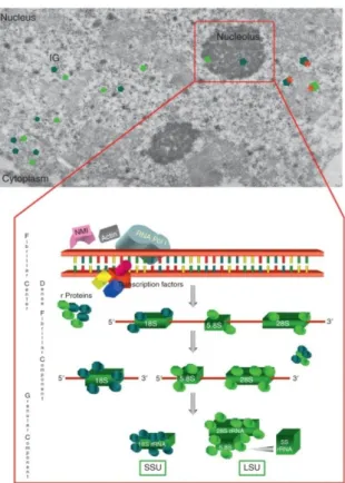
Nucleolus is the site of ribosome biogenesis. It has been shown that cancer cells are characterized by elevated rates of ribosome biogenesis and protein synthesis and show irregular shaped and hypertrophic nucleoli, compared with those of normal growing cells. Furthermore, there is evidence that changes in nucleolar morphology and function may depend both on rate and status of ribosome biogenesis and on proliferative activity of cycling cells (75. Montanaro L. et al. 2008). The nucleolus can be easily detected by contrast-phase light microscopy in living cells, as the consequence of the higher concentration of RNA and proteins in the nucleolus than in the nucleoplasm. At the electron microscopy level, mammalian cell nucleoli constantly exhibit three major components: fibrillar centers (FCs), which appears as roundish structures of varying size, with a very low electron opacity; the dense fibrillar component (DFC), which frequently constitutes a rim intimately associated with the fibrillar centers, composed of densely packed fibrils; and the granular component (GC), composed of granules that surround the fibrillar components (FIG. 2.4). (75. Montanaro L. et al. 2008). Ribosomal genes under active transcription are located at the boundary between FC and DFC. The DFC is the site where early steps of rRNA processing take place while the CG is the site of late rRNA processing and ribosome subunit building. (76. Raska I. et al., 2006). Therefore, the fibrillar centers plus the
25
associated dense fibrillar components can be considered to represent the structural-functional units of the nucleolus where rRNA molecules are produced. A large set of proteins involved both in rRNA genes transcription and in rRNA processing such as Nucleophosmin, Nucleolin, UBF and others component, which are located both in the FCs and the DFC, can be selectively visualized by a silver stain method used for the analysis of Nucleolar Organizer Region (NOR) on metaphase chromosomes (AgNOR staining). The silver-stained proteins are defined as AgNOR proteins and the quantification of the AgNOR stained structures represents a useful method to evaluate the rRNA transcriptional activity at light microscope level. (77. Derenzini M.et al. 1990).
FIG. 2.4:
FIG. 2.4: Nucleolus of established cell lines from human fallopian tube cancer. (TG cells)
The sample was stained with uranium and lead and visualized by Electron Microscopy. Asterisks indicate Fibrillar Centers (FC), while “f” surrounds the Dense fibrillar
component (DFC). Granular Component (GC) is indicated by “g”.
(From : Nucleolus, Ribosomes and Cancer. Montanaro L., Trerè D.and Massimo Derenzini. The American Journal of pathology, 173, 2. 2008).
26
2.3.2 Ribosomal RNA maturation and ribosome subunits
production.
The biosynthesis of ribosomes is a complex multistep process which requires the coordinated activity of all the three DNA dependent RNA Polymerases. In particular: RNA Polymerase I (Pol I) is responsible for transcription of a large rRNA precursor known as 47S, RNA Polymerase II (Pol II) transcribes the messenger RNA (mRNA) for ribosomal protein synthesis, the accessory factors and the small nuclear RNA (snoRNA), involved in rRNA modification and processing, and RNA Polymerase III (Pol III) provides the transcription of rRNA 5S and others non-coding RNAs (ncRNA) needed for ribosome construction. In eukaryotes the mature ribosome is constituted by two subunits. The small 40S subunit (SSU) contain a single 18S rRNA and at least 30 ribosomal proteins, while the large 60S subunit (LSU) consist of three rRNA (5S, 5.8S and 28S rRNA) as well as about 50 ribosomal proteins. (78. Woolford and Warner, 1991). (FIG.2.5)
27
FIG. 2.5
FIG. 2.5 Schematic representation of the main ribosome biogenesis steps.
(From Ribosome Biogenesis: From Structure to Dynamics, Barbara Cisterna and Marco Biggiogera International Review
of Cell and Molecular Biology, Volume 284. 2010).
The precursor rRNA 47S contains the sequences of mature rRNAs which are separated by two internal transcribed region defined as ITS1 and ITS2. The external side of each spacer is occupied by external transcribed regions known as 5’ ETS and 3’ ETS. Generation of mature ribosome subunit requires the coordinated action of a series of factors needed for the structural composition of mature ribosomes such as ribosomal proteins, or accessory factors like exoRNases or esoRNases and snoRNA, which are responsible for covalent modification of rRNA precursor in specific nucleotide sequences. SnoRNAs are short (60-300 nucleotides long), stable RNAs that localize within the nucleolus (79. Watkins and Bohnsack 2011). SnoRNAs act mainly as anti-sense guides in RNA modification and are carried by small nucleolar ribonucleoproteins (snoRNPs),
28
like Fibrillarin, a methyltransferase (C/D box snoRNAs) or Dyskerin, a pseudouridine synthase (H/ACA box snoRNA), to the site of RNA modification. Global rRNA pseudouridylation was recently shown to be important for the binding of ligands to the ribosome, as well as for translation fidelity and the Internal Ribosome Entry Site (IRES) dependent translation (80. K. Jack., 2011) and global methylation is now also known to be important for IRES-dependent translation (81. Basu A. et al., 2011). In addition, a set of snoRNAs are involved in pre-rRNA processing (U3, U8, U17, U22) and it is understood that this also requires base-pairing with the pre-rRNAs. The whole set of factors needed for ribosome biogenesis is recruited inside the nucleolus where associates with nascent rRNA precursor and leads to the formation of a 90S pre-ribosomal particle, a macromolecular structure in dynamic evolution which reaches its mature and functional form during its traffic from the nucleolus to cytoplasm. The 90S pre-ribosomal particle must be split by a series of progressive modifications and cleavages, in pre 60S and pre 40S subunits, which follow two separate maturation route (FIG.2.6).
29
FIG.2.6
FIG. 2.6: Pre-rRNA processing in Humans
(From: Mapping the cleavage sites on mammalian pre-rRNAs: Where do we stand? Mullineux S. T., Lafontaine D. (Biochimie 94, 1521-1532. 2012)
The processing pathway starts with cleavage of the 47S at both ends of the molecule, at sites 01 and 02, generating the 45S precursor which is primarily processed by two alternative pathways. In pathway 1 processing is initiated in the 5’-ETS by concomitant cleavage at sites A0 and 1. This step generates the 41S pre-rRNA which is cleaved at site 2 into 21S and 32S precursor rRNAs species. The cleavage of 41S RNA leads to the formation of SSU and LSU
pre-30
ribosomal particles. The 21S is successively trimmed to sites C and E giving rise to 18S-E pre-rRNA. The last one is then exported to the cytoplasm where it is cleaved at site 3 to generate the 18S rRNA. The maturation of the LSU require the processing of the 32S pre-rRNA which is cleaved at site 3’ in ITS2, generating the 12S pre-rRNA and the mature 28S rRNA. The 12S is cut at site 4a into 7S and then at site 4’ to generate the 5.8S rRNA. Conversely, in pathway 2, where processing starts in ITS1 at site 2, the 30S is directly matured into 21S by simultaneous processing at sites A0 and 1, or through the formation of a 26S pre-rRNA intermediate. (82. Mullineux S.T. and Lafontaine D., 2012). Furthermore, differently from the maturation of the others rRNAs, the 5S rRNA, transcribed by RNA Polymerase III, after the processing of only few nucleotides at 3’end, shuttle in the cytoplasm and interact with Rrs1 and Rrs2, which are implicated in the recruitment the rRNA 5S, within two ribosomal proteins of the large subunit (RPL11 and RPL5) in the LSU pre ribosomal particle. (83. Zhang et al., 2007). At the end of ribosome biogenesis mature ribosome subunits 40S and 60S are ready to associates with messenger RNA (mRNA) in the cytoplasm leading to the assembly of the mature 80S, which will start the protein synthesis.
31
2.4
Modulation of cell cycle regulators and control of
ribosome biogenesis
Depending on their function as positive or negative effectors of cell cycle progression, different cell cycle regulators can influence ribosome biogenesis status by stimulation or inhibition of RNA Polymerases activity. Extracellular signals, such as mitogens, upregulate the RNA Polymerases activity and lead to increase of ribosome biogenesis. Extracellular signal-regulated kinases (ERKs) are activated in response to different mitogens through a signal transducing cascade that involves Ras, Raf and ERK kinase MEK.
It has been shown that ERK phosphorylates and activates the transcription intermediary factor 1 A (TIF1A), a transcription factor required for specific promoter recruitment of Pol I. (84.. Zhao, J. et al. 2003). ERK is also able to interact with upstream binding factor I (UBF) and stimulate its function of Pol I recruitment on rDNA.(85. Stefanovsky, V. Y. et al. 2001). ERK mediated phosphorylation also increases the efficiency of specific general transcription factor 3 B (TF3B), which is necessary for Pol III mediated transcription
(86. Felton-Edkins, Z. A., 2003). The stimulation of Pol I and Pol III activity by ERK ensure that 47S rRNA, 5S rRNA and tRNAs are synthetized in a coordinated manner in response to mitogenic stimuli. Another crucial factor that finely tunes ribosome biogenesis and cell cycle progression is the oncogene Myc. Known as a target of ERK mediated signals, Myc can regulates the transcription of a huge set of genes, including those encoding ribosomal proteins, factors involved in ribosome production and translation factors. In addition Myc stimulates directly the activity of PolI by inducing the transcription of UBF or by the binding with promoter selective factor I (SL1), responsible for PolI recruitment on its target promoters.
32
(87. Poortinga, G. et al. 2004; 88. Grandori C. et al. 2005). On the other hand, Myc can also increase Pol III activity via TF3B interaction. (89. Gomez-Roman, 2003). Deregulation of Myc expression is a common feature of a series of tumors like Burkitt lymphomas, Neuroblastomas and Colon Carcinomas. It is clear that such deregulation can affect the transcription of all the three RNA Polymerases and stimulate ribosome biogenesis rates. Apart from Myc, another key regulator of protein synthesis and cell growth is mammalian target of rapamycin (mTOR). (90. Fingar and Bleins, 2004). mTOR is known as a positive regulator of protein synthesis, increasing ribosome production and translational initiation. mTOR phosphorylates UBF in its C-terminal domain promoting its binding with SL1 and directing PolI on its target genes promoters. (91. Hannan, K. M. et al., 2003). Pol III activity is also regulated by mTOR which can bind TF3B and promotes recruitment of the enzyme on target genes. In parallel, mTOR inhibits Maf 1, a repressor of 5S rRNA and tRNA transcription. (92. Kantidakis T. et al. 2010). mTOR activity is upregulated in human cancers, due to changes in upstream control elements that influence also cell cycle progression such as Ras. These events correlate again with Ribosome biogenesis and protein synthesis stimulation. On the other hand, ribosome biogenesis is negatively controlled by different tumor suppressors which are also involved in the negative control of cell cycle progression. The tumor suppressor pRB is one of the most important factors involved in G1/S passage during cell cycle progression. It represses E2F1 activity, which stimulates the expression of proteins necessary for the S-phase entry. In parallel, pRB controls the rate of ribosome biogenesis by hindering the RNA Polymerases activity. UBF and TF3B can be targeted and sequestered by pRB to achieve the inhibition of Pol I or Pol III respectively. (93. Cavanaugh, A. H. et al., 1995). Interestingly, when the demand for rRNA transcription is decreased, pRB
33
accumulates in nucleolus, while mitogenic stimulation leads to dissociation of pRB with UBF and TF3B, leading to a raise of rRNA and tRNA production. (94. Scott J. et al. 2000 ; 95. Hannan, K. M. et al. 2001). Modulation of RNA polymerases transcription can occur also in response to the activity of factors involved in cell cycle checkpoint control. The tumor suppressor p53, which is responsible for cell cycle arrest in G1/S or G2/M, binds and sequestrates both SL1 and TFIIIB. (96. Zhai W, Comai L. 2000; 97. Crighton, D. et al. 2003). Again the sequestration of transcription factors leads to a reduced production of rRNA and tRNAs. Moreover, upstream regulators of p53 may affect the Pol I and Pol III transcription. Oncogenic stress induced by Myc overexpression brings to the alternative transcription of INK4a/ARF locus, leading to the production of ARF. It is well known that ARF can bind and inhibit MDM2, thus inducing p53 stabilization. On the other hand ARF is involved also in direct regulation of ribosome biogenesis. It has been demonstrated that ARF is able to block the 47S processing by the binding of Nucleophosmin (B23). ARF binding promotes the ubiquitination and subsequent degradation of B23, and cause the arrest of rRNA maturation. This pathway provides a more efficient checkpoint for coupling p53 dependent inhibition of cell cycle progression with ribosome production. In parallel, the tumor suppressor p53 represents the main player of the stress response induced by alterations of ribosome biogenesis which also have a strong effect on cell cycle progression regulation. Indeed not only cell cycle regulators affect the activity of RNA Polymerases, but also ribosome biogenesis changes can modulate the function of key factors involved in the regulation of transition through the different cell cycle phases.
34
2.5 Ribosome biogenesis alterations effects on nucleolar
Morphology and cell cycle progression
Ribosome biogenesis control is crucial for cell cycle progression, loss of this control result in altered cell cycle and deregulated cell growth (98. Ruggero and Pandolfi. 2003). Ribosome biogenesis alterations can involve the three major components necessary for ribosome production: rRNA, ribosomal proteins and ribosome processing factors. Available evidence indicates that the mechanism of p53 stabilization after ribosome biogenesis perturbation is mainly the result of the release from the nucleolus of several ribosomal proteins that bind to MDM2 and relieve its inhibitory activity toward p53 (99. Y. Zhang and H. 2009). (FIG 2.7). In the past years it has been shown that the inhibition of rRNA transcription, triggered by drugs such as actinomycin D (100. Y. Zhang et al., 2003), 5-fluorouracil (101. Sun and Dai, 2007), mycophenolic acid (102. Sun and Dai 2008) or by TIF-IA depletion expression, lead to p53 stabilization. (103. X. Yuan et al., 2005) Similarly, alterations of rRNA processing pathway, due to an expression of a dominant negative form of Bop1, trigger again the activation of p53 pathway (104. Pestov et al., 2001). It has been postulated that, in order to stabilize p53, all cell stresses should induce a nucleolar disruption which can result in the release of the nucleolar proteins from the nucleolus to the nucleoplasm (105. Rubbi C.P., Milner j., 2003). Accordingly, the stabilization of p53 induced by the inhibition of rRNA transcription or inhibition of rRNA processing, was also thought to be caused by the release of RPs from the disrupted nucleolus. However, recent studies demonstrated that the nucleolar disruption was not a necessary condition for p53 stabilization after ribosome biogenesis
35
alterations. In fact, the down-regulation of rRNA synthesis induced by POLR1A silencing (106. Donati et al. 2011) or depletion of certain RPs, such as RPS6 (107. Fumagalli et al., 2009), caused p53 stabilization without inducing changes in the nucleolar structure organization. Recently, it was shown that after selective down-regulation of rRNA synthesis, RPs which were no more required for ribosome biogenesis, bind to MDM2 in larger amounts, thus increasing p53 stabilization. According to this model, the level of p53 stabilization appears to be the consequence of the availability of free ribosomal proteins not used for ribosome-subunit production. Therefore, it was not surprising that after stimulation of rRNA transcription, the ribosomal protein quantity that binds and inactivates MDM2 was reduced, thus resulting in a greater degradation of p53. In fact the up-regulation of rRNA synthesis in cancer cells induced by treatment with insulin or the insulin-like growth factor (IGF1) led to a decreased expression of p53. (106. Donati et al. 2011). This decrease is a consequence of the increased MDM2 mediated p53 proteasomal degradation due to the reduced availability of ribosomal proteins for MDM2 binding which were, in fact, used in a higher number for ribosome building. Among the pool of ribosomal proteins, RPL11 and RPL5 seem to exert a pivotal role in MDM2 inhibition. It was demonstrated that specific depletion of these ribosomal proteins mitigates p53 stabilization after induced ribosome biogenesis alterations. (108. Fumagalli S. et al. 2012). In addition recent studies have shown that RPL11 and RPL5 can bind MDM2 in cooperation with the 5S rRNA, which take part in 60S production with the large ribosomal proteins. (109. Horn H.F. and Vousden K.H. 2008).
36
FIG. 2.7:
FIG. 2.7: Schematic representation of ribosome biogenesis alteration effects on p53
pathway activation. (From: Deisenroth C., Zhang Y. 2010)
In summary, in cancers cells the upregulated ribosome biogenesis lead to an increased demand of ribosomal proteins for rRNA binding. Under these conditions, MDM2 ubiquitin ligase activity is not efficiently inhibited and p53 protein levels may be reduced, leading to a downregulation of the cellular tumor suppression potential. Conversely, ribosome biogenesis alterations correlate with a reduced requirement of ribosomal protein for ribosome construction which in turn are more available for MDM2 binding. In this way, after ribosome biogenesis alterations, cycling cells can activate the p53 pathway to ensure cell cycle arrest or alternatively to start apoptotic program (1. Montanaro L. 2012).
37
2.6 RNA Polymerase III and Non-coding RNAs
Eukaryotic cells utilize three distinct DNA Dependent RNA Polymerases each specialized in the transcription of specific classes of genes. Pol III synthesizes a set of small, non-coding RNAs (ncRNA) involved in regulation of essential cellular processes such as protein synthesis, ribosome production and rRNA processing. These ncRNAs include all tRNAs, 5S rRNA, splicing U6 RNA, signal recognition particle 7SL RNA, RNA components of RNase P and RNase MRP. (110. G.Dieci et al. 2007).
2.6.1 Enzyme Structure
RNA Polymerases share different subunits that are defined A, B or C by their affiliation to RNA Polymerase I, II or III. Human RNA Polymerase III is the largest and most complex among the three RNA polymerases and consists of 17 different subunits with an overall mass of 700 KDa.
The horse-shoe shaped core contains the two largest PolIII specific subunits C160 (POLR3A) and C128 (POLR3B) encoded by polR3A and polR3B genes forming the active center and the nucleic acid binding cleft. C160 and C128 are responsible for the catalytic activity of the enzyme. As shown by different studies, depletion of C160 (POLR3A) with neutralizing antibodies, inhibits in vitro RNA Polymerase III transcription and mutations in polR3B gene are closely associated with hypomyelinating leukodystrophy-7 (111. G. Bernard et al. 2011). Other core components are two assembly subunits shared with Pol I (AC40 and AC19 codified by polRC and polR1D genes), five peripheral subunits shared with Pol I and II (ABC27, ABC23, ABC14.5, ABC10β and ABC10α), and C11, a subunit that participates in RNA cleavage during backtracking. Attached to the core a
38
subcomplex, that contains subunits C17 and C25, forms an elongated stalk that provides a platform for initiation factors and is also involved in the interaction with newly synthesized ncRNAs (112. A.J. Jasiak et al. 2006). The stalk completes the group of 12 subunits that have counterparts in all three eukaryotic RNA polymerases. In addition, Pol III contains five specific subunits (C31, C34, C37, C53 and C82) organized in two distinct subcomplexes. One subcomplex corresponds to the C37/C53 heterodimer, which is the counterpart of Pol I subunits A49/A34.5 and is distantly related to Pol II initiation factor TFIIF. The C37/C53 heterodimer interacts with DNA inside the DNA-binding cleft and is involved in transcription initiation, elongation, termination and reinitiation. The other subcomplex corresponds to the C31/C82/C34 heterotrimer and is involved only in transcription initiation (113. R. Carter 2010). A structural organization of RNA Polymerase III enzyme is represented in FIG. 2.8
FIG.2.8
FIG. 2.8: Structural organization of the Pol III-specific subcomplexes.
(From: Analyzing RNA polymerase III by electron cryomicroscopy Carlos Fernández-Tornero et al. RNA Biology 8:5, 760-765. 2011)
39
2.6.2 RNA Polymerase III products
RNA Polymerase III transcribes a range of ncRNAs mainly derived from different promoters. Three types of promoter have been described as PolIII target. (FIG.2.9)
FIG.2.9:
FIG.2.9: Representation of the three class of RNA Polymerase III targeted promoters.
Most Pol III target genes such as tRNA genes, are transcribed under the control of the first class of promoters. Class one promoters are characterized by nucleotides sequence blocks defined as A and B boxes in the transcribed region. The A and B boxes are recognized by general transcription factor (TF3C). TF3C recruits TF3B, which is composed of BDP1, BRF1 subunit and of the tata binding protein, known as TBP. The second type internal promoters are characteristic of 5S rRNA gene, which needs the specific binding of an additional TF3A to a C site, compared to the first type. The last class of promoters is characteristic of U6 snoRNA genes. These promoters contain a TATA box, which is bound by TBP, and proximal sequence elements (PSEs), which is bound by small nuclear RNA activating protein complex (SNAPc) factor. (114. Orioli et al. 2011).
40
The major elements produced by the first class of promoters are tRNAs. tRNAs transport aminoacids into ribosomes and decipher triplets of nucleotides at each codon of mRNAs. High levels of tRNAs are also connected with tumorigenesis. It has been shown that tRNAs are overproduced in human ovarian cancers and overexpression of initiator tRNAimet is oncogenic. (115. Winter et al. 2000; 116. Marshall et al. 2008). In addition a recent research suggests that tRNAs can also inhibit caspase activation by attenuation of cytochrome- c induced apoptosis (117. Mei et al. 2010). Another ncRNA transcribed by Pol III is 7SK RNA, a small nuclear RNA (snoRNA) of about 330 nucleotides. 7SK RNA binds to LARP7/PIP7S to form a small nuclear ribonucleoprotein (snRNP). 7SK snRNP, within HEXIM proteins, binds transcription elongation factor b (TEFb). P-TEFb is required for phosphorylation of the C- terminal domain of Pol II, which lead to the transition of the enzyme from transcriptional initiation to elongation. HEXIM-7SKsnRNP-P-TEFb complex seems to be responsible for inhibition of the activity of P-TEFb and lead to negative modulation of Pol II mediated transcription. (118. Nguyen V.T. et al. 2001). Pol III also provides the transcription of MRP RNA, the RNA component of the mitochondrial RNA processing complex (RNase MRP). The RNase MRP is an essential eukaryotic ribonucleoprotein endoribonuclease involved in different molecular processes. The central region of MRP RNA is required for RNase MRP transport in mitochondria, where it cleaves RNA transcript to generate primers for mitochondrial DNA replication. However the snRNP exerts also nucleolar functions. It has been demonstrated in Saccharomyces Cerevisiae that RNase MRP complex possess the ability to process the rRNA precursors at the A3 site to form 5.8S rRNA. (119. Mattijssen S. et al., 2010). This study clearly demonstrates how MRP RNA function is important for a correct progression of ribosome
41
biogenesis. Since RNase MRP is highly conserved, similar function may exist also in higher eukaryotic organism, where genetic alterations of MRP RNA are coupled with disease development. Indeed, mutations in human MRP RNA gene are responsible for the development of an inherited pleiotropic syndrome, known as cartilage-hair hypoplasia. (120. Ridanpa M. et al. 2001). Moreover 7SL RNA is known as PolIII product and is a component of the signal recognition particle (SRP). 7SL RNA occupies the center of the SRP complex and offers binding sites for six proteins (SRP9, SRP14, SRP19, SRP54, SRP68 and SRP72).
In particular SRP address the nascent polypeptide chains and membrane proteins to the endoplasmatic reticulum. (121. Andrews D.W. et al. 1987).
In human cells YRNA, another PolIII transcript, associates with Ro60 and La protein to build the Ro RNPs. (122. Fabini et al. 2001). It has been observed that Ro RNPs are associated with chromosomal DNA replication and are required for DNA replication in human cells. (123. Christov C.P. 2006). In addition, PolIII is responsible for H1 RNA production which represents the RNA subunit of RNase P, a processing ribonucleoprotein involved in maturation of tRNA precursors. (124. Kikovska E. et al 2007). However, there are a number of other RNA encoding genes that are transcribed as precursors and are also processed by RNase P. Recently, RNase P from yeast was shown to be involved in a new pathway for alternative maturation of intron-encoded box C/D SnoRNAs, which are involved in rRNA covalent modification. ( 125. Coughlin D.J. et al. 2008).
Additional indications that nuclear RNase P can bind and cleave non-tRNA-like substrates in a sequence specific fashion come from studies that show the possible involvement of RNase P in pre-rRNA processing. (126. Niranjanakumari S. et al. 2007). RNAse P seems to be involved in the recognition of cleavage sites near an
42
apparent sequence consensus in the ITS1 region of rRNA precursor, although multiple minor cleavage sites were also identified. (127. Chamberlain J.R. 1996). Given the non-random association of mRNAs with RNase P and effects on the abundance of these RNAs by RNase P mutants, it appears that RNase P could be involved in mRNA turnover, as well as tRNA, snoRNA and rRNA maturation. RNA Polymerase III is also responsible for the transcription of U6, the small nuclear RNA component U6 snRNP, a component of the spliceosome machinery. (128. Kunel G. et al. 1986). Finally one of the most characterized target of PolIII is the rRNA 5S. 5S rRNA is known as the only rRNA that is transcribed out of the nucleolus and is not matured after 47S processing. It has been shown that after transcription, rRNA 5S shuttle in the cytoplasm and form a complex with RPL5. Subsequently 5SrRNA can be transported to the nucleus where, within RPL5 and RPL11 is incorporated in the large subunit processome during early steps of ribosome biogenesis. The 5S rRNA represents an essential component of 60S subunit and may exert a crucial role in ribosome production. In addition different evidence lead to the postulation that 5S rRNA RPL5 and RPL11 represent the pivotal players involved in MDM2 inhibition after ribosome biogenesis alteration. RNA polymerase III products are involved in housekeeping cellular processes such as protein synthesis (tRNA), ribosome assembly (5S rRNA) and rRNA processing (RNase P and MRP), and deregulation of these essential ncRNA could potentially results in ribosome biogenesis stress.
43
Results
3.1 POLR3A interference leads to inhibition of
RNA Polymerase III activity
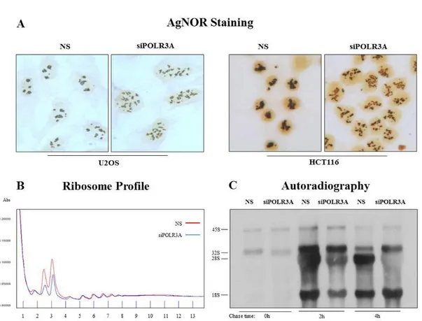
To inhibit in a selective manner the activity of RNA Polymerase III (Pol III) we deplete a catalytic subunit of the enzyme, the POLR3A, by transfection of small interfering RNA oligonucleotides (siRNA) in HCT116 and U2OS human cancer cells. We evaluate the residual expression of POLR3A mRNA by RT-real time PCR after 48H from the end of transfection procedure and observed a reduction of about 80% of analyzed mRNA respect to control cells transfected with non-silencing oligonucleotides (NS) (FIG 1A). In order to evaluate the transcriptional activity of RNA Polymerase III, we evaluated the expression of tRNAs and 5S rRNA. Since tRNAs are characterized by a long half-life, it was necessary to evaluate the expression of tRNA precursors after POLR3A interference, to obtain information about Pol III transcriptional activity. For this reason we performed a RT Real Time qPCR using a methodological approach that permits to evaluate only the amount of new synthetized tRNAs. We found that the treatment with POLR3A siRNA leads to a decrease in the level of POLR3A mRNA and a reduction of tRNAs precursors expression (FIG3.1A). Regarding the evaluation of 5S rRNA expression, 5S rRNA is present in every 60S subunit of the cell ribosomes and therefore characterized by a very long stability, thus also in this case was necessary to analyze the amount of the new synthesized rRNA. For this purpose, we incorporated in HCT116 human tumor cells, transfected with POLR3A siRNA, a radioactive analog of Uridine: the 3H-Uridine. We performed a pulse of 3H-Uridine for 1H and harvest the cells after different time of chase
44
with unlabeled Uridine. After the treatment, we extracted the total RNA and evaluated the production of newly synthetized small RNAs by autoradiography. The autoradiographic analysis showed, 48 hours after the end of POLR3A siRNA transfection, a strong reduction of newly synthetized 5S rRNA (FIG.3.1B). These preliminary results indicated that targeting POLR3A expression by RNA interference represents a good method to inhibit Pol3 mediated transcription.
FIG. 3.1:
FIG.3.1: POLR3A siRNA transfection lead to an inhibition of PolIII transcriptional activity A) RT Real Time qPCR analysis of POLR3A mRNA and tRNA precursors in HCT116 and
U2OS human tumor cell after POLR3A interference. B) Autoradiographic analysis of 3HUridine labeled small RNAs in HCT116 tumor cells after POLR3A inhibition.