Università degli Studi di Ferrara
DOTTORATO DI RICERCA IN
"Biochimica, Biologia Molecolare e Biotecnologie"
CICLO XXVI
COORDINATORE Prof. FRANCESCO BERNARDI
Mitochondrial calcium uptake
and release mechanisms as
key regulators of cell life or
death
Settore Scientifico Disciplinare MED/04
TABLE OF CONTENTS
ABBREVIATIONS ... pag. 3 INTRODUCTION ... pag. 6
Mitochondrial structure and functions ... pag. 6 Mitochondria ... pag. 6 Citric Acid Cycle, Respiratory Chain and Oxidative Phosphorylation ... pag. 8 Mitochondria and cell fate ... pag. 12 The intracellular Ca2+ signalling ... pag. 14
Ca2+ signalling and cell cycle ... pag. 17
MCU, the Mitochondrial Calcium Uniporter ... pag. 18 mPTP, the mitochondrial Permeability Transition Pore ... pag. 21 [Ca2+] measurements by the photoprotein aequorin ... pag. 23
MCU INVOLVEMENT IN CELL CYCLE ... pag. 27 Introduction ... pag. 27 Results and Discussion ... pag. 30 ROLE OF THE C SUBUNIT OF THE Fo ATP SYNTHASE IN MITOCHONDRIAL PERMEABILITY TRANSITION ... pag. 40 Introduction ... pag. 40 Results and Discussion ... pag. 41 THE MITOCHONDRIAL PERMEABILITY TRANSITION PORE IS A DISPENSABLE ELEMENT FOR MITOCHONDRIAL CALCIUM EFFLUX pag. 51 Introduction ... pag. 51 Results and Discussion ... pag. 53 MATERIALS AND METHODS ... pag. 73 REFERENCES ... pag. 79
ABBREVIATIONS
Ca2+ Calcium
[Ca2+]c Cytoplasmic calcium concentration
[Ca2+]m Mitochondrial calcium concentration
acetyl-CoA acetyl coenzyme A ADP Adenosine 5’ diphosphate ANT Adenine Nucleotide Translocase ATP Adenosine 5’ triphosphate [ATP]m mitochondrial ATP
ATP5A ATP synthase, H+ transporting, mitochondrial F1 complex, alpha subunit
Bcl-2 B cell leukemia/lymphoma 2 CaM Ca2+-calmodulin
CaMK Ca2+-calmodulin kinase Cdk Cyclin-dependent kinase CsA Cyclosporine A
DAG Diacylglycerol
DOA Dominant Optic Atrophy DRP1 Dynamin Related Protein 1 EMRE Essential MCU Regulator ER Endoplasmic Reticulum ETC Electron Transport Chain
FACS Fluorescence-Activated Cell Sorting FAD Flavin Adenine Nucleotide
FCCP Carbonyl Cyanide –p- trifluoromethoxyphenylhydrazone GFP Green Fluorescent Protein
HA Hemagglutinin
HK Hexokinase II
HtrA2 High temperature requirement protein A2 IAP Inhibitor of Apoptosis Protein
IB Intracellular Buffer
IMM Inner Mitochondrial Membrane IMS Intermembrane Space
IP3 Inositol 1,4,5-trisphosphate
MAM Mitochondria Associated Membrane MCU Mitochondrial Calcium Uniporter
MCUR1 Mitochondrial Calcium Uniporter Regulator 1 MEFs Mouse Embryonic Fibroblasts
mgluR1 matabotropic glutamate Receptor MICU1 Mitochondrial Calcium Uptake 1 m Mitochondrial membrane potential
MPT Mitochondrial Permeability Transition mPTP mitochondrial Permeability Transition Pore mtGFP mitochondrial Green Fluorescent Protein NCLX mitochondrial Na+/Ca2+ antiporter OMM Outer Mitochondrial Membrane pHH3 phosphohistone H3
PI Propidium Iodide
PiC inorganic Phosphate Carrier Pin1 Peptidyl-prolyl cis/trans isomerase PIP2 Phosphatidylinositol-4,5-biphosphate
RNA pol II RNA polymerase II RNS Reactive Nitrogen Species ROS Reactive Oxygen Species siRNA short interfering RNA TCA Citric Acid Cycle
TSPO Peripheral Benzodiazepine Receptor VDAC Voltage-Dependent Anion Channels
INTRODUCTION
Mitochondrial structure and functions
Mitochondria
Mitochondria are organelles with complex structures and functions. They are derived from an α-proteobacterium-like ancestor, due to an ancient “invasion” that occurred more than a billion years ago [1] The acquisition of mitochondria (and plastids) was a key event in the evolution of the eukaryotic cell, supplying it with bioenergetic and biosynthetic factors. At subcellular resolution mitochondria are composed of an outer membrane (OMM), mostly permeable to ions and metabolites up to 10 kDa, and a highly selective inner mitochondrial membrane (IMM), characterized by invaginations called cristae. The space between these two structures is called the intermembrane space (IMS). Together, the OMM and IMM enclose the mitochondrial matrix. The IMM is further subdivided into two compartments: the peripheral inner boundary membrane and the cristae [2]. Cristae are not simply random folds, but rather internal compartments formed by profound invaginations originating from very tiny “point-like structures” in the inner membrane. These narrow tubular structures, called cristae junctions, can limit the diffusion of molecules from the intra-cristae space towards the IMS, thus creating a microenvironment where mitochondrial electron transport chain (ETC) complexes (as well as other proteins) are hosted and protected from random diffusion. The inner boundary membrane is enriched with structural proteins and components of the import machinery of mitochondria [3]. Mitochondrial morphology in living cells is heterogeneous and can range from small spheres to interconnected tubules. This heterogeneity results from the balance between fusion and fission processes, and represents a phenomenon termed mitochondrial dynamics [4]. A growing body of evidence indicates that mitochondrial morphology is critical for the physiology of the cell and changes in mitochondrial shape have been related to many different processes such as development, neurodegeneration, calcium (Ca2+) signalling, reactive oxygen species (ROS) production, cell division, and apoptotic cell death [5]. Mitochondrial shape is controlled by the recently identified “mitochondria-shaping proteins”, which regulate the fusion-fission equilibrium of the organelle. In mammals, key components of the fusion machinery include the homologues MFN1 and MFN2 [6]. The only dynamin-like GTPase currently identified in the IMM is OPA1, a fusion protein that
is mutated in dominant optic atrophy (DOA), the most common cause of inherited optic neuropathy. Post-transcriptional mechanisms, including proteolytic processing, tightly regulate OPA1 activity. In mammalian cells, mitochondrial division is regulated by DRP1 and FIS1 [7, 8]. The large GTPase DRP1 is a cytosolic dynamin-related protein, whose inhibition or downregulation results in a highly interconnected mitochondrial network. The same phenotype is caused by the downregulation of FIS1, a protein of the OMM, proposed to act as a mitochondrial receptor for DRP1 [9]. For example, mitochondrial dynamics seem to influence production of ROS and cellular longevity. DRP1-dependent fragmentation of the mitochondrial reticulum is a crucial component for accumulation of ROS in pathological conditions [10]. How mitochondrial fission is required for ROS production and lifespan remains unclear, although a link between the two processes seems plausible. Hence, factors other than mitochondrial metabolism per se could have a role in the pathogenesis of ROS-related diseases. Interestingly, many ROS (as well as Reactive Nitrogen Species, RNS) sources and targets are localized in the mitochondria and ER with are relevant consequences for different pathways [11].
Within cells, energy is provided by oxidation of “metabolic fuels” such as carbohydrates, lipids and proteins. It is then used to sustain energy-dependent processes, such as the synthesis of macromolecules, muscle contraction, active ion transport or thermogenesis. The oxidation process results in free energy production that can be stored in phosphoanhydrine “high-energy bonds” within molecules such as nucleoside diphosphate and nucleoside triphosphate (i.e., adenosine 5' diphosphate and adenosine 5’ trisphosphate, ADP and ATP, respectively), phosphoenolpyruvate, carbamoyl phosphate, 2,3-bisphosphoglycerate, and other phosphates like phosphoarginine or phosphocreatine. Among them, ATP is the effective central link-the exchange coin-between energy producing and the energy demanding processes that effectively involve formation, hydrolysis or transfer of the terminal phosphate group.
In general, the main energy source for cellular metabolism is glucose, which is catabolized in the three subsequent processes: glycolysis, tricarboxylic acid cycle (TCA or Krebs cycle), and finally oxidative phosphorylation to produce ATP. In the first process, when glucose is converted into pyruvate the amount of ATP produced is low. Subsequently, pyruvate is converted to acetyl coenzyme A (acetyl-CoA) which enters the TCA cycle, enabling the production of NADH. Finally, NADH is used by the respiratory chain complexes to generate a proton gradient across the inner mitochondrial membrane, necessary for the production of large amounts of ATP by mitochondrial ATP synthase. In
addition, it should be mentioned that acetyl-CoA could be generated also by lipid and protein catabolism.
Citric Acid Cycle, Respiratory Chain and Oxidative Phosphorylation
The citric acid cycle (TCA) was elucidated by Sir Hans Krebs in 1940 [12]. The triose deriving from glycolysis is completely oxidized into three molecules of CO2 during a
sequence of reactions that allow the reduction of cofactors NAD and flavin adenine nucleotide (FAD), providing energy for the respiratory chain in the form of electrons. In 1949 Kennedy and Lehningher demonstrated that the entire cycle occurs inside mitochondria [12]. The first reaction of the citric acid cycle is the condensation of one Acetyl-CoA and a molecule of citrate to generate oxaloacetate and is catalysed by citrate synthase. Citrate is then transformed into isocitrate by aconitase through the formation of cis-aconitate. This step is reversible and could lead to the formation of both citrate and isocitrate. Only the fast consumption of isocitrate by its dehydrogenase can force the reaction to the proper direction. Isocitrate dehydrogenase catalyses the first irreversible oxidation leading to the decarboxylation of isocitrate, generating CO2 and α-ketoglutarate.
The second carbon leaves the cycle in the following step, when the newly generated α-ketoglutarate is immediately decarboxylated by the α-α-ketoglutarate dehydrogenase complex in a reaction similar to the pyruvate decarboxylation. In fact, both these complexes share high similarities in enzyme amino acid composition and in the organization of the different subunits. Energy released from both oxidations is used to generate NADH from NAD that directly feeds into the respiratory chain.
The following step is catalysed by succinyl-Coa synthetase and utilizes the energy derived from the CoA removal to phosphorylate GDP (or ADP) to GTP (or ATP). Selectivity for the nucleotide is determined by the isozyme involved. It has been well established that at least two isozymes of succinyl-CoA synthetase are expressed in animal tissues [13] and the proportion between them seems to be tissue specific.
The succinate generated in the previous step is the 4 carbon compound that is then converted, by three sequential reactions, to oxaloacetate to conclude the cycle. The first of these steps is the oxidation of succinate to fumarate by succinate dehydrogenase. This enzyme, tightly bound to the inner mitochondrial membrane (IMM), catalyses FAD reduction to FADH2 that provides electrons for the respiratory chain. Fumarate is then
hydratase are oncosuppressor genes and their inactivation leads to the accumulation of succinate and fumarate that spread in the cytosol and promote hypoxiainducible factor 1α (HIF1α) accumulation by inactivating prolyl hydroxilase enzymes (promoter of HIF1α degradation); HIF1α, promotes a pseudo-hypoxic condition that favours tumour development [14]. The last event that completes the citric acid cycle is the oxidation of L-malate to oxaloacetate. This reaction is performed by L-L-malate dehydrogenase, which induces the reduction of another molecule of NAD to NADH. The resulting molecule of oxaloacetate is suitable for starting another cycle through condensation with an acetyl group.
During all these processes, only one molecule of ATP (or GTP) is produced, but three molecules of NADH and one of FADH2 (plus one molecule of NADH from pyruvate
dehydrogenase), which provide electrons for respiratory chain, are also generated and subsequently result in the production of large amounts of ATP [15].
The mitochondrial respiratory chain comprises a series of protein complexes and fat-soluble compounds which act as electron carriers and the majority of which are integral membrane proteins containing prosthetic groups capable of accepting or donating one or two electrons. It is located in the IMM and is able to produce an electrochemical potential across the mitochondrial membrane by creating a concentration gradient of H+ ions between the two sides of the membrane. This potential is exploited to activate the transport channels present on the membrane and to promote the synthesis of ATP by ATP synthase. The electron carriers of the respiratory chain are organized in separated supramolecular intramembrane complexes, and each of them represents a fraction of the respiratory chain (Fig. 1).
The complexes of the respiratory chain are: Complex I: NADH dehydrogenase Complex II: Succinate dehydrogenase Complex III: cytochrome c reductase Complex IV: Cytochrome c oxidase
All complexes are formed by multiple subunits and contain all the proteins encoded by nuclear DNA that is mitochondrial, except the Complex II which is entirely encoded by nuclear DNA. Coenzyme Q (ubiquinone) transfers the electrons previously transported in complex I and complex II from NADH and FADH2 to complex III, and hence they will be
conducted to complex IV by cytochrome c. It eventually transfers electrons directly to oxygen, leading to H O production.
Coenzyme Q (also called ubiquinone) is located between Complex I and Complex III and can accept only one electron, becoming a semiquinone radical, or two electrons acquiring the fully reduced ubiquinol form. Since ubiquinone is small in size and hydrophobic, it is freely diffusible in the lipid bilayer of the IMM and can act as a bridge between less mobile electron carriers in the same membrane.
The cytochrome c is also an essential part of the ETC. It is a peripheral membrane protein of 12.5 KDa soluble in water and weakly associated with the external surface of IMM due to interaction with cardiolipin [16]. This protein transfers electrons between complexes III and IV. Like all cytochromes it has a prosthetic group (heme group) and it is constituted by a tetrapyrrolic ring with an iron atom covalently bonded to the center. Cytochrome c allows the passage of electrons through the oscillation of the iron from the ferric form Fe3+ to the ferrous form Fe2+.
The chemical mechanism which couples the proton flow with the phosphorylation is called Chemiosmotic Model and it was proposed by Peter Mitchell [17]. In agreement with this model the electrochemical energy contained in the difference of proton concentration and in the charge separation across the IMM leads to the synthesis of ATP when the proton flow reverses its direction and protons back into the matrix through a proton channel associated to the ATP synthase. The mechanism of action of ATP synthase was elucidated by the Nobel Prizes Paul Boyer and John Walker. ATP synthase could be divided in two main components: Fo that allows the channelling of protons, and F1 that catalyses ATP
phosphorylation. The Fo is embedded in the IMM, while the F1 resides in the mitochondrial
matrix and is bound to the Fo through a γ subunit (which drives conformational changes)
and a b2δ dimer (that holds Fo and F1 together). The protons flow from the intermembrane
space to the matrix through the Fo inducing its rotation; the movement is transmitted from
the γ subunit to the F1 causing conformational rearrangements. The Fo region consists of
three main subunits a (ATP6 gene), b (ATP5F1 gene) and c (ATP5G1, ATP5G2 and ATP5G3 genes), and in humans six additional subunits d, e, f, g, F6 and 8. The F1 has a
trimeric structure consisting of αβ dimers. The sequential changes are linked to the binding of substrates, phosphorylation and release of ATP. The three available dimers are never in the same conformational state and the conformational changes in one dimer drive rearrangements in the other [18]. This region consists of different protein subunits, α (ATP5A1 and ATPAF2 genes), β (ATP5B, ATPAF1 and C16orf7 genes), γ (ATP5C1 gene), δ (ATP5D gene) and ε (ATP5E gene). It has been calculated that for the synthesis of one ATP molecule, 4 protons are required [19]. Once synthetized, ATP can locate inside mitochondrial matrix or be transported into the IMS by the nucleotide exchanger adenine
nucleotide translocase (ANT) that passively exchanges ATP with ADP. Once in the IMS, ATP can freely pass the OMM through the voltage dependent anion channel (VDAC).
Figure 1: Schematic representation of mitochondrial electron transport chain [20].
A recent our work by Bonora et al. suggested moreover that the c subunit of the Fo ATP
synthase constitutes a critical component of the mitochondrial Permeability Transition Pore (mPTP) and that it is required for the MPT, mitochondrial fragmentation and cell death induced by oxidative stress or mitochondrial Ca2+ overload [21]. However this part will be discussed more extensively in the thesis.
The concentration gradient of H+ ions that is created between the two sides of the membrane is equivalent to a proton motive force and this is defined mitochondrial transmembrane potential which corresponds to -150/-180 mV in the cytosol [22]. This is essential for many processes, including the production of ATP by ATP synthase and the accumulation of Ca2+. Dysfunction of the respiratory chain are associated with mitochondrial disorders and affect tissues that require a large amount of energy as the brain, heart and skeletal muscle [23].
Mitochondrial DNA encodes thirteen polypeptides, each of which is an integral subunit of respiratory chain complexes [24].
Mitochondria and cell fate
Mitochondria are also important checkpoints of the apoptotic process, as they may release caspase cofactors [25].
Apoptosis, or programmed cell death, is a highly regulated cellular event that plays an extremely important role in the tissue homeostasis and in the development of multicellular organisms. Defects in regulation of apoptosis are often associated with pathological conditions such as neurodegenerative diseases, tumorigenesis, autoimmune syndromes and viral infections. Apoptosis occurs through two pathways: the extrinsic pathway and the intrinsic or mitochondrial mitochondrial pathway, and it requires the action of specific enzymes (caspases) and regulatory proteins (such as those of the family Bcl-2). From the biochemical point of view, apoptosis is a proteolytic event generated by the activation of a large family of proteases called caspases (cysteine aspartate-specific proteases), generally present in the cytoplasm in the form of inactive enzymes.
The main player in the apoptotic activation process is cytochrome c. The majority of cytochrome c is tightly bound to mitochondrial inner membrane, thanks to its electrostatic interactions with acidic phospholipids, but a small fraction probably exists loosely attached to inner mitochondrial membrane and available for mobilization.
This protein is an irreplaceable component of the mitochondrial electron transport chain, shuttling electrons from complexes III to IV, and is thus essential to life: the disruption of its only gene is embryonic lethal [26]. Once released in the cytoplasm, this protein drives the assembly of a caspases activating complex together with Apaf-1 (apoptosis–protease activating factor 1) and caspase 9, the so-called ‘apoptosome’. Cytochrome c, once in the cytosol, induces the rearrangement and heptaoligomerization of Apaf-1: each of these complexes can recruit up to seven caspase molecules, leading to their proteolytic self-processing and consequent activation [27]. Mitochondria contain several other proapoptotic, intermembrane space-resident proteins, such as Smac/ DIABLO, HtrA2/Omi, AIF and EndoG. DIABLO (direct inhibitor of apoptosis-binding protein with a low isoelectric point) and HtrA2 (high temperature requirement protein A2) both have an N-terminal domain that can interact and inhibit IAPs (inhibitor of apoptosis proteins).
Another event that accompanies apoptosis is the loss of mitochondrial membrane potential caused by the opening of the PTP. The opening may be triggered by several factors, such as an excessive accumulation of Ca2+, ATP depletion, oxidative stress, increased fatty acids or inorganic phosphate. PTP opening causes mitochondrial depolarization, followed immediately by mitochondria swelling, rupture of the OMM, release of cytochrome c and
other apoptotic factors, caspases activation and cell death by apoptosis. It is interesting to underline the important role of Ca2+ in this event, demonstrating how the mitochondrial Ca2+ signal represents a complex signaling pathway that mitochondria can translate in different biological consequences.
Moreover, mitochondria are the most important source of intracellular reactive oxygen species and leak from the electron transfer chain is supposed to be the main route [28]. An unexpected pathway has emerged that involves p66Shc in mitochondrial reactive oxygen species production. Intriguingly, upon phosphorylation by PKCβ and peptidyl–prolyl cis/trans isomerase (Pin1) recognition, p66Shc translocates to mitochondria [29] where it exerts its own oxidoreductase activity [30]. As a consequence, p66shc directly oxidizes cytochrome c (thus allowing electron to escape mitochondrial electron transport chain) and generates H2O2, leading to mPTP opening and in turn cell death [31] (Fig. 2).
Figure 2: Signal transduction pathway of p66Shc in oxidative condition [31].
The study of Ca2+ homeostasis in apoptotic process has highlighted some important regulators of apoptosis, such as proteins of Bcl-2 family, which are located in
endoplasmic reticulum (ER)). Indeed, Bcl-2 was found to be associated to OMM, ER and nucleus; there is also a cytoplasmic form of Bcl-2 [32]. The Bcl-2 protein family controls the intrinsic pathway of apoptosis.
Proapoptotic Bax and Bak proteins exist as inactive monomers in viable cells with Bax localizing in the cytosol, loosely attached to membranes, and Bak residing in mitochondrial fraction. Upon apoptosis induction, Bax translocate to mitochondria where it homo-oligomerizes and inserts in the outer membrane; similarly, also Bak undergoes a conformational change, which induces its oligomerization at the outer mitochondrial membrane. Together, these events trigger OMM permeabilization, the crucial process mediating the release of intermembrane space-resident caspase cofactors into the cytoplasm [33].
Mitochondria also undergo a more ‘macroscopic’ remodelling of their shape during the programed cell death. Indeed, after apoptosis induction, mitochondria become largely fragmented, resulting in small, rounded and numerous organelles. This process occurs quite early in apoptotic cell death, soon after Bax/Bak oligomerization, but before caspase activation.
Mitochondrial and ER networks are also fundamental for the maintenance of cellular homeostasis and for the determination of cell fate under stress conditions [34]. The communication between the two organelles takes place via a zone of close contact between ER and mitochondria, called MAM (mitochondria associated membrane) [35], involved in bioenergetics and cell survival.
The intracellular Ca
2+signalling
In resting cells the [Ca2+]c is carefully maintained at extremely low levels (100 nM), while
the concentration of the ion in the extracellular space is about 1 mM. There are also intracellular organelles, such as the ER and secretory granules, which contain one to ten thousand fold greater concentrations of Ca2+ than the cytoplasm. There is an electric gradient between the two sides of the plasma membrane, which stands on values between -10 and -70 mV. The cell then spends a significant proportion of this energy against electrochemical gradient to maintain the concentration of this ion on such low values. However, in response to different stimuli the amount of the cation may increase significantly, up to 1-3 μM. The rapid increase in [Ca2+] in the cytoplasm is therefore one of the main features of the complex Ca2+ signal at the cellular level. The effect of this increase was correlated with an increasing number of physiopathological events. Muscle
contraction, hormone secretion, synaptic transmission, as well as the proliferation and apoptosis are just a few examples of Ca2+-mediated processes. The fundamental question is how it is possible that different events may be regulated by a single messenger. The answer is found in the versatility of the Ca2+ signal.
In recent decades the huge amount of research done in this field has in fact revealed that the differences in amplitude, speed, and spatio-temporal characteristics of the [Ca2+] increase play an essential role in different events mediation.
This versatility is ensured by the great complexity of the systems to regulate the Ca2+ homeostasis: a lot of proteins (channels, pumps, receptors, enzymes, etc.) in fact collaborate in calcium signal activation, shutdown, and translation, and by the interaction with each other create many different spatio-temporal profiles of the signal responsible for the versatility and flexibility of the tasks of this intracellular messenger.
The processes that shape the Ca2+ signalling in all its complexity can be schematically divided into four functional units [36]:
- stimuli that produce messengers able to mobilize Ca2+ ions;
- the consequent Ca2+ signalling activation that feed Ca2+ into the cytoplasm; - the role of Ca2+ as a second messenger;
- the OFF mechanisms of Ca2+ signalling, composed of pumps and exchangers that remove Ca2+ from the cytoplasm to restore resting conditions.
It is very important to discuss now the role of Ca2+ signalling in mitochondria. In the sixties in fact a lot of studies conducted in isolated organelles have demonstrated the ability of energized mitochondria to accumulate Ca2+ [37]. This has quickly found an explanation in the chemiosmotic theory that was becoming popular in those years. It became clear that the proton gradient generated by the activity of the respiratory chain may be used not only for the synthesis of ATP, but also to accumulate cations in the matrix.
Precisely the proton gradient determines the mitochondrial potential of about -180 mV (negative inside the organelle), which provides the driving force necessary to support the Ca2+ influx into the mitochondria. It has been calculated that the equilibrium would be reached only if the [Ca2+] in the matrix reached values 106 times higher than those present in the cytosol. Furthermore, the inhibition of the respiratory chain or the collapse of the membrane potential blocks the ability of mitochondria to accumulate the cation.
The development of new indicators such as aequorin allowed to demonstrate that the increase in [Ca2+]c in live and intact cells induced by physiological stimuli is always
much higher than in the cytosolic compartment (up to 500 μM in chromaffin cells). We can say that mitochondria play an active role in the signaling pathway of Ca2+.
Mitochondrial Ca2+ uptake plays a key role in the regulation of many cellular functions, ranging from ATP production to cell death. Increases in mitochondrial calcium activate several dehydrogenases and carriers, inducing an increase in the respiratory rate, H+ extrusion, and ATP production necessary for the correct energy state of the cell. However, prolonged increase in [Ca2+]m leads to the opening of the mitochondrial permeability
transition pore (mPTP), a critical event driving to cell death by apoptosis [38].
Although it is generally accepted that cellular energy metabolism, survival and death are controlled by mitochondrial Ca2+ signals, the underlying molecular mechanisms have been completely elucidated yet.
The mitochondrial Ca2+ uptake and release mechanisms are based on the utilization of gated channels for Ca2+ uptake and exchangers for release that are dependent upon the negative mitochondrial membrane potential, which represents the driving force for Ca2+ accumulation in the mitochondrial matrix [39].
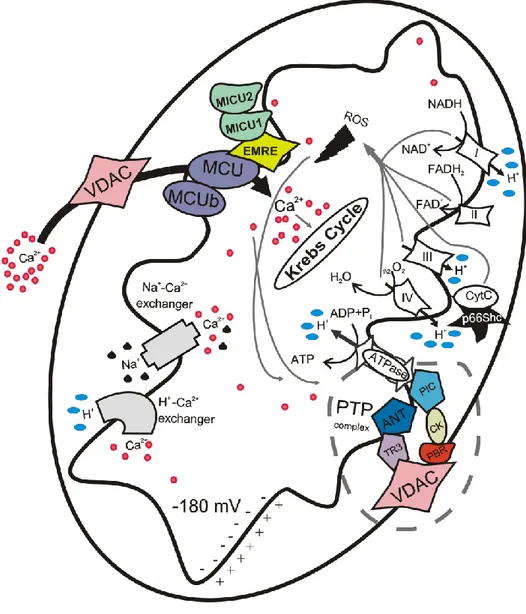
There are several proteins involved in mitochondrial Ca2+ influx and efflux, such us the mitochondrial calcium uniporter (MCU) [40, 41] and the mitochondrial Na+/ Ca2+ antiporter (NCLX) [42]. It is mostly accepted that Na+/Ca2+ exchange activity is relevant for excitable cells [43] and that NCLX inhibition or silencing does not completely arrest Ca2+ efflux [42], which indicates that other mechanisms are involved in this process. Two mechanisms have been proposed to supplement Ca2+ efflux, one based on the H+/Ca2+ antiporter [44] and another based on the mPTP [45], but evidence supporting the latter remains elusive (Fig. 3).
In particular this thesis focuses the attention on MCU and mPTP complex that are involved in the maintenance of mitochondrial Ca2+ homeostasis and more details are shown below.
Figure 3: Schematic representation of mitochondrial calcium signalling.
Ca
2+signalling and cell cycle
Ca2+ signalling plays a crucial role in every cellular process, such us the origin of life, the fertilization and cell death. Several lines of evidence suggest that the intracellular Ca2+ concentration influences mitogenic signals that control the progression of the cell cycle. Despite these strong indications, little attention is paid to the role that Ca2+ has on this process [46].
In eukaryotes, Ca2+-calmodulin (CaM) is one of the proteins more sensitive to Ca2+, which constitutes a family of Ser/Thr kinases that are able to stimulate the Ca2+-calmodulin kinase (CaMK). These proteins are involved in cell cycle regulation, in particular the G1/S phase progression and the exit from mitosis are dependent on changes in the concentration
several targets, such as ion channels and protein kinases. The selective inhibition of CaMKI leads to a block in the G1 phase due to a decrease in the cyclin D levels, an increased phosphorylation of pRb and prevention of the activation of cyclin-dependent kinases (Cdk) 4. The other isoform CaMKII is implicated in the progression into S phase, but also in the G2 and M phases [47].
Another protein involved in Ca2+ signalling is calcineurin, a heterodimer composed by the
catalytic subunit calcineurin A and the regulatory, Ca2+-binding subunit calcineurin B. This protein is activated as a result of changes in intracellular [Ca2+] and it controls the cell cycle progression with the proteins of the CaMK family. In particular, in the G1 phase inhibition of both proteins stops the cycle before the activation of the cyclin D1/Cdk4 complex. During the G1/S transition, the inhibition of calcineurin and CaMK leads to the accumulation of p21 and p27 respectively, the increase of the levels of these proteins is sufficient to inactivate Cdk2 and to block the cell cycle. Finally, CaMKII is the target of CaM during the G2/M transition and also during the metaphase/anaphase transition [48]. Changes in the mRNA of CaM are associated with a transition of cells from G1 phase to the quiescent phase. CaMs are also capable of activating DNA polymerase α and δ, indicating a role in the PCNA transcription. Ca2+ and CaM are involved also in the breakdown of the nuclear envelope, cytokinesis and in the contraction of myosin’s ring [46].
MCU, the Mitochondrial Calcium Uniporter
Ca2+ accumulation into mitochondria plays a fundamental role in many cellular processes and occurs through the uniporter, thanks to a mechanism driven by the membrane potential generated by the respiratory chain. The activity and biochemical properties of the MCU have been characterized for years, but the molecular identity of the channel has remained unknown for a long time [49].
The molecular characterization of MCU was a major challenge for several reasons, including the lack of a selective inhibitor (the uniporter was inhibited not specifically by ruthenium red) and a yeast homolog of the uniporter.
MCU has a low affinity for Ca2+; in fact, MCU takes up Ca2+ in the micromolar range and experiments in permeabilized cells report a Kd of the uniporter of 10 M [50]. In addition,
a biphasic effect of calcium on the MCU has been reported: beyond a certain level, cytosolic Ca2+ inactivates the uniporter, preventing further Ca2+ uptake and this process might avoid an excessive accumulation of the cation in mitochondria [51].
Initially the candidates proposed for the MCU were the uncoupling proteins UCP2/3 [52], but this suggestion was then retired [53]. Recently, Perocchi and colleagues [54] demonstrated that MICU1 (mitochondrial calcium uptake 1) has a key role in regulating the classically defined uniporter.
Finally in 2011 it has been discovery the molecular identity of MCU, the coiled-coil domain-containing protein 109A (CCDC109A) [40, 41, 55].
The nuclear MCU gene, located on chromosome 10, encodes for a protein of 40 kDa but it loses its target sequence when it is imported into the mitochondria. The result is a mature 35-kDa protein expressed in all tissues, capable of oligomerization, giving rise to a complex of higher molecular weight [41].
From the topological point of view, the MCU is located in the IMM, with the N-terminal and the C-terminus facing the matrix; there is a linker of 9 aa (the DIME domain) facing in the IMS between the two transmembrane domains. The DIME domain is essential for the Ca2+ transport because mutations inside it render the protein non-functional [41].
The overexpression of MCU increases mitochondrial Ca2+ uptake and sensitizes cells to apoptotic stimuli, and the employment of short interfering RNA (siRNA) silencing of MCU strongly reduced mitochondrial Ca2+ uptake. This reduction is specific for mitochondria, does not induce impairment of the electrochemical gradient or change in mitochondrial morphology and the induction of specific mutations at the level of the putative pore-forming region reduce the mitochondrial calcium uptake and blocks the channel activity of the protein [40]. Recently it was shown that miR-25, a small (22 nucleotides) noncoding regulatory RNA overexpressed in several human cancers, decreases mitochondrial Ca2+ uptake through selective MCU downregulation, conferring resistance to apoptotic challenges. MCU appears to be downregulated in human colon cancer samples, and accordingly, miR-25 is aberrantly expressed, indicating the importance of mitochondrial Ca2+ regulation in cancer cell survival [56].
About MICU1, previously identified as regulator of MCU, recently it has been shown that it is a 54-kDa single-pass membrane protein containing two highly conserved EF-hand Ca2+-binding domains. Through [57] or independently [58] of these domains MICU1 stabilizes the closed state of the MCU complex limiting mitochondrial Ca2+ entry.
MICU1 has two paralogues (MICU2 and MICU3) possessing N-terminal mitochondrial targeting sequences. MICU2 localizes in mitochondria, it has a highly conserved EF-hand domain and interacts with MICU1 and MCU [59].
that of MCU. It has two transmembrane domains with the N-terminal and the C-terminus facing the IMS but it has a lower level of expression and a different expression profile compared to the MCU. MCUb might be a dominant-negative isoform and the insertion of one or more MCUb subunits in the multimer might alter the permeability to Ca2+ [60]. However the mRNA levels of MCUb are very low when compared to MCU levels; this mRNA is highly expressed in the heart and lungs and little expressed in skeletal muscle. Variations of tissue-dependent accumulation of mitochondrial Ca2+ have been reported, and the Ca2+ influx in skeletal muscle is 28-fold greater than in cardiac mitoplasts [61]. In 2012 it has been identified also a regulator of MCU, CCDC90A or MCUR1 (mitochondrial calcium uniporter regulator 1), an integral membrane protein required for MCU-dependent mitochondrial Ca2+ uptake [62]. MICUR1 interacts with MCU but not with MICU1 and these three proteins do not exist in the same complex [62].
It has been identified also an essential MCU regulator (EMRE), it is a component of the uniporter complex and it interacts with MICU1 at IMS and with MCU oligomers in the inner membrane. [63].
Therefore the uniporter complex (uniplex) seems to be composed of MCU holomers, MCUb, MICU1, MICU2 and EMRE [63, 64].
The latest work by Patron et al. shows also that MICU1 and MICU2 finely tune the MCU by exerting opposite effects on MCU activity; at low [Ca2+] MICU2 largely shuts down MCU activity, at higher [Ca2+] MICU1 allows the response of mitochondria to Ca2+ signals generated in the cytoplasm [65].
Thus, MCU complex is important for mitochondrial Ca2+ uptake and the equilibrium of all the components manteins the normal cellular bioenergetics (Fig. 4).
Figure 4: Schematic representation of the different components of the uniporter complex [64].
mPTP, the mitochondrial Permeability Transition Pore
The mPTP is a high-conductance channel located at the contact sites between the inner and outer mitochondrial membranes. This channel is responsible for the non-selective permeability state of the mitochondrial inner membrane. The transition to this state for small molecules is referred to as the MPT. Due to osmotic forces, MPT results in a massive influx of water into the mitochondrial matrix, eventually leading to the structural break- down of the organelle [25, 66].
The molecular composition of the mPTP is not yet clear, but several proteins have been shown to be components that participate in mPTP activity, including VDAC [67], ANT [68], the inorganic phosphate carrier (PiC) [69], peptidyl prolyl isomerase F (PPIF) [70, 71], the peripheral benzodiazepine receptor (TSPO) [72], hexokinase II (HKII) [73] and several members of the Bcl-2 family. Both pro- and anti-apoptotic BCL-2 family members, including BAX, BID, BCL-2 and BCL-XL [74-76] have been shown to physically bind
to-and hence modulate the function of-PTPC components, indicating that the molecular machineries mediating MPT-driven and primary MOMP do not operate in a mutually exclusive manner.
Ca2+ ions, prooxidant and proapoptotic proteins, a decrease in the mitochondrial membrane potential, pH variations and adenine nucleotides all sensitize the opening of the pore [25, 77].
MPT resulting from mPTP opening is usually considered a transducer event in between Ca2+ or oxidative signal and different type of cell death [78, 79].
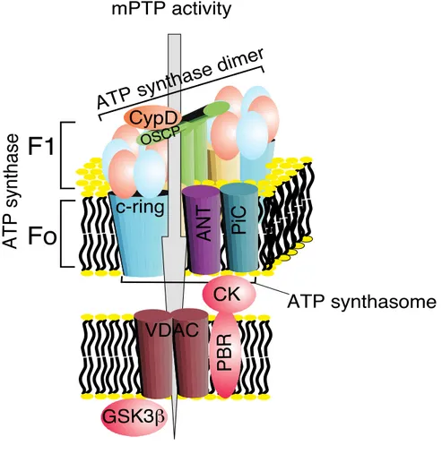
Recently, we suggested that, similar to PPIF, the c subunit of the FO ATP synthase
constitutes a critical component of the mPTP and that it is required for the MPT, mitochondrial fragmentation and cell death induced by oxidative stress or mitochondrial Ca2+ overload [21] (discussed later) (Fig. 5).
Figure 5: Schematic representation of the different components of the mPTP complex.
Nonetheless several observations have suggested that mPTP is a component of the Ca2+ efflux mechanism [80, 81] proposing a physiological role for this ambiguous complex. Unfortunately a different amount of studies have proposed the exact opposite [82, 83] leaving this supposition still unresolved.
In this thesis I will focus also my attention on the role of mPTP in mitochondrial Ca2+ homeostasis in a non-pathological context.
[Ca
2+] measurements by the photoprotein Aequorin
At the beginning of the nineties, due to the rapid development of molecular biology techniques, the aequorin cDNA was cloned and this opened the possibility to transfect the gene and obtain the expression of this protein in live cells. It was then possible to specifically localise it within the cell by including defined targeting signals in the amino acid sequence to measure variations in [Ca2+] in well-defined subcellular compartments in a highly specific manner [84].
Aequorin was extracted and purified in 1962 by the luminescent jellyfish Aequorea Victoria [85]. It consists of a monomeric apoprotein of 189 amino acids and a hydrophobic prosthetic group (coelenterazine) of 400 Da. The ability of the bioluminescent protein is closely related to the presence of the prosthetic group. The probe has three high affinity EF-hand regions able to bind Ca2+. When the cation binds aequorin there is a conformational change of the protein resulting in the bond breaking between apoprotein and coelenterazine, irreversible oxidation of the latter and photon emission (Fig. 6). In nature, the detachment of the prosthetic group in the form of celenteramide is irreversible and in vitro an active aequorin can be obtained by incubating the apoprotein with coelenterazine in the presence of oxygen and 2-mercaptoethanol. Reconstitution of an active aequorin (expressed recombinantly) can be obtained also in living cells by simple addition of coelenterazine to the medium.
The rate of photons emission is measured by a photomultiplier and it can be converted to [Ca2+]: under known conditions of pH, [Mg2+], ionic strength and temperature and aequorin exposure to solutions containing known amounts of Ca2+ it is possible to draw a sigmoidal calibration curve which correlates the [Ca2+] with the ratio of light emitted at any istant by the photoprotein and measured by the photomultiplier (L) and the light emitted from the same aequorin in the presence of saturating [Ca2+] (Lmax). [86] (Fig. 7).
Figure 7: Relationship between the free Ca2+ concentration and the rate of aequorin photon emission [86].
The rate of aequorin luminescence is independent of [Ca2+] at very high (>10-4 M) and very low [Ca2+] (< 10-7 M). However it is possible to expand the range of [Ca2+] that can be monitored with aequorin.
Until now, different recombinant aequorins were obtained, each characterized by a precise subcellular localization, by including specific targeting sequencing in the engineered cDNAs, and all targeted aequorins synthesized in our laboratory include modifications of the photoprotein N-terminal. As demonstrated by Watkins and Campbell in fact, the C- terminal proline residue of aequorin is essential for the long-term stability of the bound coelenterazine so it is not possible to alterate it [87].
It is also possible to introduce a point mutation in one of three sites able to bind Ca2+ (AEQmut) to reduce the affinity of the protein for the cation and thus to obtain accurate measurements in a wider range of concentrations (between 1 and 200 μM). Finally, for
higher [Ca2+] it is possible to use a modified,prosthetic group, the coelenterazine n, which has lower affinity for the cation [88].
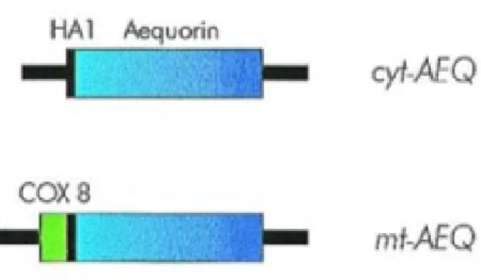
Below I briefly describe the constructs that I used in this thesis. Cytoplasm (cytAEQ)
An unmodified aequorin cDNA encodes a protein that, in mammalian cells is located in the cytoplasm and, given its small size, also diffuses into the nucleus. An alternative construct was also available that is located on the outer surface of the ER and of the Golgi apparatus. This construct was intended to drive the localization of aequorin to the inner surface of the plasma membrane given that it derives from the fusion of the aequorin cDNA with that encoding a truncated metabotropic glutamate receptor (mgluR1). The encoded chimeric protein, however, remains trapped on the surface of the ER and Golgi apparatus, with the aequorin polypeptide facing the cytoplasmic surface of these organelles. The cytoplasmic signal revealed by this chimeric aequorin is indistinguishable from that of a cytoplasmic aequorin, but it has the advantage of being membrane bound and excluded from the nucleus (Fig. 8).
Mitochondria (mtAEQ)
All mitochondrial proteins, but the 13 encoded by the organellar genome, are synthesized on cytoplasmic ribosomes and then imported into the organelle. In most cases, import depends on the presence of a cleavable signal at the N-terminus of a precursor protein. This signal (rich in basic and hydroxylated residues, and devoid of acidic ones) usually referred to as mitochondrial presequence, is removed after import by matrix proteases [89]. When added to a heterologous protein, a mitochondrial presequence is sufficient to drive its import into mitochondria. MtAEQ was the first targeted aequorin generated in the laboratory, which has been successfully employed to measure the [Ca2+] of the mitochondrial matrix of various cell types. This construct includes the targeting presequence of subunit VIII of human cytochrome c oxidase fused to the aequorin cDNA [84] (Fig. 8).
Figure 8: Schematic representation of aequorin chimeras.
Luminescence detection
The aequorin detection system is derived from that described by Cobbold and Lee [90] is based on the use of a low noise photomultiplier placed in close proximity (2-3 mm) of aequorin expressing cells. The cell chamber, which is on the top of a hollow cylinder, is adapted to fit 13-mm diameter coverslip. The volume of the perfusing chamber is kept to a minimum (about 200 μl). The chamber is sealed on the top with a coverslip, held in place with a thin layer of silicon. Cells are continuously perfused via a peristaltic pump with medium thermostated via a water jacket at 37°C. The photomultiplier (EMI 9789 with amplifier-discriminator) is kept in a dark box. During manipulations on the cell chamber, the photomultiplier is protected from light by a shutter. During aequorin experiments, the shutter is opened and the chamber with cells is placed in close proximity of the photomultiplier. The output of the amplifier-discriminator is captured by an EMIC600 photon-counting board in an IBM compatible microcomputer and stored for further analysis.
Recently it has been extensively explained with a specific protocol the subcellular Ca2+ measurements in mammalian cells using aequorin-based probes [91].
MCU involvement in cell cycle
Introduction
Cell cycle, cyclins and cyclin-dependent kinases
Cell division is a universal process for living organisms and it ensures reproduction, growth and development of the organism. The regulation of this process is the basis of cell ability to maintain genetic integrity, which is important to cell survival and proliferation [92]. At present proteins alterations involved in cell cycle regulation are of considerable interest, because they are directly involved in the tumor development. Therefore, understanding all the mechanisms that regulate cell proliferation becomes increasingly indispensable.
In mammals cell cycle consists of two main phases: interphase and mitosis. The interphase occupies most of the time of cell division and can be further divided into:
- G1 phase, which prepares the cell for the genetic material division with a size increase and synthesis of numerous proteins and organelles such as mitochondria and ribosomes. At a certain point - the restriction point - the cell is committed to division and moves into the S phase.
- S phase, during which DNA synthesis replicates the genetic material. Each chromosome now consists of two sister chromatids.
- G2 phase, which runs from the completion of S phase until the phase of mitotic division. It is characterized by a high metabolic activity and the synthesis of the mitotic spindle;
- G0 phase or "quiescent state” is the phase in which cells do not divide because they are already differentiated and do not continue in the division cycle, or it may be a waiting phase to proceed the division later. They are busy carrying out their functions in the organism. e.g., secretion, attacking pathogens. Most of the lymphocytes in human blood are in G0. However, with proper stimulation, such as encountering the appropriate antigen, they can be stimulated to reenter the cell cycle at G1.
The mitotic phase includes the most significant events of cell division and can be divided into two phases: mitosis and cytokinesis. During interphase, chromosomes are not visible individually but they are condensed in the form of chromatin, an association
of DNA and protein present in the nucleus, in the step of division the chromatin coils in individual chromosomes that will then be divided.
The mechanism of division involves the diploid somatic cells (2n): they consist of two pairs of homologous chromosomes that carry both genes that control the same characteristics.
A class of proteins directly involved in cell cycle progression are cyclins, discovered in 1983 by Tim Hunt and his colleagues [93]. Cyclins levels vary drastically through the phases of the cell cycle as a result of transcriptional changes and degradation mediated by the proteasome.
There are eight cyclins involved in cell cycle progression: cyclin A1 and A2, B1-B2-B3, C, D1-D2-D3, E1-E2, F, G1-G2, H. All these show a 150 amino acid residues region called cyclin box; this domain consisting of five helices [94] is responsible for the binding of the N-terminal part of specific Cdk [95].
The fluctuation of the expression levels of these proteins has been demonstrated by the fact that cyclins A and B are accumulated at the beginning of S phase and in the late G2 phase respectively, while in G1 phase cyclin D1 levels increase and remain high until the mitosis. While cyclins A and B contain a "destruction box", cyclins D and E contain a PEST sequence, or a segment of amino acid residues rich in proline (P), glutamic acid (E), serine (S) and threonine (T), these sequences are required for efficient proteolysis ubiquitization-mediated in the end of each phase of the cell cycle [96]. Cyclin H forms complexes with Cdk7 to constitute an enzyme known as CAK (kinase activated by Cdk); CAK is involved in the activation of CDC2 and Cdk2, respectively, by phosphorylation of residues Thr 160 and Thr 161. Together with the Cdk 7 and MAT-1 (protein menage-a - trois- 1), cyclin H form a tertiary complex which modulates transcription by acting on the activity of RNA polymerase II (RNA pol II). Cyclin T does not seem to be directly involved in cell cycle regulation, but his bond with the Cdk9 makes active in various processes such as transcription, signal transduction and differentiation [95].
Cyclin F has some similarities with cyclins A and B, it seems interact with the cyclin B and Cdk1 to form a trimeric complex. Cyclins G (G1-G2) are the p53 target and they appear to be involved in the arrest of the cycle in G2/M phases after DNA damage.
Cyclins activate specific Cdks, a family of serine/threonine protein kinases [95], and the activation occurs in specific phases of the cell cycle [96].
The formation of specific complexes cyclin/Cdk ensures the progression of the cell cycle; cyclins have a high expression at particular times and then they are degraded, ensuring the
periodic activation of the Cdk, which on the contrary have expression levels constant (Fig. 9).
Figure 9: Schematic representation of the complex cyclin/Cdk during different phases of the cell cycle [94].
So the cell cycle is one of the most regulated mechanisms of the cell.
It has long been known the role of Ca2+ as a second messenger which can be influenced by a large number of effector proteins and as explained previously mitochondrial Ca2+ homeostasis is crucial for the destiny of the cell.
Therefore it was interesting to investigate a possible link between mitochondrial Ca2+ homeostasis and cell cycle. On the other hand, mitochondrial Ca2+ uptake by MCU has a great importance for the regulation of cell life and energy production. It will be particularly interesting to investigate if MCU could be a pivotal regulator of cell cycle.
After an optimization of synchronization’s protocol for cells derived from primary culture (mouse embryonic fibroblast, MEFs), we studied the expression of MCU in different phases of cell cycle in synchronized MEFs cells, showing an increased expression of this
Moreover, we measured mitochondrial and cytosolic Ca2+ response after agonist stimulation in each phase of cell cycle, and mitochondrial membrane potential, the driving force for the Ca2+ accumulation within the mitochondria.
Finally we checked also the effect of MCU overexpression on cell cycle phases.
Our data show that MCU expression positively correlates with the mitochondrial membrane potential but, surprisingly, it also opposite correlates with agonist-dependent mitochondrial and cytosolic Ca2+ response. MCU overexpression instead does not alter cell cycle phases.
Results and discussion
Results
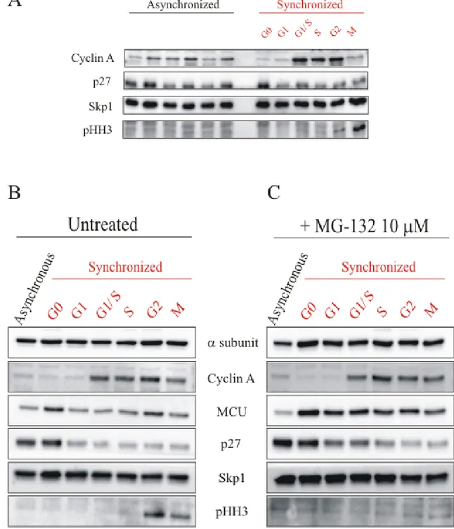
The first step of this work was an optimization of a cellular synchronization protocol to correctly discriminate the different phases of the cell cycle in MEFs cells. The synchronization procedure is based on the Serum Starvation (0.1% FBS) to allow the cells to enter in the stationary phase G0. Then the serum addiction leads the cells to re-enter in the cell cycle. The correct synchronization was assessed by western blot, using specific markers of the cycle. In particular, Cyclin A is a marker of S-G2 phase (as described previously), p27 or Cyclin-dependent kinase inhibitor 1B is a cell cycle inhibitor protein that controls the cell cycle progression at G1, phosphohistone H3 (pHH3) is a marker of M phase and Skp1 is used as protein loading control. Asynchronous cells were used as control and each cellular homogenate was taken together with the corresponding homogenate in synchronized cells. (Fig. 10A).
We have then evaluated endogenous MCU expression in each phase of cell cycle, and we use a specific antibody for the protein of interest. In this case and in the next experiments, we use only a single control in asynchronous cells, corresponding to the beginning of cell cycle in synchronized cells. It seems evident that MCU is more expressed in G0 and S-G2 phases, and this expression is specific for MCU. Indeed the expression of another mitochondrial protein, ATP5A, coding for α-subunit of F1 ATP synthase, does not change
in different phases of cell cycle (Fig. 10B).
We also have studied the MCU involvement in cell cycle by the blockade of its degradation through the use of MG-132, an inhibitor of the proteasome. We have treated MEFs cells with 10 μM MG-132 3 h before the end of each phase of cell cycle.
There are no appreciable differences in the markers of cell cycle, but it is evident that MCU is not degraded because its expression remains constant during all the phases. With these results we deduce that the non-degradation of MCU and therefore its complete stabilization has not effect on cell cycle progression (Fig. 10C).
Figure 10: (A) Western Blot of MEFs cells synchronization. On the left asynchronous MEFs cells are used as control. Cyclin A is a marker of S-G2 phases, p27 is a marker of G0-G1 phase, pHH3 is a marker of M phase, Skp1 is used as protein loading control. (B) Western Blot of MCU expression in
Therefore it is interesting to investigate a possible link between mitochondrial Ca2+ homeostasis and cell cycle. On the other hand, mitochondrial Ca2+ uptake by MCU has a great importance for the regulation of cell life and energy production.
Ca2+ is in fact a second messenger able to control multiple cellular processes such as gene transcription, muscle contraction, cell proliferation [97], learning, memory, fertilization, development and secretion [98].
The intracellular Ca2+ concentration influences signals that control cell cycle progression, despite these strong indications, little attention is paid to the role of Ca2+ in the cell cycle [46].
There are a lot of proteins that are modulated by the Ca2+ signal, due to its versatility and ability to diffuse, as the CaMK, activated by CaM, which are regulators of the G1/S transition and regulators of the exit from mitosis [92]; calcineurin also plays a role in mediating the entry into G1 phase and the continuation in the S phase [48]. In order to carry out its functions, Ca2+ undergoes fluctuations that alter its concentration during the resting state: normally Ca2+ ions are stored in mitochondria, ER and Golgi Apparatus and only in response to stimulation they are released to be poured into the cytosol. Mitochondrial and ER networks are also fundamental for the maintenance of cellular homeostasis and for the determination of cell fate under stress conditions [34]. The communication between the two organelles takes place via a zone of close contact between ER and mitochondria, called MAM [35], involved in bioenergetics and cell survival. In particular, ER is constituted by a network of connections between cisterns and tubular structures that represent an extension of the nuclear envelope. During the cell cycle this structure undergoes specific changes: during interphase it is presented as a series of tubules and cisterns, while at the time of mitosis, it only highlights the extensive network of cisterns. This alteration is due to the depolymerization of microtubules that occurs during the M phase; once anaphase occurred, cisterns are able to provide the making of the nuclear envelope, even before the appearance of the tubular structure [99].
For [Ca2+] measurements in differentintracellular compartments, we infected MEFs cells with the adenovirus for the mitochondrial and cytosolic aequorin. This experiment was conducted in asynchronous and synchronized cells. MEFs cells are stimulated with 100 μM ATP to enable Ca2+ mobilization. This agonist selectively binds the membrane receptor coupled to a trimeric G protein: in particular, the α subunit is able to activate phospholipase C which hydrolyzes phosphatidylinositol-4,5-bisphosphate (PIP2)
its receptor on the ER causes an increase of the [Ca2+] in the cytoplasm and in the mitochondrial matrix.
So [Ca2+]m measured after agonist stimulation is 37.33 ± 1.68 μM in asynchronous cells,
28.76 ± 2.85 μM in G0 phase cells, 32.4 ± 2.57 μM in G1 phase cells, 58.17 ± 4.33 μM in G1/S phase cells, 50.12 ± 4.13 μM in S phase cells, 48.44 ± 8.09 μM in G2 phase cells and 65.0 ± 6.86 μM in M phase (n=15). (Fig. 11A and 11B)
Instead [Ca2+]c measured after agonist stimulation is 2.27 ± 0.11 μM in asynchronous cells,
1.65 ± 0.1 μM in G0 phase cells, 2.41 ± 0.09 μM in G1 phase cells, 1.97 ± 0.08 μM in G1/S phase cells, 1.97 ± 0.08 μM in S phase cells, 1.77 ± 0.06 μM in G2 phase cells and 1.95 ± 0.07 μM in M phase (n=15). (Fig. 11C and 11D)
Our data suggest that, surprisingly, MCU expression opposite correlates with agonist-dependent mitochondrial and cytosolic Ca2+ response.
Figure 11: Measurements of [Ca2+] using recombinant aequorin upon agonist (100 μM ATP)
We then questioned about the possible effect of the phase-specific expression of MCU on the mitochondrial membrane potential (Ψm), since it represents the driving force for the Ca2+ accumulation within mitochondria. To measure this parameter we use the fluorescent probe Tetramethylrhodamine, methyl ester (TMRM), a cell-permanent, cation, red-orange fluorescent dye that is readily sequestered by active mitochondria, in asynchronous and synchronized MEFs cell.
The percentage of arbitrary fluorescence units (AFU) measured are -42.37 ± 1.72 in asynchronous cells, -43.27 ± 1.82 in G0 phase cells, -42.1 ± 1.79 in G1 phase cells, -28.08 ± 1.46 in G1/S phase cells, -32.38 ± 1.29 in S phase cells, -38.31 ± 1.74 in G2 phase cells and -31.35 ± 1.28 in M phase (n=80) (Fig.12).
Our data show that MCU expression in this case positively correlates with the mitochondrial membrane potential.
Figure 12: Measurements of ΔΨm with potentiometric dye TMRM in MEFs cells. The data are presented as means ± SEM *p<0.05, **p<0.01, ***p<0.001.
To evaluate the possible role of MCU in cell cycle, we decided to investigate the effects of its overexpression in the different phases.
The overexpression was achieved by cells infection with an adenovirus expressing MCU. Cell cycle was analyzed by the Tali® Image-Based Cytometer and we meausured control cells without infection (Fig. 13A), cells infected with an adenovirus expressing a
mitochondrial Green Fluorescent Protein (mtGFP) (Fig. 13B) and cells expressing MCU-GFP (Fig. 13C).
Not infected
PHASE % CELLS G1 phase 65% S phase 5% G2-M phase 30%mtGFP infection
PHASE % CELLS G1 phase 62% S phase 7% G2-M phase 31%MCU-GFP infection
PHASE % CELLS G1 phase 63% S phase 7% G2-M phase 29Figure 13: Cell cycle analysis in not incedcted (A), mtGFP infected (B) and MCU-GFP infected (C) MEFs cells by Tali® Image-Based Cytometer. The percentage of cells is referred to the number of cells in each phase of cell cycle.
MCU overexpression does not alter cell cycle progression, as the percentage of cells in each phase of cell cycle does not change.
(Fluorescence-indicated for this type of analysis. As for the previous experiment, we meausured control cells without infection (Fig. 14A), cells infected with an adenovirus expressing mtGFP (Fig. 14B) and cells expressing MCU-GFP (Fig. 14C).
Figure 14: Cell cycle analysis in not incedcted (A), mtGFP infected (B) and MCU-GFP infected (C) MEFs cells by FACS. The percentage of cells is referred to the number of cells in each phase of cell cycle.
These results confirm our previous data, the overexpression of MCU does not seem to affect cell cycle. Minor variations in cellular distribution are present only between infected and not infected cells, maybe due to the cell transfection.
Discussion
The cell cycle is the series of events leading to cell division and duplication of its genetic material. It is an evolutionarily conserved process in the succession of the events and in their regulation [95].
The proper development of an organism is due to the precise regulation and the respect of the order of cell cycle progression. Proteins alterations can trigger an uncontrolled proliferation that often leads to tumor formation, thus it is so important that all the events of the cycle are regulated on multiple levels.
The control system is constituted by the complex cyclin/Cdk. Cyclins are proteins with different expression in the cell cycle phases, as a result of transcriptional changes and proteasome degradation [96].
Cdks are a family of serine/threonine protein kinase that form specific complexes with cyclins and ensure the cell cycle progression. In fact cyclins expression in well-determined phases allows the periodic activation of the Cdk that, instead, show constant levels of expression [96].
A lot of Ca2+-binding proteins (CaM, CaMK and calcineurin) are involved in the regulation of certain phases of the cell cycle [47]. Recently, it has been identified the mitochondrial Ca2+ uniporter MCU, a protein of 40 kDa expressed in all tissues, which is located in the IMM, with the N-terminal and the C-terminus facing the matrix [40, 41]. MCU is a low affinity-channel but it has been shown that mitochondria are exposed to microdomains with high [Ca2+], which represent the contact sites between mitochondria and ER [100]. Given the high importance of this protein in the regulation of cell life, the aim of this work is the investigation of a possible MCU involvement in the cell cycle. At the beginning the attention was focused on the development of cell cycle synchronization protocols for primary cultures (MEFs cells) in order to discriminate the different phases of the cycle. We then evaluate endogenous expression of MCU in each phase of the cell cycle by western blot. MCU is more expressed in G0 and S-G2 phases, and this is specific for the protein of interest as the expression of another mitochondrial protein, ATP5A, coding for α-subunit of F1 ATP synthase, does not change in different phases of cell cycle.
We also have investigated the MCU involvement in cell cycle by the blockade of its degradation through the use of MG-132, an inhibitor of the proteasome. It is evident that MCU is not degraded because its expression remains constant during all the phases. We can deduce that the non-degradation of MCU and therefore its complete stabilization has
At this point, given the important role of the MCU in the Ca2+ homeostasis, we measured mitochondrial and cytosolic Ca2+ concentrations after agonist stimulation in each phase of cell cycle, so as to be able to understand if the phase-specific expression of MCU is related to a different Ca2+ accumulation into mitochondria. Surprisingly, MCU expression opposite correlates with agonist-dependent mitochondrial and cytosolic Ca2+ response. The Ca2+ accumulation gradient into mitochondria is governed by the strongly electronegative potential inside so we proceeded with mitochondrial membrane potential measurement in the different phases of the cell cycle. To measure this parameter we use the fluorescent probe TMRM, a cell-permanent, cation, red-orange fluorescent dye that is readily sequestered by active mitochondria, in asynchronous and synchronized MEFs cell. Data reveal a positive correlation with the phase-specific expression of the MCU, suggesting a compensatory effect of the expression of the protein of interest.
At this point, it was interesting to understand if MCU overexpression could have influence on cell cycle progression and we took advantage of the use of flow cytometry, in particular a specialized type of this technology, the fluorescence-activated cell sorting (FACS). It provides fast, objective and quantitative recording of fluorescent signals from individual cells as well as physical separation of cells in each phase of cell cycle.
MEFs cells were infected with an adenovirus expressing mtGFP or with an adenovirus expressing MCU-GFP, cells not infected were used as control. The percentage of cells in each phase of the cell cycle remains constant in all the experimental conditions, to indicate that the overexpression of MCU does not alter cell cycle progression.
Numerous other works are still in progress and not included in this thesis.
We are trying to modulate the mitochondrial membrane potential using two compounds known for their ability to alter it. The first one is FCCP (Carbonyl cyanide -p- trifluoromethoxyphenylhydrazone), a chemical uncoupler of electron transport and oxidative phosphorylation, that permeabilizes the IMM to protons, destroying the proton gradient and, in doing so, uncouples the electron transport system from the oxidative phosphorylation system. Thus electrons continue to pass through the electron transport system and reduce oxygen to water, but ATP is not synthesized in the process.
The second compound is the methyl succinate, a putative cell permeable complex II-linked substrate that, on the contrary, hyperpolarizes the mitochondrial membrane.
We are also investigating the correlation between cell cycle, MCU and cancer: it is known that miR-25 (a microRNA overexpressed in colon cancer cells) decreases mitochondrial Ca2+ uptake through selective MCU downregulation, conferring resistance to apoptotic challenges. MCU appears to be downregulated in human colon cancer samples, and
accordingly, miR-25 is aberrantly expressed, indicating the importance of mitochondrial Ca2+ regulation in cancer cell survival [56]. Therefore it is evident that MCU has a great importance in the regulation of cell fate and for this reason we are moving on a tumor cell line.
In conclusion, these experimental evidences suggest that the only activity of MCU is not sufficient for the regulation of the cell cycle.
In the light of the latest discovery about the different components of the uniporter complex [54, 59, 60, 62, 63], it is now interesting to investigate the possible involvement of these proteins in cell cycle, and preliminary data show that also in this case there is a different phase-specific protein expression. Our data suggest moreover that MCU could have a possible compensatory role in mitochondrial Ca2+ homeostasis.
Future experiments will then be directed to study the roles of all MCU’s interactors in the cell cycle progression, with a more thorough investigation in tumor cell line.
![Figure 1: Schematic representation of mitochondrial electron transport chain [20].](https://thumb-eu.123doks.com/thumbv2/123dokorg/4719970.45613/12.892.131.785.174.607/figure-schematic-representation-mitochondrial-electron-transport-chain.webp)
![Figure 2: Signal transduction pathway of p66Shc in oxidative condition [31].](https://thumb-eu.123doks.com/thumbv2/123dokorg/4719970.45613/14.892.130.793.484.966/figure-signal-transduction-pathway-p-shc-oxidative-condition.webp)
![Figure 4: Schematic representation of the different components of the uniporter complex [64]](https://thumb-eu.123doks.com/thumbv2/123dokorg/4719970.45613/22.892.181.731.86.511/figure-schematic-representation-different-components-uniporter-complex.webp)

![Figure 7: Relationship between the free Ca2+ concentration and the rate of aequorin photon emission [86]](https://thumb-eu.123doks.com/thumbv2/123dokorg/4719970.45613/25.892.273.633.309.670/figure-relationship-free-concentration-rate-aequorin-photon-emission.webp)
![Figure 9: Schematic representation of the complex cyclin/Cdk during different phases of the cell cycle [94]](https://thumb-eu.123doks.com/thumbv2/123dokorg/4719970.45613/30.892.248.657.170.698/figure-schematic-representation-complex-cyclin-different-phases-cycle.webp)