Formaldehyde solutions in simulated sweat increase human melanoma
but not normal human keratinocyte cells proliferation
M. Rizzi, PhD
a, B. Cravello
b, S. Tonello
a, F. Renò, PhD
a,⁎
a
Innovative Research Laboratory on Wound Healing, Health Sciences Department, Università del Piemonte Orientale, via Solaroli 17, 28100 Novara, NO, Italy
b
Associazione Tessile e Salute (Health and Textile Association), Corso Pella 2, 13900 Biella, BI, Italy
a b s t r a c t
a r t i c l e i n f o
Article history: Received 11 March 2016
Received in revised form 2 August 2016 Accepted 9 September 2016 Available online 10 September 2016
Our skin is in close contact with clothes most of the time thus risking potentially noxious chemicals contact. One of the potentially harmful manufacturing by-products that can be released by textiles when sweating is formal-dehyde, used as an anti-crease treatment. As it is known to be carcinogenic to humans and a potent skin sensi-tizer, the aim of this study was to investigate its effects on both normal human keratinocytes (HaCaT cells) and on a highly invasive malignant melanoma cell line (SK-MEL-28) in order to contribute to the definition of safety cut-off to be applied to the production processes.
Formaldehyde concentrations below the commonly accepted limits (10–50 μM) were obtained by diluting form-aldehyde in simulated sweat (UNI EN ISO 105-E04). The effects on cell proliferation were evaluated by cell counting, while ERK pathway activation was evaluated by western blot.
Low concentrations of formaldehyde (10μM) in both acidic and alkaline simulated sweat were able to increase malignant melanoma cell proliferation, while not affecting normal keratinocytes. Melanoma proliferation in-crease was greater in acidic (pH = 5.5) than in alkaline (pH = 8) conditions. Moreover, formaldehyde stimula-tion was able to induce ERK pathway activastimula-tion.
The data obtained suggest the need for an even increasing attention to the potentially harmful effects of textile manufacturing by-products.
© 2016 Elsevier Ltd. All rights reserved.
Keywords: Formaldehyde Melanoma Textile ERK pathway 1. Introduction
The skin represents the outer shell of the body and is crucial in per-mitting the interaction between the whole organism and the external environment. Anatomically, the skin is composed of two mutually inter-dependent layers: the dermis (the innermost) and the epidermis (the outermost). The epidermis is composed of several cell types among which keratinocytes predominate and contribute to innate immune re-sponse, thus playing a key role in protecting the organism from external threats (Grimstad et al., 2012; Köllisch et al., 2005). As any other tissue, the skin can develop benign tumors as well as malignant ones. Melano-ma originates from melanocytes, neural crest derived, a pigment pro-ducing cells located in various anatomical sites and especially in skin basal layer (Palunic et al., 2016). Melanocyte transformation into tumor cells is usually a multistep process involving the interaction of environmental, genetic, and host (i.e. previous personal or strong family history of melanoma and atypical nevi phenotype) factors (Konstantinov et al., 2016; Palunic et al., 2016). Malignant melanoma
is one of the most aggressive skin cancers and its incidence has radical-ly increased in the past decades, especialradical-ly among the Caucasian popu-lation, with an annual incidence increasing at a greater rate than any other major cancer. Although it is less common than other skin tumors (i.e. basal cell and squamous cell carcinoma) and displays an overall mortality rate of around 20%, it accounts for the majority (~ 80%) of skin cancer related deaths (Palunic et al., 2016; Singh and Salama, 2016; Jeong et al., 2011; Mueller and Bosserhoff, 2009). Such increased incidence and morbidity is clinically testified by the 10-year survival rate for metastatic patients amounting tob10% despite advanced treatments, as metastatic disease is highly resistant to conventional therapies (Konstantinov et al., 2016; Palunic et al., 2016; Ma et al., 2013; Buommino et al., 2009; Köllisch et al., 2005).
The skin is mostly in contact with clothes and accessories that may represent an often ignored source of potentially noxious compounds. A prolonged contact with potentially harmful chemicals derived from clothing as manufacturing by-products can result in skin integrity dam-age,finally resulting in allergic or sensitization reactions in the short pe-riod whereas in the long run they can induce overproliferation of neoplastic cells (Rizzi et al., 2014).
Among the potentially noxious chemicals found in textiles as manufacturing by-products there is formaldehyde (FA), a highly reactive compound widely used in many industrial processes that can
⁎ Corresponding author at: Innovative Research Laboratory for Wound Healing, Health Sciences Department, Università del Piemonte Orientale“A. Avogadro”, Via Solaroli 17, 28100 Novara, Italy.
E-mail address:fi[email protected](F. Renò).
http://dx.doi.org/10.1016/j.tiv.2016.09.009
0887-2333/© 2016 Elsevier Ltd. All rights reserved.
Contents lists available atScienceDirect
Toxicology in Vitro
enter the human body by inhalation, ingestion or through the skin (Tulpule and Dringen, 2013). For these reasons, formaldehyde exposure concerns both workers, elderly as well as children (Duong et al., 2011). The presence of FA as a manufacturing by-product in textiles is of great clinical interest as there is mounting evidence correlating such al-dehyde to adverse health effects. Moreover, FA is carcinogenic to humans, as well as a potent skin sensitizer (Duong et al., 2011; Bosetti et al., 2008; Naya and Nakanishi, 2005).
Formaldehyde, as well as other textile manufacturing by-products, could activate ERK (extracellular-signaling regulated kinases) signaling pathway and induce very different dose-dependent cellular effects, ranging from apoptosis to enhanced cell proliferation, in both normal and tumor cell lines (Rizzi et al., 2014; Aĭzenshtadt et al., 2012; Szende and Tyihák, 2010; Freick et al., 2006; Tyihák et al., 2001).
Although up to now there are no mandatory regulations for formal-dehyde presence in textiles, the mounting evidence strongly relate it to adverse health effects thus fostering many standards or voluntary labels acting in the textile industry (i.e. the European Ecolabel, the National Technical Report UNI/TR 11359 and many private marks (Oeko-Tex, Bluesigned, Aafa RLS, and so on)) to regulate its presence in thefinal product.
A previous study (Rizzi et al., 2014) demonstrated that neutral pH aqueous solutions with formaldehyde concentrations lower than nor-mally indicated limits by international standards and labels were able to induce a significant increase in melanoma cells proliferation.
As FA present on textiles as a manufacturing by-product could be re-leased from clothes upon sweat extraction, the aim of the present study was to evaluate the effects of FA diluted in simulated sweat (according to UNI EN ISO 105-E04 directive) on an in vitro model of normal keratinocyte (HaCaT) and melanoma (SK-MEL-28) human cell lines in order to contribute to the definition of safety cut-off to be applied to the production processes.
2. Materials and methods 2.1. Cell culture
Human melanoma cell line SK-MEL-28 was a kind gift of Prof. Daniela Taverna from Molecular Biotechnology Center, Department of Molecular Biotechnology and Health Sciences, University of Turin whereas HaCaT cells were purchased from Cell Lines Service GmbH (Eppelheim, Germany).
Spontaneously immortalized keratinocytes (HaCaT), isolated from human adult skin (Boukamp et al., 1988), and SK-MEL-28 cells, a highly metastatic melanoma cell line (Jeong et al., 2011), were grown as previ-ously described (Rizzi et al., 2014; Renò et al., 2013).
2.2. Simulated sweat solutions
Simulated sweat solutions were prepared according to UNI EN ISO 105-E04 directive, describing the composition of two artificial fluids with acidic (5.5) and basic (8) pH. Briefly, simulated basic sweat is com-posed of 0.5 g/L L-histidine monohydrochloride monohydrate (C6H9O2N3·HCl·H2O), 5 g/L sodium chloride (NaCl), 5 g/L sodium
phos-phate dibasic dodecahydrate (Na2HPO4·12H2O), whereas simulated
acidic sweat is composed of 0.5 g/LL-histidine monohydrochloride monohydrate (C6H9O2N3·HCl·H2O), 5 g/L sodium chloride (NaCl),
2.2 g/L sodium phosphate monobasic dehydrate (NaH2PO4·2H2O). In
both solutions pH was adjusted to the desired value by adding sodium hydroxide (NaOH).
2.3. Cell treatments
According to effects observed in a previous study, cells were treated with growing concentrations (10–50 μM) of formaldehyde (FA) (Sigma Aldrich, St. Luis, MO, USA) (Rizzi et al., 2014) aqueous solutions made in
simulated sweat, according to UNI EN ISO 105-E04 directive. Starting acidic and basic solutions were diluted in DMEM (Dulbecco's Modified Eagle's Medium) without FBS (Fetal Bovine Serum) just before cell treatment to obtain intermediate concentrations, whereas cell treat-ment was done in complete medium specific for each cell line. Tested concentrations were chosen taking into account both FA IC50
(3.03 × 10⁻⁴M) (Sakaguchi et al., 2007) and limits described in the pri-vate label Oeko-Tex and in the volunteer European label (Ecolabel) guidelines (20 ppm, corresponding to 1 mg/L or 3.33 × 10⁻⁵M in the elution solution) for such compound in textiles (Rizzi et al., 2014).
To evaluate formaldehyde effects on cell proliferation, treatment timing (2 days for HaCaT cells and 3 days for SK-MEL-28) was chosen according to the observed cell duplication time (approximately 48 h for keratinocytes and 72 h for melanoma cells).
2.4. Cell proliferation
In order to evaluate the effects of formaldehyde on cell proliferation, 5 × 10³SK-MEL-28 cells and 2 × 10³HaCaT cells were seeded in 48 well
plates and treated with test chemicals. At the end of the experiment, cells werefixed, and stained with crystal violet dye photographed at 4 × magnification using an optical microscope (Leica ICC50HD). Each experiment was performed in triplicate. The counting procedure has been performed as previously described (Rizzi et al., 2014). Cell density was expressed as percentage on control values ± standard error of the mean (S.E.M.).
2.5. Western blot
For time course experiments, 1 × 10⁶cells were treated for different
times (0 min, 5 min, 30 min, 60 min, 120 min) with the formaldehyde concentration giving the maximum proliferation result. Cell lysis, pro-tein quantification and western blot determination of ERK and phospho ERK expression was performed as previously described (Rizzi et al., 2014).
2.6. Statistical analysis
ANOVA test followed by Bonferroni's post-hoc test were done for statistical analysis. Statistical procedures were performed with the Prism 4.0 statistical software (GraphPad Software Inc., CA, USA). Proba-bility values of pb 0.05 were considered statistically significant. For pro-liferation studies, statistical analysis was performed comparing cell density observed at each concentration tested to the control. For western blotting experiments, statistical analysis was performed comparing signal intensity observed at each time point to control (T0, unstimulated) conditions.
3. Results
3.1. Formaldehyde effects on normal human keratinocytes
Experiments aimed at evaluating the effects of formaldehyde on normal human keratinocytes proliferation showed that the lowest FA concentration tested (10μM), when dissolved in simulated sweat pre-pared according to UNI EN ISO 105-E04 directive, was not able to affect normal human keratinocytes proliferation (Fig. 1). In fact, as shown in
Fig. 1, when FA was diluted in acidic solution, a slight, even if not statis-tically significant, increase in cell proliferation was observed, whereas in alkaline conditions cell proliferation did not differ from control. More-over, when the compound was tested at higher concentrations (25 and 50μM), its effects on HaCaT cells proliferation varied depending on the artificial sweat pH. In particular, 25 μM acidic FA treatment re-sulted in a nearly unvaried proliferation effect compared to control con-ditions, whereas 50μM acidic solution started to induce toxic effects
107 M. Rizzi et al. / Toxicology in Vitro 37 (2016) 106–112
(pb 0.01), resulting in a reduction of about 60% in cell proliferation (Fig. 1, light gray bars).
On the other hand, when test solutions were prepared in basic sim-ulate sweat (pH = 8), initial toxic effects started to be evident at 25μM (pb 0.05) and to worsen at 50 μM (p b 0.01), resulting in a reduction of about 60% in cell proliferation (Fig. 1, dark gray bars).
Even if a lack of any statistically significant effect of low FA concen-trations on HaCaT cells proliferation was observed, ERK (Extracellular signaling Regulated Kinase) activation pathway was investigated.
As shown inFig. 2, stimulation with 10μM FA in simulated sweat re-sulted in slight variations in ERK signaling pathway activation, depend-ing on artificial fluid pH. When the aldehyde was diluted in acidic simulated sweat, it only induced a slight and transient (approximately 20%, pb 0.01) ERK activation after 60 min stimulation, as highlighted by ERK/pERK ratio compared to basal ERK activation state (Fig. 2, light gray bars). On the other hand, FA stimulation at basic pH results in a slight decrease in ERK signaling (maximum reduction of approximately 20%), starting from 5 min stimulation (Fig. 2, dark gray bars). Such re-sults closely retrace proliferation profile with the late (60 min) slight in-crease in ERK phosphorylation occurring at acidic pH, and a slight decrease in ERK pathway activation following alkaline stimulation. 3.2. Formaldehyde effects on human melanoma cells
The effects of formaldehyde on human melanoma cells were evalu-ated in a highly metastatic cell line (SK-MEL-28) (Jeong et al., 2011). While having no appreciable effects on normal keratinocytes, both acid and alkaline stimulations at the lowest FA concentration (10μM) positively affected melanoma cell proliferation (Fig. 3). Interestingly
such effect was pH dependent, as acidic stimulation strongly increased cell proliferation (approximately 80% increase, pb 0.001) (Fig. 3, light gray bars), whereas the effect of alkaline stimulation was less marked (approximately 40% increase, pb 0.05) (Fig. 3, dark gray bars). Further-more, when FA was tested at higher concentrations (25 and 50μM), in-dependently form the simulated sweat pH, it did not significantly affect SK-MEL-28 cells proliferation.
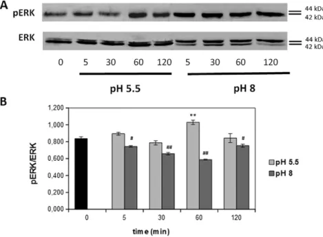
SK-MEL-28 proliferation was strongly affected by 10μM FA stimula-tion: ERK pathway was evaluated under such experimental conditions, as this intracellular signaling cascade is known to play a key role in tumor proliferation (Rizzi et al., 2014; Aĭzenshtadt et al., 2012; Chuang et al., 2000).
As shown inFig. 4, stimulation with 10μM FA in simulated acidic sweat resulted in a significantly transient increase in ERK activity (ap-proximately 50% increase, pb 0.001), with an activation peak after 5 min stimulation, as highlighted by pERK/ERK ratio (Fig. 4, light gray bars). On the other hand, when FA was diluted in basic artificial sweat, ERK activation peak (approximately 50% increase, pb 0.001) appeared later, starting after 60 min stimulation (Fig. 4, dark gray bars). Also for melanoma cells, ERK activation profile closely retraced the results ob-served in cell proliferation studies: the higher increase in cell prolifera-tion occurred after acid stimulaprolifera-tion related to an early and strong increase in ERK phosphorylation (reaching the maximum peak after 5 min stimulation) compared to basal condition. The lower proliferation increase observed after alkaline stimulation, instead, correlates with a late and sustained ERK activation (peaking after 60 min and re-maining also after 120 min), thus giving a possible explanation for observed reduced effect on cell proliferation when compared with acid stimulation.
Fig. 1. Effects of formaldehyde (FA) on HaCaT cells proliferation. A) representative images (magnification 4×) of control and FA treated cells. B) quantification of FA diluted in acidic (pH = 5.5, light gray bars) or basic (pH = 8, dark gray bars) simulated sweat effects on normal human keratinocytes cell proliferation. Results represent the mean value of three independent experiments and are expressed as mean values ± standard error of the mean (S.E.M.). ** pb 0.01; # p b 0.05; ## p b 0.01.
4. Discussion
The skin is in touch with clothes for a long time: clothing thus repre-sents an often ignored source of potentially noxious substances as many chemicals that are potentially harmful to human health may remain on textiles as manufacturing by-products and can be released upon sweat extraction. Thanks to its aqueous nature, sweat can induce chemical sol-ubilization of such compounds, making them more easily accessible to cells. Among the potentially noxious manufacturing by-products that can be released by textiles there is formaldehyde, which is widely used in textile industry in anti-crease treatments (De Groot et al., 2010). Many chemical manufacturing by-products found in textiles, such as formaldehyde, are classified as potentially carcinogenic to humans, but only some countries have mandatory legal guidelines regulating their levels in clothes and accessories. Considering formaldehyde, only some countries (i.e. Japan and Finland) have legally limited FA content for textile fabrics to reduce the risk of contact dermatitis and other ad-verse effects among their population (De Groot et al., 2010). To bridge such regulatory gap, some standards or voluntary labels (i.e. European Ecolabel, the National Technical Report UNI/TR 11359) and private marks (Oeko-Tex, Bluesigned, Aafa RLS, and others) have introduced limits to regulate their presence in thefinal textile products.
The present study aims at evaluating the effects of formaldehyde luted in simulated sweat prepared according to UNI EN ISO 105-E04 di-rective on cell proliferation and ERK signaling pathway activation in normal human keratinocytes and in a human malignant melanoma cell line.
In a previous study (Rizzi et al., 2014), it has been demonstrated that neutral pH aqueous FA solutions were able to increase cell proliferation and ERK activation in two differently invasive melanoma cell lines.
In the present study attention was focused on normal human keratinocytes (HaCaT cells) and on a highly invasive melanoma cell line (SK-MEL-28) in order to evaluate whether there are any differences in FA induced cellular responses in normal and tumorigenic cells.
To reach a more physiological experimental environment, formalde-hyde was diluted in artificial fluids mimicking sweat composition under different physiological conditions. In fact, exocrine sweat is known to be characterized by an acid pH whereas apocrine sweat is characterized by an alkaline pH (Fishberg and Bierman, 1932).
Formaldehyde is the simplest aldehyde, as it is composed of one car-bon, one oxygen and two hydrogen atoms and, because of its small size, can quickly penetrate cell walls and membranes. From a chemical point of view, it is an electrophilic molecule that can be attacked by various nucleophilic species of biological interest.
Living organisms can interact with formaldehyde of both endoge-nous or exogeendoge-nous origin, mediating different effects at cellular level, depending on its dose (Szende and Tyihák, 2010).
In vitro, FA reacts with a plenty of functional groups from both pro-teins and DNA,finally resulting in different chemical modifications, whose nature, yield and half-life strongly depend on reaction conditions (i.e. pH, temperature) (Hoffman et al., 2015).
In vitro experiments with analytically pure formaldehyde (0.1– 10 mM) showed a clear dose dependence in cellular effects. Tyihák and coworkers reported that high FA concentrations (10 mM) cause a high degree of cellular damage, moderate concentrations (1 mM) in-crease apoptosis, whereas low concentrations (0.1 mM) are able to en-hance cell proliferation and reduce apoptosis. Interestingly, these authors noticed that tumor cells seem to be more sensitive than normal cells to FA effects (Szende and Tyihák, 2010; Tyihák et al., 2001).
Formaldehyde rapidly crosses biological membranes. As it has been demonstrated that it can efficiently bind and block protein functionality of viruses, it is reasonable to assume that it can work equally well on cel-lular systems, protein complexes and associated biological processes (Suterland et al., 2008).
Chemically, formaldehyde acts as a bifunctional cross-linking agent, able to inactivate, stabilize or immobilize proteins, without perturbing their tertiary structure,finally resulting in quite stable in vivo modifica-tions, with cross-link half-lives of ~10–20 h depending on the cell type and experimental conditions (Hoffman et al., 2015; Metz et al., 2004).
Depending on the peptide sequence, formaldehyde-induced chemi-cal modifications could be represented by the reversible formation of methylol groups and Shiff bases or the more stable methylene groups. The formation of such modifications is influenced by various factors, among which local environment and pH play a key role (Tulpule and Dringen, 2013; Metz et al., 2004; Conaway et al., 1996; Steinhardt et al., 1946).
Formaldehyde not only reacts with proteins but also with DNA, resulting in the formation of crosslinked DNA-protein adducts, a well
Fig. 2. Formaldehyde (FA) induced ERK phosphorylation in HaCaT cells. A) representative western blot images obtained by stimulating the cells with 10μM FA for different times. B) densitometric quantification of ERK phosphorylation. Results represent the mean value obtained from three independent experiments and are expressed as mean values ± standard error of the mean (S.E.M). Light gray bars represent acidic stimulation while dark gray bars represent FA stimulation at basic pH. ** pb 0.01; # p b 0.05; ## p b 0.01.
109 M. Rizzi et al. / Toxicology in Vitro 37 (2016) 106–112
known abnormality in the early carcinogenic process. As low concentra-tions of FA were shown to be able to increase cell proliferation, such ef-fect could induce the accumulation of cells bearing abnormal DNA-proteins crosslinks, thus enhance its genotoxicity and carcinogenicity (Szende and Tyihák, 2010).
As an increase in cell proliferation is known to be a critical factor in chemical-induced carcinogenesis (Starr and Gibson, 1985), in the pres-ent study attpres-ention has been focused on FA ability to differpres-ently affect normal and tumor cell responses. As shown in the results section, low concentrations (10μM) of formaldehyde diluted in both acidic or basic simulated sweat were not able to increase normal human keratinocytes proliferation. In addition, it has been observed that the higher concentration tested (25 and 50μM) were able to induce a toxic response.
On the other hand, when FA diluted in both acidic or basic simulated sweat was tested at the lower concentration (10μM) on SK-MEL-28 cells, it was shown to increase cell proliferation, with a bigger increase in acidic conditions. The higher concentrations tested (25 and 50μM), indeed, did not significantly reduce cell proliferation compared to con-trol in both acidic and basic pH conditions.
Because of these results, ERK signaling pathway activation has been studied. ERK is an upstream activator ofN150 substrates involving such signaling cascade in the regulation of critical cellular responses, includ-ing transcription, translation, mitosis and apoptosis (Brys et al., 2016). In particular, ERK pathway is strongly involved in regulating cellular re-sponses such as mitogenesis and differentiation, as it has been demon-strated that its transient activation mediates cell proliferation, whereas its persistent activation mediates cell growth arrest and differentiation (Chuang et al., 2000).
Data shown in the results section correlate acidic FA (10μM) in-duced increase in SK-MEL-28 proliferation with an early transient
activation of ERK signaling pathway, thus confirming the previously de-scribed formaldehyde involvement in its activation in various normal and tumor cellular models (Rizzi et al., 2014; Aĭzenshtadt et al., 2012; Chuang et al., 2000).
Alkaline FA (10μM) induced increase in SK-MEL-28 proliferation was lower than the effect observed after acidic stimulation and ERK ac-tivity peak appeared later than at pH 5.5: it could be hypothesized that under these conditions such tardive signaling pathway activation was not sufficient to induce a greater increase in proliferation.
On the other hand, HaCaT cells stimulation with formaldehyde (10μM) at both acidic or alkaline pH did not result in a statistically sig-nificant increase in cell proliferation, even if ERK signaling pathway is activated. The lack of a clear effect on cell proliferation in this cell line might be due to an insufficient activation of the signaling pathway, as the model of normal human keratinocytes adopted (HaCaT cells) dis-plays a basal level of ERK activation greater than the one observed in SK-MEL-28 cells.
Melanoma is classified according to the presence of 4 significantly mutated genes, namely BRAF, RAS, NF1, Triple Wild Type (Triple-WT) (Konstantinov et al., 2016). In particular, epidemiological studies highlighted that nearly half of the patients with an advanced melanoma diagnosis harbor a valine to glutamine substitution in codon 600 of the serine-threonine kinase BRAF (V600E phenotype) (Brys et al., 2016; Eroglu and Ribas, 2016).
Observed results correlating FA stimulation with an increase in cell proliferation are even more interesting as the malignant melanoma ex-perimental model adopted (SK-MEL-28 cells) harbors BRAF (V600E) mutation. Such hot spot mutation, in fact, enhances cellular sensitivity to MEK inhibition,finally resulting in both cyclin D1 protein expression downregulation and G1cell cycle arrest (Konstantinov et al., 2016; Kim
et al., 2012; Solit et al., 2006).
Fig. 3. Effects of formaldehyde (FA) on SK-MEL-28 cells proliferation. A) representative images (magnification 4×) of control and FA treated cells. B) quantification of FA diluted in acidic (pH = 5.5, light gray bars) or basic (pH = 8, dark gray bars) simulated sweat effects on human malignant melanoma cell proliferation. Results represent the mean value of three independent experiments and are expressed as mean values ± standard error of the mean (S.E.M.). *** pb 0.001; # p b 0.05.
Formaldehyde metabolism is known to generate reactive oxygen species (ROS) (Pongsavee, 2013; Duong et al., 2011), known to interfere with intracellular signaling pathways activation, cell proliferation, cyto-kines and transcription factors activation and even apoptosis (Asatiani et al., 2011). Moreover, studies on neural cells showed that FA is able to interfere with glutathione (GSH) cycle, increasing its export from the cells and thus increasing oxidative stress levels (Tulpule and Dringen, 2013).
Redox state plays a key role in maintaining cell and tissues homeo-stasis and an impairment in such balance could affect cell growth and differentiation, along with cell signaling and even apoptosis (Asatiani et al., 2011). Moreover, recent studies have highlighted that cellular re-sponses to ROS vary according to their concentrations: high ROS levels mediate toxic cellular effects (i.e. protein oxidation and DNA damage) whereas low ROS levels are known to enhance cell proliferation (Valko et al., 2007; Boonstra and Post, 2004; Halliwell and Whiteman, 2004).
ROS could induce MAPK activation and in particular ERK signaling pathway activity (Son et al., 2011): such stimulation, along with chem-ical modifications that could be induced by formaldehyde treatment in cellular proteins acting as key regulators of cell survival responses may provide a plausible explanation for the observed increase in mela-noma cell proliferation.
5. Conclusion
Data presented showed that formaldehyde, a widely used aldehyde, representing a very common textile industry manufacturing by-prod-uct, could increase tumor cell proliferation. Such results not only agree with the previously described ability of neutral pH FA solutions to in-crease melanoma cells proliferation (Rizzi et al., 2014), but also show that this chemical, when diluted in simulated sweat, thus more closely mimicking the physiological condition, is able to induce a strong prolif-erative response in the highly invasive melanoma cell line SK-MEL-28.
Considering that the skin is in close contact with clothes for a long time and that sweat can extract chemical manufacturing by-products from fabrics, thus making them more easily accessible to cells, our data suggest that an even increased attention should be paid to the
potential noxious effects of such compounds, in order to define safety cut-offs to be applied to production processes.
Transparency document
TheTransparency documentassociated with this article can be found, in online version.
Acknowledgments
The study was supported by University Local Funds (fondi di ateneo 2015).
The authors are deeply thankful to Ms. Mariangela Fortunato for her precious help with the manuscript.
References
Aĭzenshtadt, A.A., Burova, E.B., Zenin, V.V., Bubkov, D.E., Kropacheva, I.V., Pinaev, G.P., 2012.Effect of formaldehyde at low concentration on proliferation and organization of cytoskeleton of cultured cells. Cell Tissue Biol. 6, 147–153.
Asatiani, N., Kartvelishvili, T., Abuladze, M., Asanishvili, L., Sapojnikova, N., 2011. Chromi-um (VI) can activate and impair antioxidant defense system. Biol. Trace Elem. Res. 142, 388–397.
Boonstra, J., Post, J.A., 2004.Molecular events associated with reactive oxygen species and cell cycle progression in mammalian cells. Gene 337, 1–13.
Bosetti, C., McLaughlin, J.K., Tarone, R.E., Pira, E., La Vecchia, C., 2008.Formaldehyde and cancer risk: a quantitative review of cohort studies through 2006. Ann. Oncol. 19, 29–43.
Boukamp, P., Petrussevska, R.T., Breitkreutz, D., Hornung, J., Markham, A., Fusening, N.E., 1988.Normal keratinization is a spontaneously immortalized aneuploidy human keratinocyte cell line. J. Cell Biol. 106, 761–771.
Brys, A.K., Gowda, R., Loriaux, D.B., Robertson, G.P., Mosca, P.J., 2016. Nanotechnology-based strategies for combating toxicity and resistance in melanoma therapy. Biotechnol. Adv.http://dx.doi.org/10.1016/j.biotechadv.2016.01.004.
Buommino, E., Baroni, A., Canozo, N., Petrazzullo, M., Nicoletti, R., Vozza, A., Tufano, M.A., 2009.Artemisin reduces human melanoma cell migration by down-regulatingαVβ3 integrin and reducing metalloproteinase 2 production. Invest. New Drugs 27, 412–418.
Chuang, S.M., Liou, G.Y., Yang, Y.L., 2000.Activation of JNK, p38 and ERK mitogen-activat-ed protein kinases by chromium(VI) is mmitogen-activat-ediatmitogen-activat-ed through oxidative stress but does not affect cytotoxicity. Carcinogenesis 21, 1491–1500.
Conaway, C.C., Whysner, J., Verna, L.K., Williams, G.M., 1996.Formaldehyde mechanistic data and risk assessment: endogenous protection from DNA adduct formation. Pharmacol. Ther. 71, 29–55.
Fig. 4. Formaldehyde (FA) induced ERK phosphorylation in SK-MEL-28 cells. A) representative western blot images obtained by stimulating the cells with 10μM FA for different times. B) densitometric quantification of ERK phosphorylation. Results represent the mean value obtained from three independent experiments and are expressed as mean values ± standard error of the mean (S.E.M). Light gray bars represent acidic stimulation while dark gray bars represent FA stimulation at basic pH. ** pb 0.01; *** p b 0.001; ## p b 0.01; ### p b 0.001.
111 M. Rizzi et al. / Toxicology in Vitro 37 (2016) 106–112
De Groot, A.C., Le Coz, C.L., Lensen, G.J., Flyvholm, A.A., Maibach, H.I., Coenraads, P.J., 2010.
Formaldehyde-releasers: relationship to formaldehyde contact allergy. Formalde-hyde-releasers in clothes: durable press chemicalfinishes. Part 1. Contact Dermatitis 62, 259–271.
Duong, A., Steinmaus, C., McHale, C.M., Vaughan, C.P., Zhang, L., 2011.Reproductive and developmental toxicity of formaldehyde: a systematic review. Mutat. Res. 728, 118–138.
Eroglu, Z., Ribas, A., 2016.Combination therapy with BRAF and MEK inhibitors for mela-noma: latest evidence and place in therapy. Ther. Adv. Med. Oncol. 8, 48–56.
Fishberg, E.H., Bierman, W., 1932.Acid-base balance in sweat. J. Biol. Chem. 97, 433–441.
Freick, P., Haas, S.R.L., Singer, M.V., Böcker, U., 2006.Low-dose exposure of intestinal ep-ithelial cells to formaldehyde results in MAP kinase activation and molecular alter-ation of the focal adhesion protein paxillin. Toxicology 219, 60–72.
Grimstad, O., Pukstad, B., Stenvik, J., Espevik, T., 2012.Oligodesoxynucleotides inhibit toll-like receptor 3 mediated cytotoxicity and CXCL8 release in keratinocytes. Exp. Dermatol. 21, 7–12.
Halliwell, B., Whiteman, M., 2004.Measuring reactive species and oxidative damage in vivo and in cell culture: how should you do it and what do the results mean. Br. J. Pharmacol. 142, 231–255.
Hoffman, E.A., Frey, B.L., Smith, L.M., Auble, D.T., 2015.Formaldehyde crosslinking: a tool for the study of chromatin complexes. J. Biol. Chem. 290, 26404–26411.
Jeong, J.B., Hong, S.C., Koo, J.S., Jeong, H.J., 2011.Induction of apoptosis and acetylation of histone H3 and H4 by arctigenin in the human melanoma cell line SK-MEL-28. Food Nutr. Sci. 2, 128–132.
Kim, Y.K., Ahn, S.K., Lee, M., 2012.Differential sensitivity of melanoma cell lines with dif-fering B-Raf mutational status to the new oncogenic B-Raf kinase inhibitor UI-152. Cancer Lett. 320, 215–224.
Köllisch, G., Kalali, B.N., Voelcker, V., Wallich, R., Behrendt, H., Ring, J., Bauer, S., Jakob, T., Mepel, M., Ollert, M., 2005.Various members of the toll-like receptor family contrib-ute to the innate immune response of human epidermal keratinocytes. Immunology 114, 531–541.
Konstantinov, N.K., Ulff-Møller, C.J., Dimitrov, S., 2016. Histone variants and melanoma: facts and hypoteses. Pigment Cell Melanoma Res. 29, 426–433.http://dx.doi.org/10. 1111/pcmr.12467.
Ma, J., Han, H., Liu, D., Li, W., Feng, X., Xue, X., Wu, X., Niu, G., Zhang, G., Zhao, Y., Liu, C., Tao, H., Gao, B., 2013.HER2 as a promising target for cytotoxicity T cells in human melanoma therapy. PLoS One 8, e73261.
Metz, B., Kersten, G.F.A., Hoogerhout, P., Brugghe, H.F., Timmersmans, H.A.M., de Jong, A., Meiring, H., ten Hove, J., Hennink, W.E., Crommelin, D.J.A., Jiskoot, W., 2004. Identifi-cation of formaldehyde-induced modifiIdentifi-cations in proteins. J. Biol. Chem. 279, 6235–6243.
Mueller, D.V., Bosserhoff, A.K., 2009.Role of miRNAs in the progression of malignant mel-anoma. Br. J. Cancer 101, 551–556.
Naya, M., Nakanishi, L., 2005.Risk assessment of formaldehyde for the general population in Japan. Regul. Toxicol. Pharmacol. 43, 232–248.
Palunic, J., Kovacevic, Z., Jansson, P.J., Kalinowski, D., Merlot, A.M., Huang, M.L.H., Lok, H.C., Sahni, S., Lane, D.J.R., Richardson, D.R., 2016.Roads to melanoma: key pathways and emerging palyers in melanoma progression and oncogenic signaling. Biochim. Biophys. Acta 1863, 770–784.
Pongsavee, M., 2013.Changes to oxidative stress levels following exposure to formalde-hyde in limphocytes. World Acad. Sci. Eng. Technol. 79, 1538–1540.
Renò, F., Rizzi, M., Invernizzi, M., Migliario, M., Cisari, C., 2013.Low doses amino-bisphosphonates stimulate keratinocyte growth inactivating glucocorticoid receptor. Eur. J. Pharm. 721, 301–304.
Rizzi, M., Cravello, B., Renò, F., 2014.Textile industry manufacturing by-products induce human melanoma cell proliferation via ERK 1/2 activation. Cell Prolif. 47, 578–586.
Sakaguchi, H., Miyazawa, M., Yoshida, Y., Ito, Y., Suzuki, H., 2007.Prediction of preserva-tive sensitization potential using surface marker CD86 and/or CD54 expression on human cell line THP-1. Arch. Dermatol. Res. 298, 427–437.
Singh, B.P., Salama, A.K.S., 2016. Updates in therapy for advanced melanoma. Cancers.
http://dx.doi.org/10.3390/cancers8010017.
Solit, D.B., Garraway, L.A., Pratilas, C.A., Sawai, A., Getz, G., Basso, A., Ye, Q., Lobo, J.M., She, Y., Osman, I., Golub, T.R., Sebolt-Leopold, J., Sellers, W.R., Rosen, N., 2006.BRAF muta-tion predicts sensitivity to MEK inhibimuta-tion. Nature 439, 358–362.
Son, Y., Cheong, Y.K., Kim, N.H., Chung, H.T., Kang, D.G., Pae, H.O., 2011.Mitogen-activated protein kinases and reactive oxygen species: how can ROS activate MAPK pathways? J. Signal. Transduct. 2011, 792639.
Starr, T.B., Gibson, J.E., 1985.The mechanistic toxicology of formaldehyde and its implica-tions for quantitative risk estimation. Ann. Rev. Pharmacol. Toxicol. 25, 745–767.
Steinhardt, J., Fugitt, C.H., Harris, M., 1946.The effect of formaldehyde on acidic and basic properties of wool. J. Biol. Chem. 165, 285–291.
Suterland, B.W., Toewa, J., Kast, J., 2008.Utility of formaldehyde cross-linking and mass spectrometry in the study of protein-protein interactions. J. Mass Spectrom. 43, 699–715.
Szende, B., Tyihák, E., 2010.Effect of formaldehyde on cell proliferation and death. Cell Biol. Int. 34, 1273–1282.
Tulpule, K., Dringen, R., 2013.Formaldehyde in brain: an overlooked player in neurode-generation? J. Neurochem. 127, 7–21.
Tyihák, E., Bocsi, J., Timár, F., Rácz, G., Szende, B., 2001.Formaldehyde promotes and in-hibits the proliferation of cultured tumor and endothelial cells. Cell Prolif. 34, 135–141.
Valko, M., Leibfritz, D., Moncol, J., Cronin, M.T.D., Mazur, M., Telser, J., 2007.Free radicals and antioxidants in normal physiological functions and human disease. Int. J. Biochem. Cell Biol. 39, 44–84.