ARTICLE OPEN ACCESS
“Better explanations” in multiple sclerosis
diagnostic workup
A 3-year longitudinal study
Massimiliano Calabrese, MD, Claudio Gasperini, MD, Carla Tortorella, MD, Gianmarco Schiavi, MD, Giovanni Frisullo, MD, Paolo Ragonese, MD, Roberta Fantozzi, MD, Luca Prosperini, MD, Pietro Annovazzi, MD, Cinzia Cordioli, MD, Massimiliano Di Filippo, MD, Diana Ferraro, MD, Alberto Gajofatto, MD,
Simona Malucchi, MD, Salvatore Lo Fermo, MD, Giovanna De Luca, MD, Maria L. Stromillo, MD, Eleonora Cocco, MD, Antonio Gallo, MD, Damiano Paolicelli, MD, Roberta Lanzillo, MD, Valentina Tomassini, MD, Ilaria Pesci, MD, Maria E. Rodegher, MD, and Claudio Solaro, MD, the RIREMS group (Rising Italian Researchers in Multiple Sclerosis)
Neurology
®
2019;92:e2527-e2537. doi:10.1212/WNL.0000000000007573Correspondence Prof. Calabrese massimiliano.calabrese@ univr.it
Abstract
BackgroundThe exclusion of other diseases that can mimic multiple sclerosis (MS) is the cornerstone of current diagnostic criteria. However, data on the frequency of MS mimics in real life are incomplete.
Methods
A total of 695 patients presenting with symptoms suggestive of MS in any of the 22 RIREMS centers underwent a detailed diagnostic workup, including a brain and spinal cord MRI scan, CSF and blood examinations, and a 3-year clinical and radiologic follow-up.
Findings
A total of 667 patients completed the study. Alternative diagnoses were formulated in 163 (24.4%) cases, the most frequent being nonspecific neurologic symptoms in association with atypical MRI lesions of suspected vascular origin (40 patients), migraine with atypical lesions (24 patients), and neuromyelitis optica (14 patients). MS was diagnosed in 401 (60.1%) patients according to the 2017 diagnostic criteria. The multivariate analysis revealed that the absence of CSF oligoclonal immu-noglobulin G bands (IgG-OB) (odds ratio [OR] 18.113), the presence of atypical MRI lesions (OR 10.977), the absence of dissemination in space (DIS) of the lesions (OR 5.164), and normal visual evoked potentials (OR 3.550) were all independent predictors of an alternative diagnosis.
Interpretation
This observational, unsponsored, real-life study, based on clinical practice, showed that diseases that mimicked MS were many, but more than 45% were represented by nonspecific neurologic symptoms with atypical MRI lesions of suspected vascular origin, migraine, and neuromyelitis optica. The absence of IgG-OB and DIS, the presence of atypical MRI lesions, and normal visual evoked potentials should be considered suggestive of an alternative disease and redflags for the misdiagnosis of MS.
RELATED ARTICLE Editorial
Differential diagnosis of multiple sclerosis: The better explanations in clinical practice Page 1037 MORE ONLINE CME Course NPub.org/cmelist
From the Departments of Neuroscience, Biomedicine and Movement (M.C., A. Gajofatto) and Neurological and Movement Sciences (G.S.), University of Verona; Department of Neurosciences (C.G., C.T.), Azienda Ospedaliera San Camillo Forlanini, Roma; Department of Basic Medical Sciences, Neurosciences and Sense Organs (C.T., D.P.), University of Bari; Policlinico Gemelli (G.F.), Rome; Dipartimento di Biomedicina Sperimentale e Neuroscienze Cliniche (BIONEC) (P.R.), Universit`a di Palermo; Istituto Neurologico Mediterraneo (R.F.), Pozzilli; Department of Neurology and Psychiatry (L.P.), Sapienza University of Rome; Multiple Sclerosis Center (P.A.), ASST Valle Olona, PO di Gallarate; Multiple Sclerosis Center (C.C.), Ospedale di Montichiari, Spedali Civili di Brescia; Clinica Neurologica (M.D.), Dipartimento di Medicina, Universit`a di Perugia; Department of Biomedical, Metabolic and Neurosciences (D.F.), University of Modena and Reggio Emilia, Modena; Neurologia 2-CRESM (S.M.), AOU San Luigi Gonzaga, Orbassano; Multiple Sclerosis Centre (S.L.), A.O.U. Policlinico-Vittorio Emanuele, Catania; Neurology Clinic (G.D.), Multiple Sclerosis Center, SS. Annunziata Hospital, Chieti; Department of Medicine, Surgery and Neuroscience (M.L.S.), University of Siena; Department of Medical Science and Public Health (E.C.), University of Cagliari; Department of Medical, Surgical, Neurological, Metabolic and Aging Science (A. Gallo), University of Campania; Department of Neuroscience, Reproductive Sciences (R.L.), University Federico II, Naples, Italy; Institute of Psychological Medicine and Clinical Neurosciences (V.T.), Cardiff University School of Medicine, UK; Ospedale di Vaio (I.P.), Centro SM, Fidenza, Parma; Ospedale San Raffaele (M.E.R.), Milan; and Department of Rehabilitation (C.S.), Mons L Novarese Hospital, Moncrivello, Italy.
Go to Neurology.org/N for full disclosures. Funding information and disclosures deemed relevant by the authors, if any, are provided at the end of the article. The Article Processing Charge was funded by the University of Verona.
This is an open access article distributed under the terms of the Creative Commons Attribution-NonCommercial-NoDerivatives License 4.0 (CC BY-NC-ND), which permits downloading and sharing the work provided it is properly cited. The work cannot be changed in any way or used commercially without permission from the journal.
Multiple sclerosis (MS) diagnostic criteria have evolved over time with an increasing use of paraclinical markers, especially MRI,1–4to enable a definite diagnosis earlier than clinical fea-tures alone would allow.5,6 Despite technological advance-ments, current diagnostic criteria still rely on key principles of MS diagnosis articulated in the mid-20th century: demonstra-tion of disseminademonstra-tion in space and in time of demyelinating lesions and exclusion of alternative explanations.1–4,7
The 2010 and, more recently, the 2017 McDonald diagnostic criteria revision3,4simplified the diagnostic process, allowing a more rapid diagnosis.5,6However, the increasing focus on an earlier diagnosis of MS, to allow initiation of disease-modifying therapies, may increase the risk of misdiagnosis, which remains an important issue in clinical practice.7,8 In daily clinical practice, the neurologist must pay extreme at-tention to clinical presentation, since MS diagnostic criteria are better applicable in patients with typical presentations suggestive of MS compared to patients with atypical or nonspecific symptoms (e.g., headache, arthralgias)9
; more-over, any diagnostic tests that suggest the possibility of an alternative diagnosis, so-called redflags,10should be carefully evaluated. Interpretation of personal history, physical exami-nation, and results of imaging and laboratory testing by a cli-nician with expertise in MS remains fundamental in correctly diagnosing MS.
Nevertheless, the differentiation of MS from other MS-mimicking conditions might be difficult. This is due to the low amount of data available on the frequency of alternative di-agnoses in real life. Clinical and paraclinical information about these diseases is also poor, thus making it even more complicated to define which examinations could be useful in the diagnostic workup to confirm that “no better explanation” exists.
We therefore designed a real-life study aimed at evaluating (1) the main diseases that mimic MS at clinical onset and their frequency, (2) the most relevant clinical and paraclinical characteristics suggesting a diagnosis different from MS, and, finally, (3) the best diagnostic workup for the exclusion of other possible explanations of clinical symptoms suggest-ing MS.
Methods
Setup of a shared diagnostic workup
A restricted subgroup of participants revised the literature in order to obtain a list of blood, CSF, and instrumental
examinations needed to identify possible alternative diagnoses.10–13 Then, a shareable list of examinations to be included in the diagnostic workup was created and submitted to each center in order to obtain a consensus statement. We considered an item accepted if agreed on by at least 70% of centers. The main reasons to reject an item were (1) not available in all centers, (2) available but not extensively used in every day clinical practice, since it requires a motivated request based on a specific clinical suspicion (e.g., MOG anti-bodies), (3) too expensive. The minimum set of examinations suggested by consensus by the panel is reported in table 1. However, each center was free to exclude some examinations if considered of no use in relation to the clinical presentation. Finally, after a revision of the literature, each alternative di-agnosis was based on the most recent international guidelines, where possible (i.e., migraine, neuromyelitis optica spectrum disorder [NMOSD], Bechet disease, Sj¨ogren syndrome, sys-temic lupus erythematous, Susac syndrome, fibromyalgia)
14–20and on common clinical practice where clear guidelines
were not available. The diagnosis of nonspecific neurologic symptoms associated with atypical MRI lesions of suspected ischemic origin was formulated, on the base of the clinical practice, in those patients presenting with nonspecific (for MS) neurologic symptoms (e.g., migraine, arthralgia, and myalgia), normal neurologic examination, no CSF immuno-globulin G oligoclonal bands (IgG-OB), normal visual evoked potentials, MRI lesions atypical for MS, without spinal cord lesions, and at least one major vascular risk factor (i.e., hypertension, patent foramen ovale, hypercholesterol-emia). The diagnosis of nonspecific neurologic symptoms associated with atypical MRI lesions was reserved for those cases where no hypothesis about the origin of the MRI lesions was possible, the symptoms were not suggestive of MS, and all the other examinations were normal. The characteristics of the typical MRI lesions are reported below.
Study population
We included all patients referred to any of the 22 participating MS centers between March and September 2014 because MS was suspected, and who required a diagnostic workup, in-cluding blood, CSF, and instrumental examinations, in order to confirm the diagnosis of MS.
Inclusion criteria
The 2 main groups were as follows:
1. Patients presenting with symptoms or signs suggestive of MS.
Glossary
ANA= antinuclear antibodies; CI = confidence interval; CIS = clinically isolated syndrome; CMV = cytomegalovirus; DIS = dissemination in space; DIT = dissemination in time; ENA = extractable nuclear antigen; IgG-OB = immunoglobulin G oligoclonal bands; MS = multiple sclerosis; NMO = neuromyelitis optica; NMOSD = neuromyelitis optica spectrum disorder; OR= odds ratio; UI = under investigation; VEP = visual evoked potentials.
2. Patients presenting with atypical symptoms or signs but referred to one of the centers because MS was suspected, and who required a specialized diagnostic assessment. Usually these patients were referred to one of our MS centers by a general neurologist or by an emergency department physician (i.e., colleagues with-out specific experience in MS) and were included, following a general neurologic examination, if the hypothesis of an MS diagnosis was not immediately rejected.
Exclusion criteria
The only exclusion criterion was an already established di-agnosis of MS.
Clinical and paraclinical examinations
Information about family history, type of first clinical manifestation (motor, sensory, cortical, visual, spinal cord, or cerebellar), and the co-occurrence of atypical symptoms or signs (i.e., arthralgia, myalgia, fever, oral ulceration, xerophthalmia, xerostomia, rash, migraine, livedo retic-ularis, epilepsy, gastrointestinal disorders, signs of neu-ropathy, history of abortion, or thrombosis) were collected.
During the diagnostic assessment, each patient underwent a complete neurologic examination, assessed by means of the Expanded Disability Status Scale21 and a 1.5T brain MRI according to recent Italian guidelines.22
For each MRI lesion, the size, morphology, and location were evaluated. Oval, asymmetric white matter lesions perpendic-ular to the ventricles (Dawson fingers) or in the periven-tricular, juxtacortical, infratentorial or spinal cord region, and having a diameter >6 mm, were considered typical of MS.22,23 All other lesions were considered atypical. We also evaluated the number of contrast-enhancing lesions and the dissemi-nation in space of the lesions; IgG-OB were investigated through isoelectric focusing.24IgG-OB testing in the CSF was paired with the serum to confirm that IgG-OB were unique to CSF.
Visual evoked potentials (VEP) were performed, in accor-dance with International Society for Clinical Electrophysiol-ogy of Vision international recommendations,25and latency and morphology were recorded.
Finally, blood-based examinations were carried out in order to exclude alternative diagnoses (table 1).
Local ethics committees approved the study. The present study was conducted in accordance with specific national laws and the ethical standards laid down in the 1964 Declaration of Helsinki and its later amendments.
Study design and follow-up
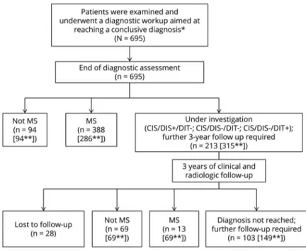
Theflowchart of the study is summarized in figure 1. At the end of the diagnostic workup, all recruited patients were classified into 3 groups according to their diagnosis: 1. MS according to the 2010 revisions of McDonald
diagnostic criteria3
2. Not MS (specification of the alternative diagnosis was required)
3. Under investigation (UI): in case a conclusive diagnosis could not be formulated, and additional follow-up was deemed necessary
The latter was further divided into the following:
3a. Clinically isolated syndrome (CIS)/dissemination in space (DIS)+: patients with a CIS and CIS of the lesions but lacking dissemination in time (DIT) of lesions and a better explanation for signs and symptoms
3b. CIS/DIS−/DIT−: patients with CIS but lacking DIS and DIT; this group included optic neuritis and myelitis (with negative anti-aquaporin-4 antibodies) with normal brain MRI scans
3c. CIS/DIS−/DIT+: patients with CIS lacking DIS but with evidence of DIT (e.g., patients with optic neuritis and 2 periventricular brain lesions, one of which enhanced by gadolinium)
Each patient without a conclusive diagnosis at the end of the initial diagnostic workup (3a, 3b, and 3c groups) underwent a clinical and radiologic 3-year follow-up, in order to assess
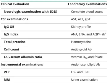
Table 1 Minimum set of examinations required to exclude alternative diagnoses in patients presenting with clinical symptoms suggestive of demyelinating diseases of the CNS
Clinical evaluation Laboratory examinations Neurologic examination with EDSS Complete blood count
CSF examinations AST, ALT, gGT
IgG-OB Kidney profile
IgG index ANA, ENA, and AQP4 aba
Total proteins Homocysteine
Cell count Antithyroid Ab
CSF/serum albumin ratio Vitamin B12and folate
Instrumental examinations Antiphospholipid Ab
VEP ESR and CRP
MRI Urine examination
Abbreviations: Ab = antibodies; ALT = alanine transaminase; ANA = antinu-clear antibodies; AQP4 = anti-aquaporin 4; AST = aspartate transaminase; CRP = C-reactive protein; EDSS = Extended Disability Status Scale; ENA = extractable nuclear antibodies; ESR = erythrocyte sedimentation rate; gGT = γ-glutamyltransferase; IgG-OB = immunoglobulin G oligoclonal bands; VEP = visual evoked potentials.
aAnti-AQP4 antibodies were mandatory in case of symptoms/signs
whether DIS or DIT occurred. During this period, each pa-tient was evaluated at least every 6 months and underwent a new MRI at least every year. At the end of the 3 years, based on the clinical and radiologic follow-up, each patient of the previous UI group was reclassified as MS, not MS (with a definitive alternative diagnosis), or still as UI. Since the 2017 revised diagnostic criteria for MS were published during the course of the study,4each diagnosis was revised, and each patient reclassified according to the new criteria.
Recurrent optic neuritides or myelitides that did not show DIS at the end of the follow-up were included in the not-MS group.
Statistical analysis
The χ2test was applied to test the effect of IgG-OB, type, number, and location of MRI lesions and VEP abnormalities in identifying MS vs not MS patients both at the end of the initial diagnostic workup and at the end of the follow-up period. For each variable, we also calculated sensitivity, specificity, and accuracy.
A logistic multivariate analysis was performed, using the oc-currence of an alternative diagnosis at the end of the study as dependent variable. As independent variables at baseline, we included the presence of atypical MRI lesions, the location of the lesions (periventricular, juxtacortical, infratentorial, spinal cord), DIS of lesions, the number of contrast-enhancing lesions, the presence/absence of IgG-OB, the VEP results, the presence of atypical symptoms, and any significantly abnor-mal blood assay.
Data availability statement
The entire dataset, including all data used in this study, and completely anonymized, is located in a Dropbox folder and will be shared following request by qualified investigators.
Results
Results after the initial diagnostic workup
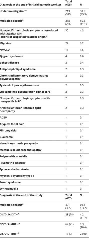
According to inclusion/exclusion criteria, 695 patients (481 female, mean age 40.6 years) were enrolled in this study. At the end of the initial diagnostic workup, an alternative di-agnosis was formulated in 94 patients (13.5%); among these, nonspecific neurologic symptoms associated with atypical MRI lesions of suspected ischemic origin (4.3% of the total di-agnoses and 31.9% of the alternative didi-agnoses) was the most frequent alternative diagnosis. Migraine associated with atypical MRI lesions (3.2% of the total diagnoses and 23.4% of the alternative diagnoses) and NMOSDs (1.6% of the total di-agnoses and 11.7% of the alternative didi-agnoses) were the other most frequent diagnoses (table 2 for more details).
According to the 2010 revision of diagnostic criteria, an MS diagnosis was possible in 286 (41.2%) patients while a con-clusive diagnosis was not reached in 315 (45.3%) patients, who remained under investigation. Among these 315 patients without a definite diagnosis, 206 showed DIS of MRI lesions (CIS/DIS+) and 102 showed both DIS and IgG-OB. Fol-lowing the publication of the 2017 revision of MS diagnostic criteria, the latter group was reclassified as MS. Therefore, among the 695 patients included in the study, the application of the 2017 revision of diagnostic criteria has allowed the
FigureStudy algorithm
*Including patients with symptoms or signs suggestive of multiple sclerosis (MS) presenting to one of the centers or patients sent to one of our centers by their physician or by the local emergency department with suspected MS. **Within the bracket number according to 2017 diagnostic criteria. CIS/DIS+ = clinically isolated syndrome with dis-semination in space of the lesions; CIS/DIS− = clinically iso-lated syndrome without dissemination in space of the lesions.
Table 2 Main diagnoses of the patients included in the study at the end of the diagnostic workup or at the end of the 3-year follow-up period
Diagnosis at the end of initial diagnostic workup Total (695) % Under investigationa 213 (315) 30.6 (45.3) Multiple sclerosisa 388 (286) 55.8 (41.1) Nonspecific neurologic symptoms associated
with atypical MRI
lesions of suspected vascular originb
30 4.3 Migraine 22 3.2 NMOSD 11 1.6 Sj¨ogren syndrome 4 0.6 Behçet disease 3 0.4 Antiphospholipid syndrome 2 0.3
Chronic inflammatory demyelinating polyneuropathy
2 0.3
Systemic lupus erythematosus 2 0.3 Subcombined degeneration spinal cord 2 0.3 Nonspecific neurologic symptoms with
nonspecific MRIc
2 0.3
Arteritic anterior ischemic optic neuropathy
2 0.3
ADEM 1 0.1
Atypical facial pain 1 0.1
Fibromyalgia 1 0.1
Glaucoma 1 0.1
Hereditary spastic paraplegia 1 0.1 Metabolic leukoencephalopathy 1 0.1
Polyneuritis cranialis 1 0.1
Psychiatric disorder 1 0.1
Spinocerebellar ataxia 1 0.1
Myotonic dystrophy type 1 1 0.1
Susac syndrome 1 0.1
Syringomyelia 1 0.1
Diagnosis at the end of the study Total (667) % Multiple sclerosisa 401 (355) 60.1 (53.2) CIS/DIS+/DIT2a 28 (78) 4.2 (11.7) CIS/DIS2/DIT2a 62 (71) 9.3 (10.6) CIS/DIS2/DIT+a 13 (0) 2.0 (0)
Table 2 Main diagnoses of the patients included in the study at the end of the diagnostic workup or at the end of the 3-year follow-up period(continued) Diagnosis at the end of initial diagnostic workup
Total (695) % Nonspecific neurologic symptoms associated
with atypical MRI
lesions of suspected vascular originb
40 6.0
Migraine with atypical MRI lesions 24 3.6
NMOSD 14 2.1
Nonspecific neurologic symptoms with atypical MRIc
10 1.5
Recurrent optic neuritis 7 1.0
Behçet disease 7 1.0 Sj¨ogren syndrome 6 0.9 ADEM 6 0.9 Psychiatric disorder 5 0.7 Antiphospholipid syndrome 5 0.7 Fibromyalgia 4 0.6
Systemic lupus erythematosus 4 0.6 Hereditary spastic paraplegia 4 0.6
Susac syndrome 4 0.6
Chronic inflammatory demyelinating polyneuropathy
3 0.4
Arteritic anterior ischemic optic neuropathy 3 0.4 Subcombined degeneration spinal cord 3 0.4
Glaucoma 2 0.3
Undifferentiated connective tissue disease 2 0.3
Atypical facial pain 2 0.3
Cerebellar hamartoma 1 0.1 Metabolic leukoencephalopathy 1 0.1 Myasthenia gravis 1 0.1 Polyneurits cranialis 1 0.1 Recurrent myelitis 1 0.1 Spinocerebellar ataxia 1 0.1
Myotonic dystrophy type 1 1 0.1
Syringomyelia 1 0.1
Abbreviations: ADEM = acute disseminated encephalomyelitis; CIS/DIS+ = clini-cally isolated syndrome with dissemination in space of the lesions; CIS/DIS− = clinically isolated syndrome without dissemination in space of the lesions; DIT = dissemination in time; NMOSD = neuromyelitis optica spectrum disorder.
aData are according to 2017 diagnostic criteria (and 2010 diagnostic
criteria).
bThis group included patients with atypical MRI lesions without IgG-OBs and
spinal cord lesions, with normal VEP and with several vascular risk factors (i.e., hypertension, patent foramen ovale, hypercholesterolemia).
cThis group included patients where no hypothesis about the origin of the
identification of 388 (55.8%) patients with MS (instead of 286, according to the 2010 criteria) while a conclusive di-agnosis was missing in only 213 (30.6%) patients (instead of 315, table 2 for more details).
Results at the end of the study
Among the 315 patients without a definite diagnosis during the initial diagnostic workup, 28 were lost to follow-up, while 287 completed a 3-year clinical and radiologic follow-up (mean 36.6 ± 4.1, range 34–40 months).
At the end of the follow-up, an alternative diagnosis was made in an additional 69 patients. Among the 667 patients who completed the study, an alternative diagnosis was formulated in 163 (24.4%) patients (figure 1 for more details). Non-specific neurologic symptoms associated with atypical MRI lesions of suspected ischemic origin (24.5% of the alternative diagnoses) and migraine associated with atypical MRI lesions (14.7% of the alternative diagnoses) were still the main al-ternative diagnoses. Table 2 summarizes the total alal-ternative diagnoses at the end of the study.
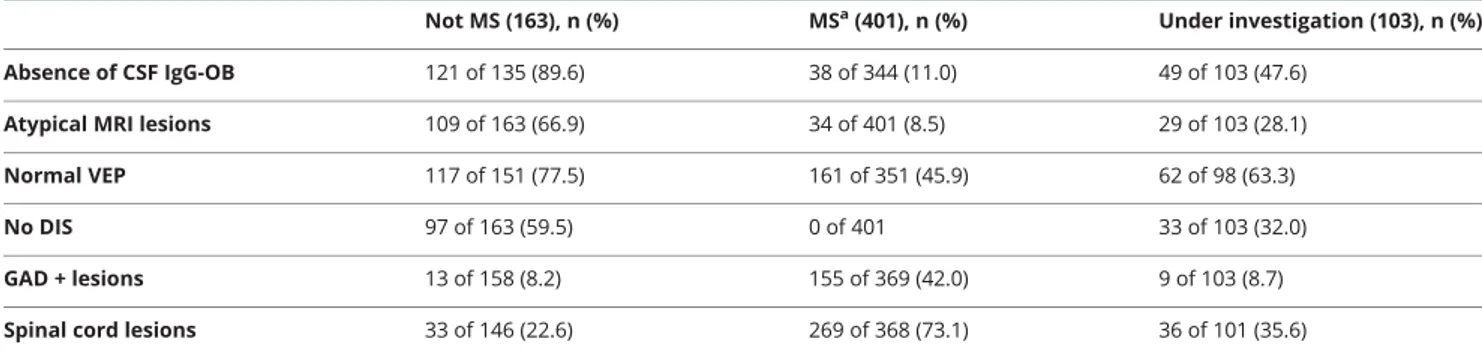
Table 3Role of the main paraclinical tests in the identification of alternative diseases
Not MS (163), n (%) MSa(401), n (%) Under investigation (103), n (%)
Absence of CSF IgG-OB 121 of 135 (89.6) 38 of 344 (11.0) 49 of 103 (47.6) Atypical MRI lesions 109 of 163 (66.9) 34 of 401 (8.5) 29 of 103 (28.1)
Normal VEP 117 of 151 (77.5) 161 of 351 (45.9) 62 of 98 (63.3)
No DIS 97 of 163 (59.5) 0 of 401 33 of 103 (32.0)
GAD + lesions 13 of 158 (8.2) 155 of 369 (42.0) 9 of 103 (8.7)
Spinal cord lesions 33 of 146 (22.6) 269 of 368 (73.1) 36 of 101 (35.6)
Abbreviations: DIS = dissemination in space of the lesions; GAD = gadolinium; IgG-OB = CSF IgG oligoclonal bands; MS = multiple sclerosis; VEP = visual evoked potentials.
aThe diagnosis of MS was formulated according to the 2017 diagnostic criteria.
Table 4Sensitivity, specificity, and predictive values of atypical MRI lesions, lack of dissemination in space (DIS) of lesions, absence of CSF oligoclonal bands, and normal visual evoked potentials (VEP), for the not multiple sclerosis (MS) diagnosis
No. of
patients Sensitivity Specificity PPV NPV Based on the patients who received a not-MS diagnosis at the end of
diagnostic workup
Presence of atypical MRI lesions 77 66.9
(62.7–71.0) 76.3(72.3–79.9) 40.8(36.6–45.3) 90.4(87.5–92.8) No MRI DIS 91 59.5 (55.2–63.9) 75.4 (71.3–79.0) 35.4 (31.3–39.8) 89.1 (86.0–91.6) Normal VEP 84 77.5 (76.2-79-0) 45.1 (43.2–48.8) 17.9 (15.1–19.3) 92.5 (90.1–95.2) Absence of CSF IgG-OB 67 89.6 (86.5–91.7) 69.8 (66.2–73.2) 31.6 (27.0–35.6) 97.7 (96.2–98.6) Based on the patients who received a not-MS diagnosis at the end of
3-year follow-up
Presence of atypical MRI lesions 46 73.9
(70.1–75.3) 66.7 (63.8–68.9) 69.4 (67.3–71.8) 70.4 (68.4–73.2) No MRI DIS 63 67.2 (65.2–70.0) 81.5 (79.7–83.6) 100 64.8 (62.3–67.7) Normal VEP 56 64.3 (62.1–67.6) 38.4 (36.3–40.1) 80.6 (78.6–82.9) 20.2 (18.3–23.1) Absence of CSF IgG-OB 51 80.4 (78.2–82.1) 62.0 (60.1–64.3) 79.1 (77.3–81.5) 68.8 (66.1–70.9) Abbreviations: IgG-OB = immunoglobulin G oligoclonal bands; NPV = negative predictive value; PPV = positive predictive value.
The diagnosis of MS was made according to the 2010 di-agnostic criteria in 355 patients, while in 149 patients it was not possible to reach a conclusive diagnosis. Among these, 78 patients showed only DIS of MRI lesions (CIS/DIS+/ DIT−) while 71 did not show DIS or DIT (CIS/DIS−/ DIT−). There were no CIS/DIS−/DIT+ patients. How-ever, following the application of the 2017 revision of di-agnostic criteria, 401 patients were reclassified as MS, whereas only 103 were still under investigation. Among these 103 patients, 28 patients showed only DIS of MRI lesions (CIS/DIS+/DIT−), 62 did not show DIS or DIT (CIS/DIS−/DIT−), and 13 showed only DIT (CIS/DIS−/ DIT+) (table 2).
The role of clinical presentation and paraclinical tests
Among the 695 patients included in the study, 653 patients presented with symptoms suggestive of MS (168 sensory, 145 visual, 127 brainstem or cerebellar, 101 motor, 21 cor-tical, 91 multifocal), 42 patients presented only with atypical symptoms, and 92 patients showed concomitant typical and atypical symptoms. The main atypical symptoms were mi-graine, arthralgia, and myalgia. No significant difference in the type of thefirst clinical manifestation or in the presence of atypical symptoms was observed between groups (data not shown).
The CSF was examined in 593 (85.3%) patients, VEPs were collected in 610 (87.8%) patients, brain MRI was collected in all patients, and the complete set of laboratory exami-nations was carried out in 602 (86.7%) patients. In addi-tion, gadolinium was administered in 635 (91.3%) patients and spinal cord MRI was performed in 623 (89.6%) patients.
The results of the main paraclinical tests in the 3 groups (MS, not MS, and UI) at the end of the study are reported in table 3. The sensitivity and specificity of the main par-aclinical tests are reported in table 4. The presence of atypical MRI lesions and the absence of DIS and of CSF IgG-OB showed the highest accuracy in the identification of not MS patients (table 4 for more details). The re-lationship between the site of MRI lesions and the final diagnosis is reported infigure e-1 (doi.org/10.5061/dryad. 33770ms).
The role of hematologic, immunologic, and rheumatologic screening
Among the blood tests performed in the not MS group of patients, abnormal results were present in 67 patients: ab-normal values of antinuclear antibodies/extractable nuclear antigen (ANA/ENA) antibodies (>1:640 dilutions) were observed in 13 and anti-aquaporin-4 antibodies in 12 patients, homocysteine was increased in 10 patients, anti-thyroid antibodies were observed in 9, and antiphospholipid anti-bodies in 8 patients; abnormally low levels of B12and folate
were observed in 8 patients and antineutrophil cytoplasmic
antibodies and immunoglobulin M antibodies against Epstein-Barr virus, cytomegalovirus (CMV), varicella-zoster virus, herpes simplex virus 1, measles, and rubella in 1 patient each.
The abnormal results of blood-based examinations were crucial for the conclusive diagnosis only in 24 cases: anti-aquaporin-4 antibodies drove the diagnosis in 12 of the 14 neuromyelitis optica (NMO) cases, increased levels of homocysteine (6 cases) and the presence of antiphospholipid antibodies (2 cases) were helpful in the diagnosis of non-specific neurologic symptoms associated with atypical MRI lesions of suspected vascular origin and antiphospholipid syndrome, respectively; the evidence of low vitamin B12levels
was helpful in the identification of 2 cases of subacute com-bined degeneration of the spinal cord, while the presence of ANA/ENA antibodies was helpful in 2 cases of systemic lupus erythematosus.
Predictors of alternative diagnoses
The multivariate analysis revealed that the absence of IgG-OBs (p <given-names> 0.001, odds ratio [OR] 18.113, 95% confidence interval [CI] 15.123–21.463), the presence of atypical MRI lesions (p <given-names> 0.001, OR 10.977, 95% CI 9.011–13.103), the absence of DIS of the lesions (p = 0.002, OR 5.164, 95% CI 3.226–7.338), and normal visual evoked potentials (p = 0.008, OR 3.550, 95% CI 2.011–5.121) were all independent predictors of an alterna-tive diagnosis and should be considered red flags for the misdiagnosis of MS. Of note, the presence of abnormal blood examinations did not reach statistical significance.
Discussion
The concept of no better explanation is a key element of all MS diagnostic criteria revisions1–4,10; however, little has been done in recent years to gather information on diseases that are part of the differential diagnosis of MS in real life. Diagnostic criteria were created to predict the development of MS in patients with a CIS suggestive of inflammatory demyelination and, therefore, with a clinical presentation typical for MS. When used in a different context, however, these criteria might not perform as well in distinguishing MS from other disorders since they could be fulfilled by several other neu-rologic diseases, leading to possible misdiagnoses and in-appropriate treatments.
The European Magnetic Resonance Network in MS (MAG-NIMS) expanded the criterion of the no better explanation over a decade ago by establishing, mostly on the basis of literature, some red flags that should help clinicians in the differential diagnosis of MS.10 Recently, the same group
suggested a diagnostic algorithm13that incorporates features that have been identified as useful in differentiating MS from NMOSD and imaging features that suggest alternative di-agnoses in the current MS diagnostic criteria. This review
summarized several new developments in the MS imaging field that have occurred in the last decade; it described the coexistence of age-related changes and vascular diseases that have been recognized in patients with MS and pose major diagnostic challenges;finally, it highlighted those features that distinguish MS from the newly recognized antibody-mediated syndromes of NMOSD and acute demyelinating encephalo-myelitis. The authors stated that several challenges will be faced in the near future to differentiate MS from all those diseases that can mimic it clinically and radiologically. They concluded that real-world studies on this topic are necessary and desirable.
The frequency of MS mimics in real life and, hence, their relevance and effect on everyday clinical practice has remained largely unexplored. Consequently, a precise and evidence-based diagnostic workup aimed at the exclusion of other diseases mimicking MS has not yet been developed.
To better define the concept of no better explanation, we performed a prospective longitudinal observational study that involved 22 highly specialized MS centers. Since this is a real-life study we decided to include all consecutive patients who required a diagnostic assessment to confirm/ exclude the diagnosis of MS: this included patients with a CIS suggestive of MS but also patients without typical MS signs or symptoms referred to one of our centers with a suspicion of MS based on the evaluation of a general physician or a non-MS neurologist.
We also defined, by consensus, the minimum set of laboratory/paraclinical examinations that should be per-formed in patients enrolled in the study, in order to ex-clude most common better explanations of the clinical presentation (table 1). Since this is a real-life study, each clinician was still free to exclude some examinations and to include additional examinations, according to the clinical presentation. For example, as reported in table 1, anti-aquaporin-4 antibodies were searched only in case of optic neuritis or myelitis. This is why not all the examinations are available for each patient. Nevertheless, the majority of the patients underwent a comprehensive battery of blood tests. Finally, since the current diagnostic criteria at the time of this study were those described in 2011 by Polman et al.,3 we decided to describe the classification of each patient on the basis of both the 20103and the 20174 di-agnostic criteria.
Thefirst result of this study was, therefore, a portrait of the pathologic conditions that mimic MS in the everyday clinical practice of highly specialized MS centers: among these, nonspecific neurologic symptoms associated with atypical MRI lesions of suspected ischemic origin, mi-graine, and NMOSD are by far the most common. Re-garding these diagnoses, it has to be underlined that, differently from NMOSD and migraine, for which specific diagnostic guidelines are available, the diagnosis of
nonspecific neurologic symptoms (i.e., a CIS with symp-toms not suggestive of MS) associated with atypical MRI lesions of suspected ischemic origin is mainly based on common clinical practice. In order to make this diagnosis more consistent across the centers, we asked each center to include in this group only patients with at least one major vascular risk factor and no evidence of MS or other diseases.
Our results are mostly in line with those of a previous study7 aimed at characterizing patients misdiagnosed with MS: as in our study, migraine, NMOSD, CIS, and nonspecific MRI changes were among the most frequent alternative diagnoses. The main differences between the 2 studies refer to the fre-quency of functional disorders andfibromyalgia, which could be explained by differences in the recognition of these dis-orders as well as patient enrollment procedures. As also highlighted by Solomon et al.,7,8 fibromyalgia was reported significantly more often by the Mayo Clinic neurologists than by other centers.
The second interesting result was the observation that in 30% of the cases, a conclusive diagnosis was not reached at the end of the initial diagnostic workup even after applying 2017 revised MS diagnostic criteria.4 Moreover, after extending the clinical and radiologic follow-up up to 3 years, we still had 15% of participants without a definitive di-agnosis. As expected, 2010 diagnostic MS criteria performed significantly worse than 2017 criteria, with more than 20% of patients still without a diagnosis at the end of the fol-low-up.
From a clinical point of view, and in order to define the best diagnostic workup for those patients with a CIS suggestive of MS, it is of interest to analyze the sensitivity and specificity of the radiologic and immunologic examinations currently used in clinical practice.
The presence of atypical lesions, or lesions located in the subcortical or in the infratentorial white matter and the absence of IgG-OB, characterized the 163 patients who received an alternative diagnosis. A similar finding was reported for the CIS/DIS− patients. This result was con-firmed by the multivariate analysis run in the followed up subjects that showed that the absence of CSF IgG-OB, DIS of the lesions, and the presence of atypical MRI lesions were the most relevant predictors of an alternative di-agnosis, and should be considered red flags for the mis-diagnosis of MS.
As far as the blood tests are concerned, the presence of abnormal examinations did not reach statistical significance at the multivariate analysis. This is in line with previous evidence showing the inconsistent significance for con-version to clinically definite MS of blood tests such as EBNA1 and CMV IgG dosages.26 We observed very few cases in which one or more blood tests contributed to the
final diagnosis, with the exception of anti-aquaporin-4 antibodies that allowed the identification of several NMO cases, as already reported.27We do not suggest, therefore, a comprehensive battery of blood tests in all patients with clinical and radiologicfindings suggestive of MS, but only in those patients showing redflags suggestive of alternative diagnosis.
This study is not without limitations. The ascertainment bias could be thefirst limitation of this study. This is why, in order to limit its effect, we included both patients presenting directly to one of the MS centers with symptoms or signs suggestive of MS and patients presenting with atypical symptoms or signs who were referred to one of our MS centers by the general physician or by non-MS neurologist. Moreover, the entire battery of blood-based or paraclinical examinations was not performed in all patients. Although a minimum set of laboratory/paraclinical examinations was highly recom-mended, as all centers had very good expertise on MS, they were free to exclude or add examinations in relation to patients’ manifestations. The third possible limitation con-cerns the MRI protocol, which was not standardized across the centers. However, it has to be pointed out that all centers followed recent Italian guidelines15; this guarantees a suffi-cient quality of the images that were also comparable between centers.
Finally, as already acknowledged, the 3-year clinical and ra-diologic follow-up was not enough to make afinal—MS/no MS—diagnosis in all subjects. We will therefore continue to follow-up all patients without a conclusive diagnosis. This observational, unsponsored, real-life study, based on common clinical practice, showed that, among diseases mimicking MS, nonspecific neurologic symptoms associated with atypical MRI lesions of suspected vascular origin, mi-graine associated with atypical MRI lesions, and NMOSD were the main alternative diagnoses.
The absence of IgG-OBs and of DIS, the presence of atypical MRI lesions, and normal VEP should be considered redflags for the misdiagnosis of MS. Despite the inclusion of several blood tests in the study, only a few of these (in particular, anti-aquaporin-4 antibodies) proved to be relevant in obtaining a conclusive alternative diagnosis.
Study funding
RIREMS meetings during the planning and the conduction of the project were supported by an unrestricted contribution by Merck Serono. The sponsor only contributed to the logistics of the meetings but had no role in the planning, study design, or conduction of the project.
Disclosure
M. Calabrese has served on scientific advisory boards for Biogen, Teva, Genzyme, Merck, Bayer, and Novartis and has received travel and/or speaker honoraria from Merck, Roche,
Biogen, Novartis, and Genzyme. C. Gasperini has received compensation for consulting from Bayer HealthCare Phar-maceuticals and Biogen Idec and as a speaker for lectures from Biogen Idec, Bayer HealthCare Pharmaceuticals, Genzyme, Merck Serono, Novartis, and Teva Pharmaceutical Industries. C. Tortorella received honoraria for speaking and travel grant from Biogen, Sanofi-Aventis, Merck Serono, Bayer-Schering, Teva, Genzyme, Almirall, and Novartis. G. Schiavi, G. Fri-sullo, P. Ragonese, and R. Fantozzi report no disclosures relevant to the manuscript. L. Prosperini: consulting fees from Biogen, Novartis, and Roche; speaker honoraria from Biogen, Genzyme, Merck Serono, Novartis, and Teva; travel grants from Biogen, Genzyme, Novartis, and Teva; research grants from the Italian MS Society (Associazione Italiana Sclerosi Multipla) and Genzyme. P. Annovazzi received honoraria for lecturing and participation in advisory boards and/or travel expenses for attending congresses and meetings from Merck, Biogen, Teva, Sanofi-Genzyme, Aldmirall, Mylan, Roche, and Novartis. C. Cordioli and M. Di Filippo report no disclosures relevant to the manuscript. D. Ferraro has served on scientific advisory boards for Biogen, Roche, and Novartis and has re-ceived travel and/or speaker honoraria from Merck, Teva, Biogen, Novartis, and Sanofi-Genzyme. A. Gajofatto received research funding and advisory board compensation from Merck-Serono. S. Malucchi, S. Lo Fermo, G. De Luca, and M. Stromillo report no disclosures relevant to the manuscript. E. Cocco serves on scientific advisory boards and received honoraria for speaking from Aldmirall, Bayer, Biogen, Merck Serono, Novartis, Sanofi-Genzyme, and Teva. A. Gallo, D. Paolicelli, R. Lanzillo, V. Tomassini, I. Pesci, and M. Rodegher report no disclosures relevant to the manuscript. C. Solaro have received honoraria and travel grants from TEVA, Merck-Serono, Biogen, Almirall, GW Pharma, and Genzyme. Go to Neurology.org/N for full disclosures.
Publication history
Received by Neurology August 22, 2018. Accepted infinal form January 30, 2019.
AppendixAuthors
Name Location Role Contribution
Massimiliano Calabrese, MD Department of Neuroscience, Biomedicine and Movements, University of Verona, Italy
Author Designed and conceptualized study; role in the acquisition, analysis, and interpretation of data; drafted the manuscript for intellectual content Claudio Gasperini, MD Department of Neurosciences Azienda S. Camillo Forlanini Roma, Italy
Author Designed and conceptualized study; role in the acquisition, analysis, and interpretation of data; drafted the manuscript for intellectual content
Appendix (continued)
Name Location Role Contribution
Carla Tortorella, MD Department of Basic Medical Sciences Neurosciences and Sense Organs, University of Bari, Italy
Author Designed and conceptualized study; role in the acquisition, analysis, and interpretation of data; drafted the manuscript for intellectual content Gianmarco Schiavi, MD Department of Neuroscience, Biomedicine and Movements, University of Verona, Italy
Author Designed and conceptualized study; role in the acquisition, analysis, and interpretation of data; drafted the manuscript for intellectual content Giovanni Frisullo, MD Policlinico Gemelli, Rome, Italy
Author Designed and conceptualized study; role in the acquisition and interpretation of data; drafted the manuscript for intellectual content Paolo Ragonese, MD Dipartimento di Biomedicina Sperimentale e Neuroscienze Cliniche (BIONEC), Universit`a d Palermo, Italy
Author Designed and conceptualized study; role in the acquisition and interpretation of data; drafted the manuscript for intellectual content Roberta Fantozzi, MD Istituto Neurologico Mediterraneo, Pozzilli, Italy
Author Designed and conceptualized study; role in the acquisition and interpretation of data; drafted the manuscript for intellectual content Luca Prosperini, MD Department of Neurology and Psychiatry, Sapienza University of Rome, Italy
Author Designed and conceptualized study; role in the acquisition and interpretation of data; drafted the manuscript for intellectual content Pietro Annovazzi, MD Multiple Sclerosis Center, ASST Valle Olona, PO di Gallarate (VA), Italy
Author Designed and conceptualized study; role in the acquisition and interpretation of data; drafted the manuscript for intellectual content Cinzia Cordioli, MD Ospedale di Montichiari Spedali Civili di Brescia, Italy
Author Designed and conceptualized study; role in the acquisition and interpretation of data; drafted the manuscript for intellectual content
Appendix (continued)
Name Location Role Contribution
Massimiliano Di Filippo, MD
Clinica Neurologica, Universit`a di Perugia, Italy
Author Designed and conceptualized study; role in the acquisition and interpretation of data; drafted the manuscript for intellectual content Diana Ferraro, MD Department of Biomedical Metabolic and Neurosciences, University of Modena, Italy
Author Designed and conceptualized study; role in the acquisition and interpretation of data; drafted the manuscript for intellectual content Simona
Malucchi, MD
AOU San Luigi Gonzaga Orbassanoe, Italy
Author Designed and conceptualized study; role in the acquisition and interpretation of data; drafted the manuscript for intellectual content Salvatore Lo fermo, MD Multiple Sclerosis Centre A.O.U. Policlinico-Vittorio Emanuele, Italy
Author Designed and conceptualized study; role in the acquisition and interpretation of data; drafted the manuscript for intellectual content Giovanna de Luca, MD Neurology Clinic, Multiple Sclerosis Center SS Annunziata Hospital, Chieti, Italy
Author Designed and conceptualized study; role in the acquisition and interpretation of data; drafted the manuscript for intellectual content Marialaura Stromillo, MD Department of Medicine, University of Siena, Italy
Author Designed and conceptualized study; role in the acquisition and interpretation of data; drafted the manuscript for intellectual content Eleonora Cocco, MD Department of Medical Science and Public Health, University of Cagliari, Italy
Author Designed and conceptualized study; role in the acquisition and interpretation of data; drafted the manuscript for intellectual content Antonio Gallo, MD Department of Medical, Surgical, Neurologic, Metabolic and Aging Science, University of Campania, Naples, Italy
Author Designed and conceptualized study; role in the acquisition and interpretation of data; drafted the manuscript for intellectual content
References
1. Poser CM, Paty DW, Scheinberg L, et al. New diagnostic criteria for multiple sclerosis: guidelines for research protocols. Ann Neurol 1983;13:227–231.
2. McDonald WI, Compston A, Edan G, et al. Recommended diagnostic criteria for multiple sclerosis: guidelines from the International Panel on the Diagnosis of Mul-tiple Sclerosis. Ann Neurol 2001;50:121–127.
3. Polman CH, Reingold SC, Banwell B, et al. Diagnostic criteria for multiple sclerosis: 2010 revisions to the McDonald criteria. Ann Neurol 2011;69:292–302. 4. Thompson AJ, Banwell BL, Barkhof F. Diagnosis of multiple sclerosis: 2017 revisions
of the McDonald criteria. Lancet Neurol 2018;17:162–173.
5. Brownlee WJ, Swanton JK, Altmann DR, Ciccarelli O, Miller DH. Earlier and more frequent diagnosis of multiple sclerosis using the McDonald criteria. J Neurol Neu-rosurg Psychiatry 2015;86:584–585.
6. Gaetani L, Prosperini L, Mancini A, et al. Revisions of McDonald criteria shorten the time to diagnosis of multiple sclerosis in clinically isolated syndromes. J Neurol 2017 2018;265: 2684–2687.
7. Solomon AJ, Bourdette DN, Cross AH, et al. The contemporary spectrum of multiple sclerosis misdiagnosis: a multicenter study. Neurology 2016;87:1393–1399. 8. Solomon AJ, Naismith RT, Cross AH. Misdiagnosis of multiple sclerosis: impact of
the 2017 McDonald criteria on clinical practice. Neurology 2018;92:26–33. 9. Brownlee WJ, Hardy TA, Fazekas F, Miller DH. Diagnosis of multiple sclerosis:
progress and challenges. Lancet 2017;389:1336–1346.
10. Charil A, Yousry TA, Rovaris M, et al. MRI and the diagnosis of multiple sclerosis: expanding the concept of“no better explanation.” Lancet Neurol 2006;5:841–852. Review. 11. Miller D, Weinshenker B, Filippi M, et al. Differential diagnosis of suspected multiple
sclerosis: a consensus approach. Mult Scler J 2008;14:1157–1174.
12. Katz Sand I. Classification, diagnosis, and differential diagnosis of multiple sclerosis. Curr Opin Neurol 2015;28:193–205.
13. Filippi M, Rocca MA, Ciccarelli O, et al. MRI criteria for the diagnosis of multiple sclerosis: MAGNIMS consensus guidelines. Lancet Neurol 2016;15:292–303. 14. The International Classification of Headache Disorders. Cephalalgia 2018;38:
1–211.
15. Wingerchuk DM, Banwell B, Bennett JL, et al. International consensus diagnostic criteria for neuromyelitis optica spectrum disorders: International Panel for NMO Diagnosis. Neurology 2015;85:177–189.
16. Davatchi F, Assaad-Khalil S, Calamia KT, et al. The International Criteria for Behçet’s Disease (ICBD): a collaborative study of 27 countries on the sensitivity and specificity of the new criteria. J Eur Acad Dermatol Venereol 2014;28:338–347.
17. Shiboski CH, Shiboski SC, Seror R, et al. A consensus and data-driven methodology involving three international patient cohorts. Ann Rheum Dis 2017;76:9–16. 18. Kleffner I, D¨orr J, Ringelstein M, et al. Diagnostic criteria for Susac syndrome.
J Neurol Neurosurg Psychiatry 2016;87:1287–1295.
19. Petri M, Orbai AM, Alarc´on GS, et al. Derivation and validation of the systemic lupus international collaborating clinics classification criteria for systemic lupus eryth-ematosus. Arthritis Rheum 2012;64:2677–2686.
20. Heymann RE, Paiva ES, Martinez JE, et al. New guidelines for the diagnosis of fibromyalgia. Rev Bras Reumatol Engl Ed 2017;57(suppl 2):467–476.
21. Kurtzke JF. Rating neurologic impairment in multiple sclerosis: an expanded disability status scale (EDSS). Neurology 1983;33:1444–1452.
22. Filippi M, Rocca MA, Bastianello S, et al. Guidelines from the Italian Neurological and Neuroradiological Societies for the use of magnetic resonance imaging in daily life clinical practice of multiple sclerosis patients. Neurol Sci 2013;34: 2085–2093.
23. Barkhof F, Filippi M, Miller D, et al. Comparison of MRI criteria atfirst presentation to predict conversion to clinically definite multiple sclerosis. Brain 1997;120:2059–2069. 24. Kostulas VK, Link H, Lefvert AK. Oligoclonal IgG bands in cerebrospinalfluid:
principles for demonstration and interpretation based onfindings in 1114 neuro-logical patients. Arch Neurol 1987;44:1041–1044.
25. Odom JV, Bach M, Brigell M, et al. ISCEV standard for clinical visual evoked potentials: (2016 update). Doc Ophthalmol 2016;133:1–9.
26. Geraldes R, Ciccarelli O, Barkhof F, et al,on behalf of the MAGNIMS study group. The current role of MRI in differentiating multiple sclerosis from its imaging mimics. Nat Rev Neurol 2018;14:213.
27. Kuhle J, Disanto G, Dobson R, et al. Conversion from clinically isolated syndrome to multiple sclerosis: a large multicentre study. Mult Scler 2015;21:1013–1024.
Appendix (continued)
Name Location Role Contribution
Damiano Paolicelli, MD Department of Basic Medical Sciences, University of Bari, Italy
Author Designed and conceptualized study; role in the acquisition and interpretation of data; drafted the manuscript for intellectual content Roberta Lanzillo, MD Department of Neuroscience, University Federico II, Naples, Italy
Author Designed and conceptualized study; role in the acquisition and interpretation of data; drafted the manuscript for intellectual content Valentina Tomassini, MD Institute of Psychological Medicine and Clinical Neurosciences, Cardiff University School of Medicine, UK
Author Designed and conceptualized study; role in the acquisition and interpretation of data; drafted the manuscript for intellectual content Ilaria Pesci, MD Ospedale di Vaio Centro SM, Fidenza, Italy
Author Designed and conceptualized study; role in the acquisition and interpretation of data; drafted the manuscript for intellectual content Mariaemma Rodegher, MD Ospedale San Raffaele, Milan, Italy
Author Designed and conceptualized study; role in the acquisition and interpretation of data; drafted the manuscript for intellectual content Claudio Solaro, MD Department of Rehabilitation, Mons L Novarese Hospital, Moncrivello, Italy
Author Designed and conceptualized study; role in the acquisition, analysis, and interpretation of data; drafted the manuscript for intellectual content