1
INDEX
1. INTRODUCTION
pag. 51.1 MicroRNAs pag. 5
1.2The biology of microRNA pag. 6
1.2.1 Biogenesis pag. 6
1.2.2 Mechanisms of action pag. 8
1.3 Role and expression of microRNA pag. 11
1.3.1 Profiles of microRNAs in normal tissue and cells pag. 11
1.3.2 Stem cells biology pag. 11
1.3.3 Germline pag. 12
1.3.4 Cardiac muscle pag. 12
1.3.5 Hematopoiesis and immunity pag. 13
1.3.6 Nervous system pag. 13
1.3.7 Extracellular miRNA pag. 14
1.4 MicroRNAs in cancer pag. 16
1.4.1 miRNAs and tumorigenesis pag. 16
1.4.2 OncomiRs and metastamiRs pag. 17
1.4.3 Oncosuppressor microRNAs pag. 19
1.4.4 Role of secreted miRNAs in tumor progression pag. 21
1.4.5 miRNAs and cancer drug resistance pag. 23
2
1.4.7 miRNAs can distinguish different malignant phenotypes of the same
tumor pag. 25
1.4.8 miRNAs reflect the origin of tumor tissues pag. 25
1.4.9 Circulating miRNAs, new tumor markers pag. 25
1.4.10 miRNAs related cancer diagnosis, polynorphisms and AMO pag. 26
1.4.11 miRNAs and tumor therapy pag. 28
1.5 Renal cancer pag. 32
1.5.1 Malignant renal cell tumor pag. 32
1.5.2 Begnin renal cell tumor pag. 40
1.6 The role of microRNAs in kidney disease pag. 41
1.6.1 MicroRNAs in renal physiology pag. 41
1.6.2 MicroRNAs implicated in kidney disease and cancer pag. 43 1.6.3 MicroRNAs and their target gene networks in renal cell carcinoma pag. 46 1.6.4 MicroRNAs in human kidney cancer subtypes pag. 48
2. MATERIALS AND METHODS
pag. 492.1 Cell cultures pag. 49
2.2 Rna extraction pag. 49
2.2.1 Cells and tissues after nephrectomy pag. 49
2.2.2 Paraffin-embedded tissues pag. 49
2.3 Synthesis of cDNA pag. 50
3
2.5 Western Blotting pag. 52
2.5.1 Cellular total extract pag. 52
2.5.2 Electophoresis and immunoblotting pag. 52
2.6 Transfection with miR501-5p and antagomiR pag. 53
2.7 Cell cycle analysis pag. 53
2.8 Apoptosis analysis pag. 53
2.8.1Hoechst staining pag. 53
2.8.2 Caspase-3 assay pag. 53
2.9 Proliferation cells analysis pag. 54
2.10 Immunofluorescence pag. 54
2.11 Immunoprecipitation pag. 55
2.12 Statistical analysis pag. 55
3. AIM
pag. 564. RESULTS AND DISCUSSION
pag. 574.1 MicroRNA501-5p expression in renal carcinoma pag. 57
4.2 Molecular role of miR501-5p pag. 60
4.3 Analysis of miR501-5p upregulation in kidney carcinoma cells (KJ29) pag. 62 4.4 MicroRNA501-5p upregulation promotes cell proliferation pag. 63 4.5 MiR501-5p upregulation stimulates mTOR kinase activity pag. 64
4
4.6 MiR501-5p, regulating the pathway of mTOR, modulates the expression of
MDM2 and p53 pag. 64
4.7 MiR501-5p evaluation in KJ29 transfected with antagomiR pag. 68 4.8 Analysis of cell cycle and caspase-3 in KJ29 cells transfected with antagomiR
pag. 69 4.9 Western blot analysis of mTOR, p53 and MDM2 in KJ29 transfected with
antagomiR pag. 72
4.10 Expression of p53 and MDM2 in ccRCC tissues pag. 75
5. CONCLUSION
pag. 775
1. INTRODUCTION
1.1 microRNAs
MicroRNAs (miRNAs) are a class of endogenous non-protein-coding small RNAs that are evolutionarily conserved and widely distributed among species (June et al., 2011).
The first microRNA was identified by Lee et al. and Wightman et al. simultaneously that discovered an approximately 22-nucleotide, non-protein-coding small RNA in C. elegans in 1993 (Reinhart et al.,2000) : lin-4, a gene known to control the timing of C. elegans larval development, does not code for a protein but instead produces a pair of small RNAs (Lee et al., 1993). One RNA is approximately 22 nt in length, and the other is approximately 61 nt: the longer one was predicted to fold into a stem loop proposed to be the precursor of the shorter one. Researchers noticed that these lin-4 RNAs had antisense complementarity to multiple sites in the 3 UTR of the lin-14 gene (Lee et al., 1993; Wightman et al., 1993). This complementarity fell in a region of the 3 UTR previously proposed to mediate the repression of the lin-14 by the lin-4 gene product (Wightman et al., 1991). These complementarity sites for regulation of lin-14 by the lin-4 showed also that this regulation substantially reduced the amount of LIN-14 protein without noticeable change in levels of lin-14 mRNA. Together, these discoveries supported a model in which the lin-4 RNAs pairing to the lin-14 3 UTR to specify translational repression of the lin-14 message is part of the regulatory pathway that triggers the transition from cell divisions of the first larval stage to those of second (Lee et al., 1993; Wightman et al., 1993).
In 2000, another non-coding small RNA, let-7, was discovered to regulate nematode development. Similarly to lin-4, let-7 is an endogenous non-coding RNA with a length of 22 nt that regulates the transformation of nematode from late-stage larva to adult nematode (Reinhart et al.,2000).
The majority of human miRNA-coding genes are located in the introns, non-coding exons, or 3 untranslated regions (3 UTRs) of protein-coding genes (Rodriguez et al., 2004). A small number of miRNA genes are located within non coding transcripts in the genome (Bartel, 2004).
miRNA genes usually occur in clusters. Multiple miRNA genes are transcribed into a common precursor miRNA (pri-miRNA) that is processed into multiple mature miRNAs.
miRNA expression exhibits temporal and tissue-specific patterns. For example lin-4 is mainly expressed at the first and second larval stage in C.elegans, whereas let-7 is primarily expressed at the third and fourth larval stages and the adult stage. In human, miR-1 is only expressed in myocardial and skeletal muscle cells, whereas miR-122 is only expressed in hepatocytes.
6
miRNAs are highly conserved during evolution. The broad distribution and evolutionary conservation of miRNAs suggest their indispensable regulatory functions in vital biological processes (June T. et al,2011).
1.2 THE BIOLOGY OF MICRORNA
1.2.1 Biogenesis
In mammals, the majority of miRNAs are located within introns of either protein-coding or non-coding host genes, while others, depending on the occurrence of alternative splicing, are present either in an exon or an intron. A significant number of miRNAs are also assembled in clusters in which two or three miRNAs are generated from a common parent mRNA. Many of discovered miRNAs are specifically expressed as tissue-stage and/or developmental stage miRNAs, and this can be attributed to regulatory sequences present in their promoters (Babak P. et al., 2004; Barad O. et al., 2004; Zhao Y. et al., 2005; O’donnel K.A. et al., 2005).
Two converging pathways have been discovered for the biogenesis of miRs in animals (Figure 1). In the first, the canonical pathway, transcription of miR genes yields transcripts, termed primary miRs (pri-miRs), that are up to several thousands of bases long (Cai X. et al. 2004). pri-miRs have a characteristic hairpin morphology, comprising a loop and an imperfectly paired stem incorporating the mature miR sequence on one of the strands near the loop. Transcription of miR genes is polymerase II-dependent (some miRs that are interspersed among repetitive DNA elements, however, are polymerase III dependent) (Borchert GM. et al., 2006) and is regulated by transcription factors.
7 Figure 1: Schematic of microRNA biogenesis and action.
Many miR genes are polycistronic in that they encode two or more stem-loops that can each be processed into distinct mature miRs (Bartel, 2004). As the pri-miR is transcribed, a nuclear enzyme called Drosha (bound to a cofactor, the DiGeorge syndrome critical region 8 -DGCR8-) processes the pri-miRNA by cropping the distal stem portion. Important for their recognition in later processing, cleavage by Drosha introduces staggered cuts on each side of the RNA stem, resulting
8
in a 5 phosphate and a two-nucleotide overhang at the 3 end. This produces a shorter hairpin, called precursor miR (pre-miR) (Lee et al., 2003). The pre-miRs can then be transported to the cytoplasmic compartment of the cell by exportin-5 (Yi R.et al., 2003). Final processing is carried out by the miR-induced silencing complex (miRISC)-loading complex (miRLC) (Maniataki E. et al., 2005). The miRLC is an agglomeration of proteins that: 1. removes the loop portion of the pre-miR (by an enzyme called Dicer) (Hutvagner G. et al., 2001) to form a double-stranded pre-miR duplex, 2. strips away what is called the passenger (or miRNA*) strand from the duplex to leave a mature miR, and 3. transfers the mature miR from Dicer to another protein of the miRLC, called Argonaute (Ago) (Hutvagner G. et al., 2008). The effector of miRNA-mediated RNA silencing is the miRISC, which is composed of the mature miR attached to an Ago protein and a GW182 protein (Eulalio A. et al., 2009).
In 2007, a second pathway was identified in which the miRs (termed mirtrons) derive from introns that are the correct size to form pre-miRs directly (Ruby J.G. et al., 2007 Okamura K. et al., 2007). The mirtrons are spliced out of their host gene to form looped intermediates (or lariats) that are then debranched and refolded into the usual stem-loop structure of pre-miRs; mirtrons, therefore, bypass the Drosha processing step.
From here, mirtrons access the canonical biogenesis pathway described above. To date, only a small number of mirtrons have been found in primates (Berezikov E. et al., 2007). However, some mammalian mirtrons might have a longer 5-tail (tailed mirtrons), so the introns that potentially contain this type of miR might be more numerous than first thought (Babiarz J.E. et al., 2008).
1.2.2 Mechanisms of action
The effects of miRNAs are produced mainly within the cytoplasm of the cells through their base pairing with complementary sequences present at the 3- untranslated region (UTR) of target mRNAs (Fig. 2, right box). The binding specificity between miRNAs and these mRNAs is dictated by only 6-7 out of the approximately 22nt that compose a miRNA. This sequence is called the seed sequence and is located at the 5-end of the miRNA molecule, often in multiple copies (Pillai et al., 2005). The rest of molecule usually binds with only partial complementarity, producing characteristic mismatch bulges, especially in the central region and to a lesser extent at the 3-end. Occasionally, the pairing of the miRNA seed can be marginally suboptimal, but miRNA-mRNA annealing can be stabilized by a higher degree of complementarity at the 3-end. Of relevance are the thermodynamic properties of UTR target sites (Zhao et al., 2005). In fact, while an unstructured secondary configuration located in an accessible region may facilitate miRNA pairing, a more stable and complex structure may interfere with the binding of miRNAs even with high sequence
9
complementarity. However, in particular cellular conditions, an unfolding of these stable secondary structures might be promoted, thus rendering the same site accessible. RNA-binding proteins or miRNAs could, therefore, function cooperatively to alter the complexity of regions by binding specific neighboring of other miRNAs. This might introduce another level of miRNA target selection (Catalucci et al., 2008).
Figure 2: Schematic representation of miRNA biogenesis (left box) and mechanisms (right box).
Differences in pairing seem also important for the type of post-translational control that would be produced. When binding is partial, miRNAs are responsible for the reduced translation of targeted mRNAs (Fig. 2, 1 Translation Repression). The exact mechanism of action is still not clear since steps both before and after translation initiation have been reported as the point of repression. In fact, inhibition of initiation factor (IF) 4E-dependent initiation, (Humphreys et al, 2005 and olsen et al., 1999), elongation (Lee at al., 1993; Wightman et al., 1993 and Maroney et al., 2006) and degradation of nascent protein (Nottrott et al., 2006 and Tang, 2005), among others, have been reported (Fig. 2, 1a-c).
When miRNAs bind with precise complementarity to target mRNAs, they behave similarly to siRNAs and signal for mRNA degradation (Zhao et al., 2005; Schmitter et al., 2006). In contrast, because miRNAs are transported into the cytoplasm, miRNA-mediated mRNA degradation occurs not via an siRNA-like mechanism of endonucleolytic cleavage but rather through the normal pathway of deadenylation followed by decapping and subsequent degradation by exonuclease
10
activity (Fig.2, 2. mRNA Degradation). It has also been shown that miRISC components localize to structures called processing bodies (P-bodies) (Li uet al., 2005). These are cytoplasmic foci containing enzymes important in the normal pathway of mRNA degradation.Within these P-bodies, translationally repressed mRNA is either sequestered in storage structures or processed for degradation (Fig. 2).
MicroRNAs have been recently implicated in a cell to cell communication mechanism whereby cells are involved in an exchange of genetic material (Valadi et al., 2007). Many types of cells are known to release proteins into the extracellular environment via exosomes.These structures have now been demonstrated to contain molecules of RNA, including miRNAs (Fig. 2, 3. Secretion). This finding has raised the exciting possibility that cells can modify gene expression not only of other nearby cells but also, if released into the circulation, of cells at distant sites, with miRNAs acting akin to hormones.
All the above described functions depend on post-translational repression (either via reduced translation efficiency or degradation of the targeted mRNA), thought to be the sole mechanism through which miRNAs act. However, this view has been recently challenged. Although relatively small in size, miRNAs can still contain specific sequences at the 3-end that are responsible for controlling their post-transcriptional behavior. In fact, miRNAs, such as miRNA-29b, may contain a distinctive 3-hexanucleotide terminal motif responsible for its relocation back into the nucleus during the cycling phase of cells (Fig. 2, 4. Nuclear Translocation) (Hwang et al., 2007). The exact function of nuclear relocated miRNAs, however, is not yet understood, but it has been speculated to involve either transcriptional control or splicing regulation. Furthermore, differential precursor processing has been documented for some miRNAs, such as miRNA-138, that, while having a ubiquitous expression of the pre miRNA, are characterized by selective maturation occurring in certain cell types or at a particular developmental stage (Obernosterer et al 2006). Evidence points to the presence of an inhibitor that binds to the pre-miRNA and that prevents pre-miRNA processing by Dicer. Accumulation of the pre-miRNA within the cytoplasm might not only represent an additional level of control of miRNA expression (thought to occur primarily at the transcriptional level) but it also suggests the presence of a novel function for miRNA precursors (Fig. 2, 5. Accumulation of Unprocessed Precursor).
11
1.3 ROLE AND EXPRESSION OF MICRORNAs
Studies in human cells and model organisms have begun to reveal the mechanisms of microRNA activity, and the wide range of normal physiological functions they influence. Their alteration in pathologic states from cancer to cardiovascular disease is also increasingly clear (Boyd 2008).
1.3.1 Profiles of miRNAs in normal human tissue and cells
About one-third of microRNAs show substantial tissue specificity, while the others may vary in expression level but are not particulary tissue or cell type-specific. Among the most specific miRNA expression patterns are that of miR-122 in the liver, miR-375 in pancreatic isle tissue, miR-142 and miR-223 in the ematopoietic system, and miR-1 and miR-133 in muscle (Landgraf et al., 2007; Poy et al., 2004; Sempere et al., 2004). The utility of some tissue-specific microRNAs for identifying the tissue of origin of poorly differentiated metastases of unknown primary tumors in human patients has been demonstrated (Lu et al., 2005). Some studies on mouse model knockout for different microRNAs have shown as some animal tissue and cell lineage may be more dependent on microRNA regulation than others (Boyd et al., 2008).
1.3.2 Stem cells biology
A small mumber of microRNAs are expressed mainly in embryonic stem cells (ES), and attention has naturally focused on their roles in maintaining the pluripotent undifferentiated state and other characteristics of ES cells (Houbaviy et al., 2003; Suh et al., 2004). Some evidence for such roles comes from the poor proliferation and differentiation potential of Dicer-null murine ES cells, and from the small number of germline stem cells produced in Drosophila mutants of Dicer1 or its accompanying RNA-binding protein, Loquacious (Hatfield et al., 2005; Forstemann et al., 2005; Murhison et al., 2005; Park et al., 2007). In contrast, mouse ES cells null for the microprocessor complex member DGCR8, which is also required for microRNA biogenesis, showed a less severe impairment of division in culture while still being unable to differentiate normally into other cell types when appropriately stimulated (Wang et al., 2007). Together, the mammalian ES cell studies of Dicer and DGCR8 mutants shows these proteins having overlapping but not identical functions, suggesting that one or both may be involved in other pathways besides the microRNA pathway as it is currently understood (Boyd et al., 2008).
12 1.3.3 Germline
Tissue-specific ablation of Dicer gene in mouse oocytes results in abnormalities culminating in meiosis I arrest with severe defects in meiotic spindle formation and chromosome organization. In addition, mRNAs that are normally degraded in meiotic maturation in oocytes, and that have predicted target sites for oocyte-expressed microRNAs, were present at elevated levels in Dicer-null oocytes, suggesting that the microRNAs may partecipate in specifying their destruction (Murchison et al., 2007).
1.3.4 Cardiac Muscle
MicroRNAs are estimated to comprise at least 1% of animal genes (Berezikov et al., 2005) and regulate 30% of the human genome (Lewis et al., 2005), making them one of the most abundant classes of regulators (Stark et al., 2005), with a pattern of expression that is often perturbed in disease states (Lu et al., 2005; Care et al., 2007; Alvarez et al., 2005). A large array of miRNAs can be found within tissues of an organism, and at least one miRNA is specifically expressed per tissue (Wienholds et al., 2005). In muscle, for example, miRNA-133 has been found to be preferentially expressed. Other miRNAs, such as miRNA-1, have also been found to be muscle specific (Sempere et al., 2004; Basjerville et al., 2005). To date, only miRNA-208a has been found to be purely cardiac specific.
The miRNA-1 family is one of the most highly conserved and consists of miRNA-206 (which is not expressed in cardiac muscle) and two closely related transcripts, miRNA-1-1 and miRNA-1-2 (Wienholds et al., 2005; Brennecke et al., 2005; Sokol et al., 2005). The miRNA-1 family is found as part of a polycistronic unit that is transcribed together with components of the miRNA-133 family, comprised of miRNA-133a- 1, miRNA-133a-2, and miRNA133-b paralogs. Chromosome 2 contains the miRNA-1-1/miRNA-133a-2 intergenic bicistron, while miRNA-206/miRNA-133b (also intergenic) is found on chromosome 1. On the other hand, miRNA-1-2/miRNA-133a-1 is intronic and is located on chromosome 18; these miRNAs are found on the opposite strand of the nonmuscle-specific protein-encoding gene, Mindbomb (Mib1), between exons 12 and 13, demonstrating the complex characteristics of miRNA genetics (Rao et al., 2006).
At the regulatory level, mammalian cardiac miRNA-1 is controlled by the serum response factor (SRF), which recruits a coactivator, myocardin, to muscle-specific genes that control differentiation (Zhao et al., 2005). This is slightly different from that occurring in skeletal muscle where miRNA-1 expression requires myogenic transcription factors, such as myogenic differentiation 1 (MyoD), myocyte enhancer factor 2, and myogenin. In addition, the presence of putative transcription factor binding sites in between miRNA-1-1 and miRNA-133a-2 suggests the possibility that the individual
13
miRNAs contained in the polycistronic unit may be independently regulated (Rao et al., 2006). Concurrently, similar transcriptional control has been shown for skeletal muscle-specific expression of miRNA-133 (Chen et al. 2006). However, in cardiac muscles, where expression of MyoD and myogenin is not observed, modulation of miRNA- 133 levels is mainly regulated by SRF, as shown for miRNA-1. In addition, miRNA-133 has been reported to repress SRF, suggesting a possible regulatory loop (Chen et al. 2006).
1.3.5 Hematopoiesis and Immunity
Normal hematopoiesis and immunity are guided by microRNA-mediated gene regulation. miR-181 is expressed in hematopoietic tissues, and at lower levels in several other tissues such as muscle (Naguibneva et al 2006). Overexpression of miR-181 in murine hematopoietic progenitor cells increases the relative proportion of B cells to T cells in peripheral blood; interpretation of the mechanisms of this effect is complicated by the role played by this microRNA in modulating thymocyte T cell receptor signalling and clonal selection, but is clearly influences the final balance of the two major lymphoid lineages (Chen et al., 2004; Li et al., 2007; Neilson et al., 2007).
Immunological studies in mice have shown that miR-155 is expressed in germinal center B cells, and is required for normal germinal center formation, antibody titers in response to antigen, and plasma producing increased levels of IL-4 and less interferon gamma than controls (Thai et al., 2007; Rodriguez et al., 2007).
In both mouse and human cells, the earliest stages of erythroid cell differentiation are accompanied by dramatic upregulation of miR-451 (Zhan et al., 2007; Masaki et al., 2007). Indeed, even peripheral red blood cell preparations show significant amounts of miR-451, perhaps indicating a role for this microRNA in the final regulation of mRNA in reticulocytes (Rathjen et al., 2006), cell differentiation or persistence (Thai et al., 2007; Rodriguez et al., 2007; Vigorito et al., 2007). Normal functioning of T lymphocytes and dendritic cells also appears to depend on this microRNA (Thai et al., 2007; Rodriguez et al., 2007). In absence of miR-155, helper T cells deviate toward the TH2 phenotype, producing increased levels of IL-4 and less interferon gamma than controls (Thai et al., 2007).
1.3.6 Nervous System
miR-9, miR-124 and miR-128 are among the microRNAs most highly and specifically expressed in the mammalian brain (Cao et al., 2006; Lagos-Quintana et al., 2002). Deletion of the well-conserved miR-9 in fruit flies causes duplication of sensory neurons and sensory organs in development (Li et al 2006). The effects of overexpression or temporarily knocking
14
down miR-124 in chick neural tubes, in contrast, are somewhat controversial, and show, at most, a modest impact of this microRNA on neuronal differentiation (Cao et al., 2007; Visvanathan et al., 2007). Neuronal differentiation in neuroblastoma and embryonal carcinoma cell lines in vitro is enhanced by miR-124, at least in part due to miR-124 inhibiting expression of regulator of alternate mRNA splicing, PTBP1 (Makeyev et al., 2007). Given the apparent rich diversity of microRNAs expressed at lowel levels in the mammalian CNS, subtle phenotypes or highly cell type-specific microRNA roles may be the rule in this organ (Berezikov et al., 2006).
1.3.7 Extracellular miRNA
While the majority of miRNAs are found intracellularly, a number of miRNAs have recently been detected outside of cells, including in various body fluids (e.g., serum, plasma, saliva, urine and milk) (Chen et al., 2008; Lawrie et al., 2008; Mitchell et al., 2008; Park et al., 2009; Chen et al., 2010; Hanke et al., 2010). Furthermore, alterations in the level and composition of these extracellular circulating miRNAs are tightly correlated with various health problems, including cancers (Chen et al., 2008; Lawrie et al., 2008; Mitchell et al., 2008; Park et al., 2009; Hanke et al., 2010), diabetes (Chen et al., 2008) and tissue injury (Ji et al., 2009; Laterza et al., 2009; Wang et al., 2009). These results firmly establish the quantification of circulating miRNAs as an extremely promising biomarker to assess and monitor the body’s pathophysiological status.
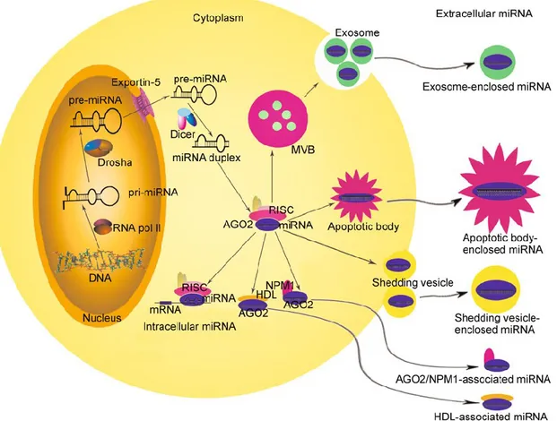
Extracellular miRNAs circulate in the body fluids with a high concentration and sufficient integrity despite high extracellular ribonuclease (RNase) activity, indicating that extracellular miRNAs are likely packaged in some manner to shield them from digestion. Indeed, naked miRNAs added to plasma are immediately degraded, whereas circulating miRNAs are stable for hours under the same conditions (Mitchell et al., 2008). Further studies have also shown that circulating miRNAs are protected from and resistant to harsh conditions such as extreme temperatures, extreme pHs or freeze-thaw cycles (Chen et al., 2008). This phenomenon raises intriguing questions regarding the mechanism of circulating miRNA stability. At present, there are at least three possibilities for the remarkable stability of circulating miRNAs and their sources: (1) they are passively leaked from broken cells during tumorigenesis or tissue injury (Chen et al., 2008; Mitchell et al., 2008); (2) they are packaged in small membranous vesicles, including exosomes, shedding vesicles and apoptotic bodies, which accounts for the release of miRNAs into circulation and offers protection against RNase activity (Valadi et al., 2007; Zernecke et al., 2009; Kosaka et al., 2010; Zhang et al., 2010); (3) they are protected by the formation of a protein-miRNA complex (Fig. 3).
15
Figure 3. A model of the secretion of miRNAs. After being processed to the mature form, some miRNAs can bind to
complementary sequences on target mRNAs to repress translation or trigger mRNA cleavage. Other miRNAs are packaged and transported to the extracellular environment via three different pathways: (1) they are enclosed within membranous vesicles, including exosomes, shedding vesicles and apoptotic bodies; (2) they are associated with lipoproteins, such as HDL; (3) they are associated with RNA-binding proteins, such as AGO2 and NPM1. AGO2, Argonaute 2; NPM1, nucleophosmin1.
Some studies have demonstrated that many extracellular miRNAs are detectable in conjunction with proteins or lipoproteins, but devoid of membrane vesicles, suggesting the existence of non-vesicleenclosed, RNA-binding protein-associated miRNAs in extracellular fluids, including those bound to AGO2, nucleophosmin1 (NPM1), and high-density lipoprotein (HDL) (Wang et al., 2010; Arroyo et al., 2011; Turchinovich et al., 2011; Vickers et al., 2011). Recent studies indicate that miRNAs released from donor cells that are either enclosed in vesicles (exosomes, shedding vesicles and apoptotic bodies) or associated with lipoproteins (HDL) are active and can function as secreted molecules to influence the recipient’s cell phenotype (Zernecke et al., 2009; Kosaka et al., 2010; Zhang et al., 2010; Vickers et al., 2011). The difference between vesicle enclosed and lipoprotein-associated miRNAs is largely unknown. Their different secretion mechanisms suggest that they may originate from different cell types and, therefore, have different fates and functions. For example, miRNAs detected only in the lipoprotein-associated fractions may be generated by cells with lipoprotein transport pathways. By contrast, miRNAs that are predominantly packaged in
16
vesicles may originate from cell types known to generate abundant vesicles. However, miRNAs associated with other types of RNA-binding proteins (e.g., AGO2 and NPM1) may also be actively released from donor cells and taken up by recipient cells, although the direct evidence remains elusive.
1.4 MICRORNAs IN CANCER
1.4.1 miRNAs and tumorigenesisTumors result from pathological changes caused by dysregulation of cell proliferation and apoptosis. Abnormal expression of oncogenes and tumor suppressor genes has been widely accepted as the molecular mechanism of tumorigenesis. However, this traditional concept is being challenged by the discovery of non-coding RNAs. Recent studies have shown that miRNAs play important regulatory roles in tumorigenesis. miRNAs are expressed in a variety of tumors, exhibiting abnormal functions; they are involved in tumorigenesis and tumor development by negatively regulating protein-coding genes related to these processes. On one hand, downregulation or null expression of a tumor suppressor miRNA can lead to the expression of the miRNA target genes that promote tumorigenesis, causing excessive cell proliferation and abnormal differentiation and resulting in tumorigenesis (Jun et al., 2011).
On the other hand, overexpression of oncogenic miRNAs can lead to decreased expression of the target genes with tumor suppressor functions, thereby promoting tumorigenesis and tumor development. For example, overexpression of miR-221 or miR-222 inhibits the expression of the Kit protein, resulting in the dedifferentiation of thyrocytes and tumorigenesis (He et al., 2005). Deficiency of miR-15a and miR-16-1 expression can lead to overexpression of Bcl-2, an important anti-apoptotic factor, resulting in decreased apoptosis, thereby promoting tumorigenesis and tumor development. Some studies have found that miR-150 is significantly overexpressed in gastric cancer tissues and promotes gastric cancer cell proliferation through EGR2 (Wu et al., 2010). Moreover, the expression of miR-34 is decreased in gastric cancer. Restoring miR-34 expression in the gastric cancer cells with a mutant p53 significantly inhibits the protein expression of Bcl-2, Notch, and HMGA2, causing cell cycle arrest in G1 phase and cell growth inhibition. It also increases the activity of caspase-3 to promote apoptosis and inhibits the formation and growth of gastric cancer stem cell spheroids (Ji et al., 2008).
An interesting phenomenon found in recent studies of tumor miRNA expression profiles is that the miRNAs abnormally expressed in tumors usually exhibit reduced expression levels compared to their expression levels in normal tissues (Lu et al., 2005). This phenomenon may reflect a higher proliferation rate and a less differentiated state of tumor cells. Another explanation is that the cells
17
with low levels of miRNAs are selected for during tumorigenesis due to their proliferative and survival advantages. These two possibilities are not mutually exclusive. In fact, both of them are supported by experimental evidence. For instance, after differentiation induction, HL60 cells show significantly increased miRNA expression, consistent with the fact that the differentiation state of the cells is maintained by the enhancement of miRNA transcription. Moreover, studies in lung cancer models have indicated that inhibiting miRNA biogenesis by genetic approaches or RNAi can promote tumorigenesis and tumor development (Kumar et al., 2007). In addition, c-Myc can induce the universal silencing of miRNA transcription (Chang et al., 2008), providing a possible mechanism for miRNA downregulation in malignant cells. These findings suggest that the majority of miRNAs may function as tumor suppressors in tumorigenesis.
1.4.2 OncomiRs and metastamiRs
In general, a miRNA able to promote cancer targets mRNAs encoding tumor suppressor proteins, while miRNAs exhibiting tumor suppressor properties usually target mRNAs encoding oncoproteins. miRNAs which have been demonstrated to play a crucial role in the initiation and progression of human cancer are defined as oncogenic miRNAs (oncomiRs) (Ge et al., 2013; Rather et al., 2013; Shu et al., 2011; Haug et al., 2011; Tang et al., 2011; Ma et al., 2010; Mercatelli et al., 2008). Moreover, miRNAs have been firmly demonstrated to be involved in cancer metastasis (metastamiRs) (Hurst et al., 2009; Wotschofsky et al., 2012; Taylor et al., 2013; Welch et al., 2013). Thus, therapeutic strategies involving miRNA silencing have been suggested, based on the roles of these small non-coding RNAs as oncogenes (Ge et al., 2013; Rather et al., 2013; Shu et al., 2011; Haug et al., 2011; Tang et al., 2011; Ma et al., 2010; Mercatelli et al., 2008).
Another very interesting feature of miRNAs has been found by studying cancer-associated miRNAs in different experimental model systems; cancer-specific miRNAs are present in extracellular body fluids and may play a crucial role in the cross-talk between cancer cells and surrounding normal cells (Moldovan et al., 2013; Chen et al., 2012; Kosaka et al., 2011; Chen et al 2012; Ramachandran et al., 2012; Muralidharan et al, 2010). Of note, evidence of the presence of miRNAs in serum, plasma and saliva supports their potential as an additional set of biomarkers for cancer. As previously mentioned, extracellular miRNAs are protected, for example, by exosome-like structures, small intraluminal vesicles shed from a variety of cells (including cancer cells), with a biogenesis connected with the endosomal sorting complex required for transport machinery in multivesicular bodies. These extracellular structures, originally considered as a ‘garbage bag’ devoted to discarding degraded proteins, are now considered to play an important role as an intercellular communication tool. It is still unclear as to whether these exosome-associated miRNAs
18
occur as a result of tumor cell death and lyses, or are actively excreted from tumor cells into the microenvironment. However, this novel secretory machinery of miRNAs may be involved in tumor-associated features, such as the enhancement of angiogenesis, the increase of cytokine secretion and migration to pre-metastatic niche. Table 1 illustrates a summarized list of oncomiRs and metastamiRs.
Cells/Tissues miRNA target Modulated
mRNA Effects following antagomiR treatement Authors Human
glioblastoma miR-27a FOXO3a
Suppression of U87 growth in vitro and in vivo
Ge et al. Cutaneous squamous cell carcinoma (SCC) miR-155 CDC73 Decreased cell viability, increased apoptosis and marked regression of xenografts in nude mice Rather et al. Malignant
astrocytoma cells miR-335 Daam1
Growth arrest, cell apoptosis, invasion repression and marked regression of astrocytoma xenografts Shu et al. Neuroblastoma miR-92 DKK3 Increased released of the tumor suppressor Dickkopf-3 (DKK3), a secreted protein of the DKK family of Wnt regulators Haug et al. Glioma miR-381 LRRC4 Decreased cell proliferation and
tumor growth Tang et al. Breast cancer miR-10b Hoxd10 Suppression of
19
metastases
Prostate cancer miR-221/miR-222 p27 tumor growth Reduction of Mercatelli et al.
Table 1: Exaples of oncomiRs suitable for antagomiR-based miRNA targeted therapy of cancer.
1.4.3 Oncosuppressor microRNAs
In addition to oncogenic activities, miRNAs exhibit, oncosuppressor properties by targeting mRNAs encoding oncoproteins (Scheibner et al 2012; Endo et al., 2013; Liang et al., 2013; Thomas et al., 2012; Ibrahim et al., 2011; Wiggins et al., 2010; Trang et al., 2011; Wu et al., 2013; Huang et al., 2013). Piovan et al recently explored the interaction between certain miRNAs and transcriptional factors involved in determining cell fate, including the well known ‘genome guardian’, p53 (Piovan et al., 2012). They demonstrated that miR-205, an oncosuppressive miRNA lost in breast cancer, is directly transactivated by the oncosuppressor p53. Moreover, evaluating miR-205 expression in a panel of cell lines belonging to the highly aggressive triple-negative (estrogen receptor (ER), progesterone receptor (PR) and Her2/neu) breast cancer subtype, which still lacks an effective targeted therapy and is characterized by an extremely undifferentiated mesenchymal phenotype, the authors demonstrated that this miRNA is critically downregulated compared with a normal cell line. The re-expression of miR-205 strongly reduced cell proliferation, cell cycle progression and clonogenic potential in vitro, and inhibited tumor growth in vivo. The tumor suppressor activity of miR-205 is partially exerted by targeting E2F1, one of the master regulators of cell cycle progression, and LAMC1, a component of the extracellular matrix involved in cell adhesion, proliferation and migration. In another study, Lee et al. (Lee et al., 2011), demonstrated that an estrogen-downregulated miRNA, miR-34b, acts as an oncosuppressor that targets cyclin D1 and Jagged-1 (JAG1) in an ERα-positive/wild-type p53 breast cancer cell line (MCF-7), as well as in ovarian and endometrial cells, but not in ERα-negative or mutant p53 breast cancer cell lines (T47D, MBA-MB-361 and MDA-MB-435). The negative association between ERα and miR-34b expression levels has also been found in ERα-positive breast cancer patients. In addition, the overexpression of miR-34b has been shown to inhibit ERα-positive breast tumor growth in an orthotopic mammary fat pad xenograft mouse model. Table 2 illustrates a summarized list of oncosuppressor miRNAs (Iorio et al., 2012; Xu et al., 2013; He et al., 2013; Iorio et al., 2009).
20
Tumor type miRNA Modulated mRNA
Effects following pre-miRNA
administration Authors
Acute leukemia miR-27a Bax and Bad
Inibition of cell growth due, at least in part, to increased cellular apoptosis Scheibner et al. Oral squamous cell carcinoma (OSCC)
miR-596 LGAL3BP Growth inhibition Endo et al.
Breast cancer miR-302 AKT1 and RAD52
Sensitized radioresistant breast cancer cells to ionizing radiation Liang et al. Chronic myelogenous leukemia (CML) cells
miR-33a Pim-1 Decelerated cell
proliferation Thomas et al.
Colon carcinoma miR-33a Pim-1 Reduced tumor
proliferation Ibrahim et al.
Colon carcinoma miR-145 c-Myc and ERK5
Reduced tumor proliferation and
increased apoptosis
Ibrahim et al.
Lung cancer miR-34a
Repression of c-Met, Bcl-2; partial repression
of CDK4
Block of tumor
growth Wiggins et al.
Lung cancer miR-let7
Negative regulation of the cell cycle oncogenes RAS, MYC and HMGA2 Reduction of
tumor growth Trang et al.
Non-small cell lung adenocarcinomas,
miR-29b CDK6,
21
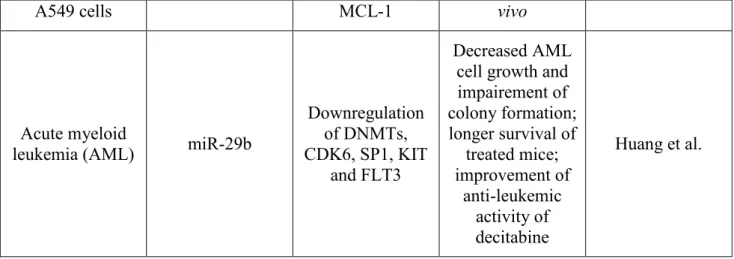
A549 cells MCL-1 vivo
Acute myeloid
leukemia (AML) miR-29b
Downregulation of DNMTs, CDK6, SP1, KIT
and FLT3
Decreased AML cell growth and impairement of colony formation; longer survival of treated mice; improvement of anti-leukemic activity of decitabine Huang et al.
Table 2: miRNAs acting as tumor suppressor genes and are suitable for replecement theraphy of cancer.
1.4.4 Role of secreted miRNAs in tumor progression
The tumor microenvironment plays a critical role in cancer progression. Cancer cells can influence normal cells to abandon their homeostatic activities and instead support the neoplastic nature of the tumor. The dynamic crosstalk between cancer cells and normal cells in the microenvironment is crucial to the progression of disease. Crosstalk can occur through secreted molecules and paracrine signalling (Muralidharan-Chari et al., 2010). The secreted molecules are no longer limited to cytokines, chemokines, growth factors or other protein molecules but now also include secreted miRNAs. Skog et al. reported that particular mRNAs and miRNAs are highly enriched in microvesicles from primary human glioblastoma cells (Skog et al., 2008). Glioblastoma-derived RNA contained in microvesicles is functional and is taken up by and processed in the human brain microvascular endothelial cell line HBMVEC, generating a functional protein (Skog et al., 2008). These results suggest that the tumor-derived microvesicles can modify the surrounding normal cells by changing their translational profile. In addition, the tumor-specific mRNA and miRNAs characteristic of gliomas, such as EGFRvIII and miR-21, could be detected in serum microvesicles of glioblastoma patients (Skog et al., 2008).
This finding led to the hypothesis that tumor cells use exosomes to transport genetic information, including miRNAs, to surrounding cells, thereby supporting tumor growth and progression. Furthermore, Ohshima et al. revealed that the let-7 miRNA family was enriched in the extracellular exosomes from a metastatic gastric cancer cell line AZ-P7a, while low metastatic AZ-521, as well as other cancer cell lines, showed no such enrichment (Ohshima et al., 2010). Because let-7 miRNAs generally function as tumor suppressors that target oncogenes such as RAS and high mobility group A2 (HMGA2), they proposed that cancer cells selectively secrete let-7 miRNAs into
22
the extracellular environment via exosomes, reducing the anti-tumorigenic effect within the cells and facilitating oncogenesis and metastasis (Ohshima et al., 2010).
Therefore, miRNAs can not only promote the development of primary tumors, but also affect tumor progression, including tumor metastasis (Ma and Weinberg, 2008; Dumont and Tlsty, 2009; Nicoloso et al., 2009; Zhang et al., 2009; Baranwal and Alahari, 2010; Ding et al., 2010; Khew-Goodall and Khew-Goodall, 2010; Li et al., 2010; Ma et al., 2010; Sachdeva and Mo, 2010; Santarpia et al., 2010; Schmittgen, 2010; Tian et al., 2010; Zhang et al., 2010 ). Both mutation (Gardner and Vinther, 2008) and misexpression (Santarpia et al., 2010; Zhang et al., 2010) of miRNAs can affect their normal functions, leading to abnormal expression of their target genes. Thus, tumor metastasis may be affected when the target genes are related to tumor cell migration, invasion, resistance, and other metastatic phenotypes. Among several known miRNAs promoting tumor metastasis, miR-10b and miR-373 are particularly prominent. miR-10b is a direct target gene of the transcription factor Twist1 that promotes epithelial-mesenchymal transition and tumor metastasis. The expression of miR-10b is markedly upregulated in human breast cancer cells of high metastatic potential; the invasiveness of these cells decreases by 10-fold if the activity of miR-10b is blocked by an antisense oligonucleotide. Overexpression of miR-10b in breast cancer cells with low metastatic potential leads to a significant increase in the invasiveness of the tumor cells. Six weeks after orthotopic inoculation of highly miR-10b-expressing breast cancer cells into the breast of immunodeficient young female mice, tumors were found at the inoculation sites in all of the inoculated mice, together with apparent interstitial and vascular infiltration; distant metastasis appeared in all of these mice 11 weeks after the inoculation. No invasion or metastasis was observed when using low miR-10b expressing breast cancer cells. miR-10b promotes the invasion and metastasis of breast cancer cells through inhibiting its target gene HOXD10 to increase the expression of RhoC (Ma et al., 2007). miR-335 can induce cell morphological changes to reduce the invasiveness and metastasis of breast cancer cells through directly regulating its targets, the progenitor cell-regulating transcription factor SOX4 and the extracellular matrix component cadherin C (Tavazoie et al., 2008). miR-29c can inhibit the metastasis of nasopharyngeal carcinoma by regulating a variety of extracellular matrix proteins, including inhibiting collagen and laminin 1 (Sengupta et al., 2008). miR- 373 was identified from in vitro screening for enhanced tumor cell migration phenotypes (Huang et al., 2008), and its role in promoting metastasis has been confirmed in vivo. It should be noted that miR-373 also exhibits the characteristics of an oncogene during the tumorigenesis of testicular germ cell tumors (Voorhoeve et al., 2006). It has been suggested that its oncogenic characteristics of promoting metastasis are a result of regulating different target genes.
23 1.4.5 miRNAs and cancer drug resistance
In addition to the findings that miRNAs are associated with tumor pathogenesis and development, miRNAs are closely related to the drug resistance of tumor cells (Lwin et al., 2010; Ma et al., 2010; Zheng et al., 2010; Akao et al., 2011). miRNAs affect the sensitivity of tumor cells to cytotoxic drugs (Xia et al., 2008), biologically targeted drugs (Miller et al., 2008; Weiss et al., 2008), endocrine drugs (Garofalo et al., 2008; Zhao et al., 2008), and cytokine drugs (Kovalchuk et al., 2008). For example, Kovalchuk et al. (2008) found that the expression of miR-451 is significantly reduced in the doxorubicin-resistant breast cancer cell line MCF27/DOX and that restoring miR-451 expression by transfection can increase the sensitivity of MCF27/DOX cells to doxorubicin. Si et al. (2007) discovered that a miR-21 inhibitor can significantly increase the sensitivity of breast cancer cells to topotecan. The studies by Venturini et al. (2007) showed that miR-17-19b transfection can remarkably increase the imatinib induced apoptosis of the acute myeloid leukemia cells K562.
miRNAs appear to play important roles in tumor drug resistance by regulating the expression of drug resistance proteins, and targeted intervention of miRNAs may effectively reverse tumor drug resistance (Table 3).
Cancer type miRNA Target gene Reference
Breast cancer miR-451 miR221/222 miR-200 family miR-328 Mdr1/Pglycoprotein P27(Kip1) E-cadherin BCRP/ABCG2 Kovalchuk et al. Miller et al. Tryndyak et al Pan et al. Gastric cancer miR-15a
miR-16 BCl-2 Xia et al.
Non-small cell
lung cancer miR221/222 Kit, p27 Zhao et al.
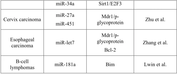
Ovarian cancer miR-214 miR-130a miR-27a miR-451 PTEN M-CSF Mdr1/p-glycoprotein Zhu et al.
Prostate cancer miR-34a miR-148a
Sirt-1
MSK1 Fujita et al. Colon cancer miR-519c ABCG2 Ibrahim et al.
24
miR-34a Sirt1/E2F3 Cervix carcinoma miR-27a
miR-451
Mdr1/p-glycoprotein Zhu et al.
Esophageal carcinoma miR-let7 Mdr1/p-glycoprotein Bcl-2 Zhang et al. B-cell
lymphomas miR-181a Bim Lwin et al.
Table 3: Reported studies on miRNAs and tumor drug resistance.
1.4.6 miRNAs and tumor diagnosis
miRNAs can be used to identify benign and malignant lesions and to determine the prognosis of cancer patients. High-throughput miRNA detection technologies have been emerging in recent years, including miRNA chips, miRNA expression profiling using magnetic bead–based flow cytometry, and miRNA qPCR assays. These methods have been used to identify a variety of tumor-specific miRNAs through parallel comparison between cancer tissues and the adjacent tissues. miRNAs are differentially expressed between tumor cells and normal cells, as well as among the tumor cells originating from different tissues. The discovery of miRNA expression features can help not only distinguish between benign and malignant lesions, but also determine the degree of tumor malignancy and prognosis, thereby providing the basis for personalized therapy (Alvarez-Garcia and Miska, 2005; Calin et al., 2005). For example, Calin et al. (2005) selected 13 out of 190 miRNAs as a set of miRNA expression markers for chronic lymphocytic leukemia; this miRNA set can not only distinguish between malignant and normal B lymphocytes, but also identify the chronic lymphocytic leukemia cases with high expression of 70-kD zeta-associated protein (ZAP70) or a mutated IgV (H) gene, which are associated with high malignancy, strong invasiveness, and poor prognosis. In non-small cell lung cancer, miR-155 upregulation and let-7 downregulation indicate poor prognosis. In colon cancer patients, high expression of miR-21 indicates poor prognosis. In other studies, it has been found that a characteristic spectrum composed of seven miRNAs (miR-10b, miR-21, miR- 223, miR-338, let-7a, miR-30a-5p, miR-126) is sufficient to predict the survival of gastric cancer patients (Li et al., 2010). Because miRNAs are relatively stable, it becomes more and more promising to use miRNAs as new tumor markers.
25
1.4.7 miRNAs can distinguish different malignant phenotypes of the same tumor
Different malignant phenotypes of tumors originating from the same tissue can result in markedly different therapeutic outcomes and prognosis. miRNAs can distinguish not only normal cells from tumor cells, but also the different malignant phenotypes of tumor cells originating from the same tissue. For example, Budhu et al. (2008) examined the miRNA expression profiles in cancer tissues and the corresponding adjacent tissues from 241 hepatic cancer cases using miRNA microarray. These cancer specimens included both invasive and non-invasive samples. Their results showed that the levels of 20 miRNAs can accurately predict hepatic cancer metastasis, which are closely related to post-operative relapse and the survival of hepatic cancer patients. Compared to their expression in drug-sensitive gastric cancer cells, miR-15a and miR-16b were downregulated in drug-resistant gastric cancer cells (Xia et al., 2008), and miR-218 was downregulated in the gastric cancer cells with high metastatic potential (Tie et al., 2010).
1.4.8 miRNAs reflect the origin of tumor tissues
By examining the samples from 540 cases, including 363 cases of six types of malignancies with high incidence rates and 177 normal controls, Volinia et al. (2006) discovered that specific miRNA expression profiles can not only distinguish normal cells from tumor cells, but also reflect the origin of tumor tissues. Lu et al. (2005) analyzed the miRNAs in 334 samples from 217 mammalian species and found that tumors originating from the organs that develop from embryonic endoderm, such as stomach, intestine, and liver, displayed similar miRNA expression patterns, whereas the miRNA expression patterns of leukemia were significantly different from those of solid tumors, suggesting that characteristic miRNA expression profiles possess diagnostic value in the identification of tumor tissue origin and exhibit great potential in the diagnosis of metastatic tumors with unknown primary tumor origin. miRNA expression profiles can also accurately reflect the progression and differentiation state of tumor tissues, especially for the poorly differentiated malignant tumors. Compared with the previously reported mRNA expression profiles, miRNA expression profiles provide a more accurate reflection of tumor status (Alvarez- Garcia and Miska, 2005).
1.4.9 Circulating miRNAs, new tumor markers
The discovery of circulating miRNAs further promoted investigations of miRNAs as biological markers in tumor diagnosis and prognosis. Lawrie et al (2008) first observed high expression of 21 in sera from patients with diffuse large B cell lymphoma and the close correlation of miR-21 expression with disease recurrence and patient survival. Mitchell et al. (2008) isolated RNAs of 18-24 nucleotides from the plasma of healthy volunteers and constructed a small RNA library. They
26
found 91 known and 4 unknown miRNAs by sequencing analysis. Subsequently, 25 patients with metastatic prostate cancer and 25 healthy volunteers were separated into different groups, whose sera were examined. They found that the plasma miR-141 levels in prostate cancer patients were significantly higher than those in the normal control group. Thus, prostate cancer patients can be effectively distinguished from healthy individuals based on their miR-141 expression levels, indicating that miR-141 can be used as a circulating miRNA marker for the detection of prostate cancer. Chen et al. (2008) analyzed the miRNA expression in the sera from patients with lung cancer, colon cancer, or diabetes using Solexa sequencing and quantitative PCR.
The results showed that these diseases all have their characteristic serum miRNA expression patterns. For example, 63 miRNAs were expressed in the sera of patients with lung cancer, but not in the sera of healthy individuals. Ten miRNAs were common to the sera of lung cancer and colon cancer patients. Resnick et al. (2009) examined the miRNA expression profile in plasma from eight ovarian cancer patients and found 23 differentially expressed miRNAs, among which 10 molecules had been previously reported in solid ovarian tumors. Verification in plasma from 19 ovarian cancer patients by qRT-PCR showed that miR-21, miR-29a, miR-92, miR-93, and miR-126 were remarkably upregulated in these patients, whereas miR-155, miR-127, and miR-99b were significantly downregulated.
Nevertheless, that study suggests that specific plasma miRNAs may serve as molecule markers that are detectable earlier than the traditional tumor markers used in tumor diagnosis and in efficacy assessment. These studies demonstrated that miRNAs are broadly present in the sera of healthy individuals and of patients with different diseases, including cancer. The types and quantities of miRNAs present in serum and plasma vary with physiological and pathological conditions. Specific miRNA expression profiles in the sera of tumor patients provide a new method for early diagnosis, classification, prognosis, efficacy assessment, and relapse monitoring of tumors, bringing new hope to noninvasive tumor diagnosis (Tie et al., 2011).
1.4.10 miRNAs related cancer diagnosis, polymorphisms and AMO
As already mentioned, the important roles of miRNAs in cancer and their potential applications as useful and effective targets have generated great interest in cancer gene therapy strategies, as well as diagnosis, classification, prognosis and risk factor evaluations. Based on microarrays for miRNA expression profiling studies, differences in miRNA expression could be detected between normal and cancer tissues, which can classify different tumor types and tumor grades (Visone et al., 2007; Yanaihara et al., 2006; Trang et al., 2008). Certain miRNA signatures are correlated with prognosis and can potentially be used to determine the specific course of treatment. Michael et al. (2003) found aberrant miRNA expression in solid tumors as they identified 28 different miRNAs in
27
colonic adenocarcinoma compared with normal mucosa. miR-143 and -145 were significantly down-regulated in the cancer. Similar situation was detected in other cancers, as miR-221, -222, and -146 in papillary thyroid carcinoma (He et al., 2005), miR-21 and -155 in pancreatic cancer (Bloomston et al., 2007), and miR-141 in prostate cancer (Mitchell et al., 2008). Through analyzing the expression of 217 miRNAs in 334 samples that included primary tumors, tumor-derived cell lines and normal tissues, Lu et al. (2005) found that miRNA profiles can distinguish between normal and cancer tissues, separate different cancer types, stratify the cancer differentiation state and cluster sample groups according to their embryonic lineage.
Single nucleotide polymorphisms (SNPs) within the miRNA coding genes or within miRNA target genes are likely to be deleterious and can affect an individual‘s risk to develop diseases such as cancers. Yu et al. (2007) found that 12 miRNA-related SNPs showed an aberrant allele frequency in human cancers. Chin et al. (2008) identified an SNP in let-7 complementary site 6 (LCS6) in the KRAS 3’ UTR that is associated with smoking-induced lung cancer risk. This variant allele is found in 20% of the 74 non-small cell lung carcinoma patients in the study. These unique miRNA expression signatures might be the hallmarks of tumor progressions and prognosis evaluations. Many miRNAs, have great potential in tumorigenesis, tumor invasion, metastasis, malignant progression, and poor prognosis. In in vitro and in vivo experiments, it has been confirmed that knockdown of certain miRNAs could change the tumor progression and biological characteristics as potential therapeutic targets (Bonci et al., 2008). In cell culture and xenograft mice models, synthetic anti-miRNA oligonucleotides (AMOs) with 2‘-O-methyl modification have been shown to effectively inhibit endogenous miRNAs. Krützfeldt et al. (2005) studied the utility of AMOs in vivo through intravenous injection of modified AMOs to target the liver-specific miR-122. Impressively, a single injection of 240 mg·kg-1 body weight conferred specific miR-122 silencing for up to 23 days. As an alternative to 2-hydroxyl-modified AMOs, lock nucleic acid based oligonucleotides (LNA-antimiR) have been shown to be more stable and less toxic in inhibiting endogenous miRNAs in vivo. Kota et al. (2099) showed that the systemic delivery of a single miRNA could cause tumor regression in a mice model of liver cancer. They delivered adeno-associated virus 8 (AAV8)-expressing miR-26a intravenously in Myc-induced mice harboring preformed liver tumors. After 3 weeks, they observed a significant regression of tumors in mice with the miR-26a treatment. These findings indicate a possibility of specific miRNAs-target therapy.
28 1.4.11 miRNAs and tumor therapy
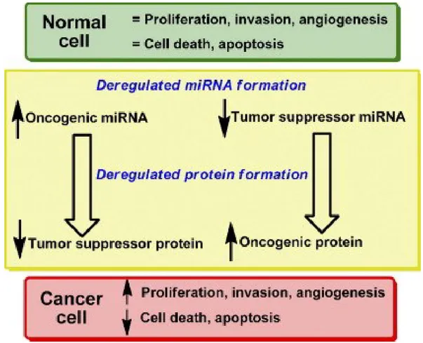
With the continuing discovery of the roles of miRNAs in tumorigenesis and tumor development, people have started to explore the use of miRNAs in tumor treatment. A single miRNA can simultaneously regulate multiple protein-coding genes and a number of signaling pathways associated with tumor growth and proliferation, metastasis, drug resistance, and other malignant phenotypes (Fig. 4).
Figure 4: Deregulated microRNA biogenesis and tumorigenesis. Both reduced expression of miRNA acting as tumor
suppressor and increased expression of miRNA acting as oncogenic miRNA can alter the synthesis of either oncogenic protein or tumor suppressor protein and lead to tumor formation.
Therefore, miRNAs exhibit advantages over individual protein-coding genes for treating tumors that involve alterations in multiple genes. For example, miR-31 can simultaneously regulate five proteins involved in tumor metastasis, RhoA, RDX, MMP16, Fzd3, and ITGA5, in breast cancer cells. It inhibits multiple steps in metastasis, including tumor cell migration, invasion, and colony formation in distant organs. Exogenous overexpression of miR-31 can significantly suppress the metastasis of breast cancer cells both in vitro and in vivo (Valastyan et al., 2009, 2010; Valastyan and Weinberg, 2010). In head and neck tumors, miR-204 can simultaneously regulate more than 60 target proteins involved in multiple tumor-associated signaling pathways (Lee et al., 2010), suggesting that miR-204 is a promising target for the treatment of head and neck tumors.
29
As already mentioned, associated miRNAs can be divided into two categories: tumor-promoting miRNAs and tumor-suppressing miRNAs. Tumor-tumor-promoting miRNAs are highly expressed in tumors. A number of methods can be used to downregulate or to suppress the expression of tumor-promoting miRNAs, including antagomirs, antimiRNA oligonucleotides and miRNA sponges. Tumor suppressing miRNAs are not expressed or are expressed at low levels in tumors. Thus, tumor treatment can be exerted by introducing corresponding exogenous miRNAs. The fact that a single miRNA can regulate multiple target genes provides new approaches for gene therapy based on RNAi. Because tumorigenesis and tumor progression are often regulated by multiple genes, an artificial miRNA can be designed to target multiple genes that are highly expressed in tumors, thereby simultaneously suppressing the expression of multiple oncoproteins. This effect cannot be achieved by traditional RNAi technology. Moreover, miRNA genes are often distributed in clusters. For example, the pri-miRNA of the miR-17-92 cluster contains seven independent mature miRNAs. Therefore, we can simulate the structure of the miR-17-92 precursor, design a structure for the expression of multiple miRNAs from a single promoter, and induce the interference of multiple oncogenes (Tie et al., 2011).
Altered expression of miRNAs is apparent in virtually all tumor types and includes blood-borne malignancies as well as solid tumors. The functional consequence of miRNA deregulation became evident as the introduction or repression of a single miRNA can effectively contribute to tumorigenesis or tumor progression. Numerous functional studies using cultured cancer cells and mouse models of cancer have identified miRNAs that function as conventional tumor suppressors or oncogenes. Examples of miRNAs with oncogenic activity are miR-155 and miR-17–92; in contrast, miR-15a, miR-16, as well as miRNAs of the miR-34 and let-7 families, are tumor-suppressor miRNAs (Calin et al., 2006;Johnson et al., 2007; Johnson et al., 2005; Kumar et al., 2008; Esquela-Kersher et al., 2008; Trang et al., 2009; He et al., 2007). The tumor-suppressive or oncogenic activity for many of these miRNAs is not limited to a particular tumor type, in agreement with the supposition that conventional cancer genes function as such regardless of tissue origin. The deregulation of some of these miRNAs also correlates with tumor differentiation status, disease stage, and patient outcome, further suggesting that aberrant miRNA function has a direct impact on tumor development. For instance, low let-7 levels and high miR-155 levels are indicative of poor survival of patients with non–small cell lung cancer (Yanaihara et al., 2006). Other miRNAs have specifically been implicated in early tumorigenesis or metastasis, representing unique opportunities for therapeutic intervention that will depend on the context and requirement of therapy.
The therapeutic application of miRNAs involves two strategies. One strategy is directed toward a gain of function and aims to inhibit oncogenic miRNAs by using miRNA antagonists, such as
anti-30
miRs, locked-nucleic acids (LNA), or antagomiRs. These miRNA antagonists are oligonucleotides with sequences complementary to the endogenous miRNA. They carry chemical modifications that enhance the affinity for the target miRNA and trap the endogenous miRNA in a configuration that is unable to be processed by RISC, or alternatively, leads to degradation of the endogenous miRNA. Small molecule inhibitors specific for certain miRNAs are also being developed to inhibit miRNA function. The second strategy, miRNA replacement, involves the reintroduction of a tumor-suppressor miRNA mimic to restore a loss of function. Although the inhibitory approach is more commonly accepted and conceptually follows rules that also apply to small molecule inhibitors and short interfering RNAs (siRNA), miRNA replacement represents a new opportunity to explore the therapeutic potential of tumor suppressors (Johnson et al., 2007;Esquela-Kersher et al., 2008; Trang et al., 2009; Kota et al., 2009). Therapeutically restoring the levels of tumor suppressors in tumor tissues has been investigated in the past by gene therapy; however, a practical application of this approach is still pending. Because the definition of tumor suppressors was restricted to protein-encoding genes, gene therapy usually involves the delivery of a relatively large DNA plasmid or viral vector that encodes the desired protein. Often, vector size, inefficient delivery to target tissues, and the requirement for nuclear localization represent technical challenges that limit this approach to local, rather than systemic, administration (McCormick et al., 2001; Roth 2006). Thus, despite a strong scientific rationale for cancer treatment, logistic obstacles associated with gene therapy leave the full therapeutic benefit of using tumor suppressors unanswered. miRNAs provide a new opportunity because, unlike proteins, miRNA mimics are substantially smaller, will merely have to enter the cytoplasm of target cells to be active, and can be delivered systemically using modes and technologies that are also used for siRNAs. Therefore, the delivery hurdle for miRNA mimics seems to be less an impediment than it is for protein encoding DNA. In addition, several other key observations support the concept of miRNA replacement therapy:
- the majority of differentially expressed miRNAs is suppressed in tumor tissue relative to normal tissues, indicating that the probability for miRNAs as tumor suppressors is greater than the probability as oncogenes (Lu et al., 2005);
- inhibition of endogenous miRNA processing induces oncogenic transformation and augments tumorigenesis, suggesting that the tumor suppressive role of miRNAs prevails over an oncogenic role (Kumar et al., 2007).
Another advantage of miRNA mimics is the fact that a miRNA mimic has the same sequence as the depleted, naturally occurring miRNA and, therefore, is expected to target the same set of mRNAs that is also regulated by the natural miRNA. Nonspecific off-target effects are unlikely as miRNA mimics are expected to behave like the natural counterpart for which the proper miRNA-mRNA
31
interactions have evolved over a billion years. The strongest rationale for exploring the therapeutic potential of miRNAs, however, is based on the observation that a single miRNA can regulate multiple oncogenes and oncogenic pathways that are commonly deregulated in cancer (Esquela-Kersher et al., 2006). Therefore, miRNAs act in accordance with our current understanding of cancer as a “pathway disease” that presumably can only be successfully treated when intervening with multiple oncogenic pathways (Check, 2008). The inhibitory effects induced by miRNAs on any particular target may be mild and may merely lead to a subtle reduction of protein expression; however, the simultaneous down-regulation of a broad set of targets has far-reaching biological consequences that determine the course of the cellular phenotype. The rapid and coordinated manipulation of protein levels across multiple pathways endows these regulatory RNAs with the ability to instantly switch between cellular programs. By restoring the expression of tumor suppressive miRNAs, miRNA replacement therapy seeks to reinstate those cellular programs that are active in normal cells and interfere with oncogenic programs necessary for the malignant phenotype (Bader et al., 2010).
To date, few tumor suppressor miRNAs have been discovered for which the proof of concept of miRNA replacement therapy has been shown in preclinical animal models of cancer (Bader et al., 2010).
Therapeutic miRNA mimics may be better tolerated by normal cells than cancer cells because: - pathways activated or repressed by the miRNA mimic are already activated or repressed by the endogenous miRNA;
- administration of therapeutic miRNA mimics is only an insignificant incremental increase of what is already present in normal cells;
- normal cells are not addicted to oncogenic pathways and manage to recover from the therapeutic dose used;
- normal cells have the ability to regulate the activity or presence of the miRNA mimic, whereas cancer cells do not.
As therapeutic programs advance miRNAs closer to the clinic, it will become critical to study miRNA-induced effects in normal cells and to assess potential toxicity in higher species. Taken together, miRNA replacement has emerged as a highly promising therapeutic strategy. It encompasses several conceptual aspects of traditional gene therapy and technical features of siRNA therapeutics. However, given the fundamental differences in the approach, mechanism of action, and outcome, miRNA mimics should be viewed as a new class of therapeutics. Available data, showing that miRNAs can function as bona fide tumor suppressors and that synthetic versions of these miRNAs robustly interfere with tumor growth in animal models, strongly support the