A CASE OF HYPERSENSITIVITY PNEUMONITIS
IN A WORKER EXPOSED TO TEREPHTHALIC ACID
IN THE PRODUCTION
OF POLYETHYLENE TEREPHTHALATE
PIETRO SARTORELLI1, GABRIELE D’HAUW1, DONATELLA SPINA2, LUCA VOLTERRANI3,
and MARIA ANTONIETTA MAZZEI3
1 University of Siena, Siena, Italy
Department of Medical Biotechnology, Unit of Occupational Medicine, University Hospital of Siena
2 University Hospital of Siena, Siena, Italy
Unit of Pathology
3 University of Siena, Siena, Italy
Department of Medicine, Surgery and Neuroscience, Unit of Diagnostic Imaging, University Hospital of Siena Abstract
Occupational hypersensitivity pneumonitis (OHP) is an interstitial lung disease caused by sensitization to an inhaled antigen. Polyethylene terephthal-ate (PET) is mainly used for disposable beverage bottles. A clinical case of hypersensitivity pneumonitis (HP) in a 66-year-old patient in the follow-up as a worker formerly exposed to asbestos is presented. At the first visit in 2012 a diagnosis of asbestosis and pleural plaques was formulated. In 2017 the high resolution computed tomography was performed demonstrating a slight progression of the pulmonary fibrosis, while physical examinations revealed inspiratory crackles on auscultation, and lung function tests showed a decreased diffusing capacity for carbon monoxide. The radiological and histological pictures were compatible with HP. From 1992 to 2013 the patient worked in a chemical company that produced PET for disposable beverage bottles. A diagnosis of OHP was made, and the most likely causative agents were terephthalic acid and dimethyl terephthalate. To the best of the authors’ knowledge, this is the first report of an OHP case in PET production. Int J Occup Med Environ Health. 2020;33(1):119 – 23
Key words:
asbestos, extrinsic allergic alveolitis, polyethylene terephthalate, terephthalic acid, pulmonary fibrosis, pleural plaques
Funding: publication of this work was supported by the Tuscan Region (project DGRT 396/2016, entitled “Health surveillance for former asbestos exposed workers,” project manager: Elisabetta Chellini, Ph.D.).
Received: April 16, 2019. Accepted: September 10, 2019.
Corresponding author: Pietro Sartorelli, University of Siena, Department of Medical Biotechnology, Unit of Occupational Medicine, University Hospital of Siena, Viale
INTRODUCTION
According to the position paper of the European Academy of Allergy and Clinical Immunology (EAACI) [1], occupa-tional hypersensitivity pneumonitis (OHP) “is an immuno-logic lung disease with variable clinical presentation and outcome resulting from lymphocytic and frequently
granu-lomatous inflammation of the peripheral airways, alveoli, and surrounding interstitial tissue which develops as the result of a non-IgE-mediated allergic reaction to a variety of organic or low molecular weight agents that are present in the work environment.” As demonstrated for idiopathic pulmonary fibrosis (IPF), a multidisciplinary approach
in-At the first visit in 2012, the patient’s ventilatory capacity was not reduced, even if he reported symptoms of chronic bronchitis (i.e., cough and phlegm for at least 3 months/year) in the last 15 years. The clinical examination of the chest was negative. The high resolution computed tomography (HRCT) showed pulmonary fibrosis with subtle thickening of the inter- and intra-lobular septa in the subpleural regions of both lungs. Pleural thickening was also present (Figure 1a). The mineralogical analysis of the bronchoalveolar lavage flu-id (BALF) by scanning electron microscopy (SEM) showed an amphiboles concentration of 472 ff/ml BALF, while chrys-otile fibres and asbestos bodies were under the detection lim-it. This result was comparable with the concentration levels of amphiboles in BALF, measured in the same laboratory in patients suffering from asbestosis [4]. At the end of the inves-tigations, a diagnosis of asbestosis and pleural plaques (PPs) was formulated.
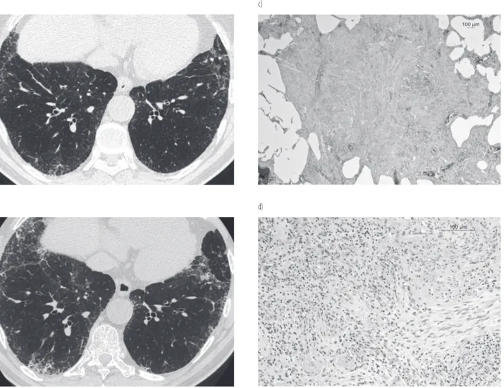
In 2017, during the follow-up for asbestosis and PPs related to the past asbestos exposure, another HRCT examination was performed demonstrating a slight progression of the pulmonary fibrosis not only in the extension but also in the profusion of CT features, both in the upper and lower lobes, in relation to the previous HRCT performed in 2015 (Fig-ure 1b). Physical examinations revealed inspiratory crackles at the pulmonary bases on auscultation. The patient recently experienced the onset of progressive dyspnea. Moreover, his lung function tests showed a decreased diffusing capacity for carbon monoxide (CO). The mineralogical analysis showed an amphiboles concentration of 320 ff/ml BALF (chrysotile fibres and AB were again under the detection limit). The to-tal number of cells in BALF was 90 700/ml, with 71% of mac-rophages, 14% of lymphocytes, 6% of neutrophils and 9% of eosinophils, and with a CD4/CD8 ratio of 1.68.
For a better definition of the case, pneumological advice was requested that suggested performing a surgical lung biopsy. The histological examination carried out on the lung biopsies showed bronchiolocentric lymphocytic inter-stitial pneumonitis with scattered eosinophils and an orga-cluding clinicians, radiologists, pathologists, and
occupa-tional physicians is strongly recommended to improve the diagnosis of OHP [2]. Today the frequency of some well known forms of the disease, such as the farmer’s lung, has decreased, while new causal agents are observed.
Polyethylene terephthalate (PET) is a clear, strong and lightweight thermoplastic polymer resin of the polyester family, mainly used for packing liquids and foods. It is produced from ethylene glycol (EG) and terephthalic acid (TPA) or dimethyl terephthalate (DMT). The first step in the polymerization sequence is an esterification of acid with glycol, with water being liberated in the process. The second step in the polymerization sequence is the polycondensa-tion of bis-hydroxyethyl terephthalate (BHET) with the lib-eration of EG. Many catalysts are used during the polycon-densation as well as during the initial esterification or trans-esterification (e.g., antimony compounds such as trioxide or triacetate). Certain health effects have been recognized for phthalates and derivatives. In particular, a number of reports have suggested that endocrine disruptors may leach into the contents of bottles made from PET [3].
A clinical case of a patient with a history of both occupa-tional asbestos and TPA exposure is presented. During the follow up as an asbestos-exposed worker, the diagnosis of OHP was formulated.
CASE REPORT
The patient was a 66-year-old male, non-smoker, in the follow-up since 2012 as a worker formerly exposed to as-bestos. He worked as a maintenance mechanic in the ship engine room in 1973–1992. His work included the removal of asbestos insulation of various parts of the engine room before repairing the installations and insulating them again with asbestos. In 1992–2013 he worked in a chemi-cal company that produced PET for disposable beverage bottles. He worked at the extrusion line of PET sheets where he was exposed to fumes produced in the polymer-ization process.
The patient, according to the pulmonologist, was treated with corticosteroids and azathioprine.
DISCUSSION
Several cases of asthma occurred in workers exposed to phthalic anhydride, which is mainly used in alkyd and epoxy resins [5]. In some cases, even phthalates seem to be able to cause asthmatic reactions. The case of a sub-ject who developed asthma and alveolitis-type reaction nizing pneumonia pattern. Rare and poorly formed
inter-stitial granulomas were present. In the most advanced ar-eas, destructive scarring with architectural distortion, but with centrilobular and periseptal prevalence, and without honeycombing, were seen (Figure 1c and 1d). No asbestos bodies were detected. The clinical picture was compatible with hypersensitivity pneumonitis (HP). A diagnosis of OHP was, therefore, made. The possible environmental causes were not documented from the exposure history.
Figure 1. Examinations of a 66-year-old patient, a worker formerly exposed to asbestos. High resolution computed tomography scans
showing a moderate increase in fibrotic changes in 2 different CT, performed in a) 2012 (the early fibrotic changes were distributed in the subpleural region, along the peribronchiolar structures) and b) 2015. Histological specimens confirmed c) a centrilobular fibro-myomatosis with the peripheral area of myxoid connective tissue (Hematoxylin and eosin stain [HE stain], 25×), d) associated with bronchiolitis obliterans: infiltrates of lymphocytes and eosinophils (HE stain, 50×).
a) c)
d) b)
dard test and the diagnosis is made through a combination of procedures [1]. No single historical symptom, physical examination finding, diagnostic laboratory data or radio-logic finding is diagnostic for HP, even if all these features support the diagnosis [9]. The relevant antigen to hyper-sensitivity pneumonitis cannot be identified in up to 20% to 30% of patients [11]. According to Hanak et al. [16], the diagnostic criteria for HP included the following:
1) the presence of respiratory symptoms,
2) the radiologic evidence of a diffuse lung disease, 3) a known exposure, or a positive serologic test result, to an inciting antigen,
4) no other identifiable cause for the lung disease. If there was no identifiable inciting antigen, 1 of the fol-lowing 2 criteria was required:
1) a lung biopsy specimen that demonstrated features of HP, or
2) BALF lymphocytosis and high-resolution computed tomographic evidence of ground-glass opacities or centri-lobular nodules bilaterally.
In the present case symptoms, the radiologic and patho-logic findings suggested the HP diagnosis in the presence of occupational exposure to a potential causal agent. The lack of BALF lymphocytosis is compatible with the chron-ic HP. Although the specifchron-ic compound could not be de-termined with certainty, as is always the case of chemicals, in the present case OHP is likely linked to occupational exposure to TPA or DMT.
CONCLUSIONS
To the best of the authors’ knowledge, this is the first re-port of an OHP case in PET production.
ACKNOWLEDGMENTS
The authors would like to thank Dr. David Bennet (Department of Medicine, Surgery and Neuroscience, Unit of Respiratory Diseases and Lung Transplant, University of Siena), Dr. Ricca-rdo Romeo and Dr. Antonietta Gerardina Sisinni (Unit of
Oc-after being exposed to polyester containing polyethyl-ene terephthalate and polybutylpolyethyl-ene terephthalate was described [6]. Heated polyvinyl chloride (PVC) fumes containing phthalates possibly contribute to the devel-opment of asthma in adults [7]. Few cases of alveolitis have been linked to occupational phthalates exposure. Allergic alveolitis was observed following exposure to an epoxy polyester powder paint containing low amounts of trimellitic anhydride and phthalic anhydride [8]. An OHP case was observed in a yacht manufacturing worker: among the various chemicals the patient was ex-posed to, the most likely causative agents were dimethyl phthalate and styrene [9].
When an uncommon cause is involved, the clinical and occupational histories are the cornerstones to the diag-nosis of OHP [1]. In literature an overlap between IPF and fibrotic HP was found, with a number of patients who were diagnosed with HP after the IPF had previously been diagnosed [10]. The distinction of HP from other inter-stitial lung diseases is important for the management and therapy because the HP treatment involves, first of all, antigen avoidance to reduce the inflammatory/immune response. As HP must be considered in all cases of inter-stitial lung diseases, a detailed environmental exposure history is mandatory [11,12]. In the present case, the oc-cupational history was puzzling because of the asbestos exposure at levels compatible with the onset of asbestosis proven also by the presence of PPs [13]. On the one hand, the progression, with a visible year-to-year increase in the disease symptoms, and the radiographic findings revealed by HRCT, also through multiplanar reformat using the spiral acquisition technique, with the loss of pulmonary function in the absence of asbestos exposure, suggested a diagnosis different from asbestosis [14,15]. On the other hand, HP may show an adverse outcome even after avoid-ing exposure to the causal agent [1].
The EAACI Task Force stated that the diagnosis of OHP in general often remains challenging as there is no gold
stan-polyester powder paint containing low amounts (< 1 %) of acid anhydrides. Eur Respir J. 1997;10(4):948–51.
9. Volkman KK, Merrick JG, Zacharisen MC. Yacht-maker’s lung: A case of hypersensitivity pneumonitis in yacht manu-facturing. WMJ. 2006;105(7):47–50.
10. Morell F, Villar A, Montero MÁ, Muñoz X, Colby TV, Pip-vath S, et al. Chronic hypersensitivity pneumonitis in patients diagnosed with idiopathic pulmonary fibrosis: a prospective case-cohort study. Lancet Resp Med. 2013;1(9):685–94, https://doi.org/10.1016/S2213-2600(13)70191-7.
11. Ohshimo S, Bonella F, Guzman J, Costabel U. Hypersen-sitivity pneumonitis. Immunol Allergy. Clin North Am 2012;32(4):537–56, https://doi.org/10.1016/j.iac.2012.08.008. 12. Mazzei MA, Sartorelli P, Bagnacci G, Gentili F, Sisinni AG,
Fausto A, et al. Occupational Lung Diseases: Underre-ported Diagnosis in Radiological Practice. Semin Ultra-sound CT MR. 2019;40(1):36–50, https://doi.org/10.1053/ j.sult.2018.10.019.
13. Mazzei MA, Contorni F, Gentili F, Guerrini S, Mazzei FG, Pinto A, et al. Incidental and Underreported Pleural Plaques at Chest CT: Do Not Miss Them-Asbestos Exposure Still Ex-ists. Biomed Res Int. 2017;2017:6797826, https://doi.org/10. 1155/2017/6797826.
14. American Thoracic Society. Diagnosis and initial manage-ment of nonmalignant diseases related to asbestos. Am J Respir Crit Care Med. 2004;170(6):691–715.
15. Abbritti M, Mazzei MA, Bargagli E, Refini RM, Penza F, Perari MG, et al. Utility of spiral CAT scan in the follow-up of patients with pulmonary Langerhans cell histiocytosis. Eur J Radiol. 2012;81(8):1907–12, https://doi.org/10.1016/ j.ejrad.2011.04.018.
16. Hanak V, Golbin JM, Ryu JH. Causes and presenting fea-tures in 85 consecutive patients with hypersensitivity pneu-monitis. Mayo Clin Proc. 2007;82(7):812–6.
cupational Medicine, University Hospital of Siena, University of Siena, Siena, Italy) for their kind collaboration.
REFERENCES
1. Quirce S, Vandenplas O, Campo P, Cruz MJ, de Blay F, Kos-chel D, et al. Occupational hypersensitivity pneumonitis: an EAACI position paper. Allergy. 2016;71(6):765–79, https:// doi.org/10.1111/all.12866.
2. Raghu G, Remy-Jardin M, Myers JL, Richeldi L, Ryerson CJ, Lederer DJ, et al. Diagnosis of Idiopathic Pulmonary Fibro-sis. An Official ATS/ERS/JRS/ALAT Clinical Practice Guide-line. Am J Respir Crit Care Med. 2018;198(5):e44–68, https:// doi.org/10.1164/rccm.201807-1255ST.
3. Sax L. Polyethylene Terephthalate May Yield Endocrine Dis-ruptors. Environ Health Perspect. 2010;118(4):445–8, https:// doi.org/10.1289/ehp.0901253.
4. Paolucci V, Romeo R, Sisinni AG, Scancarello G, Volterrani L, Mazzei MA, et al. Asbestos Exposure Biomarkers in the Fol-low-up of Asbestos-Exposed Workers. Ind Health. 2018;56(3): 249–54, https://doi.org/10.2486/indhealth.2017-0125.
5. Venables KM. Low molecular weight chemicals, hypersensi-tivity and direct toxicity: the acid anhydrides. Br J Ind Med. 1989;46(4):222–32.
6. Cartier A, Vandenplas O, Grammer LC, Shaughnessy MA, Malo JL. Respiratory and systemic reaction following ex-posure to heated electrostatic polyester paint. Eur Respir J. 1994;7(3):608–11.
7. Jaakkola JJ, Knight TL. The role of exposure to phthal-ates from polyvinyl chloride products in the development of asthma and allergies: a systematic review and meta-analysis. Environ Health Perspect. 2008;116(7):845–53, https://doi.org/ 10.1289/ehp.10846.
8. Piirilä P, Keskinen H, Anttila S, Hyvönen M, Pfäffli P, Tuo-mi T, et al. Allergic alveolitis following exposure to epoxy
This work is available in Open Access model and licensed under a Creative Commons Attribution-NonCommercial 3.0 Poland License – http://creativecommons.org/ licenses/by-nc/3.0/pl/deed.en.