UNIVERSITA’ DEGLI STUDI DI MESSINA
DOTTORATO DI RICERCA in
BIOLOGIA APPLICATA E MEDICINA SPERIMENTALE (XXIX ciclo)
Curriculum: Medicina Sperimentale
Coordinatore: Prof. Salvatore CUZZOCREA
1
H NMR-BASED METABOLOMICS:
A POWERFUL TOOL TO UNRAVEL THE MECHANISMS OF
TOXICITY OF MERCURY
Tesi di Dottorato
Dott.ssa Tiziana CAPPELLO
Tutor
Chiar.ma Prof.ssa Maria MAISANO
Co-Tutor
Chiar.ma Dott.ssa Patrícia PEREIRA
SSD:
BIO/06 – ANATOMIA COMPARATA E CITOLOGIA
Summary
Environmental metabolomics is a high-throughput approach that has shown considerable potential in mechanistic research. Indeed, it is one of the most powerful “-omics” techniques that allow the simultaneous evaluation of a broad number of endogenous small metabolites, and offer the potential to unravel subtle alterations in biological pathways providing insights into the mechanisms that underlie various physiological conditions, identify new metabolite biomarkers as defensive or adaptive response, and thus provide an overview of the metabolic status of a biological system.
Mercury (Hg) is a hazardous pollutant because of its persistence, high toxicity and tendency to bioaccumulate and biomagnify throughout food chains, with implications to humans. Despite the efforts on evaluating the biological impact of Hg on aquatic organisms, the toxicity mechanism of Hg still need to be clarified, and its understanding requires new ways. In this regard, the use of environmental metabolomics may serve as a powerful tool focusing on metabolites involved in various metabolic pathways, which changes might reveal insights into the Hg toxicity mechanisms. Given the similarity of fish responses with higher vertebrates, fish can be used to screen potentially hazardous effects to humans. Considering that oxidative stress has been described as a key pathway to initiate Hg toxicity in fish, the combination of metabolomics and conventional oxidative stress biomarkers could be an innovative and promising approach to disclose Hg-induced toxicity.
Hence, this strategy was applied to the liver and gills of the golden grey mullet Liza aurata inhabiting an Hg contaminated system (Aveiro Lagoon, Portugal), with the aim to unmask the mechanisms of Hg toxicity. Furthermore, this thesis has also the merit to provide a detailed comparative analysis of the differential bioaccumulation of Hg and metabolic responses in
S
UM M AR Yfish organs, which allowed to discern tissue-specific toxicological effects of Hg in fish liver and gills, attributable to their differential physiological functions.
In mullet liver, the major site for xenobiotic bioaccumulation and detoxification processes, metabolomics revealed that Hg accumulation has multiple levels of impact, interfering with membrane stabilization/degradation/repair processes, osmoregulation, energy metabolism, gene expression and antioxidant protection. Also, the oxidative stress biomarkers in liver revealed that Hg triggers adaptive responses of antioxidant system depicted in increased GST and CAT activities as well as GSHt content, compensating GPx activity depletion. Therefore, the combined use of metabolomics and oxidative stress endpoints allowed a better understanding of Hg hepatotoxicity, identifying GSH as a first line of defence against Hg and providing evidences of oxidative insults in cell membranes. Nonetheless, the induction of lipid peroxidation in mullet liver was efficiently prevented.
In mullet gills, metabolomics enabled an integrated description of numerous metabolites involved in various metabolic pathways. Interferences with the ion-osmoregulatory processes were revealed, with reduced taurine and glycerophosphocholine, along with increased creatine level. Impairments in energy metabolism were observed, as well as occurrence of protein catabolism supported by the increased levels in amino acids and alanine, the latter mainly involved in nitrogenous waste excretion. Vulnerability in the respiratory gas exchange activity was suggested by augmentation of isobutyrate, a known biomarker of anoxia. The combination of metabolomics and pro-oxidant status evaluation indicated the occurrence of massive GSH oxidation under Hg stress, and an inability to carry out its regeneration (GR activity was unaltered) or de novo synthesis (depletion in GSH constituent amino acids). The prevention of lipid peroxidative damage occurred, and it may be associated with the enhancement of membrane stabilization/repair processes, resulting from depletion in precursors of phosphatidylcholine. Though the exposure to Hg in
gills led to a state of vulnerability due to limitations of antioxidant system
S
UMM
AR
towards a physiological accommodation, the membranes were able to answer to oxidative insult through the enhancement of stabilization/repair processes.
Finally, from the detailed comparison of the differential bioaccumulation of Hg and metabolic responses in fish liver and gills, tissue-specific toxicological mechanisms of Hg, attributable to differential structural properties and physiological functions of the two organs under examination, were also detected. Specifically, Hg accumulation in fish tissues provoked severe disturbances in ion-osmoregulatory processes, mainly in gills, as highlighted by the depletion of osmolytes, namely taurine and glycerophosphocholine. However, the decrease in taurine level recorded in liver was associated to the metal-chelating property of taurine, thus resulting in the occurrence of hepatic detoxification processes. Differential disorders in energy-producing metabolic pathways were found between gills and liver, resulting in an enhancement of anaerobic metabolism as strategy to replenish insufficient energy supply in gills and promotion of gluconeogenesis in liver. Interferences with protein metabolism were also detected in both organs, depicting an ongoing protein catabolism in gills and, conversely, augmented protein synthesis in liver for repair of Hg-damaged proteins or activation of cytoprotective mechanisms to counteract Hg toxicity. Additionally, differential alteration in membrane stabilization/repair processes and perturbation of antioxidant defence system were also pointed out between mullet gills and liver.
The novel concurrent use of metabolomics and conventional oxidative stress endpoints, coupled with Hg tissue burdens characterization, demonstrated to be a sensitive and effective tool towards a mechanistically based assessment of Hg toxicity in fish, providing novel insights into the mechanisms underlying Hg-induced oxidative stress. Overall, the metabolomics approach demonstrated its effectiveness in the elucidation of the mechanisms of Hg toxicity in wild fish, and allowed to discern tissue-specific toxicological effects of Hg in fish gills and liver, attributable to their differential physiological functions, and potentially observable in humans.
S
UM M AR YRiassunto
La metabolomica ambientale rappresenta un approccio all’avanguardia che ha mostrato un potenziale considerevole nella ricerca meccanicistica. La metabolomica è la tecnica “-omica” che consente di effettuare una valutazione simultanea di un ampio numero di piccole molecole endogene, e offre il potenziale di mettere in luce minime alterazioni in pathways biologici fornendo quindi delucidazioni nei meccanismi che sottintendono diverse condizioni fisiologiche, di identificare nuovi biomarkers di stress metabolici come risposte di difesa o adattamento a diverse cause di stress, e di fornire quindi una panoramica dello stato metabolico di un sistema biologico.
Il mercurio (Hg) è un inquinante pericoloso a causa della sua persistenza, alta tossicità e tendenza a bioaccumulare e biomagnificare attraverso le reti trofiche, con conseguenze per l’uomo. Nonostante i numerosi studi condotti per valutare l’impatto biologico del Hg in organismi acquatici, i meccanismi di tossicità del Hg non sono ancora chiari, e la loro comprensione richiede nuovi strumenti di indagine. In questo contesto, l’uso della metabolomica ambientale può rappresentare uno valido strumento per lo studio dei metaboliti coinvolti in vari pathways metabolici, le cui variazioni possono rivelare indicazioni sui meccanismi di tossicità del Hg. Data la similarità delle risposte osservate in bassi vertebrati, come i pesci, con gli alti vertebrati, i pesci possono essere utilizzati per mettere in evidenza effetti potenzialmente pericolosi nell’uomo.
Considerando che lo stress ossidativo è stato descritto come il pathway chiave che innesca la tossicità del Hg nei pesci, la combinazione della metabolomica e di convenzionali biomarkers di stress ossidativo potrebbe rappresentare un approccio innovativo e promettente per svelare la tossicità indotta da Hg.
Pertanto, questa strategia è stata applicata al fegato e alle branchie del cefalo dorato Liza aurata proveniente da un’area contaminata da Hg (Aveiro Lagoon, Portogallo), con lo scopo di comprendere i meccanismi di tossicità del Hg.
R
IA S S U N T OInoltre, questa tesi ha anche il merito di fornire una dettagliata analisi comparativa del bioaccumulo differentiale del Hg e delle risposte metaboliche osservate nei campioni tissutali dei pesci, che hanno permesso di distinguere effetti tossicologici tessuto-specifici del Hg nel fegato e nelle branchie dei pesci, attribuibili alle loro diverse funzioni fisiologiche.
Nel fegato dei cefali, il sito principale per il bioaccumulo e i processi di detossificazione degli xenobiotici, la metabolomica ha rivelato che l’accumulo di Hg ha multipli livelli di impatto, interferendo con i processi di stabilizzazione/degradazione/riparo delle membrane, osmoregolazione, metabolismo energetico, espressione genica, e protezione antiossidante. Inoltre, i biomarkers di stress ossidativo hanno mostrato che il Hg innesca nel fegato risposte adattative del sistema antiossidante, risultanti nell’aumento delle attività del GST e CAT, e del contenuto del GSHt, che compensa la riduzione nell’attività del GPx. Pertanto, l’uso combinato della metabolomica e degli endpoints dello stress ossidativo hanno consentito una approfondita comprensione dell’epatotossicità del Hg, identificando il GSH come la prima linea di difesa contro il Hg e fornendo evidenze dell’insulto ossidativo nelle membrane cellulari. Tuttavia, l’induzione della perossidazione lipidica nel fegato dei cefali è stata efficacemente prevenuta.
Nelle branchie dei cefali, la metabolomica ha fornito una descrizione integrata di numerosi metaboliti coinvolti in vari pathways metabolici. In dettaglio, interferenze con i processsi di iono-osmoregolazione sono stati osservati mediante la riduzione della taurine e glicerofosfocolina, con incremento della creatina. Danni nei pathways energetici sono stati osservati, così come il catabolismo proteico, supportato dall’aumento di amminoacidi e di alanina, coinvolta principalmente nell’escrezione dei rifiuti azotati. Vulnerabilità nell’attività respiratoria è stata anche suggerita dall’incremento dell’isobutirato, un noto biomarker di anossia. L’uso combinato della metabolomica e della valutazione dello stato pro-ossidante hanno indicato una massiva ossidazione del GSH in risposta a Hg, e una incapacità della sua
R
IA S S U N T Origenerazione (attività di GR era inalterata) o de novo sintesi (riduzione dei precursori del GSH, glicina e glutammato). Il danno perossidativo lipidico è stato prevenuto, probabilmente per un’attivazione dei processi di stabilizzazione/riparazione delle membrane, resultante dalla riduzione dei precursori della fosfatidilcolina. Sebbene l’esposizione al Hg nelle branchie ha portato a uno stato di vulnerabilità dovuto a limitazioni del sistema antiossidante verso un’accomodazione fisiologica, le membrane sono state in grado di rispondere all’insulto ossidativo mediante l’attivazione di processsi di stabilizzazione/riparazione.
Infine, dal confronto dettagliato del bioaccumulo differenziale del Hg e delle risposte metaboliche osservate nel fegato e nelle branchie dei pesci, meccanismi tossicologici tessuto-specifici del Hg, attribuibili alle diverse proprietà strutturali e funzioni fisiologiche dei due organi in eame, sono stati evidenziati. Specificamente, l’accumulo del Hg nei tesuti dei pesci ha provocato severi disturbi nei processi iono-osmoregolatori, principalmente nelle branchie, come evidenziato dalla riduzione in osmoliti, quali taurina e glicerofosfocolina. Comunque, la riduzione nel livello della taurina registrato nel fegato è stato associato alla proprietà di chelazione dei metalli della taurina, e risulta quindi nell’attuazione di processi epatici di detossificazione. Alterazioni differenziali nei pathways energetici sono stati osservati tra branchie e fegato, risultatnti nelle branchie nell’attivazione del metabolismo anaerobico come strategia per rifornire una riserva energetica insufficiente, e nel fegato nella promozione della gluconeogenesi. Interferenze nel metabolismo proteico sono state anche individuate in entrambi gli organi, indicando un catabolismo proteico in corso nelle branchie e, al contrario, un’attivazione della sintesi proteica nel fegato per la riparazione delle proteine danneggiate da Hg o per l’attivazione di meccanismi citoprotettivi al fine di contrastare la tossicità del Hg. In aggiunta, alterazioni diffeernziali nei processi di strabilizzazione/riparazione delle membrane e disturbi nel sistema di difesa
antiossidante sono stati anche osservati fra branchie e fegato.
R
IAS S U N T O
Pertanto, l’innovativo uso combinato della metabolomica e biomarker convezionali di stress ossidativo, in congiunzione con la caratterizzazione del Hg nei tessuti, si è dimostrato uno strumento sensibile ed efficace nella valutazione meccanicistica della tossicità del Hg nel fegato e nelle branchie dei pesci, fornendo nuove informazioni sui meccanismi che sottintendono il danno ossidativo indotto dal Hg. In generale, la metabolomica ha dimostrato la sua efficacia nella delucidazione dei meccanismi di tossicità del Hg nei pesci in natura, e ha permesso di distinguere effetti tossicologici tessuto-specifici del Hg fra le branchie e il fegato del cefalo dorato, attribuibili alla loro diverza funzione fisiologica, e potenzialmente osservabili nell’uomo.
R
IA S S U N T OList of abbreviation
1-D One-dimensional
1H NMR Protonic Nuclear Magnetic Resonance
ADP Adenosine Diphosphate
ANOVA One-way Analysis of Variance
AAS Atomic Aborption Spectrometry
ATP Adenosine Triphosphate
BCCA Branched Chain Amino Acids
CAT Catalase CH3CH2OH Ethanol CK Creatine Kinase CYP Cytochrome P450 D2O Deuterated Water DCM Dichloromethane DDT Dichlorodiphenyltrichloroethane DHg Dissolved Mercury
DMeHg Dissolved Methylmercury
DSS 2,2-dimethyl-2-silapentane-5-sulfonate DTNB 5,5´-dithiobis-2-nitrobenzoic Acid DTPA Diethylene Triamine Pentaacetic Acid ERA Ecological/environmental Risk Assessment
EU European Union GC Gas Chromatography GCS γ-glutamylcysteine Synthetase GPx Glutathione Peroxidase
L
IST O F A B B R E VI AT IONGR Glutathione Reductase GSH Glutathione GSHt Total Glutathione GSSG Oxidized Glutathione GST Glutathione S-transferase Hg Mercury
HMDB Human Metabolome DataBase
HPLC High Performance Liquid Chromatography
HPLC-UV High Performance Liquid Chromatography-Ultraviolet
iHg Inorganic Mercury
LAR Largo do Laranjo
LPO Lipid Peroxidation
MeHg Methylmercury
MS Mass Spectrometry
MS-222 Tricaine methanesulfonate
MTs Metallothioneins
NADPH Nicotinamide Adenine Dinucleotide Phosphate
NMR Nuclear Magnetic Resonance
PAHs Polycyclic Aromatic Hydrocarbons
PCA Principal Components Analysis
PCBs Polychlorinated Biphenyls
PMS Post Mitochondrial Supernatant
RF Radio Frequency
RLP Rickettsiales-like Prokaryote
RNA Ribonucleic Acid
ROS Reactive Oxygen Species
RVD Regulatory Volume Decrease
SJ São Jacinto
SOD Superoxide Dismutase
SVD Singular Value Decomposition
L
IS T O F A B B R E V IA T IO NTBA Thiobarbituric Acid
TBARS Thiobarbituric Acid Reactive Substances
TCA Trichloroacetic Acid
TMSP Sodium 3-trimethylsilyl-2,2,3,3-d4-propionate
TNB 5-thio-2-nitrobenzoicacid
XOD Xanthine Oxidase
L
IS T O F A B B R E V IA T IO NList of figures
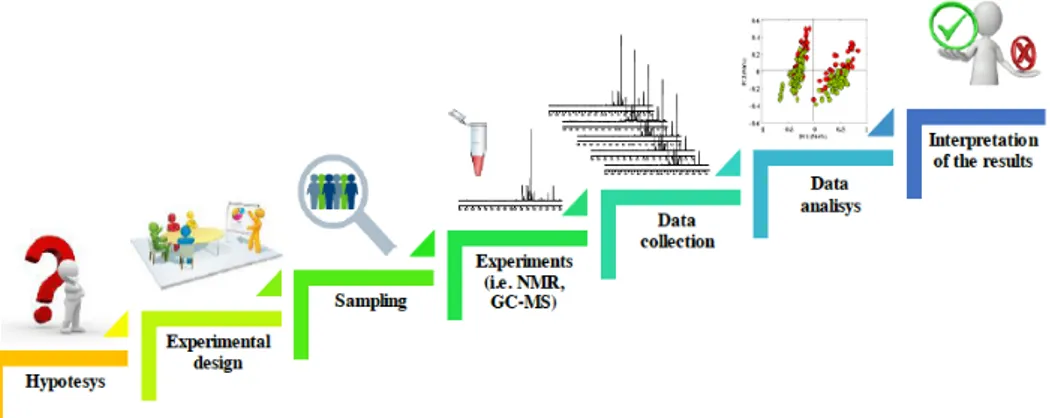
Fig. 1.1. Typical workflow of a metabolomic strudy: hypothesis, experimental design, sampling, analytical platforms, data collection, multivariate data analysis, and interpretation of the results, representing the basic steps in metabolomics.
26 Fig. 3.1. Location of the sampling sites at Aveiro lagoon (Portugal): São
Jacinto (SJ) (40°41′00′′ N, 8°42′44′′ W); Laranjo (LAR) (40º43’28.98’’ N, 8º37’35.80’’ W).
45
Fig. 4.1 Total Hg (tHg), MeHg, inorganic Hg (µg g-1, dry weight) and %
of MeHg (in relation with tHg) in the liver of L. aurata captured in Laranjo (LAR) and São Jacinto (SJ) at Aveiro lagoon. Mean, standard deviation, standard error, outliers (ο) are presented. Significant differences are indicated by ‘a’ vs. SJ. 60
Fig. 4.2 A representative 1-D 500 MHz 1H NMR spectrum of liver tissue
extracts from L. aurata collected from SJ, with (A) representing the aliphatic region and (B) a vertical expansion of the aromatic region. Keys: (1) DSS, (2) taurocholic acid, (3) leucine, (4) isoleucine, (5) valine, (6) lactate, (7) alanine, (8) unknown #1, (9) acetate, (10) glutamate, (11) glutamine, (12) succinate, (13) glutathione, (14) creatine, (15) malonate, (16) phosphocholine, (17) glycerophosphocholine, (18) taurine, (19) unknown #2, (20) glycine, (21) inosine, (22) glucose, (23) glycogen, (24) uracil, (25) uridine, (26) fumarate, (27) tyrosine, (28) unknown #3, (29) phenylalanine, (30) niacinamide, and (31) hypoxanthine. 62
Fig. 4.3
Principal components analysis (PCA) of 1H NMR spectra of liver
extracts showing separation (PC1 vs. PC2) between L. aurata collected from SJ (blue square) and those from LAR (red triangle).
63
Fig. 4.4
Corresponding PC2 loading plot showing the metabolic differences between individuals collected from SJ and LAR. Keys: (1) taurocholic acid, (2) branched chain amino acids: isoleucine, leucine, valine, (3) lactate, (4) alanine, (5) glutamate, (6) glutathione, (7) succinate, (8) phosphocholine, (9) glycerophosphocholine, (10) taurine, (11) glucose, (12) inosine, (13) tyrosine, and (14) hypoxanthine. 63
Fig. 4.5 Oxidative stress responses (CAT, GPx, GR, GST and SOD activities and GSHt and TBARS content) in the liver of L. aurata captured in Laranjo (LAR) and São Jacinto (SJ) at Aveiro lagoon. Mean, standard deviation, standard error, outliers (ο) and extreme values (ø) are presented. Significant differences are indicated by
‘a’ vs. SJ. 66
Fig. 5.1 Total Hg (tHg), MeHg, inorganic Hg (µg g-1, dry weight) and %
of MeHg (in relation with tHg) in the gills of L. aurata captured in Laranjo (LAR) and São Jacinto (SJ) at Aveiro lagoon. Mean, standard deviation, standard error, outliers (ο) are presented. Significant differences are indicated by ‘a’ vs. SJ.
85
Fig. 5.2 A representative 1-D 500 MHz 1H NMR spectrum of gill tissue
extracts from L. aurata collected from SJ, with (A) representing the aliphatic region and (B) a vertical expansion of the aromatic region. Keys: (1) leucine, (2) isoleucine, (3) valine, (4) isobutyrate, (5) lactate, (6) alanine, (7) lysine, (8) arginine, (9) N6-acetyl lysine, (10) glutamate, (11) glutamine, (12) glutathione, (13) acetone, (14) aspartate, (15) creatine, (16) malonate, (17) choline, (18) phosphocholine, (19) glycerophosphocholine, (20) taurine, (21) unknown #1, (22) glycine, (23) serine, (24) betaine, (25) inosine, (26) glucose, (27) UDP-glucose, (28) uracil, (29) uridine, (30) fumarate, (31) tyrosine, (32) phenylalanine, (33) niacinamide, and (34) hypoxanthine.
86
Fig. 5.3 Principal components analysis (PCA) of 1H NMR spectra of gill
extracts showing separation (PC1 vs. PC2) between L. aurata collected from SJ (blue square) and those from LAR (red
triangle). 87
Fig. 5.4 Corresponding PC1 loading plot showing the metabolic differences between gills of L. aurata collected from SJ and LAR. Keys: (1) branched chain amino acids: isoleucine, leucine, valine, (2) isobutyrate, (3) lactate, (4) alanine, (5) glutamate, (6) glutathione, (7) creatine, (8) choline, (9) phosphocholine, (10) glycerophosphocholine, (11) taurine, (12) glycine, (13) serine, (14) uracil, (15) inosine, and (16) fumarate. 88
Fig. 5.5 Oxidative stress responses (CAT, GPx, GR, GST and SOD activities and GSHt and TBARS content) in the gills of L. aurata captured in Laranjo (LAR) and São Jacinto (SJ) at Aveiro lagoon. Mean, standard deviation, standard error, outliers (ο) and extreme values (ø) are presented. Significant differences are indicated by
List of tables
Table 3.1.
Total mercury and methylmercury levels in coastal seawater (579), three estuarine sediments (MESS-2, IAEA-405 and BCR-580) and fish protein (DORM-4), as well the respective certified values. Mean levels and the associated uncertainty are presented.
47 Table
3.2.
Water temperature (T), salinity, dissolved oxygen (DO), total dissolved Hg (DHg), dissolved methylmercury (DMeHg), and the percentage of MeHg with respect to total mercury. Data measured at low-tide are presented for São Jacinto (SJ) and Laranjo (LAR) at Aveiro lagoon. Means and associated standard deviations are presented. 48
Table. 3.3.
Total Hg, methylmercury (MeHg) and the percentage of MeHg with respect to total Hg in surface sediment. Data are presented for São Jacinto (SJ) and Laranjo (LAR) at Aveiro lagoon. Means and associated standard deviations are presented. 49
Table 4.1
Relative changes in metabolite concentrations between LAR and SJ golden grey mullets (p < 0.05a; p < 0.01b; p < 0.005c; Student’s t test). (s: singlet; d: doublet; t: triplet; dd: doublet of doublets; q: quartet; m: multiplet) 64
Table 5.1
Relative changes in metabolite concentrations in gills between LAR and SJ golden grey mullets (p < 0.05a; Student’s t test). (s: singlet;
d: doublet; t: triplet; dd: doublet of doublets; q: quartet; m: multiplet) 89
Table 6.1
Total Hg (tHg), MeHg, inorganic Hg (µg g-1, dry weight) and % of MeHg (in relation with tHg) in the gills and liver of L. aurata captured in Laranjo (LAR) and São Jacinto (SJ) at Aveiro lagoon. Data are presented as mean (± standard deviation).
107
Table 6.2
Relative changes in gill and liver metabolite concentrations between LAR and SJ golden grey mullets (p < 0.05a; p < 0.01b; p < 0.005c; Student’s t test). (- indicates no changes in metabolite
level). 109
Table of contents
Chapter I: Introduction
1.1. General introduction……….……….…………...18
1.2. Environmental monitoring…..…...…………...……….………....…20
1.3. Environmental metal contamination..………...……....23
1.3.1. Toxicity of mercury…..…………...………..…………..25
1.4. Metabolomics……….………...………...….………...26
1.4.1. Metabolomics workflow………..29
1.4.2. NMR-based metabolomics…...…...……..…...…………...…..30
1.4.3. Chemical shifts in NMR spectra………...………...33
1.4.4. Metabolomics in environmental monitoring…...…..……...……34
Chapter II: Aims of the thesis
2. Aims of the thesis...………...…………..…………38Chapter III: Environmental availability of mercury in the
Aveiro Lagoon, Portugal
3.1. General introduction………..……...……...………...403.2. Materials and Methods……….………...………...….………...43
3.2.1. Study area characterization………..43
3.2.2. Sampling………..…45
3.2.3. Mercury in the water column………...46
3.2.4. Mercury in sediments………...47
3.3. Results………...48
3.3.1. Water and sediment characteristics………..48
Chapter IV: Metabolic and oxidative stress responses in liver of
wild mullet Liza aurata
4.1. General introduction………...…51
4.2. Materials and Methods………...………...…53
4.2.1. Chemicals………...53
4.2.2. Mercury in fish liver………...…...………53
4.2.3. Metabolomics analysis………..……....……….54
4.2.3.1. Tissue metabolite extraction………...…...……….54
4.2.3.2. 1H NMR metabolomics and spectral pre-processing….55 4.2.4. Oxidative stress endpoints……….….………...56
4.2.5. Data analysis………..…...……….59
4.3. Results………...……...………..……...………60
4.3.1. Mercury levels in the liver…………...………...60
4.3.2. Metabolomics analysis………...…….…………...61
4.3.2.1. 1H NMR spectroscopy of liver tissue extracts………...61
4.3.2.2. Pattern recognition analysis of 1H NMR spectra…...61
4.3.3. Oxidative stress endpoints………..……...65
4.4. Discussion………..………..……….66
4.4.1. Discussion on metabolic responses to mercury…...…...……...66
4.4.2. Discussion on oxidative stress responses to mercury…………70
4.5. Conclusions………...………..………...………..73
Chapter V: Metabolic and oxidative stress responses in gills of
wild mullet Liza aurata
5.1. General introduction……….……..………...……..765.2. Materials and Methods………...………....…77
5.2.1. Chemicals………...77
5.2.2. Mercury in fish gills………...…...……….77
5.2.3. Metabolomics analysis………..……....……….78
5.2.3.1. Tissue metabolite extraction………...…...……….78
5.2.3.2. 1H NMR metabolomics and spectral pre-processing…...79
5.2.4. Oxidative stress endpoints……….…….………...80
5.2.5. Data analysis………..…...……….83
5.3. Results………...……...……..………...………84
5.3.1. Mercury levels in the gills……...………...84
5.3.2. Metabolomics analysis………...…….………...85
5.3.2.1. 1H NMR spectroscopy of gill tissue extracts……..…….85
5.3.2.2. Pattern recognition analysis of 1H NMR spectra…...87
5.3.3. Oxidative stress endpoints…………...………..……...90
5.4.1. Discussion on metabolic responses to mercury…...……..…...91 5.4.2. Discussion on oxidative stress responses to mercury……...…97 5.5. Conclusions………..………..………...……....102
Chapter VI: Tissue-specific responses to mercury in wild mullet
Liza aurata
6.1. General introduction………….……..…….………...105 6.2. Comparative analysis of mullet tissue responses….…...…….…106 6.3. Discussion………...……..…...110 6.3.1. Disturbances in osmoregulation and ionoregulation………...111 6.3.2. Changes in energy metabolism………....112 6.3.3. Interferences with protein metabolism…………....…………113 6.3.4. Alteration in membrane stabilization processes…...114 6.3.5. Perturbation in antioxidant defence system……….115 6.4. Conclusions…………...………...……...………...……116
Chapter VII: Conclusions
7. Conclusions………...…………...…………..……119
Chapter VIII: References
8. References………...………...…………123
Chapter IX: Appendix
9.1. Curriculum Vitae of Dr. Tiziana Cappello…….………...…....142 9.2. Scientific Production………..………..…….…154
CHAPTER I
1. Introduction
1.1. General introduction
The transitional ecosystems between land and sea, a narrow strip at the edge of both environments, contains some of the most productive and valuable habitats of the world (Valiela, 1995). Coastal lagoons are among these important ecosystems, since several organisms use lagoon habitats for nesting, feeding, reproduction or sheltering (Barnes, 1980). Despite a general decrease in the anthropogenic pressure on coastal ecosystems observed recently in developed countries, coastal lagoons are still undergoing major human impact (Lotze et al., 2006). The most important environmental concerns are associated with unplanned development (urbanization and industrialization), unregulated discharges (municipal sewage and industrial waste) and depletion of resources (over-fishing and bad use of agricultural land). Those activities could lead to the enhancement of contaminant availability and massive algal growth due to eutrophication, associated with an increase in the duration of intermittent periods of lower oxygenation (Rabouille et al., 2007). In fact, the deficient water renewal in coastal lagoons may slow down the dilution process and enhance sediment accumulation and/or retention of contaminants that are transferred to the biota (Mucha et al., 2004). Moreover, the fate of waterborne contaminants in shallow waters is regulated by resuspension and deposition, two physical processes that strongly depend on tidal currents and wind, which have low expression in some lagoon systems.
Therefore, there are concerns about risk to aquatic organisms inhabiting transitional ecosystems, because these organisms are exposed to high concentrations of environmental contaminants due to the intense anthropogenic activities. The major aim of environmental science is to make robust, practical and relatively low cost procedures for risk assessment, and to predict consequences of toxic compounds (Rice, 2003). Methods to effectively
I.
G
EN ER A L IN TR O D U C TI O Nmonitor and quantify these effects are essential to provide an indication of ecosystem health status, an issue of both urgent and international concern.
Traditional approaches addressing this issue frequently use the concept of “biomarkers” to be applied in sentinel species, which may be both invertebrates and lower vertebrates, in order to provide an early warning system of exposure and toxic effects in the ecosystem. Sentinel species can be defined as biological indicators that accumulate a pollutant in their tissues (Beeby, 2001), offering a potentially simple solution to both the problem of measuring bioavailability and of summarizing complex patterns of contamination. Sentinel species should be widely distributed, easy to identify in the field, abundant and large enough to provide material for biomarker analysis (Beeby, 2001; Galloway et al., 2004; Phillips, 1977). The application of biomarkers in environmental research has been recently reinforced with the introduction of “-omics” technologies, which can offer greater insights into the effects of external insults on a biological system at a molecular level.
Among the “-omics” technologies, environmental metabolomics, which involves the study of low molecular weight metabolites, is in fact a cutting edge approach to assessing the health of organisms and discover novel metabolic biomarkers as organismal defensive or adaptive responses to various stress, thus providing an overview of the metabolic status of a biological system (Kell, 2004). In order to achieve a more detailed insight of the organismal health status, mainly in regard to mercury pollution, the aim of this thesis is to integrate environmental metabolomics to a combined approach of metal accumulation and conventional and well-established oxidative stress biomarkers, with the purpose to unravel the toxicity mechanisms of mercury.
I.
G
EN ER A L IN TR O D U C TI O N1.2. Environmental monitoring
To a varying extent, human activities have adverse impacts on the health status of marine environments. As far as threats to the marine environment are concerned, pollution is by far the more significant. The internationally recognised definition of pollution for the marine sector was developed by the Group of Experts on the Scientific Aspects of Marine Environmental Protection (GESAMP, 1993) and reads: “Introduction of man, directly or indirectly, of substances or Energy into the marine environment (including estuaries) resulting in such deleterious effects as harm to living resources, hazard to human health, hindrance to marine activities including fishing, impairment of quality for use of seawater, and reduction of amenities.”
Common class of pollutants arising from natural and anthropogenic sources include heavy metals, polycyclic aromatic hydrocarbons (PAHs), polychlorinated biphenyls (PCBs), organic solvents and dioxins. Some of these compounds may have deleterious effects on both the target organism, and on many non-target species, including humans. Following exposure to such chemicals, these pollutants can be absorbed directly into the bodies of aquatic organisms such as fish and invertebrates via respiratory, as they remove oxygen from the water for respiration (De Zwaan and Eertman, 1996), dermal and oral routes, through the digestive tract following ingestion of contaminated food and water. These processes may also affect humans by their consumption of contaminated seafood (Martin et al., 1996). Therefore, there is an urgent need to understand the biological effects that these pollutants have on vulnerable organisms, such as those living in aquatic environments.
Connections must be established between external levels of exposure, internal levels of tissue contamination and early adverse effects. Many of the hydrophobic organic compounds and their metabolites, which contaminate aquatic ecosystems, have yet to be identified and their impact on aquatic life has yet to be determined. Therefore, the exposure, fate and effects of chemical
I.
E
NVI R ONM E NT AL M ONI T OR IN Gcontaminants or pollutants in the aquatic ecosystem have been extensively studied by environmental toxicologists. Indeed, in the early 20th century, researchers proposed the use of living organisms, in parallel with physico-chemical analysis, to evaluate the health state of the aquatic system (Amiard et al., 1998). In this context, biomonitoring was defined as the systematic use of biological responses to assess changes in the environment (Cairns and Van der Schalie, 1980).
Linking the adverse effects of environmental contaminants in individual animals to their ecosystem-level consequences is a key challenge in regulatory risk assessment (Moore et al., 2004). Ecological/environmental risk assessment (ERA) is defined as the procedure by which the likely or actual adverse effects of pollutants and other anthropogenic activities on ecosystems and their components are estimated with a known degree of certainty using scientific methodologies (Depledge and Fossi, 1994; Van der Oost et al., 2003). The risk assessment process identifies and quantifies the risk resulting from a special use or occurrence of a chemical compound, and seeks a solution to the problem, whereas risk analysis determines the risk of a specific situation (Van der Oost et al., 2003). ERA has become increasingly important since environmental scientists as well as the general public have learned that chemicals which are not toxic to humans can have deleterious effects on natural resources which are generally valued (Bascietto et al., 1990). Although ERA is generally performed by predictive methods, the interest in the assessment of pollution that began in the past and may have ongoing consequences in the future is increasing. These so-called retrospective ERAs are primarily concerned with establishing the potential relationship between a pollutant source and an ecological effect caused by exposure of organisms to the pollutant (Suter, 1993).
The responses to pollutant stress within a biological system (Bayne et al., 1985) have triggered the research to establish early-warning signals, or biomarkers, reflecting the adverse biological responses towards anthropogenic
I.
E
NVI R ONM E NT AL M ONI T OR IN Genvironmental toxins (Bucheli and Fent, 1995). A biomarker is defined as a change in a biological response starting at the subcellular level (e.g. interference with molecular pathways) and ultimately leading to adverse effects at higher levels of biological organization (De Coen and Janssen, 2003), which can be related to exposure to or toxic effects of environmental chemicals (Peakall, 1994). In principle, these early warning biomarkers should be capable of predicting reduced performance, impending pathology and damage to health (Moore et al., 2004). Hence, biomarkers should be able to identify those organisms that have been, or are being, exposed to certain chemicals or those organisms that are suffering, or will suffer, future impairments of ecological relevance (Forbes et al., 2006). In an environmental context, biomarkers thus offer promise as sensitive indicators demonstrating that toxicants have entered organisms, have been distributed between tissues, and are eliciting a toxic effect at critical targets (McCarthy and Shugart, 1990).
Van Gastel and Van Brummelen (1996) redefined the terms ‘biomarker’, ‘bioindicator’ and ‘ecological indicator’, linking them to different levels of biological organization. They considered a biomarker as any biological response to an environmental chemical at the sub-individual level, measured inside an organism or in its products (urine, faeces, hair, feathers, etc.), indicating a deviation from the normal status that cannot be detected in the intact organism. A bioindicator is defined as an organism giving information on the environmental conditions of its habitat by its presence or absence or by its behavior, and an ecological indicator is an ecosystem parameter, describing the structure and functioning of ecosystems. Since many of the biomarkers are short-term indicators of long-term adverse effects, these data may permit intervention before irreversible detrimental effects become inevitabile (McCarthy and Shugart, 1990). Various biochemical parameters in aquatic organisms have been tested for their responses to toxic substances and their potential use as biomarkers of exposure or effect. Biomarkers, which have been
investigated most extensively, are enzymes involved in the detoxication of
I.
E
NVI R ONM E NT AL M ONI T OR IN G
xenobiotics and their metabolites (biotransformation enzymes, antioxidant enzymes), as reported by Van der Oost et al. (2003).
Nevertheless, despite the common use of biomarkers in ecotoxicology, some authors have claimed that such approaches have failed to live up to their promise. Forbes et al. (2006) listed a number of different limitations that have blighted numerous biomarker studies, and made a series of demands on how such experiments could be improved. In order to achieve a more detailed insight of the organismal health status, the application of biomarkers has recently been reinforced with the introduction of “-omics” technologies, which can offer greater insights to the pollutant effects at a molecular level.
1.3. Environmental metal contamination
An increasing variety of industrial and agricultural chemicals are introduced in coastal ecosystems. Among the best studied contaminants, there are chemicals of both longstanding and more recent concern, such as polycyclic aromatic hydrocarbons (PAHs), organochlorine pesticides (e.g. dichlorodiphenyltrichloroethane, DDT), industrial products (e.g. polychlorinated biphenyls, PCBs), dioxins, nitroaromatic compounds, organometallic compounds, pesticides, estrogenic compounds, and many metals including Cd, Cr, Cu, Fe, Hg and Zn (Livingston, 2001). Metals are of great environmental concern, since they tend to concentrate in aquatic organisms, are virtually non-degradable, and thus produce long lasting effects upon the environment even after their major sources have been removed.
Metals are introduced into coastal systems through fluvial inputs, direct effluent discharges and by the atmosphere. After discharged metals are mostly adsorbed on suspended particles and finally accumulate in the sediment, which can serve as sink or source of metals to the overlying water. Many aquatic
I.
E
NVI R ONM E NT AL M E T AL C ONT AM INAT IONorganisms spend a major portion of their life in or on sediment with the possibility to take up those metals. Moreover, numerous studies have shown that sediment-water interaction in aquatic systems play an important role on controlling metals transport processes (De Domenico et al., 2013, 2011; Gomez et al., 1999; Point et al., 2007; Thouzeau et al., 2007). Indeed, the mineralization of organic matter plays a fundamental role in the sediment-water exchanges of metals.
A number of metals are used by living organisms to stabilize protein structures, facilitate electron transfer reactions, and are essential cofactors for oxidative phosphorylation, in gene regulation and free-radical homeostasis. For example, Cu, Zn and Fe are essential as constituents of the catalytic sites of several enzymes (Siegel, 1973). Nevertheless, other metals like Pb, Hg and Cd may displace or substitute essential metals and interfere with the proper functioning of enzymes and associated cofactors. Trace elements may accumulate in aquatic organisms through different mechanisms: directly from water, via uptake from suspended particles and sediment, or by the consumption of lower trophic level organisms. The former is an essential point to consider in evaluating adverse effects on ecosystems (Van der Oost et al., 2003). In view of that, there are several works that use the accumulation of metals in organisms as mean to assess the environmental health status (Fernandes et al., 2007; Morrison et al., 2007; Pereira et al., 2009).
The absorption of metals by aquatic animals involves their transfer to the circulatory system by epithelial barrier of gills, digestive organs or integument. Dissolved metals are mainly taken up by exposed body surfaces such as the gills, whereas particulate metals are mostly ingested and then taken up after solubilization in the gut. Uptake of essential metals such as Ca, Cu, Fe and Zn, often involves specific pathways, such as calcium channels and specific membrane carriers for Fe and Cu (Sunda and Huntsman, 1998). For nonessential metals (e.g. Cd and Hg) specific uptake mechanisms are not known and, thus appear to follow existing pathways for essential metals
I.
E
NVI R ONM E NT AL M E T AL C ONT AM INAT ION(Sunda and Huntsman, 1998).
Sequestration of metals in an immobilized form occurs throughout the various organs involved in pathways for metal uptake, transport, utilization and release. One of the best studied intracellular structures are the metallothioneins. These are low-molecular-weight cytosolic proteins rich in -SH groups, with high affinity for metal ions, known to be involved in metal homeostasis and over-expressed in organisms experiencing high metal contamination (Viarengo et al., 1998). Their expression in tissues is regarded as an indicator of metal contamination and widely used as a tool for biomonitoring programs (Fasulo et al., 2008; Viarengo et al., 1998).
Aquatic organisms utilize a variety of mechanisms to eliminate metals. The kinetic of metal release is complex and reflects the diverse compartments from which metals must be mobilized. Additionally, physical and chemical parameters, such as temperature and salinity, may affect the rate of release in aquatic animals, which can use several pathways to release metals (Mieiro et al., 2014, 2011; Pereira et al., 2014).
1.3.1. Toxicity of mercury
Among trace elements, mercury (Hg) is worldwide recognized as a hazardous pollutant mainly due to its persistence in water and sediments (Luoma and Rainbow, 2008), high toxicity to living organisms and tendency to bioaccumulate and biomagnify throughout food chains (Renzoni et al., 1998). The main sources of Hg to aquatic ecosystems derive both from natural processes (e.g. geological emissions) as well as anthropogenic activities, such as fossil fuel combustions (Pacyna et al., 2001), mining and smelting operations, and chlor-alkali industries (Driscoll et al., 2013; UNEP, 2011).
I.
E
NVI R ONM E NT AL M E T AL C ONT AM INAT IONDespite the great efforts on evaluating the biological impact of Hg on aquatic organisms (De Domenico et al., 2013, 2011; Mieiro et al., 2014, 2011), the toxicity mechanisms of Hg still need to be clarified. In recent field and laboratory studies, oxidative stress has been described as a key pathway to initiate Hg toxicity in fish (Elia et al., 2003; Larose et al., 2008; Mieiro et al., 2014, 2011; Monteiro et al., 2010). Hence, both the modulation of antioxidant enzymes and changes in glutathione (GSH) content have been frequently employed as useful biomarkers for monitoring Hg contamination on aquatic organisms (Brandão et al., 2015; Guilherme et al., 2008; Mieiro et al., 2011).
However, these currently used biochemical assays are often inconclusive on elucidation of the mechanisms underlying the Hg-induced oxidative stress in fish. Hence, understanding the Hg toxicity requires new ways. In this regard, the use of environmental metabolomics may serve as a powerful tool focusing on a number of key metabolites, which changes might reveal insights into the mechanisms of oxidative damage due to Hg. Although metabolomics has been extensively used for mechanistic research, no studies have been developed to specifically investigate this scientific question. Therefore, the present thesis aims to addresses this specific issue and represents a very recent example of the potential of metabolomics to clarify the toxicity mechanisms of Hg, providing insights on the metabolic and oxidative stress responses in wild fish.
1.4. Metabolomics
The “-omics” sciences are readily increasing disciplines aimed at the study of biological systems (Berry et al., 2011). They include, among the others, genomics, transcriptomics, proteomics, and metabolomics. Whereas genomics, transcriptomics, and proteomics are based on the analysis of the genome, gene expression and proteins, respectively, metabolomics is deemed as the end point
I.
M
ETA B O LO M IC Sof the “-omics cascade” (Dettmer and Hammock, 2004).
Indeed, one of the most recent additions to the “-omics” family is metabolomics, which in 2002 was defined by Fiehn as “the qualitative and quantitative study of the metabolome in a biological system” (Fiehn, 2002). Metabolomics is focused on the study of endogenous low molecular weight metabolites (<1000 Da), whose production and levels vary with the physiological, developmental, or pathological state of cells, tissues, organs or whole organisms (Lin et al., 2006; Viant, 2007). The metabolome describes the composition of low molecular weight metabolites at the time of sampling, and includes compounds such as lipids, sugars, and amino acids that can provide important clues about the health of individuals and a functional measure of cellular status at that moment in time (Lin et al., 2006; Schmidt, 2004).
There are several advantages for the application of this technique. One of the greatest advantages of metabolomics is that the metabolome is often the first to respond to anthropogenic stressors, where in some cases no changes in the transcriptome and proteome occur (Viant, 2007). Hence, it has been suggested that metabolomics may provide the most functional information of the “-omics” technologies (Sumner et al., 2003), as transcript and protein changes do not necessarily lead to a biochemical change in the study organism (Fiehn et al., 2002, 2000). Furthermore, the metabolome, a term coined by Oliver et al. (1998) to describe the set of metabolites synthesised by an organism in a fashion analogous to that of the genome and proteome, represents the final “-omic” level in a biological system, and metabolites represent functional entities, unlike messenger RNA molecules, which are further upstream of biological processes (Raamsdonk et al., 2001). Metabolites thus have a clear function in the life of the biological system and are also contextual allied with the further advantage that there are far fewer metabolites than genes or gene products to be studied (Raamsdonk et al., 2001). However, it should be duly noted that all “-omics” technologies provide valuable information, and integration of these techniques promises to provide the most
I.
M
ETA B O LO M IC Scomplete understanding of biological systems.
Basically, metabolomic studies can be divided in targeted and untargeted analyses (Fernández-Peralbo and Luque de Castro, 2012). In untargeted approaches, significant metabolites are, by definition, unknown prior to analysis, while in targeted analysis, the physico-chemical characteristics of the metabolites are known and an exhaustive separation of them from the matrix is usually required for quantification. Therefore, metabolomics investigations can be designed as targeted studies looking for specific metabolite changes, although this requires some prior knowledge on the metabolic action of whatever toxicant is being tested. Otherwise, an alternative metabolomic approach is where the global metabolome is analysed, although constrained by the efficiency and sensitivity of the techniques used to extract and detect the metabolites. This method is a relatively non-targeted approach where there is little, if any, prior selection of which metabolic components to measure. Thus, a similar study design can be used in both a screening mode and for mechanistic exploration (Keun, 2006).
A further differentiation of metabolomic analyses can be done based on the scientific application, namely metabolic profiling, metabolic fingerprinting, metabolic footprinting, and metabolomics (Oldiges et al., 2007). Metabolic profiling is the quantitative analysis of a group of pre- defined metabolites, like members of a particular pathway. Metabolic fingerprinting and metabolic footprinting are metabolomic studies focused on the classification of samples by analysing their intracellular metabolites (endometabolome) and extracellular metabolites (exometabolome), respectively. Lastly, metabolomics can be defined as the complete analysis of the entire cellular metabolome, in which all the metabolites are quantified and identified. While target analysis, metabolic profiling, and metabolomics are all quantitative approaches that require unique identification of all metabolites, metabolic fingerprinting and metabolic footprinting are semi-quantitative approaches and even unknown metabolites
can be used to get deeper insights into samples metabolic profiles (untargeted).
I.
M
ETA B O LO M IC S
1.4.1. Metabolomics workflow
Independently of the field of application, metabolomic studies have to pass through numerous steps in order to achieve profitable and reliable results. Indeed, a typical workflow of a metabolomic strudy includes hypothesis, experimental design, sampling, analytical platforms, data collection, multivariate data analysis, and interpretation of the results, which represent the basic steps in metabolomics.These aspects are summarized in Figure 1.1.
Figure 1.1. Typical workflow of a metabolomic strudy: hypothesis, experimental
design, sampling, analytical platforms, data collection, multivariate data analysis, and interpretation of the results, representing the basic steps in metabolomics.
Among these aspects, sample selection and preparation is a crucial point in metabolomic experiments. A correct sampling provides a real snapshot of the metabolome at a certain point in time, hence the necessity to adopt procedures fostering an unbiased sampling. Strategies of sampling and sample preparation vary according to the experimental setup, thus different strategies for the metabolites sampling can be performed. Extracellular metabolites present in human or animal biofluids are sampled using either non-invasive (urine) or invasive (serum, plasma) methods. As a matter of fact, the process of sampling
I.
M
ETA B O LO M IC Scan change the metabolome composition (Dunn and Ellis, 2005).
Samples storage is another crucial point in metabolomic analysis as the continued freezing/defrosting of samples could damage their molecular stability. The inhibition of the enzymatic activity, aimed to preserve sample biochemical composition, is normally achieved through the freeze clamping or freezing in liquid nitrogen followed by storage at -80 °C (Dunn and Ellis, 2005).
In regard to metabolite extraction, the adopted procedures dictate the nature and levels of the extracted metabolites. For non-targeted approaches, the objective is to extract the maximum number of metabolites from many chemical classes in a quantitative and non-biased manner with minimal losses of metabolites. For metabolic profiling, extraction is generally performed by the disruption of cell walls and subsequent distribution of metabolites into polar (methanol, water) and non-polar (chloroform, hexane, ethyl acetate) solvents followed by the removal of the cellular residue.
The preparation of samples for the analysis is also dependent on the metabolomics strategy employed. Targeted analyses require separation of the metabolome into chemical classes, whereas for metabolic profiling and fingerprinting analyses, samples are mainly analysed directly without further separation of metabolites into subclasses (Emwas et al., 2013).
However, in spite of the great variability related to the sampling procedure, it is generally recognized that the impact of unpredictable biological variability is much higher than that related to the analytical one.
1.4.2. NMR-based metabolomics
Metabolites separation and identification is made possible thanks to several
advanced analytical techniques. Nuclear Magnetic Resonance (NMR)
I.
M
ETA B O LO M IC S
spectroscopy, Mass Spectrometry (MS), and chromatographic methods, such as Gas Chromatography (GC) and High Performance Liquid Chromatography (HPLC), are the most commonly used platforms (Dunn and Ellis, 2005). All techniques have advantages and drawbacks and there is not an analytical technique completely suitable for metabolomic studies. In particular, NMR and MS have been demonstrated to be complementary and powerful analytical approaches for the complete characterization of the metabolome (Pan and Raftery, 2007).
By virtue of its numerous advantages, NMR spectroscopy is largely used in metabolomic studies for the analysis of bulk metabolites. High-resolution NMR spectroscopy is a quantitative technique that can report on hundreds of compounds in a single measurement. NMR can provide information on metabolites that comprise nuclei such as 1H, 13C, 31P. These nuclei can exist at different energy states in a strong magnetic field because they possess nuclear spin, allowing the generation of valuable structural information. NMR spectroscopy (and most spectroscopic techniques) is based on the principle that lines that can be seen in the spectra are due to transitions between these energy states or levels. Such a transition can be caused by a photon of light whose frequency, υ, is related to the energy gap, Δ E, between the two levels according to:
Δ E = hυ
where h is the universal constant, namely Plank’s constant.
The appearance of multiplets and other peaks in an NMR spectrum are predicted by using different rules to this approach but are still related to energy levels. The splitting of energy levels in a magnetic field is dependent on the nuclear spin of the atom. For atoms that possess spin (such as 1H, 13C, 31P), the net spin of the nucleus can be determined from the number of protons plus neutrons. When the number of protons and number of neutrons are even, there
I.
M
ETA B O LO M IC Sis no spin. If the number of protons plus neutrons is odd then the nucleus has half integer spin (i.e. 1/2, 3/2, 5/2) and if the number of protons and neutrons are both odd then the nucleus has an integer spin (i.e. 1, 2, 3). The overall spin, I, is important and a nucleus of spin I will have 2I + 1 possible orientations. When a magnetic field is applied, the energy levels split with each level given a magnetic quantum number, m. In a magnetic field a nucleus with spin 1/2 will have two orientations: m = +1/2, a low energy state aligned parallel to the magnetic field; and m = -1/2, a high energy state aligned anti-parallel to the magnetic field. The initial populations of the energy levels are determined by thermodynamics, as described by the Boltzmann distribution and means the lower energy level will contain slightly more nuclei than the higher level. However, these nuclei can be excited into the higher level by electromagnetic radiation with the frequency of radiation needed determined by the difference between the energy levels.
In an NMR experiment, each nucleus has a magnetic moment and their relative contributions add up to create a net magnetic field along the direction of the applied field (B0). This is called bulk magnetisation and can be represented by a vector pointing along the direction of the applied field (z). If this magnetisation vector is tipped away from the z axis, which can be brought about by the application of a radiofrequency pulse, it rotates about the direction of the magnetic field sweeping out a constant angle. The vector is said to precess about the field in a motion known as Larmor precession at the Larmor frequency (Keeler, 2005). The precession of the magnetisation vector is what is detected in an NMR experiment, known as the free induction signal. If these signals were to be plotted they would represent simple oscillations at the Larmor frequency, and Fourier transformation of these signals can produce an NMR spectrum.
The magnetisation can be rotated away from its equilibrium position along the z axis using the idea of resonance by applying a small magnetic field along
the x axis that is resonant with the Larmor frequency. Different types of pulses
I.
M
ETA B O LO M IC S
exist and when the transmitter frequency is exactly the same as the Larmor frequency, the pulse is said to be exactly on resonance (Keeler, 2005). Pulses can be modified by changing the time at which they are applied. This alters the angle (known as “flip angle of the pulse”) through which the magnetisation has been rotated. Commonly used flip angles are 90º where the process is called a 90º pulse, and 180º where the magnetisation is taken all the way from +z to –z. In practical NMR spectroscopy multiple resonances are usually present in the spectrum with different Larmor frequencies. A sufficiently strong radio frequency (RF) pulse is therefore needed to overcome the induced field to move all the signals at different Larmor frequencies away from equilibrium and this is called a hard pulse. When the pulse is weaker it is possible to excite a single signal within the spectrum and can be done by choosing a radio frequency identical to a selected signal reducing the influence of the pulse on other signals. These pulses are known as selective pulses or soft pulses and can be used to help suppress resonances that are not of interest in the sample (for example solvent-based resonances).
1.4.3. Chemical shifts in NMR spectra
Depending on the local chemical environment, different protons in a molecule resonate at slightly different frequencies, and frequencies at which NMR absorptions (lines) occur scale linearly with the magnetic field strength. As this frequency shift and the fundamental resonant frequency are directly proportional to the strength of the magnectc field, the shift can be converted into a dimensionless value known as the chemical shift. The chemical shift is then reported relative to a reference resonance frequency (a commonly used compound is sodium 3-trimethylsilyl-2,2,3,3-d4-propionate (TMSP), or 2,2-dimethyl-2-silapentane-5-sulfonate (DSS), which is used in this thesis). The
I.
M
ETA B O LO M IC Sdifference between the frequency of the signal and the frequency of the reference is divided by the frequency of the reference signal to give the chemical shift and is expressed in parts per million (ppm) (Keleer, 2005).
The chemical shift can be used to obtain structural information. For the 1 H-NMR spectrum of ethanol (CH3CH2OH), three specific signals are expected at three specific chemical shifts: one for the CH3 group, one for the CH2 group and one for the OH group. The three methyl groups average out during the course of the NMR experiment and the protons become degenerate forming a peak at the same chemical shift. The size and shape of peaks also give further information and the CH3 peak would be three times as large as the OH, and the CH2 peak only twice the size of the OH peak but 2/3 the size of the CH3 peak. The integrated areas under the peaks are also important, and in a complex mixture they can be used to predict the concentration of a particular compound.
1.4.4. Metabolomics in environmental monitoring
Nuclear magnetic resonance (NMR) spectroscopy-based metabolomics, when linked with pattern recognition techniques and data mining tools, can detect differences in the profile of metabolites (metabolite biomarkers) in response to environmental stressors, diseases or exposure to toxicants (Cappello et al., 2016a, 2016b, 2015, 2013b; Fiehn, 2002; Hines et al., 2007; Liu et al., 2015; Tuffnail et al., 2009; Viant et al., 2003; Xu et al., 2015), thus providing an overview of the metabolic status of a biological system. Among the pattern recognition techniques, Principal Component Analysis (PCA) is a multivariate statistical technique which can be used to identify correlations amongst a set of variables and to transform the original set of variables to a new set of uncorrelated variables called principal components (PCs). PCs are linear combinations of the original variables and are derived in decreasing
I.
M
ETA B O LO M IC Sorder of importance. Therefore, the first PC accounts for the maximum variation among the samples, and subsequent PCs are chosen to account for progressively decreasing variance (Chatfield and Collins, 1980; Jolliffe, 1986; Martens and Naes, 1989). PCA is a variable-directed technique and therefore does not use any a priori knowledge of the groupings within samples in the data set. It is an unsupervised method, and thus plots of PCs are thought to display the natural relationships between the samples on a score plot.
Metabolite profiling, originally developed for human biomedical applications (Nicholson et al., 1988) has now been increasingly employed in several research areas, including plant science (Kim et al., 2010), food quality (Tarachiwin et al., 2008), microbial metabolomics (Boroujerdi et al., 2009) and environmental metabolomics (Viant, 2009). Because metabolomics can provide valuable information on how xenobiotics influence physiological functions, this technique has also been applied to experimental studies of selective exposure on various aquatic organisms, both invertebrates (Cappello et al., 2013b; Wu and Wang, 2010) and fish (Cappello et al., 2016a, 2016b; Iacono et al., 2010; Santos et al., 2010).
In ecotoxicology, metabolomics is of particular value for the risk assessment of chemicals in the environment (Lin et al., 2006). Recently, there have been numerous studies applying metabolomic techniques to this area of research. Various studies have been published in aquatic species on the effects of pesticides and other xenobiotics using NMR-based metabolomics. Experiments with Japanese medaka (Oryzias latipes) have shown that metabolic changes in embryos exposed to dinoseb correlated with traditional toxic endpoints such as reduced growth and heart rates, abnormal development and post-exposure mortality (Viant et al., 2006a). Results from 1H NMR metabolomics were also in agreement with findings from a previous study using in vivo 31P NMR and HPLC-UV, in which phosphocreatine utilisation for compensation of ATP loss could be used as an indicator of medaka embryotoxicity (Pincetich et al., 2005).
I.
M
ETA B O LO M IC SThe advantages of NMR-based metabolomics has also been proven in studies involving salmon (Oncorhynchus tshawytscha) alevins exposed to dinoseb, diazinon and esfenvalerate where identifiable metabolic changes were detected with greater sensitivity using NMR compared to HPLC-UV (Viant et al., 2006b). Furthermore, the metabolic fingerprints of eyed egg and alevin extracts revealed both dose-dependent and mechanism-specific responses induced by pesticides.
Metabolomics applications to disease monitoring in wild species have been shown by studies in Californian red abalone (Haliotis rufescens) (Rosemblun et al., 2006; Viant et al., 2003). NMR metabolomics was successfully able to distinguish between healthy, stunted and diseased groups afflicted with a disease known as withering syndrome associated with a Rickettsiales-like prokaryote (RLP) infection. The ratio between the metabolites homarine and glucose in foot muscle was able to effectively predict the disease status of RLP infected animals. Other environmental stressors such as increased water temperature were also assessed and judged to be a contributing factor in disease development since RLP infection alone did not cause animals to develop withering syndrome.
A further parasitic infection investigated using NMR-based metabolomics involved Atlantic salmon (Salmo salar) exposed to the bacterium Aeromonas salmonicida. The metabolic response consisted primarily of changes in lipoprotein and choline levels for infected salmon highlighting the potential of metabolomics for studying the disease status in this and other wild species (Solanky et al., 2005).