Age-related differences in plasma BDNF levels after prolonged bed rest
XCecilia Soavi,1Uroš Marušicˇ,2Juana Maria Sanz,1Mario Luca Morieri,1Edoardo Dalla Nora,1 Bostjan Šimunicˇ,2Rado Pišot,2Giovanni Zuliani,1and Angelina Passaro1
1Medical Science Department, University of Ferrara, Ferrara, Italy; and2Science and Research Centre, University of Primorska, Capodistria, Slovenia
Submitted 7 January 2016; accepted in final form 1 March 2016
Soavi C, Marušicˇ U, Sanz JM, Morieri ML, Dalla Nora E, Šimunicˇ B, Pišot R, Zuliani G, Passaro A. Age-related differences in plasma BDNF levels after prolonged bed rest. J Appl Physiol 120: 1118–1123, 2016. First published March 3, 2016; doi:10.1152/japplphysiol.01111.2015.—Brain-derived neurotrophic factor (BDNF) is a member of the family of neurotrophins and has been implicated in brain resistance to insults. Murine studies have demonstrated increased hippocampal concentra-tion after acute immobilizaconcentra-tion and decreased concentraconcentra-tion after chronic immobilization. In humans, chronic stress and sedentary lifestyle result in decreased plasma BDNF levels, but there no data exist regarding acute immobilization. The aim of our study was to evaluate age-related responses [comparing 7 younger subjects (age 23 ⫾ 3 yr) and 8 older subjects (age 60 ⫾ 4 yr)] of plasma BDNF before (baseline data collection, BDC) and after 14 days (BR14) of horizon-tal bed rest (BR). At BDC, BDNF levels were not different between the two groups (P⫽ 0.101), although at BR14, BDNF levels were higher in older subjects (62.02 ⫾ 18.31) than in younger subjects (34.36 ⫾ 15.24 pg/ml) (P ⫽ 0.002). A general linear model for repeated measures showed a significant effect of BR on BDNF (P⫽ 0.002). The BDC BDNF levels correlated with fat-free mass in both populations (ALL) (R⫽ 0.628, P ⫽ 0.012), (older, R ⫽ 0.753, P ⫽ 0.031; younger, R⫽ 0.772, P ⫽ 0.042), and with total cholesterol in ALL (R⫽ 0.647, P ⫽ 0.009) and older study subjects (R ⫽ 0.805,
P⫽ 0.016). At BR14, BDNF correlated with total cholesterol (R ⫽
0.579, P⫽ 0.024) and age (R ⫽ 0.647, P ⫽ 0.009) in ALL. With an increase in age, the brain could become naturally less resistant to acute stressors, including the detrimental effects of prolonged bed rest, and thus the increase in BDNF in the older study group might reflect a protective overshooting of the brain to counteract the negative effects in such conditions.
brain-derived neurotrophic factor; bed rest; acute stress; aging brain
NEW AND NOTEWORTHY
Blood concentrations of brain-derived neurotrophic factor (BDNF) increase after 14 days of bed rest in older but not younger people, and may indicate that the aging brain is less resistant to acute stresses, and increases in BDNF could represent the protective response to the acute stress of bed rest. MANY ELDERLY INDIVIDUALS AREnecessarily bedridden at a
cer-tain point in their life; for example, an acute medical condition may require hospitalization, with negative effects on metabo-lism and both motor and cognitive performance. In fact, bed rest was demonstrated to induce insulin resistance, increase total cholesterol and triglyceride plasma levels, impair micro-vascular regulation, and increase basal arterial tone leading to hypertension (1, 16, 50). Moreover, bed rest has been associ-ated with a rapid decrease in muscle size due to a reduction in
muscle protein synthesis and an increase in protein degradation (57); in addition, prolonged bed rest was shown to act as a stressful agent on the brain, negatively affecting brain structure (23), manifesting changes in brain electrocortical activity (30) and detrimentally effecting executive functioning and mood (24, 25).
Brain-derived neurotrophic factor (BDNF) is a growth factor member of the neurotrophin family. It binds specifically to the tyrosine receptor kinase B (TrkB), a tyrosine kinase receptor, thus mediating neurotrophic signaling (8). During normal de-velopment BDNF plays a critical role in cell differentiation, migration, neuronal survival, dendritic arborization, synapto-genesis, and synaptic plasticity. But also following insults, in developing/adult brain, BDNF plays an important role in the repair of damage and resistance to insults (8).
Even if the cellular sources of BDNF found in human plasma are not yet clearly defined, there is some evidence suggesting a pivotal role in cerebral output. Pan et al. (34) showed that in mice, BDNF can cross the blood-brain barrier bidirectionally, and in studying rat models, Karege et al. (18) demonstrated similar hippocampal and serum levels during maturation and aging processes in which these changes were compatible with the changes in hippocampal BDNF mRNA expression, but not with BDNF mRNA expression in platelets. On the basis of these results serum concentration of BDNF has already been used as indirect measure of BDNF activity in the central nervous system (9). In humans, release of BDNF from the brain has been observed at rest, and a 2- to 3-fold increase occurred during exercise, and Rasmussen et al. (40) estimated that in both conditions brain contribution determined 70-80% of total circulating BDNF.
Alternatively, although BDNF is upregulated in contracting muscle fibers, it has been demonstrated that muscles are not a source of circulating BDNF (31, 37).
Low levels of plasma BDNF have been associated with Alzheimer and other forms of dementia (56), diabetes (20, 58), depression (5, 17), and acute coronary syndrome (28); we have recently suggested the existence of a synergistic effect of late-onset Alzheimer disease and diabetes on BDNF plasma levels (36).
Interestingly, in murine models, a different behavior of BDNF was noticed in response to chronic and acute immobi-lization. Chronic immobilization, as well as other chronic stressors, reduce BDNF concentration in rat hippocampus (41, 42, 49, 54, 55), whereas Marmigère et al. (29) demonstrated that in a very acute phase of immobilization stress (15– 60 min), a transient increase in hippocampal BDNF mRNA ex-pression occurs, followed by a reduction to levels lower than basal at 180 min; similarly, an increase in hippocampal BDNF content was found at 180 min, which rapidly returned at basal levels when the immobilization stimuli lasted up to 300 min.
Address for reprint requests and other correspondence: A. Passaro, Medical Science Dept., Univ. of Ferrara, Via Ariosto 35, 44121, Ferrara, Italy (e-mail: [email protected]).
The same result can be obtained acutely by exposing rats to damaging factors such as hypoxia, ischemia, and neurotoxic substances (3), or other kinds of acute stress such as forced swimming in cold water (47). In this context, acute immobili-zation can be considered as an acute stress factor that induces activation of short-term protective mechanisms, whereas chronic stressors as chronic immobilization can lead to exces-sive stimulation and functional distortion of neuroendocrine systems by reducing BDNF expression (33).
Similarly, studies with humans have showed a reduction in serum BDNF levels in sedentary subjects (45), such as people exposed to chronic stress (7, 35), but data are lacking about plasma BDNF responses to acute stressors. Thus we hypothe-sized that plasma levels of BDNF could be associated with long-term bed rest in human subjects and that there will be age-related differences. To test this hypothesis we used data collected during 14 days of horizontal bed rest in which seven younger and eight older healthy adults were tested before and after 14 days of horizontal bed rest.
MATERIALS AND METHODS
Population. We designed a study that consisted of two groups of
healthy volunteers: younger subjects [n ⫽ 7, age 19–28 yr (mean 23.3⫾ 3.X)] and older subjects (n ⫽ 8, mean age 59.5 ⫾ 4.X yr). After 3 days of an ambulatory period (regulated hospital diet and daily activities), all subjects underwent horizontal bed rest for 14 days in standard air conditioned hospital rooms at the Orthopedic Hospital of Valdoltra (Slovenia). Continuous surveillance and 24-h medical care was provided during the entire bed rest period, and all subjects received an individually controlled, normal caloric diet: for each subject, resting energy expenditure (estimated by means of bioimpe-dentiometric measures) was multiplied by a factor of 1.2, with caloric content consisting of 60% carbohydrates, 25% fats, and 15% proteins (4). Subjects performed all daily activities in bed, and were allowed to freely communicate, watch television, listen to radio, read, use a computer, and receive visitors.
Exclusion criteria included smoking; regular alcohol consumption; ferromagnetic implants; history of deep vein thrombosis with D-dimer levels at enrollment⬎500 g/l; acute or chronic skeletal, neuromus-cular, metabolic, or cardiovascular disease; and pulmonary embolism. All subjects gave their written informed consent. The study was performed in accordance with the ethical standards of the 1964 Declaration of Helsinki.
Biological samples and measurements. Blood samples were
col-lected from each subject after an overnight fasting at enrollment [i.e., baseline data collection (BDC)] and after 14 days of forced bed rest (BR14) and centrifuged in the absence or presence of EDTA to obtain serum and plasma, respectively. Aliquots were stored at ⫺80°C. Levels of HDL cholesterol after precipitation of lipoproteins con-taining apo-B (6), total cholesterol, and triglycerides (TG) were assayed in serum using the Trinder method. The coefficient of variation was⬍2% for total and HDL cholesterol and ⬍5% for TG for intrabatch and interbatch, respectively. LDL cholesterol plasma levels were calculated using the Friedewald formula (12). Plasma glucose was measured using standard enzymatic methods (FAR, Italy). The intra-assay coefficient of variation was ⬍3%. Fasting insulin levels were assayed using an ultrasensitive insulin ELISA kit manufactured by Mercodia (Sweden), and the intra-assay coefficient of variation was ⬍3%. Fasting insulin resistance was evaluated by calculating the homeostasis model assessment (HOMA-IR) (32). TNF-␣ plasma levels were measured using an ELISA Kit (Invitrogen, USA). The intra-assay coefficient of variation was⬍5.2%. C-reactive protein plasma levels were measured using an immunoturbidimetric test (High sensitive C-reactive protein, Roche, Italy). The intra-assay
coefficient of variation was⬍2%. BDNF plasma levels were mea-sured by means of ELISA (Promega, Italy), following the manufac-turer’s instructions.
At BDC and BR14 all subjects were tested with bioimpedentio-metric measures for body composition using a tetrapolar impedance-meter (BIA101; Akern, Florence, Italy) according to the manufactur-er’s instructions (27). All measurements were collected by the same trained staff member after subjects had fasted for 8 h.
Statistical analysis. Continuous variables were expressed as means
(SD) or, when necessary, as median (range), and categorical variables as the number/percentage. Means were compared by one-way ANOVA using the Bonferroni test for post hoc analysis, whereas medians were compared using the nonparametric Kruskal-Wallis test. Normality of distribution was tested with the Shapiro-Wilk test. Variations in BDNF plasma levels and the other variables of interest between BDC and BR14 were analyzed by general linear model repeated measures, within-subjects and between-subjects test.
Correlations between continuous variables were tested by multi-variate linear regression analysis. Variables with nonnormal distribu-tion were analyzed after log transformadistribu-tion or with a nonparametric test (Spearman’s test).
Multivariate linear regression analysis (stepwise forward method) was used to test the association between the plasma BDNF levels and other variables previously selected by univariate analysis. Dichoto-mous variables were included as dummy variables (0, absent; 1, present).
Statistical analysis was performed using SPSS 22.0 software (SPSS, Chicago, IL) and statistical significance was set P⬍ 0.05. RESULTS
Baseline characteristics of all subjects are shown in Table 1. In addition to the expected difference in age (P⬍ 0.001), the group of older individuals had significantly higher plasma levels of total (P⬍ 0.001) and LDL cholesterol (P ⬍ 0.001), TG (P⫽ 0.038), and C-reactive protein (P ⫽ 0.050) compared with the younger group.
Effect of bed rest on BDNF. At baseline, plasma BDNF
levels were not significantly different between the two groups (older, 37.53⫾ 18.58 pg/ml; younger, 22.38 ⫾ 13.84 pg/ml;
P⫽ 0.101).
At BR14, BDNF levels were significantly different be-tween the two groups (older, 62.02 ⫾ 18.31 pg/ml vs. Table 1. Baseline characteristics of the two groups
Younger, n⫽ 7 Older, n⫽ 8 P
Demographic and anthropometric characteristics
Age, yr 23.3⫾ 3 59.5⫾ 4 ⬍0.001
Body mass index, kg/m2 24.0⫾ 2.3 26.9⫾ 4.2 0.128
Fat-free mass, kg 60.9⫾ 3.9 59.8⫾ 6.9 0.722 Fat mass, kg 14.0⫾ 6.2 19.9⫾ 4.9 0.062 Metabolic profile Total cholesterol, mg/dl 151.1⫾ 15.2 212.4⫾ 24.4 ⬍0.001 LDL cholesterol, mg/dl 89.0⫾ 11.6 143.0⫾ 18.6 ⬍0.001 HDL cholesterol, mg/dl 43.1⫾ 7.4 42.1⫾ 7.7 0.804 Triglycerides, mg/dl 95.0⫾ 35.5 136.5⫾ 33.9 0.038 Insulin, mU/l 5.42⫾ 1.58 5.69⫾ 1.11 0.697 Glucose, mg/dl 77.2⫾ 6.1 79.6⫾ 9.5 0.565 HOMA-IR 1.02⫾ 0.25 1.12⫾ 0.27 0.440 Inflammation/stress markers C-reactive protein, mg/dl 0.05⫾ 0.02 0.11⫾ 0.06 0.050 TNF␣ 0.98⫾ 1.32 1.45⫾ 2.65 0.680 BDNF, pg/ml 22.38⫾ 13.84 37.53⫾ 18.58 0.101
BDNF, blood-derived neurotrophic factor; HOMA-IR, homeostasis model assessment.
younger, 34.36⫾ 15.24 pg/ml, P ⫽ 0.002), and the difference was also maintained when BDC BDNF levels were used as a covariate (older vs. younger P⫽ 0.040). To evaluate the effect of bed rest on BDNF levels, we performed a general linear model for repeated measures, which showed a significant bed rest effect (P⫽ 0.002) but no interaction effect (P ⫽ 0.215). Post hoc analysis showed that after bed rest (BR14) there was an increase in plasma BDNF in older subjects (P ⫽ 0.009), whereas a trend toward an increase was observed in the younger group (P ⫽ 0.118) (see Fig. 1).
Linear regression analysis between BDNF levels and other variables. To investigate which anthropometric/metabolic
pa-rameters could influence BDNF basal and final levels, we evaluated the correlation between BDNF levels and other variables (age, body mass index, fat-free mass, fat mass, intracellular water, extracellular water, total body water, total cholesterol, HDL cholesterol, LDL cholesterol, TG, glucose, insulin, HOMA-IR, C-reactive protein, and TNF␣).
At baseline (BDC) in the study population, plasma BDNF levels were positively correlated with fat-free mass (R⫽ 0.628,
P ⫽ 0.012), and total (R ⫽ 0.647, P ⫽ 0.009) and LDL
cholesterol levels (R⫽ 0.681, P ⫽ 0.005); the positive corre-lation with fat-free mass was maintained in both groups (younger, R⫽ 0.772, P ⫽ 0.042; older, R ⫽ 0.753, P ⫽ 0.031), and in the older group the correlation with total cholesterol level was also confirmed (R⫽ 0.805, P ⫽ 0.016).
At BR14, BDNF plasma levels were found to correlate with total cholesterol (R⫽ 0.579, P ⫽ 0.024) and with age (R ⫽ 0.647, P ⫽ 0.009); no variables were significantly correlated with plasma BDNF in either group.
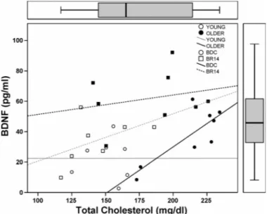
The variation in BDNF plasma levels from BDC to BR14 (⌬BDNF) did not correlate with any variables. Figure 2 shows the correlation between total cholesterol and plasma BDNF in both groups at BDC and BR14.
Finally, we produced two multiple linear regression models using BDC plasma levels of BDNF, BR14 plasma BDNF
levels, and total cholesterol and fat-free mass as dependent and independent variables in both groups separately (younger/ older). We found only that one or the other group predicted final BDNF plasma levels (R2 ⫽ 0.658, P ⫽ 0.008,
unstan-dardized beta coefficient 27.656).
We also performed a second multiple linear regression model using plasma BDNF, total cholesterol, and fat-free mass (both at BDC and BR14) as a dependent variable and as independent variables for one or the other of the two groups, and another variable, time, obtained by assigning “0” to all the detections collected at BDC from each subject, and “1” to all the detections collected at BR14. In doing so we evaluated the influence of the time on BDNF levels, which means the effect of undergoing bed rest. The results are shown in Table 2.
DISCUSSION
The present study investigated the age-related differences in plasma BDNF levels after prolonged physical inactivity (bed rest) acting as an acute stressor on the human body. To our knowledge, this was the first study aiming to investigate plasma BDNF fluctuations in younger and older healthy sub-jects undergoing 14 days of strict bed rest; therefore, it is not possible to carry out a straight-forward comparison with other studies. However, in animal studies, BDNF hippocampal levels were demonstrated to increase in murine models during acute stresses such as immobilization, explicating a protective role (13, 29, 39). On the other hand, chronic immobilization
(re-Fig. 1. Variation in plasma levels of brain-derived neurotrophic factor (BDNF) from baseline data collection (BDC) to day 14 of bed rest (BR14) in the study population.
Fig. 2. Correlation between plasma BDNF and total cholesterol levels in the older and younger study groups both at BDC and BR14.
Table 2. Multiple linear regression model indicating
predictors of BDNF plasma levels. Model R2 P for model Predictors
Nonstandard
coefficient P for variable
1 0.229 0.001 Group 20.506 0.001 2 0.341 ⬍0.001 Group 20.506 ⬍0.001 Time 13.109 0.010 3 0.497 ⬍0.001 Group 10.492 0.061 Time 18.054 ⬍0.001 Total cholesterol 0.232 0.001
peated immobilization stress) as well as other chronic stressors, has led to reduced BDNF concentrations in rat hippocampus (41, 42, 49, 54, 55). Here, we show an increase in plasma BDNF after bed rest in older subjects, but not in younger subjects.
Our aim was not to replicate a murine model of immobili-zation with humans; clearly, the two situations cannot be compared, either with time intervals nor intensity of stress. It is clear that mice experience a very strong psychological stress rather than a physical stress during forced immobilization; on the contrary, human volunteers experience little or no psycho-logical stress but substantial physical stress, as shown in recent research by our group (38). Nevertheless, the finding that acute and chronic stresses modulate BDNF expression differently in mice led us to hypothesize that such a modulation could also take place in humans.
Even if there is no agreement about the cellular source of human plasma BDNF, there is some evidence, both in humans and animals, that the brain plays a central role in determining circulating BDNF levels (18, 40), and that its contribution to total circulating BDNF has been considered to be about 70 – 80% (40). A massive presence of BDNF in platelets and in skeletal muscle was also described, but whereas platelet release upon activation does not seem to explain the variations in circulating BDNF occurring with aging or exercise (26, 40), skeletal muscle was demonstrated not to be a source of circu-lating BDNF (31, 37). On the basis of all these results, blood concentrations of BDNF have already been used as an indirect measure of BDNF activity in the central nervous system (9).
We hypothesize that with increasing age the brain might become less resistant to acute stresses such as bed rest, so that a condition such as bed rest could become more stressful for the brain, and therefore a greater effort to counteract its negative effects could be required.
Our analyses showed a positive correlation between BDNF and fat-free mass, both in the older and younger groups, and in both groups at BDC. Several studies that have examined changes in BDNF levels in humans revealed an augmentation of BDNF after an acute bout of exercise (43, 14) or, for instance, after 5 wk (59) and 3 mo (46) of endurance training, a condition that is well known to influence muscle mass in younger and older individuals (15, 48, 53). Alternatively, studies have shown lower BDNF values in sedentary individ-uals (45). Therefore, our results might reflect that people with higher levels of fat-free mass likely were more physically active. The loss of such a correlation at the end of bed rest (BR14) might reflect the different behaviors of the two vari-ables during bed rest: in fact, whereas an increase is observed in BDNF, a decrease in fat-free mass was observed at the end of bed rest in both study populations (60.3 ¡ 56.5 kg, P ⬍ 0.001) and in the older group (59.8 ¡ 56.7 kg, P ⬍ 0.001). A similar decrease in muscle mass has been confirmed in other bed rest studies involving younger (11) and older individuals (19).
Moreover, total cholesterol was found to positively correlate with BDC plasma BDNF levels in the whole study population and in our older study group, but not in the younger study group; this might reflect a different significance in total cho-lesterol plasma levels at different ages. It is well known from the literature that although a higher level of cholesterol is a risk factor for all causes and cardiovascular mortality in younger subjects (10), in older people the same levels seem to be
associated with lower mortality (a concept known as “reverse epidemiology”). This probably derives from the fact that in older people, lower levels of total cholesterol are the result of chronic inflammatory conditions rather than a healthier life-style, so that higher levels of total cholesterol actually reflect better overall health (2).
Interestingly, at the end of bed rest (BR14), this positive correlation between BDNF and total cholesterol is no longer present: this could reflect the behavior of cholesterol as an acute-phase molecule. In acute stress conditions, cholesterol plasma levels decrease (44), in this context, considering bed rest to be a stressful condition, although BDNF increases to counteract its negative effect on the brain, total and LDL cholesterol levels decrease, thereby losing the positive corre-lation.
On the other hand, the role of bed rest as an acute stress condition is confirmed by the behavior of several variables: first, total cholesterol, which decreases from BDC to BR14 both in the general population (183.79 ¡ 165.14 mg/dl, P ⬍ 0.001) and in our group of older study subjects (212.36 ¡ 183.51 mg/dl, P ⬍ 0.024), but not in our group of younger subjects (151.11 ¡ 144.14, P ⬍ 0.23), but in addition, TNF␣, a well-known inflammatory marker, increases after bed rest both in the general population (1.23 ¡ 1.46 mg/dl, P ⬍ 0.001), and it also did so in our older study subjects (1.45 ¡ 1.59 mg/dl, P ⬍ 0.001) and our younger study subjects (0.98 ¡ 1.30 mg/dl, P ⬍ 0.001). Even C-reactive protein shows an increase in plasma concentration after bed rest in the general population (0.080 ¡ 0.236 mg/dl, P ⬍ 0.033).
Age emerged as a variable at BR 14 that correlated with plasma BDNF and reflects the significant increase in BDNF observed in the older group but not the younger group. On the other hand, the multiple linear regression models demonstrate that age (i.e., one or the other group) is an independent predictor of final plasma BDNF levels, which is influenced, as expected, by bed rest (i.e., “time” factor), but also by total cholesterol. Despite what already has been said regarding cholesterol and its behavior as an acute-phase molecule, only a few studies exist in literature that link BDNF with cholesterol, and most of them refer to brain content of cholesterol and not to plasma levels (21, 51, 52). Our result seems in line with a recent study conducted in a cohort of patients suffering from bipolar disorder (22), but data are lacking for the general population, so we aim to study a larger population to confirm this result.
The potential mechanisms involved in plasma BDNF ex-pression during prolonged bed rest are further discussed. Dur-ing acute stress, as durDur-ing intense, prolonged physical activity that induces hypoxia, the response of the central nervous system [in particular the cortex and hippocampus, as suggested by Rasmussen et al. (40)] is an increased release of BDNF. If confirmed, our data seem to suggest that with prolonged bed rest, the central nervous system increase in BDNF production is a stereotypical response to repair the damage induced by acute stress, and its release in circulation counteracts the negative effects of acute stress on metabolism.
Study limitations. We acknowledge some limitation of this
study. First, the small number of subjects makes it difficult to derive any conclusions. However, it is necessary to keep in mind that bed rest studies that include older individuals are rare, and bed rest studies are very costly, labor-intensive, and
limited by hospital capacity. The total costs are estimated at⬎$20,000 per participant. However, despite having such a small population sample size, we were able to detect variables that were correlated with moderate significance. We hypothe-size that an even stronger statistical significance could be reached with a larger study population.
Finally, because we did not perform functional neuroimag-ing and did not obtain samples of cerebrospinal fluid from subjects, we have only surrogate evaluations of brain stress, such as TNF␣, and C-reactive protein.
ACKNOWLEDGMENTS
We thank the study participants for their time and effort to ensure the success of the project. We acknowledge the excellent assistance of the entire staff of the Orthopaedic Hospital Valdoltra (Koper, Slovenia). We also thank the research team and the students of the Applied Kinesiology Department of the University of Primorska for their help and logistical support, and many other researchers and colleagues from various institutions and countries who contributed to the smooth undertaking of the study.
GRANTS
This study was a part of the research project “Physical Activity and Nutrition for Quality Ageing” (PANGeA), which is supported by Cross-border Cooperation Program Slovenia-Italy 2007–2013 grant 042-2/2009.
DISCLOSURES
No conflicts of interest, financial or otherwise, are declared by the authors. AUTHOR CONTRIBUTIONS
C.S., U.M., M.L.M., E.D.N., B.S., R.P., and A.P. conception and design of research; U.M., M.L.M., B.S., R.P., and A.P. performed experiments; C.S., J.M.S., E.D.N., and A.P. analyzed data; C.S., U.M., J.M.S., E.D.N., R.P., G.Z., and A.P. interpreted results of experiments; C.S., U.M., and A.P. prepared figures; C.S. and A.P. drafted manuscript; C.S., U.M., J.M.S., M.L.M., E.D.N., B.S., R.P., G.Z., and A.P. edited and revised manuscript; C.S., U.M., J.M.S., M.L.M., E.D.N., B.S., R.P., G.Z., and A.P. approved final version of manu-script.
REFERENCES
1. Aciero PJ, Smith DL, Calles-Escandon J. Effects of short-term inactiv-ity on glucose tolerance, energy expenditure, and blood flow in trained subjects. J Appl Physiol 84: 1365–1373, 1998.
2. Ahmadi SF, Streja E, Zahmatkesh G, Streja D, Kashyap M, Moradi H, Molnar MZ, Reddy U, Amin AN, Kovesdy CP, Kalantar-Zadeh K. Reverse epidemiology of traditional cardiovascular risk factors in the geriatric population. J Am Med Dir Assoc 16: 933–999, 2015.
3. Binder DK, Scharfman HE. Brain-derived neurotrophic factor. Growth Factors 22: 123–131, 2004.
4. Biolo G, Agostini F, Simunic B, Sturma M, Torelli L, Preiser JC, Deby-Dupont G, Magni P, Strollo F, di Prampero P, Guarnieri G, Mekjavic IB, Pisot R, Narici MV. Positive energy balance is associated with accelerated muscle atrophy and increased erythrocyte glutathione turnover during 5 wk of bed rest. Am J Clin Nutr 88: 950 –958, 2008. 5. Brunoni AR, Lopes M, Fregni F. A systematic review and meta-analysis
of clinical studies on major depression and BDNF levels: implications for the role of neuroplasticity in depression. Int J Neuropsychopharmacol 11: 1169 –1180, 2008.
6. Burstein M, Scholnick HR, Morfin R. Rapid method for the isolation of lipoproteins from human serum by precipitations with polyanions. J Lipid Res 11: 583–595, 1970.
7. Chui DH, Marcellino M, Marotta F, Sweed H, Solimene U, Vignali AI, Xiao W, Ayala A, Cagnuolo U, Zerbinati N. A double-blind RCT testing beneficial modulation of BDNF in middle-aged, life style-stressed sub-jects: a clue to brain protection? J Clin Diagn Res 8: MC01–MC06, 2014. 8. Cohen-Cory S, Kidane AH, Shirkey NJ, Marshak S. Brain-derived neurotrophic factor and the development of structural neuronal connec-tivity. Dev Neurobiol 70: 271–288, 2010.
9. Cui H, Jin Y, Wang J, Weng X, Li C. Serum brain-derived neurotrophic factor (BDNF) levels in schizophrenia: a systematic review. Shanghai Arch Psychiatry 24: 250 –261, 2012.
10. Eckel RH, Cornier MA. Update on the NCEP ATP-III emerging cardio-metabolic risk factors. BMC Med 12: 115, 2014.
11. Ferrando AA, Lane HW, Stuart CA, Davis-Street J, Wolfe RR. Prolonged bed rest decreases skeletal muscle and whole body protein synthesis. Am J Physiol Endocrinol Metab 270: E627–E633, 1996. 12. Friedewald WT, Levy RI, Fredrickson DS. Estimation of the
concen-tration of low-density lipoprotein cholesterol in plasma, without use of preparative ultracentrifuge. Clin Chem 18: 499 –502, 1972.
13. Givalois L, Marmigère F, Rage F, Ixart G, Arancibia S, Tapia-Arancibia L. Immobilization stress rapidly and differentially modulates BDNF and TrkB mRNA expression in the pituitary gland of adult male rats. Neuroendocrinology 74: 148 –153, 2001.
14. Gold SM, Schulz KH, Hartmann S, Mladek M, Lang UE, Hellweg R, Reer R, Braumann KM, Heesen C. Basal serum levels and reactivity of nerve growth factor and brain-derived neurotrophic factor to standardized acute exercise in multiple sclerosis and controls. J Neuroimmunol 138: 99 –105, 2003.
15. Häkkinen K, Alen M, Kraemer WJ, Gorostiaga E, Izquierdo M, Rusko H, Mikkola J, Häkkinen A, Valkeinen H, Kaarakainen E, Romu S, Erola V, Ahtiainen J, Paavolainen L. Neuromuscular adapta-tions during concurrent strength and endurance training versus strength training. Eur J Appl Physiol 89: 42–52, 2002.
16. Hamburg NM, McMackin CJ, Huanf AL, Shenouda SM, Widlansky ME, Schulz E, Gocke N, Ruderman NB, Keaney JF Jr, Vita JA. Physical inactivity rapidly induces insulin resistance and microvascular dysfunction in healthy volunteers. Arterioscler Thromb Vasc Biol 27: 2650 –2656, 2007.
17. Karege F, Perret G, Bondolfi G, Schwald M, Bertschy G, Aubry JM. Decreased serum brain-derived neurotrophic factor levels in major de-pressed patients. Psychiatry Res 109: 143–148, 2002.
18. Karege F, Schawald M, Cisse M. Postnatal developmental profile of brain-derived neurotrophic factor in rat brain and platelets. Neurosci Lett 328: 261–264, 2002.
19. Kortebein P, Symons TB, Ferrando A, Paddon-Jones D, Ronsen O, Protas E, Conger S, Lombeida J, Wolfe R, Evans WJ. Functional impact of 10 days of bed rest in healthy older adults. J Gerontol A Biol Sci Med Sci 36: 1076 –1081, 2008.
20. Krabbe KS, Nielsen AR, Krogh-Madsen R, Plomgaard P, Rasmussen P, Eriksturp C, Fischer CP, Lindegaard B, Petersen AM, Taudorf S, Secher NH, Pilegaard H, Bruunsgaard H, Pedersen BK. Brain-derived neurotrophic factor (BDNF) and type 2 diabetes. Diabetologia 50: 431– 438, 2007.
21. Lee BH, Kim YK. Potential peripheral biological predictors of suicidal behaviour in major depressive disorder. Prog Neuropsychopharmacol Biol Psychiatry 35: 842–847, 2011.
22. Lee SY, Chen SL, Chang YH, Chen PS, Huang SY, Tzeng NS, Wang CL, Lee IH, Wang TI, Chen KC, Yang YK, Hong JS, Lu RB. Correlation of plasma brain derived neurotrophic factor and metabolic profiles in drug-naïve patients with bipolar II disorder after a twelve-week pharmacological intervention. Acta Physiatr Scand 131: 120 –128, 2015. 23. Li K, Guo X, Jin Z, Ouyang X, Zeng Y, Feng J, Ma L. Effect of simulated microgravity on human brain gray matter and white matter -evidence from MRI. PLoS One 10: e0135835, 2015.
24. Lipnicki DM, Gunga HC, Belavy DL, Felsenberg D. Bed rest and cognition: effects on executive functioning and reaction time. Aviat Space Environ Med 80: 1018 –1024, 2009.
25. Liu Q, Zhou R, Chen S, Tan C. Effects of head-down bed rest on the executive functions and emotional response. PLoS One 7: e52160, 2012. 26. Lommatzsch M, Zingler D, Schuhbaeck K, Schloetcke K, Zingler C, Schuff-Werner P, Virchow JC. The impact of age, weight and gender on BDNF levels in human platelets and plasma. Neurobiol Aging 26: 115– 123, 2005.
27. Lukaski HC, Johnson PE, Bolonchuk WW, Lykken GI. Assessment of fat-free mass using bioelectrical impedance measurements of the human body. Am J Clin Nutr 41: 810 –817, 1985.
28. Manni L, Nikolova V, Vyagova D, Chaldakov VN, Aloe L. Reduced plasma levels of NGF and BDNF in patients with acute coronary syn-dromes. Int J Cardiol 102: 169 –171, 2005.
29. Marmigère F, Givalois L, Rage F, Arancibia S, Tapia-Arancibia L. Rapid induction of BDNF expression in the hippocampus during immo-bilization stress challenge in adult rats. Hippocampus 13: 646 –655, 2003.
30. Marusic U, Meeusen R, Pisot R, Kavcic V. The brain in micro- and hypergravity: the effects of changing gravity on the brain electrocortical activity. Eur J Sport Sci 14: 813–822, 2014.
31. Matthews VB, Aström MB, Chan MH, Bruce CR, Krabbe KS, Pre-lovsek O, Akerström T, Yfanti C, Broholm C, Mortensen OH, Pen-kowa M, Hojman P, Zankari A, Watt MJ, Bruunsgaard H, Pedersen BK, Febbraio MA. Brain-derived neurotrophic factor is produced by skeletal muscle cells in response to contraction and enhances fat oxidation via activation of AMP-activated protein-kinase. Diabetologia 52: 1409 – 1418, 2009.
32. Matthews DR, Hosker JP, Rudenski AS, Naylor BA, Treacher DF, Turner RC. Homeostasis model assessment: insulin resistance and beta-cell function from fasting plasma glucose and insulin concentrations in man. Diabetologia 28: 412–419, 1985.
33. Nowacka M, Obuchowicz E. BDNF and VEGF in the pathogenesis of stress-induced affective diseases: an insight from experimental studies. Pharmacol Rep 65: 535–546, 2013.
34. Pan W, Banks WA, Fasold MB, Bluth J, Kastin AJ. Transport of brain-derived neurotrophic factor across the blood-brain barrier. Neuro-pharmacology 37: 1553–1561, 1998.
35. Paska AV, Zupanc T, Pregelj P. The role of brain-derived neurotrophic factor in the pathophysiology of suicidal behaviour. Psychiatr Danub 25: Suppl 2, 341–344, 2013.
36. Passaro A, Dalla Nora E, Morieri ML, Soavi C, Sanz JM, Zurlo A, Fellin R, Zuliani G. Brain-derived neurotrophic factor plasma levels: relationship with dementia and diabetes in the elderly population. J Gerontol A Biol Sci Med Sci 70: 294 –302, 2015.
37. Pedersen BK, Pedersen M, Krabbe KS, Bruunsgaard H, Matthews VB, Febbraio MA. Role of exercise-induced brain-derived neurotrophic factor production in the regulation of energy homeostasis in mammals. Exp Physiol 94: 1153–1160, 2009.
38. Pisot R, Marusic U, Biolo G, Mazzucco S, Lazzer S, Grassi B, Reggiani C, Toniolo L, di Prampero PE, Passaro A, Narici MV, Mohammed S, Rittweger J, Gasparini M, Gabrijelcˇicˇ M, Simunic B. Greater loss in muscle mass and function but smaller metabolic alterations in older compared to younger men following two weeks of bed rest and recovery. J Appl Physiol. First published January 28, 2016; doi:10.1152/ japplphysiol.00858.2015.
39. Rage F, Givalois L, Marmigère F, Tapia-Arancibia L, Arancibia S. Immobilization stress rapidly modulates BDNF mRNA expression in the hypothalamus of adult male rats. Neuroscience 112: 309 –318, 2002. 40. Rasmussen P, Brassard P, Adser H, Pedersen MV, Leick L, Hart E,
Secher NH, Pedersen BK, Pilegaard H. Evidence for a release of brain-derived neurotrophic factor from the brain during exercise. Exp Physiol 94: 1062–1069, 2009.
41. Rasmusson AM, Shi L, Duman R. Downregulation of BDNF mRNA in the hippocampal dentate gyrus after re-exposure to cues previously asso-ciated with foot-shock. Neuropsychopharmacology 27: 133–142, 2002. 42. Roceri M, Hendriks W, Racagni G, Ellenbroek BA, Riva MA. Early
maternal deprivation reduces the expression of BDNF and NMDA recep-tor subunits in the rat hippocampus. Mol Psychiatry 7: 609 –616, 2002. 43. Rojas Vega S, Strüder HK, Vera Wahrmann B, Schmidt A, Bloch W,
Hollmann W. Acute BDNF and cortisol response to low intensity exercise and following ramp incremental exercise to exhaustion in humans. Brain Res 1121: 59 –65, 2006.
44. Rott D, Klempfner R, Goldenberg I, Leibowitz D. Cholesterol levels decrease soon after myocardial infarction. Isr Med Assoc J 17: 370 –373, 2015.
45. Schmolesky MT, Webb DL, Hansen RA. The effects of aerobic exercise intensity and duration on levels of brain-derived neurotrophic factor in healthy men. J Sports Sci Med 12: 502–511, 2013.
46. Seifert T, Brassard P, Wissenberg M, Rasmussen P, Nordby P, Stallknecht B, Adser H, Jakobsen AH, Pilegaard H, Nielsen HB, Secher NH. Endurance training enhances BDNF release from the human brain. Am J Physiol Regul Integr Comp Physiol 298: R372–R377, 2010. 47. Shi SS, Shao SH, Yuan BP, Pan F, Li ZL. Acute stress and chronic stress change brain-derived neurotrophic factor (BDNF) and tyrosine kinase-coupled receptor (TrkB) expression in both young and aged rat hippocam-pus. Yonsei Med J 51: 661–671, 2010.
48. Sipila S, Suomeinen H. Effects of strength and endurance training on thigh and leg muscle mass and composition in elderly women. J Appl Physiol 78: 334 –340, 1995.
49. Smith MA, Makino S, Kvetnansky R, Post RM. Stress and glucocorti-coids affect the expression of brain derived neurotrophic factor and neurotrophin-3 mRNAs in the hippocampus. J Neurosci 15: 1768 –1777, 1995.
50. Stuart CA, Shoiraw RE, Prince MJ, Peters EJ, Wolfe RR. Bed-rest-induced insulin resistance occurs primarily in muscle. Metabolism 37: 802–806, 1998.
51. Suzuki S, Kiyosue K, Hazama S, Ogura A, Kashihara M, Hara T, Koshimizu H, Kojima M. Brain-derived neurotrophic factor regulates cholesterol metabolism for synapse development. J Neurosci 27: 6417– 6427, 2007.
52. Suzuki S, Numakawa T, Shimazu K, Koshimizu H, Hara T, Hatanaka H, Mei L, Lu B, Kojima M. BDNF-induced recruitment of TrkB receptor into neuronal lipid rafts: roles in synaptic modulation. J Cell Biol 167: 1205–1215, 2004.
53. Tarpenning KM, Hamilton-Wessler M, Wiswell RA, Hawkins SA. Endurance training delays age of decline in leg strength and muscle morphology. Med Sci Sports Exerc 36: 74 –78, 2004.
54. Ueyama T, Kawai Y, Nemoto K, Sekimoto M, Tone S, Senba E. Immobilization stress reduced the expression of neurotrophins and their receptors in the rat brain. Neurosci Res 28: 103–110, 1997.
55. Vaidya VA, Marek GJ, Aghajanian GK, Duman RS. 5HT2A receptor-mediated regulation of brain derived neurotrophic factor mRNA in the hippocampus and the neocortex. J Neurosci 17: 2785–2795, 1997. 56. Ventriglia M, Zanardini R, Bonomini C, Zanetti O, Volpe D,
Pasqual-etti P, Gennarelli M, Bocchio-Chiavetto L. Serum brain derived neu-rotrophic factor levels in different neurological diseases. Biomed Res Int 2013: 901082, 2013.
57. Wiggs MP. Can endurance exercise preconditioning prevention disuse muscle atrophy? Front Physiol 6: 63, 2015.
58. Zhen YF, Zhang J, Liu XY, Fang H, Tian LB, Zhou DH, Kosten TR, Zhang XY. Low BDNF is associated with cognitive deficits in patients with type 2 diabetes. Physiopharmacology 227: 93–100, 2013. 59. Zoladz JA, Pilc A, Majerczak J, Grandys M, Zapart-Bukowska J,
Duda K. Endurance training increases plasma brain-derived neurotrophic factor concentration in young healthy men. J Physiol Pharmacol 59, Suppl 7: 119 –132, 2008.