Basic Thyroidology / Original Paper
Eur Thyroid J 2016;5:224–230DOI: 10.1159/000452488
Immunohistochemical Expression of Estrogen
Receptor-α and Progesterone Receptor
in Patients with Papillary Thyroid Cancer
Giacomo Sturniolo
a
Carles Zafon
c
Mariacarla Moleti
b
Josep Castellví
d
Francesco Vermiglio
b
Jordi Mesa
c
a Department of Human Pathology of the Adult and Developmental Age “G. Barresi”, and b Department of Experimental Medicine, University of Messina, Messina , Italy; c Department of Endocrinology, Diabetes, and Metabolism Research Unit, Vall d’Hebron Institut de Recerca (VHIR), and d Department of Pathology, Hospital Vall d’Hebron, Universitat Autò noma de Barcelona, Barcelona , Spain
dition, ER-α expression significantly correlated with remis-sion of the disease. In fact, of the 192 patients followed up, 50/153 (32.7%) disease-free patients were ER-α positive, in contrast to only 3/39 (7.7%) with evidence of disease persis-tence/recurrence (χ 2 = 8.5, p = 0.0036). PR expression was not associated with any of the parameters analyzed.
Conclu-sions: The present study confirmed recent data indicating
that ER-α and PR expression is a common finding in thyroid tumor tissue. However, in contrast to previous reports, we observed an association between ER-α expression and a more favorable outcome in PTC patients.
© 2016 European Thyroid Association Published by S. Karger AG, Basel
Introduction
The incidence of differentiated thyroid cancer (DTC) is about 3 times higher in women than in men [1, 2] and decreases after menopause [3] . The age-standardized (per 100,000) incidence of thyroid cancer ranges from 2 to 10 in females and from 1 to 3 in males in different popula-tions. The higher prevalence in females, particularly
Keywords
Thyroid cancer · Estrogen receptor · Progesterone receptor · Sexual hormone receptors
Abstract
Background: Papillary thyroid cancer (PTC) prevalence is nearly 3 times higher in females than in males. This gender difference suggests that growth and progression of PTC might be influenced by female sex hormones. Objectives: To analyze the expression of both estrogen receptor (ER)-α and progesterone receptor (PR) by immunohistochemistry in 203 PTC patients. Methods: ER-α and PR expression was eval-uated in paraffin-embedded tumor tissue samples of 45 males and 158 females followed up for 7.2 ± 3.7 years.
Re-sults: ER-α was expressed in 52 (25.6%) patients (41 females
and 11 males) and PR in 94 (46.3%) patients (75 females and 19 males). ER-α and PR were coexpressed in 31 (15.3%) pa-tients (27 females and 4 males). ER-α expression correlated significantly with tumor size in the whole sample (ER-α pos-itive 22.8 ± 11.8 mm vs. ER-α negative 15.1 ± 12.4 mm; p = 0.02) and in the subgroup of women (ER-α positive 18.8 ± 12.8 mm vs. ER-α negative 14.9 ± 12.3 mm; p = 0.048). In
Received: May 10, 2016
Accepted after revision: October 14, 2016 Published online: November 24, 2016
ing the reproductive period, is observed in all regions and in all ethnic groups [4] . Since thyroid cancer and most benign thyroid diseases (Graves disease, endemic goiter, and Hashimoto thyroiditis) are significantly more fre-quent in women of childbearing age [5] , many studies have been designed to examine the influence of reproduc-tive and hormonal factors on the etiology of thyroid dis-eases [3, 6–8] . Epidemiological reports demonstrate gen-der differences in the development of thyroid cancer, thus suggesting that sex hormones, particularly estrogens, may be somewhat involved in its growth and progression [6–9] . Some studies showed the use of oral contraceptives to be associated with a moderately increased risk of thy-roid cancer, although other studies did not confirm this association [10–12] . Also, an increased risk of thyroid cancer has been reported in women using estrogens for gynecological impairments, but not in women given low-dose estrogen therapy for menopausal hormone replace-ment [11, 13, 14] .
Despite the higher incidence of DTC in females, wom-en gwom-enerally show a more favorable outcome than males. Indeed, different cohort studies reported a better “over-all” and “disease-specific” survival, and fewer relapses in women than in men. Specifically concerning papillary thyroid carcinoma (PTC), although the overall outcome seems comparable in the two sexes, analysis of subgroups aged under and over 55 years indicates a more favorable prognosis in females at a younger age [13, 14] .
All the above suggests a possible role for estrogens in the development and progression of DTC, and several studies have investigated the expression of estrogen re-ceptor (ER) subtypes in thyroid cancers. However, results are not consistent yet, likely because of the existence of confounding factors in the populations selected [15, 16] .
The purpose of our study was to determine the immu-nohistochemical expression of ER-α and progesterone re-ceptor (PR) in a large series of PTC patients, and to cor-relate the above expressions with epidemiological and clinical features.
Patients and Methods
The study included 203 consecutive patients diagnosed with the classical variant of PTC between 2000 and 2009 who underwent to-tal thyroidectomy, radioiodine ablation ( 131 I) of postsurgical resid-ual thyroid tissue (whenever deemed necessary), and levothyroxine (LT 4 ) treatment at TSH-suppressive or -semisuppressive doses. For the purpose of this study, patients were divided into 2 groups ac-cording to tumor size: the PTC group (113 patients with PTC >10 mm) and the micro-PTC group (90 patients with PTC ≤ 10 mm).
Suspicious nodules were identified at intraoperative histopa-thology and then treated in formalin for 24 h. Cutting and selection of tissue specimens were performed, followed by histological eval-uation of paraffembedded tissue. The diagnostic protocol in-cluded the assessment of: (i) histotype; (ii) tumor size; (iii) tumor margins; (iv) multifocality; (v) level of lymph node metastases, if any; and (vi) TNM staging.
Tissue-Arrayer was used for all tumor samples by coring tissue from the paraffin-embedded block in order to obtain tumor spec-imens with a diameter of 0.6–1.5 mm. Tumor sections were then transferred to a paraffin block for immunohistochemical studies.
Immunohistochemical determination was performed using automated protocols on a Ventana BenchMark Ultra Immuno-stainer. For antigen retrieval, buffer CC1 was used for 28 and 20 min for ER and PR determination, respectively. The sections were incubated with the following monoclonal primary antibod-ies: ER-α: clone SP1, Ventana-Roche, incubation for 28 min; PR: clone 1E2, Ventana-Roche, incubation for 16 min. A section of breast tissue was used as a positive control in all the samples. As described for breast cancer, the sections were assessed semiquan-titatively using the system of the H-score , which measures both percentage of positive cells and intensity of expression [17] .
Disease free was defined as follows: (i) no clinical evidence of tumor; (ii) no imaging evidence of tumor by radioactive iodine (RAI) imaging and/or neck US; and (iii) serum Tg levels <0.2 ng/ mL during TSH suppression or <1 ng/mL after stimulation in the absence of interfering antibodies [18] .
Quantitative variables are expressed as means ± SD and cate-gorical variables as frequencies and proportions. Quantitative variables were compared using the Student t test; qualitative vari-ables were analyzed by χ 2 test. p < 0.05 was considered to be statis-tically significant. Statistical analyses were performed using the SSPS statistical package (SPSS, Chicago, IL, USA).
Informed consent was obtained from the subjects, and the study protocol was approved by the institute’s committee on hu-man research.
Results
Epidemiological and Clinical Features
The main clinical and epidemiological characteris-tics of the 203 patients, either as a whole or as subgroups (PTC group and micro-PTC group), are reported in Ta-ble 1 . The mean age at diagnosis, the percentage of sub-jects younger than 45 years, and gender distribution were not different in the two subgroups. As expected, the disease was confined to the thyroid in the vast ma-jority of patients from the micro-PTC group, with evi-dence of extrathyroidal extension in 4/90 patients only. Conversely, almost one-third (32/113) of the patients in the PTC group showed extrathyroidal extension (mini-mal in 29/113 and beyond the thyroid capsule in 3/113). In addition, a significantly higher rate of patients from the same group had multifocal disease and lymph node metastases at presentation. Finally, 10/113 patients
from the PTC group and 1 patient from the micro-PTC group presented distant metastases (pulmonary in all cases).
Overall, 11 patients were lost to the follow-up. Of the remaining 192 patients (148 females and 44 males), 39/192 (20.3%; 33 from the PTC group and 6 from the micro-PTC group) showed persistent or recurrent dis-ease.
Immunohistochemical Expression of ER-α and PR
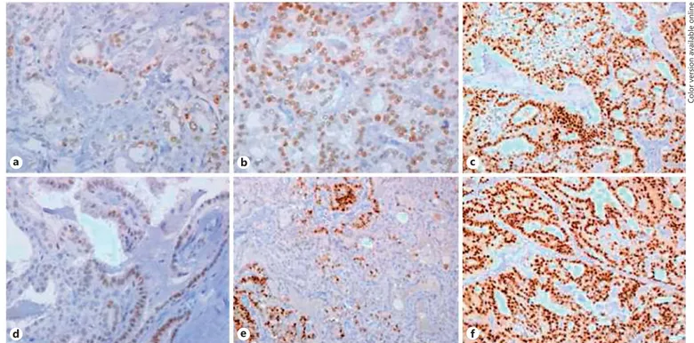
Table 2 summarizes data on ER-α and PR expression in the whole sample and in the 2 subgroups. In particular, ER-α expression was found in 52/203 (25.6%) cases ( Fig. 1 a–c), whereas PR expression was found in 94/203 (46.3%) cases ( Fig. 1 d–f).
ER-α expression was found to be similar in both sexes (41/158 females vs. 11/45 males, p = 0.83), and no differ-ences were observed in mean ages of ER-α-positive or -negative patients (45.9 ± 14.8 vs. 49.1 ± 13.9, p = 0.087). Similar results were found when the PTC and micro-PTC groups were analyzed, either separately or comparatively.
Conversely, ER-α expression significantly correlated with tumor size, either in the whole sample (ER-α positive 22.8 ± 11.8 mm vs. ER-α negative 15.1 ± 12.4 mm; p = 0.02) or in the subgroup of women (ER-α positive 18.8 ± 12.8 mm vs. ER-α negative 14.9 ± 12.3 mm; p = 0.048). When the 2 groups (PTC and micro-PTC group) were considered for analysis, the correlation between ER-α ex-pression and tumor size could not be further confirmed in the PTC group (ER-α positive 26.7 ± 12.6 mm vs. ER-α
Table 1. Epidemiological and clinical features of the study patients
Characteristics Whole sample
(n = 203) PTC group (n = 113) Micro-PTC group (n = 90) p value
Mean age at diagnosis ± SD, years Range 48.2±14.3 20–85 47.2±15.1 22–85 49.5±13.1 20–79 0.13 Age <45 years, n (%) 90 (44.3) 52 (46) 38 (42.2) 0.59 Females, n (%) Female/male ratio 158 (77.8) 3.5:1 85 (75.2) 3:1 73 (81.1) 4.3:1 0.31 Mean tumor size ± SD, mm
Range 16.97±13.93 1–80 26.35±12.65 11–80 5.92±2.88 1–10 <0.0001 Tumor extension T1a, n (%) 86 (42.4) – 86 (95.5) – T1b, n (%) 51 (25.1) 51 (45.1) – – T2, n (%) 30 (14.8) 30 (26.5) – – T3, n (%) 32 (15.7) 29 (25.6) 3 (3.3) <0.0001 T4a, n (%) 4 (1.9) 3 (2.6) 1 (1.1) 0.78 T4b, n (%) – – – – Multifocality, n (%) 75 (36.9) 49 (43.3) 26 (28.8) 0.034
Lymph node metastases
NX, n (%) 127 (62.6) 57 (50.4) 70 (77.7) 0.0001 N0, n (%) 28 (13.8) 19 (16.8) 9 (10) 0.161 N1, n (%) 48 (23.6) 37 (32.7) 11 (12.2) 0.0006 131I ablation, n (%) 141 (69.4) 94 (83.2) 47 (52.2) <0.0001 Staging I, n (%) 150 (73.9) 66 (58.4) 84 (93.3) <0.0001 II, n (%) 8 (3.9) 8 (7.1) 0 III, n (%) 13 (6.4) 12 (10.6) 1 (1.1) 0.014 IVa, n (%) 21 (10.3) 17 (15) 4 (4.4) 0.014 IVb, n (%) 0 0 0 IVc, n (%) 11 (5.4) 10 (8.8) 1 (1.1) 0.035 Follow-up, years 7.2±3.7 7.7±3.9 6.7±3.2 0.02 Disease persistence/recurrence, n (%) 39 (20.3) 33 (29.2) 6 (6.7) 0.0001
To assess disease persistence/recurrence, 192 patients were followed up in the total group, and 106 and 86 patients in the PTC and micro-PTC groups, respectively. p values: PTC vs. micro-PTC.
negative 26.2 ± 12.7 mm; p = 0.41), whereas it was sig-nificant in the micro-PTC group (ER-α positive 7.05 ± 2.6 mm vs. ER-α negative 5.6 ± 2.9 mm; p = 0.031).
No relationship was found between ER-α expression and lymph node metastases, either when analysis was performed in the whole sample or when the subgroups were considered.
Finally, ER-α expression significantly correlated with remission of the disease. In fact, of the 192 patients fol-lowed up, 50 of 153 (32.7%) disease-free patients were ER-α positive, in contrast to only 3 of 39 (7.7%) patients with evidence of persistence/recurrence (χ 2 = 8.5, p =
0.0036). The same result was confirmed when the out-come of the PTC group patients was considered. Thus, of a total of 106 PTC patients followed up, 30 of 74 (40.5%) disease-free patients and 3 of 32 (9.4%) patients with evi-dence of persistent or recurrent disease were ER-α positive
(χ 2 = 8.72, p = 0.0031). When analyzed by gender, ER-α expression was found to be significantly associated with remission in females only in the whole sample (χ 2 = 7.05,
p = 0.0079) and in the PTC group (χ 2 = 7.45, p = 0.0063).
Concerning PR expression, no correlation was found between PR positivity and gender, patient age, tumor size at diagnosis, presence of lymph node metastases, and risk of disease progression or recurrence in both sexes.
Finally, both ER-α and PR were coexpressed in 27 of 158 (17.1%) women and 4 of 45 (8.9%) males ( p = 0.26). Among females, coexpression was significantly associat-ed with a younger age at diagnosis (ER-α+PR+ positive 42.1 ± 12.5 years vs. ER-α+PR– 48.5 ± 13.8 years; p = 0.013). Moreover, a nonstatistically significant trend to-wards a lower risk of progression or recurrence was ob-served in women with ER-α and PR coexpression (χ 2 =
3.79, p = 0.051).
Table 2. Immunohistochemical expression of ER-α and PR in the study patients
ER-α+ PR+ ER-α–PR+
Whole group (n = 203)
Patients, n (%) 52 (25.6) 94 (46.3) 31 (15.3)
Females, n (%) 41 (20.2) 75 (36.9) 27 (17.1)
Males, n (%) 11 (5.1) 19 (9.3) 4 (8.9)
Mean age ± SD, years Range 45.9±14.8 20–85 47.3±14.5 20–80 42.7±13.1 20–67 Mean tumor size ± SD, mm
Range 22.8±11.8 3–50 16.7±13.7 2–80 19.03±11.2 3–50 Disease persistence/recurrence, n (%) 3 (1.6) 16 (8.3) 2 (1.04) PTC group (n = 113) Patients, n (%) 34 (26.5) 56 (46.3) 24 (21.2) Females, n (%) 27 (23.8) 42 (37.2) 21 (18.6) Males, n (%) 7 (6.2) 14 (12.4) 3 (8.9)
Mean age ± SD, years Range 44.6±14.3 22–85 45.4±15.2 22–80 40.9±11.4 22–62 Mean tumor size ± SD, mm
Range 26.7±12.6 11–50 24.4±13.6 11–80 22.7±10.4 11–50 Disease persistence/recurrence, n (%) 3 (2.8) 13 (12.3) 2 (1.9) Micro-PTC group (n = 90) Patients, n (%) 18 (20) 38 (42.2) 7 (7.8) Females, n (%) 14 (15.6) 33 (36.7) 6 (6.7) Males, n (%) 4 (4.4) 5 (5.5) 1 (1.1)
Mean age ± SD, years Range 49.3±15.8 20–79 50.2±13.1 20–73 49.1±17.4 20–67 Mean tumor size ± SD, mm
Range 7.05±2.6 3–10 6.4±2.7 2–10 7.6±2.7 3–10 Disease persistence/recurrence, n (%) 0 3 (3.5) 0
To assess disease persistence/recurrence, 192 patients were followed up in the total group, and 106 and 86 patients in the PTC and micro-PTC groups, respectively.
Discussion
Cellular signaling of estrogen is mediated classically via 2 nuclear soluble intracellular receptors: ER-α and ER-β. Other regulatory mechanisms include a nonge-nomic way and an independent ligand interaction [19] .
Several human tissues, including the breast, ovary, prostate, lung, colon, pancreas, and thyroid, express both ER isoforms [19] . Specifically concerning the thyroid, ERs have been described in both neoplastic and nonneo-plastic thyroid tissue, though the results are not consis-tent to date, likely because of differences in the sensitivity of the techniques employed [15, 16] . The role of estrogens in the development and evolution of DTC has been wide-ly anawide-lyzed in several studies [20–28] . A recent meta-analysis including >5,000 patients reported that meno-pausal women had a reduced thyroid cancer risk, where-as increwhere-asing age at first pregnancy/birth wwhere-as where-associated with a higher cancer risk [29] . Similar to other epithelial tumors, DTC also expresses both ER isoforms, with ER-α activation being associated with increased estrogen-de-pendent cell proliferation and, in contrast, ER-β likely promoting apoptotic actions and other suppressive func-tions in thyroid tumors [22, 30–33] . Overall, there is
evi-dence that ER-α expression is greater in tumors than in normal thyroid tissues, whereas ER-β expression is sig-nificantly lower in neoplastic than in nonneoplastic thy-roid tissue [28, 32] .
Concerning PR, of the few studies which so far inves-tigated its expression in thyroid tumors, most assessed PR in conjunction with ER [21–24, 34, 35] .
In the present study, we evaluated the immunohisto-chemical expression of ER-α and PR in a large series of 203 classical PTC. In particular, the study sample includ-ed similar proportions of micro-PTC (<10 mm) and PTC (>1 mm). This balance allowed us to comparatively inves-tigate differences, if any, in ER-α and PR expression in these two clinical entities, and to correlate the immuno-histochemical findings to the disease outcome.
We found ER-α and PR to be expressed in 25.6 and 46.3% of the DTC patients, respectively, with 15.3% of tumor samples coexpressing both receptors. In particu-lar, both ER-α and PR (either individually or in conjunc-tion) were found to be expressed in PTC and micro-PTC to a similar extent, with no differences among sexes. When analyzed in the whole group, a positive correlation was found between ER-α expression and tumor size, a finding seemingly in line with the trend reported in the
a b c
d e f
Fig. 1. Immnunohistochemical ER-α (a–c) and PR (d–f). The intensity of expression (brown color) progressively
increases from the right to the left panel.
literature [34] . Unexpectedly, however, the above corre-lation could not be further confirmed when PTC and mi-cro-PTC were analyzed separately, with a definite trend towards an increase in ER-α expression with increasing tumor size found in micro-PTC only. Moreover, a reverse relationship was found between ER-α expression and a poor clinical outcome, since a significantly higher pro-portion of tumors from disease-free patients, either in the whole sample or in the PTC group alone, proved to be ER-α positive. This finding is apparently inconsistent with current epidemiological and clinical evidence, over-all suggesting that thyroid tumor development and pro-gression may be influenced by estrogens. However, a study investigating the tissue expression of ER-α along with the coregulatory proteins SRC-1 (steroid receptor coactivator) and NCoR (nuclear corepressor) in different subtypes of thyroid cancers demonstrated that expression of ER-α and NCoR was significantly associated with well-differentiated tumors and a reduced incidence of disease recurrence. In contrast, the coactivator protein SRC-1, mostly expressed in poorly differentiated tumors, was as-sociated with invasion, poor differentiation, tumor recur-rence, and reduced disease-free survival [31] .
In conclusion, presently available data strongly sup-port the hypothesis that ER signaling plays a role in the development and progression of differentiated thyroid cancer. In our patients with micro-PTCs and PTCs of larger sizes, ER-α expression was found to be similar and was associated with a better outcome. The role of differ-ent patterns of ER isoform expression in thyroid tumor subtypes, along with ER-α and ER-β ratios, and the ex-pression of coregulatory proteins modulating ER func-tion should be further investigated to better understand the pathogenesis and the natural history of differentiated thyroid cancers.
Statement of Ethics
This research complies with the guidelines for human studies and animal welfare regulations.
Disclosure Statement
The authors have no conflicts of interest and any sponsorship or funding arrangements to disclose.
References
1 Correa P, Chen VW: Endocrine gland cancer.
Cancer 1995; 75: 338–352.
2 Rosenthal DS: Changing trends. CA Cancer J
Clin 1998; 48: 3–4.
3 Manole D, Schildknecht B, Gosnell B, Adams E, Derwahl M: Estrogen promotes growth of human thyroid tumor cells by different mo-lecular mechanisms. J Clin Endocrinol Metab
2001; 86: 1072–1077.
4 Parkin DM, Whelan SL, Ferlay J, Teppo L, Thomas DB: Cancer Incidence in Five Conti-nents, Vol VIII. Lyon, IARC, 1997, IARC Sci Publ 143.
5 Vanderpump MPJ: The epidemiology of
thy-roid disease. Br Med Bull 2011; 99: 39–51.
6 Zeng Q, Chen GG, Vlantis AC, van Hasselt CA: Oestrogen mediates the growth of human thyroid carcinoma cells via an oestrogen
re-ceptor-ERK pathway. Cell Prolif 2007; 40
921–935.
7 Zeng Q, Chen G, Vlantis A, Tse G, van Hasselt C: The contributions of oestrogen receptor isoforms to the development of papillary and anaplastic thyroid carcinomas. J Pathol 2008;
214: 425–433.
8 Dong W, Zhang H, Li J, Guan H, He L, Wang Z, Shan Z, Teng W: Estrogen induces meta-static potential of papillary thyroid cancer cells through estrogen receptor α and β. Int J
Endocrinol 2013; 2013: 941568.
9 Hiasa Y, Nishioka H, Kitahori Y, Yane K, Na-kaoka S, Ohshima M, Konishi N, Nishii K, Kitamura M, Matsunaga T: Immunohisto-chemical analysis of estrogen receptors in 313 paraffin section cases of human thyroid
tis-sue. Oncology 1993; 50: 132–136.
10 Preston-Martin S, Bernstein L, Pike MC, Mal-donado AA, Henderson BE: Thyroid cancer among young women related to prior thyroid disease and pregnancy history. Br J Cancer
1987; 55: 191–195.
11 Persson I, Yuen J, Bergkvist L, Schairer C: Cancer incidence and mortality in women ceiving estrogen and estrogen-progestin re-placement therapy – long-term follow-up of a
Swedish cohort. Int J Cancer 1996; 67: 327–
332.
12 La Vecchia CL, Ron E, Franceschi S, Dal Maso L, Mark SD, Chatenoud L, Braga C, Preston-Martin S, McTiernan A, Kolonel L, Mabuchi K, Jin F, Wingren G, Galanti MR, Hallquist A, Lund E, Levi F, Linos D, Negri E: A pooled analysis of case-control studies of thyroid cancer. III. Oral contraceptives, menopausal replacement therapy and other female
hor-mones. Cancer Causes Control 1999; 10: 157.
13 Ron E, Kleinerman RA, Boice JD Jr, LiVolsi VA, Flannery JT, Fraumeni JF Jr: A popula-tion-based case-control study of thyroid
can-cer. J Natl Cancer Inst 1987; 79: 1–12.
14 Jonklaas J, Nogueras-Gonzalez G, Munsell M, Litofsky D, Ain KB, Bigos ST, Brierley JD, Cooper DS, Haugen BR, Ladenson PW, Mag-ner J, Robbins J, Ross DS, Skarulis MC, Stew-ard DL, Maxon HR, Sherman SI; National Thyroid Cancer Treatment Cooperative Study Group: The impact of age and gender on papillary thyroid cancer survival. J Clin
Endocrinol Metab 2012; 97: 878–887.
15 Santin AP, Furlanetto TW: Role of estrogen in thyroid function and growth regulation. J
Thyroid Res 2011; 2011: 875125.
16 Derwahl M, Nicula D: Estrogen and its role in
thyroid cancer. Endocr Relat Cancer 2014; 21:
273–283.
17 Detre S, Saccani Jotti G, Dowsett M: A ‘‘quickscore’’ method for immunohistochem-ical semiquantitation: validation for oestro-gen receptor in breast carcinomas. J Clin
Pathol 1995; 48: 876–878.
18 Haugen BR, Alexander EK, Bible KC, Doherty GM, Mandel SJ, Nikiforov YE, Pacini F, Ran-dolph GW, Sawka AM, Schlumberger M, Schuff KG, Sherman SI, Sosa JA, Steward DL, Tuttle RM, Wartofsky L: 2015 American Thy-roid Association Management Guidelines for Adult Patients with Thyroid Nodules and Dif-ferentiated Thyroid Cancer: The American Thyroid Association Guidelines Task Force on Thyroid Nodules and Differentiated
19 Heldring N, Pike A, Andersson S, Matthews J, Cheng G, Hartman J, Tujague M, Ström A, Treuter E, Warner M, Gustafsson JA: Estro-gen receptors: how do they signal and what
are their targets. Physiol Rev 2007; 87: 905–
931.
20 Mazzaferri EL, Young RL, Oertel JE, Kem-merer WT, Page CP: Papillary thyroid carci-noma: the impact of therapy in 576 patients.
Medicine (Baltimore) 1977; 56: 171–196.
21 Lewy-Trenda I: Estrogen and progesterone receptors in neoplastic and non-neoplastic
thyroid lesions. Pol J Pathol 2002; 53: 67–72.
22 van Hoeven KH, Menendez-Botet CJ, Strong EW, Huvos AG: Estrogen and progesterone receptor content in human thyroid disease.
Am J Clin Pathol 1993; 99: 175–181.
23 Money SR, Muss W, Thelmo WL, Boeckl O, Pimpl W, Kaindl H, Sungler P, Kirwin J, Wa-clawicek H, Jaffe BM: Immunocytochemical localization of estrogen and progesterone
re-ceptors in human thyroid. Surgery 1989; 106:
975–979.
24 Bur ME, Perlman C: Estrogen and progester-one receptor expression in tumors of the
thy-roid. Am J Clin Pathol 1991; 96: 409–410.
25 Molteni A, Warpeha RL, Brizio-Molteni L, Fors EM: Estradiol receptor binding protein in head and neck neoplastic and normal
tis-sue. Arch Surg 1981; 116: 207–210.
26 Clark OH, Gerend PL, Davis M, Goretzki PE, Hoffman PG: Estrogen and thyroid-stimulat-ing hormone (TSH) receptors in neoplastic and non-neoplastic human thyroid tissue. J
Surg Res 1985; 38: 89–96.
27 Mizukami Y, Michigishi T, Nonomura A, Hashimoto T, Noguchi M, Matsubara F: Es-trogen and esEs-trogen receptors in thyroid
car-cinomas. J Surg Oncol 1991; 47: 165–169.
28 Magri F, Capelli V, Gaiti M, Villani L, Zerbini F, La Manna L, Rotondi M, Chiovato L: ER-alpha and ER-beta expression in differentiat-ed thyroid cancer: relation with tumor pheno-type across the TNM staging and peri-tumor
inflammation. Endocrine 2015; 49: 429–435.
29 Caini S, Gibelli B, Palli D, Saieva C, Ruscica M, Gandini S: Menstrual and reproductive history and use of exogenous sex hormones and risk of thyroid cancer among women: a meta-analysis of prospective studies. Cancer
Causes Control 2015; 26: 511–518.
30 Dong W, Li J, Zhang H, Huang Y, He L, Wang Z, Shan Z, Teng W: Altered expression of es-trogen receptor β2 is associated with different biological markers and clinicopathological factors in papillary thyroid cancer. Int J Clin
Exp Pathol 2015; 8: 7149–7156.
31 Kavanagh DO, McIlroy M, Myers E, Bane F, Crotty TB, McDermott E, Hill AD, Young LS: The role of oestrogen receptor α in human thyroid cancer: contributions from coregula-tory proteins and the tyrosine kinase receptor
HER2. Endocr Relat Cancer 2010; 17: 255–
264.
32 Chen GG, Vlantis AC, Zeng Q, Van Hasselt CA: Regulation of cell growth by estrogen sig-naling and potential targets in thyroid cancer.
Curr Cancer Drug Targets 2008; 8: 367–377.
33 Chen GG, Zeng Q, Tse GM: Estrogen and its
receptors in cancer. Med Res Rev 2008; 28:
954–974.
34 Vannucchi G, De Leo S, Perrino M, Rossi S, Tosi D, Cirello V, Colombo C, Bulfamante G, Vicentini L, Fugazzola L: Impact of estrogen and progesterone receptor expression on the clinical and molecular features of papillary
thyroid cancer. Eur J Endocrinol 2015; 173:
29–36.
35 Messuti I, Corvisieri S, Bardesono F, Rapa I, Giorcelli J, Pellerito R, Volante M, Orlandi F: Impact of pregnancy on prognosis of differ-entiated thyroid cancer: clinical and
molecu-lar features. Eur J Endocrinol 2014; 170: 659–
666.