Received: April 20, 2018; Revised: July 19, 2018; Accepted: July 31, 2018
© The Author(s) 2018. Published by Oxford University Press. All rights reserved. For Permissions, please email: [email protected]. doi:10.1093/carcin/bgy104
Advance Access publication August 6, 2018 Original Article
1274
Original Article
Chemoprevention of inflammation-related colorectal
cancer by silymarin-, acetyl-11-keto-beta-boswellic
acid-, curcumin- and maltodextrin-enriched dietetic
formulation in animal model
Bruna Girardi
1,†, Mariabeatrice Principi
2,†, Maria Pricci
1, Floriana Giorgio
1, Andrea Iannone
2,
Giuseppe Losurdo
2,, Enzo Ierardi
2, Alfredo Di Leo
2,*
,and Michele Barone
21
THD SpA, Correggio, Reggio Emilia, Italy and
2Gastroenterology Section, Department of Emergency and Organ
Transplantation, University of Bari, Bari, Italy
*To whom correspondence should be addressed. Tel: +39 080 5592577; Fax: +39 080 5593088; Email: [email protected]
†These authors contributed equally to this work.
Abstract
On the basis of preliminary in vitro experience, we assessed whether an enriched nutritional formulation with estrogen
receptor (ER)-beta agonist and anti-inflammatory properties may prevent inflammation-associated colorectal cancer (CRC)
in an animal model. Study sample enclosed 110 C57BL/6J male mice. Forty underwent dietary supplement safety assessment
(20 standard diet and 20 enriched formulation). Seventy were treated with azoxymethane (AOM)/dextran sulfate sodium and
divided into two groups: 35 received standard diet and 35 enriched formulation (curcumin, boswellic acids, silymarin and
maltodextrins). Miniature colonoscopy demonstrated colitis and solid lesion development in five mice/group 100 days after
first AOM injection. Mice were killed after 10 days. In each group, four subgroups received intraperitoneal bromodeoxyuridine
(BrdU) injection at 24th/48th/72nd/96th hour before killing. Anti-inflammatory effect and chemoprevention were evaluated by
lesion number/size, histological inflammation/dysplasia/neoplasia assessment, pro-inflammatory cytokine messenger RNA
(mRNA), ER-beta/ER-alpha/BrdU immunohistochemistry and TUNEL immunofluorescence. Standard formulation assumption
was associated with colon shortening compared with enriched one (P = 0.04), which reduced solid lesion number and size
(P < 0.001 for both), histological inflammation score (P = 0.04), pro-inflammatory cytokine mRNA expression (P < 0.001), number
of low-grade dysplasia (LGD; P = 0.03) and high-grade dysplasia (P < 0.001) areas. CRC was observed in 69.6% in standard and
23.5% in enriched formulation assuming animals (P < 0.001). Enriched formulation induced lower ER-alpha expression in CRC
(P < 0.001) and higher ER-beta expression in LGD (P < 0.001) being associated to higher epithelial turnover (BrdU; P<0.001) in
normal mucosa and increased apoptosis in LGD and CRC (P < 0.001 for both). Our results are promising for a successful
anti-inflammatory and chemopreventive effect of enriched formulation in CRC arising from inflamed tissue.
Introduction
Colorectal cancer (CRC) is the final outcome of a
multi-step process that, in most cases, goes through the
aden-oma–carcinoma sequence pathway (1,2). The final evolution
into cancer is mainly the consequence of three different
pathways:
a
.Long-standing inflammation of the colon, such as
ulcera-tive colitis (inflammatory model of CRC)
b.
Congenital genetic alterations leading to
adenoma–carcin-oma onset, such as familial adenadenoma–carcin-omatous polyposis and
Lynch syndrome (inherited model of CRC)
c. Sporadic recurrent adenomas (sporadic CRC)
Since the discovery of estrogen receptors (ERs) in neoplastic
colonic tissue, several epidemiological and clinical studies
sup-ported the hypothesis of a protective role of these hormones
against the development of colorectal malignancy, thus
sug-gesting their potential use in the prevention of CRC (3–7). ERs
show two main forms: ER-alpha and ER-beta. The first mainly
drives the estrogen-mediated proliferative activity, whereas
the second carries out the opposite effect. Indeed, ER-beta, the
prevalent form in the gut, acts as the most suitable mediator of
estrogenic antiproliferative action in this site (8–10).
Some substances of plant origin exert an effect in reducing
intestinal neoplasia development in animal models through
ER-beta agonist effect, such as silymarin, a phytoestrogen
com-pound derived from milk thistle (Silybum marianum) (11–14).
Boswellia serrata is a plant with anti-inflammatory properties.
Interestingly, acetyl-11-keto-beta-boswellic acid (AKBA), a
com-ponent of the gum resin of B. serrata, has been recognized as a
promising agent for the prevention of intestinal tumorigenesis
in a mouse model of inherited carcinogenesis, i.e.
adenoma-tous polyposis coli multiple intestinal neoplasia (min) animals
(15,16). Curcumin, derived from the root of Curcuma longa, has
shown anti-inflammatory and antineoplastic activity by the
modulation of several important molecular targets, such as
transcription factors, enzymes, cell cycle proteins, cytokines,
receptors and adhesion molecules (17,18). Maltodextrins are
polymers derived from starch hydrolysis, used as dietary
sup-plement to satisfy the energetic demands of the body under
intense exercise. Interestingly, they have been shown to exert
an anti-oxidative activity (19).
The hypothesis of this study was that the combinations of
phytochemicals may exert beneficial health effects beyond what
provided by a single phytochemical (20,21). For this purpose, we
preliminarily compared in vitro the effect of each component
of the nutritional combination with the complete mixture on
cultured cancer cell proliferation. Therefore, dietary
compo-nent amounts were administered in vivo below the maximum
order of magnitude of the effective concentrations of the single
substances in animals. The formulation was based on
bioavail-ability data obtained by the daily intake amount of the
compo-nents achieving high plasma concentrations and total intestinal
absorption (22).
On these bases, we performed this study using a
well-stand-ardized animal model in which intestinal carcinoma is induced
by a combination of a carcinogen [azoxymethane (AOM)] plus an
inflammatory agent [dextran sulfate sodium (DSS)], namely the
AOM/DSS model (23–26). The endpoint was to assess whether
an enriched nutritional formulation based on the combination
of silymarin, AKBA and curcumin (enriched dietary supplement)
may exert an inhibitory activity on colonic carcinogenesis (19).
In addition, the anti-inflammatory properties of enriched
diet-ary supplement were estimated.
Materials and methods
Animals
For this study, 110 C57BL/6J male mice (age 8 weeks and body weight 22 g) were purchased from Charles River (Calco, Italy). They were kept under con-trolled conditions of temperature, air and light (from 7 a.m. to 7 p.m.) and received food and water ad libitum. Animals did not receive any surgical or hor-monal manipulation but were kept anatomically and physiologically intact. All animals received care in compliance with the Guide for the Care and Use of Laboratory Animals (27). The study protocol was approved by the local eth-ics committee for animal experimentation (University of Bari, Italy, no. 6/12).
Dietary features
Of the 110 animals, 40 (20 receiving standard diet and 20 modified diet— enriched formulation, Correggio, Italy) were used for the preliminary assessment of the safety of dietary supplement. The remaining 70 animals (35 receiving standard diet and 35 modified diet—enriched formulation, Correggio, Italy) were used for the evaluation of anti-inflammatory and chemoprevention effect of enriched formulation. The feeding procedures, used in both phases of the study, were as follows:
i.
Standard diet (Harlan Teklad Rodent diet): 18.5% proteins, 3%
oils and fats, 6% fibers, 7% crude ash and 65.5% of
non-nitrog-enous compounds—wheat, maize, toasted soybean meal, corn
gluten feed, wheat straw, fish meal, lucerne meal, mineral
dicalcium phosphate, calcium carbonate, sodium chloride,
whey powder, soybean oil, yeast and hazelnut skins,
poly-vita-min complex (Mucedola srl, Settimo Milanese, Milan, Italy).
ii. Modified diet (enriched formulation): the standard diet was
enriched by a formulation containing silymarin (4 g %),
AKBA (3 g %), curcumin (2 g %), maltodextrins (69.553 g %)
and excipients (soluble fibres 16.667 g %, citric acid 1 g
%, silicon dioxide 1 g %, lignans 0.5 g %, sucralose 0.280 g
% and orange flavour 2 g %) and was administered at the
cumulative dose of 22.4 mg/100 g of body weight.
The enrichment dietetic formulation was established taking into consid-eration the following:
a.
Mice with a body weight of 20–40 g usually eat about 5 g of
daily food amount (16.7 g/100 g of body weight) (28).
b.
Daily intake was 0.892 mg of silymarin, 0.672 mg of AKBA and
0.448 mg of curcumin per 100 g of body weight for animal.
In detail, these amounts were administered below the
max-imum order of magnitude of the effective concentrations of
the single substances against cancer in animals (29,30).
c. Our supposition was that these substances could exert
beneficial effects beyond what provided by the single
phytochemical.
Safety test of enriched formulation
The following parameters were monitored within a 24-week period: viabil-ity/mortality, body weight, daily assumed food amount and welfare status. At the end of survey period, animals were killed and abdominal organs were examined by two expert pathologists in blind; in addition, the whole colon was histologically evaluated.
Anti-inflammatory and chemoprevention
assessment of enriched formulation
As reported, 70 animals underwent AOM/DSS treatment and were divided into two groups: 35 of them were fed a standard diet and 35 by the modi-fied (enriched formulation) diet.
All received the carcinogenic treatment (23–26). In detail, AOM (Sigma– Aldrich Chemie GmbH, Germany) was administered by intraperitoneal injection at the dose of 10 mg/kg of body weight at the 10th week of age and 5 mg/kg at the 12th week. Moreover, DSS (molecular weight 40 000; Sigma–Aldrich Chemie GmbH) was added at the concentration of 2% ad
Abbreviations
AOM
azoxymethane
AKBA
acetyl-11-keto-beta-boswellic acid
BrdU
bromodeoxyuridine
CRC
colorectal cancer
DSS
dextran sulfate sodium
ER
estrogen receptor
HGD
high-grade dysplasia
LGD
low-grade dysplasia
LI
labelling index
TUNEL
terminal deoxynucleotidyl
transferase-mediated dUTP nick end labelling
libitum in drinking water for three cycles of 7 days. The first dose was given one week after the initial AOM injection, the second dose was added in water after the successive AOM administration and the third dose 7 days after the end of second DSS assumption.
Endoscopic procedure
The endoscopic evaluation was performed 100 days after the first AOM injection to verify the development of colitis and solid lesions in five mice per group. The endoscopic observation was performed using a spe-cifically developed high-resolution miniature video endoscopic system (COLOVIEW miniendoscopic system; Karl Storz, Tuttlingen, Germany). Before the examination, animals were fasted for 3 days, and received poly-ethylene glycol intestinal washing (3 g in 200 ml drinking water). The dos-age was obtained adapting the human dose for colonoscopy preparation to mice weight (macrogol 3 g, simethicone 0.29 g, sodium sulphate 0.09 g, sodium bicarbonate 0.07 g and potassium chloride 0.04 g). Then, they were anaesthetized using intraperitoneal injection of tiletamine–zolazepam 250 mg/5 ml water at the dose of 20 mg/kg of body weight plus xylazine 200 mg/1 ml water at the dose of 10 mg/kg of body weight, diluted in saline solution (NaCl 0.9%). The endoscopic procedure was observed on a colour monitor and digitally recorded on a memory card (model 8402 ZX; Karl Storz). In addition, we computed an endoscopic score (range 0–5), which has been validated for murine models of inflammatory cancer (31).
Animal killing and autoptic tissue collection
All mice were killed 110 days after the first AOM injection. All animals received intraperitoneal administration of bromodeoxyuridine (BrdU, 50 mg/kg). In both groups, mice were divided into four subgroups and killed under metaphane anaesthesia at 24th, 48th, 72th or 96th hour after the injection. The colon from each animal was collected, washed and dissected along the longitudinal axis to evaluate both macroscopic and microscopic inflammatory and neoplastic lesions along the whole organ. After the macroscopic evaluation, the colon was fixed in 10% buffered for-malin and embedded in paraffin. Colon samples from each animal were frozen at −80°C for molecular biology analysis.
Histological evaluation
Sections (4 µm thick) were sequentially stained with haematoxylin and eosin for histological examination. Inflammation score (range 0–5) was evaluated according to Yu et al. (32). For each animal, the number of low-grade dysplasia (LGD) and high-low-grade dysplasia (HGD) and CRC areas were recorded and expressed as mean ± SD/animal. All tissues were examined by two expert pathologists in blind. In detail, dysplasia is defined by the reduction/disappearance of goblet cells and the presence of some major pathological microscopic changes: anisocytosis (cells of unequal size), poikilocytosis (abnormally shaped cells), nuclear hyperchromatism (exces-sive pigmentation) and the presence of mitotic figures (unusual number of cells that are dividing). Moreover, a glandular architecture derange-ment could be observed. LGD is indicated by a relatively preserved glan-dular architecture with a minimal distortion and stratified atypical pencil‐shaped nuclei mainly limited to the basal portion of the cells. The degree of cytological and architectural transformation is markedly more advanced in HGD, which is characterized by the complete loss of nuclear polarity. Despite the high risk of neoplastic evolution, differently from CRC, HGD has not yet spread and is isolated within the basal membrane (33).
Real-time polymerase chain reaction assay of
cytokine mucosal expression
This method was used to assess the anti-inflammatory effect of enriched formulation on frozen colonic mucosal samples. Therefore, pro-inflamma-tory cytokine (interferon gamma, interleukin-6 and tumour necrosis factor alpha) gene expression was assessed by real-time polymerase chain reaction (RT–PCR). This assay was performed by quantifying molecular transcription levels, thus estimating the amount of the mRNA codifying for their synthesis expressed by a numerical value represented by the fold-change compared with control. RNA was extracted from five sections of 10 µm, using QIAgen RNA mini kit (QIAGEN GmbH, Germany). Two-step reverse transcription PCR was performed using first-strand complementary DNA with a final con-centration of 1× TaqMan gene expression assay, i.e. tumour necrosis factor
alpha and glyceraldehyde 3-phosphate-dehydrogenase (Applied Biosystems, Foster City, CA). The final reaction volume was 25 µl and analysed in tripli-cate (all experiments were repeated twice). A non-template control (RNAse-free water) was included on every plate. A further validation of our method was performed by enclosing in each assay fresh samples from at least three healthy mouse colonic mucosa. Specific thermal cycler conditions were used by RT–PCR system (Applied Biosystems). A standard curve plus validation experiment were performed for each primer/probe set. A series of six serial dilutions (20 to 0.1 ng/µl) of tissue cDNA were used as a template.
ER-beta and ER-alpha nuclear expression
ER-beta and ER-alpha nuclear expression was assessed in colonic epithe-lial cells. For this purpose, an anti-ER-beta monoclonal ‘in home-made’ chicken (courtesy Prof. J.A.Gustaffson, Houston, TX) and an anti-ER-alpha polyclonal mouse (Santa Cruz, CA) antibody were used. Briefly, after antigen retrieval (shaking conditions in phosphate buffered saline with TWEEN 0.025% followed by enzymatic unmasking—proteolytic enzyme for auto-stainer, Dako, Copenhagen, Denmark) for ER-beta and by micro-wave irradiation in citric buffer at pH 6.0 for ER-alpha, slides were incu-bated with primary antibodies overnight at 4°C. Both antibodies were diluted 1:50 with buffered saline. After washing in phosphate-buffered saline, samples were incubated for 20 min (two steps) at room temperature with a horseradish peroxidase anti-mouse/anti-chicken pol-ymer-based visualization kit (Biocare Medical, Concord, CA); 3,3'-diamin-obenzidine tetrahydrochloride (Vector Laboratories, Burlingame, CA) was used as chromogen, and Harris haematoxylin (Sigma, Italy) for nuclear counterstain. As suggested by the manufacturer, sections from normal large bowel and human breast cancer tissue specimens were used as posi-tive controls for ER-beta and ER-alpha expression, respecposi-tively.
Epithelial proliferation evaluation by BrdU
immunostaining
BrdU immunostaining was performed using the BrdU Labeling and Detection kit II (Roche Diagnostics, Mannheim, Germany) following the manufacturer’s instructions. Immunohistochemical analysis was per-formed in the normal colon near the neoplastic lesions. For the analysis, 10 well-oriented crypts and 10 randomly fields were selected. DNA syn-thesis in the cells, their proliferation in the crypts and migration towards mucosal-free surface were evaluated, using the highest labelled cell within the crypt as marker of the percentage of covered axis (34). The analysis was performed in four subgroups in both enriched and standard diet in relation to the time of killing after BrdU injection (i.e. 24, 48, 72 and 96 h).
Apoptosis evaluation by TUNEL
immunofluorescence
Apoptosis was studied using the terminal deoxynucleotidyl transferase-mediated dUTP nick end labelling (TUNEL) method (In Situ Cell Death Detection Kit; Roche Diagnostics). In brief, after de-waxing, sections were treated with 0.1 M citrate buffer (pH 6.0) cooled to an internal temperature of subboiling in the microwave 350 W for 10 min. Successively, the slides were incubated with TUNEL probe at 37°C for 1 h. After washing, the sections were counterstained with TO-PRO-3 iodide diluted 1:3000 (Invitrogen Molecular Probes). All sections were observed with confocal microscopy magnification.
Immunohistochemical evaluation methods
All sections were observed at ×400 magnification. At least 10 well-ori-ented crypts were considered for the count of positive cells. Results were expressed as labelling index (LI), i.e. percentage of positive cells. For each animal, the cell count was carried out in the areas of CRC, HGD, LGD and normal tissue. Because of the large size of surgical resection, each sample contained all the four mentioned pictures. The evaluation was performed by two expert observers in a blinded approach.
Statistical analysis
The comparison of continuous variables in the two groups (enriched and standard diet) was performed by Student’s t-test for unpaired data. The comparison of ER-alpha, ER-beta and TUNEL expression in normal mucosa, LGD, HGD and CRC, was performed using one-way analysis of variance corrected by Bonferroni’s test. Fisher’s exact test and chi-squared
test for trend were used for the analysis of categorical data. The level of statistical significance was established at P < 0.05 (two tails). The stat-istical analysis was performed using the statstat-istical software GraphPad Prism version 5.00 for Windows (GraphPad Software, San Diego, CA).
Results
Preliminary in vitro study
We previously in vitro compared the effect of each component
of the nutritional combination with the complete mixture on
cultured colonic cancer cell proliferation. Each substance
(sily-marin, AKBA and curcumin) demonstrated a significant
antipro-liferative effect on colonic cancer cultured cells compared with
a control sample. In addition, the antiproliferative effect of the
combination of the three substances was significantly higher
than single concentration and double composition (Appendix 1,
Supplementary Figure 1, available at Carcinogenesis Online).
Safety test
In the safety preliminary test, no enriched diet-related adverse
event or mortality was observed. No difference between the two
groups was found in body weight, daily assumed food amount
and welfare status. In detail, no statistically significant
differ-ence was observed in total body weight (in grams; mean ± SD:
32.00 ± 1.41 for enriched and 31.78 ± 1.28 for standard diet group;
P = 0.72) at the time of killing. Daily food assumption reflected
what estimated (16.7 g/100 g of body weight), when the amount
of the provisions before and after 24 h was weighted and divided
for the number of animals (five) occupying a single cage. At
the time of killing, liver weight did not differ between the two
groups (g/100 g of body weight: 4.00 ± 1.27 for enriched diet and
4.26 ± 0.6 for standard diet; P = 0.75). In Supplementary Figure 2,
available at Carcinogenesis Online, we reported liver pictures
from both groups demonstrating the absence of organ injury.
Anti-inflammatory and chemoprevention
assessment of enriched formulation
Macroscopic findings
Supplementary Figure 3, available at Carcinogenesis Online, shows
endoscopic picture of polypoid lesions in both groups. Moreover,
the same illustration demonstrates that the endoscopic score
was higher in the standard diet compared with the enriched diet
group (3.1 ± 1.2 versus 2.2 ± 1.2, respectively; P = 0.03).
At autopsy examination, in the standard diet group, the total
length of the colon was shortened compared with the enriched
diet group (82.3 ± 0.4 versus 90.8 ± 0.5 mm, respectively; P = 0.04).
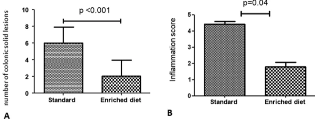
As reported in Figure 1A, there was a significant difference in
the total number of solid lesions (mean ± SD/animal) observed
in the colon at the moment of killing (6.0 ± 1.9 in the
stand-ard versus 2.1 ± 1.8 in the enriched diet group; P < 0.001). In the
standard diet group, lesion size was >7 mm in 35/150 (23.3%),
4–7 mm in 70/150 (46.6%) and <3 mm in 45/150 (30.1%) of the
total number of solid lesions. In the enriched diet group, the
lesion size was >7 mm in 1/53 (1.8%), 4–7 mm in 16/53 (30.3%)
and <3 mm in 36/53 (67.9%) of the total number of lesions. In
addition, mean size of polyps was higher in the standard than
enriched diet group (5.3 ± 1.9 versus 3.8 ± 1.7 mm, respectively;
P < 0.001).
Microscopic findings
As shown in Figure 1B, there was a significant difference in the
histological score of inflammation, evaluated according to Yu
et al. (range 0–5), between the two groups (4.4 ± 0.2 points in the
standard and 1.8 ± 0.3 points in the enriched diet group; P = 0.04).
The mean number of LGD areas per mouse was 3.5 ± 0.8 in the
standard and 2.6 ± 0.5 in the enriched diet group (P = 0.03). The
number of HGD areas was 1.8 ± 0.2 in the standard and 0.3 ± 0.2
in the enriched diet group (P < 0.001). The number of CRC areas
was 1.8 ± 0.4 in the standard and 0.4 ± 0.2 in the enriched diet
group (P = 0.006). Results are summarized in Figure 2A–C.
CRC was observed in 23/33 (69.6%) of the standard and 8/34
(23.5%) of the enriched diet group (P < 0.001).
Representative pictures of LGD, HGD and CRC are shown
in Supplementary Figure 4A, B and C, respectively, available at
Carcinogenesis Online.
Pro-inflammatory cytokine expression
Pro-inflammatory cytokine mRNA expression was significantly
higher in the standard than in the enriched diet group. In detail,
interferon gamma showed a value of 6.8 ± 2.3 (standard)
ver-sus 3.6 ± 2.8 (enriched diet) fold-change (P < 0.001); interleukin-6
showed a value of 14.0 ± 5.4 (standard) versus 4.0 ± 0.4 (enriched
diet) fold-change (P < 0.001); tumour necrosis factor-alpha
showed a value of 27.1 ± 6.7 (standard) versus 5.0 ± 0.4 (enriched
diet) fold-change (P < 0.001). Such findings are summarized
in
Supplementary Figure 5, available at Carcinogenesis Online,
where real-time PCR representative pictures have been reported.
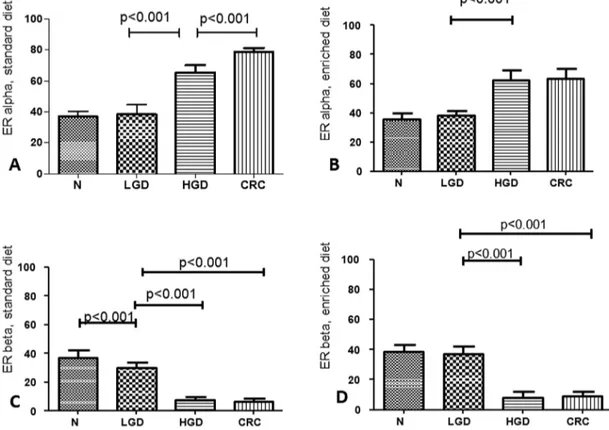
Estrogen receptors
Immunohistochemical ER-alpha and ER-beta expression is
reported in Figure 3. For ER-alpha LI values, statistical analysis
showed: Normal tissue (N) = LGD < HGD < CRC in the standard
(P < 0.001; Figure 3A) and N = LGD < HGD = CRC in the enriched
diet group (P < 0.001; Figure 3B). For ER-beta LI values,
statis-tical analysis showed: N > LGD > HGD = CRC in the standard
(P < 0.001; Figure 3C) and N = LGD > HGD > CRC in the enriched
Figure 1. (A) Total number of colonic solid lesions at autopsy (mean ± SD/animal); (B) histological inflammation score according to Yu et al.
diet group (P < 0.001; Figure 3D). Interestingly, enriched dietary
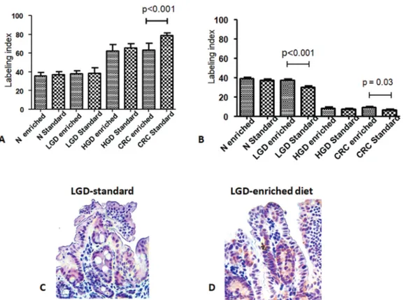
supplementation induced a lower ER-alpha expression in CRC
(P < 0.001, Figure 4A) and a higher ER-beta expression in LGD
areas (P < 0.001, Figure 4B) compared with standard diet. Figure 4
clearly shows the different ER-beta immunohistochemical
expression in LGD between standard and enriched diet group.
Figure 2. Mean number ± SD/mouse of areas of microscopic pre-neoplastic and neoplastic lesions. CRC, colorectal carcinoma; HGD, high-grade dysplasia; LGD, low-grade dysplasia.
Figure 3. (A) Estrogen receptor (ER)-alpha immunohistochemical expression in the standard group; (B) ER-alpha immunohistochemical expression in the enriched diet group; (C) ER-beta immunohistochemical expression in the standard group; (D) ER-beta immunohistochemical expression in the enriched diet group.
Epithelial DNA synthesis, proliferation and migration
In both standard and enriched diet groups, we observed in
nor-mal mucosa that positive cells were confined in the lower half of
the crypts after 24 h, whereas a progression towards the upper
half was observed after 48 h. In adddition, in the standard but
not in the enriched diet group, we found still few positive cells
near the free surface at 72nd and 96th h.
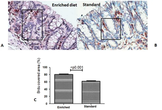
Figure 5 illustrates the percentage of crypt axis covered by
the highest labelled cell at the 48th hour after injection. This
parameter was significantly higher in the enriched than in the
standard diet group (80.6% ± 5.1% versus 61.7% ± 5.2%,
respect-ively; P<0.001).
In LGD areas of the two groups, we found positive cells in
both lower and upper half even at 24th hour. The same picture
was seen after 48, 72 and 96 h with a decreasing LI.
In HGD and CRC, the complete derangement of the crypt
architecture did not allow to evaluate the position of
BrdU-positive cells.
Epithelial apoptosis
Immunofluorescent TUNEL expression is reported in Figure 6.
Statistical analysis showed: N < LGD > HGD = CRC in the
stand-ard (P < 0.001) and N < LGD > HGD > CRC in the enriched diet
group (P < 0.001). Moreover, a significant higher level of
epithe-lial apoptosis was observed in the enriched compared with the
standard diet group in LGD and CRC areas.
Discussion
The aim of this study was to assess whether an experimental
diet formulation, enriched with silymarin, AKBA and curcumin,
could exert a protective role on colonic solid lesion development
in a mouse model of colitis-associated CRC. The hypothesis of
this study was that the combinations of phytochemicals may
exert beneficial health effects beyond what provided by a
sin-gle phytochemical (20,21). For this purpose, we preliminarily
proved in vitro the effect of each component of the nutritional
combination with the complete mixture on cultured cancer cell
proliferation, demonstrating that the antiproliferative effect
of the combination of the three substances was significantly
higher than the single concentration and the double
compos-ition. We selected the AOM/DSS model because it mimics the
stages of colon carcinogenesis, observed in the inflammatory
bowel disease-related CRC. This murine model supports the
hypothesis that chronic inflammation in inflammatory bowel
disease plays a critical role in epithelial malignancy
develop-ment in the human large bowel (35–39). AOM treatdevelop-ment followed
by repeated cycles of DSS is described to result in the onset of
chronic inflammation followed by the development of CRC in a
very high percentage of treated mice (23–26).
A first relevant result of our study was the finding of a
marked anti-inflammatory effect of the modified diet, since
enriched diet group showed a reduced inflammatory
histo-logical score as well as an impressive decrease of mucosal
pro-inflammatory cytokine mRNA expression. Moreover, we
observed in standard diet group a significant shortening of
the colon presumably as a result of long-term active
inflam-mation and consequent fibrosis, despite this feature was not
demonstrated by a specific histological staining. However, this
study could not clarify whether the anti-inflammatory effect of
enriched diet was due to an interaction with DSS, being both
substances orally administered, or to an effect of dietary
formu-lation on inflamed intestinal mucosa. On the other hand, it is
possible that anti-inflammatory effect of enriched dietary
sup-plementation could have affected the subsequent development
of dysplastic and neoplastic lesions. Indeed, we found colonic
Figure 4. (A) Comparison of estrogen receptor (ER)-alpha immunohistochemical expression between the enriched and standard diet groups; (B) comparison of ER-beta immunohistochemical expression between the enriched- and standard diet groups; immunohistochemical ER-beta staining in areas of low-grade dysplasia in standard (C) and enriched (D) diet group (positive nuclei are stained brownish and negative ones counterstained blue, magnification: ×400).
Figure 5. Percentage of crypt axis covered by the highest bromodeoxyuridine (BrdU) labelled cell at the 48th hour after injection in normal colonic mucosa. Positive cells are stained red-brownish and negative ones blue (magnification: ×400).
Figure 6. Immunofluorescent TUNEL expression in low-grade dysplasia areas of both standard and enriched diet group. Positive cells are stained green and negative ones blue (magnification: ×600).
carcinomas in 70.1% of the animals receiving a standard diet,
whereas the enriched diet group showed a marked decrease in
the number, size and volume of all colonic solid lesions. Further,
a small percentage (23.1%) developed carcinomas, whereas only
dysplastic changes were observed in the remaining animals.
The antineoplastic effect of our dietary treatment was also
related to the modulation of ERs. As expected, ER-alpha showed
a massive expression in HGD and CRC areas. Moreover, in the
same districts, ER-beta demonstrated a progressive decrease
with the sequence of carcinogenesis, in accordance with
pre-vious evidence from literature in both sporadic and
inflam-matory bowel-related CRC (40–43). Interestingly, enriched diet
group showed a higher expression of ER-beta receptors than
the standard diet group in LGD. This result is in agreement with
Calabrese et al. (44), although observed in a different model of
intestinal carcinogenesis (familial adenomatous polyposis). In
this study, an increase in ER-beta immunohistochemical signal
was found in polyps showing LGD after a treatment of 3 months
with phytoestrogens, anti-inflammatory substances and
insol-uble indigestible fibres. Therefore, this stage could represent the
checkpoint for the neoplastic progression and ER-beta agonist
effect of enriched formulation may increase apoptotic process,
thus slowing the progression to carcinoma. At this regard, the
highest TUNEL expression was observed in LGD areas
com-pared with normal and HGD/CRC districts in both the standard
and enriched diet group. Additionally, enriched diet induced a
significantly higher expression of both ER-beta and TUNEL in
LGD compared with standard one. This finding strongly agrees
with what we demonstrated previously, i.e. a co-localization in
colonic cells of ER-beta and caspase 3, which represents a
well-known early marker of programmed cellular death (45,46).
A further relevant finding of our study concerns the effect of
enriched diet on epithelial renewal (i.e. proliferation in crypts
and migration to free surface) evaluated by BrdU
immunohis-tochemical staining. In both the standard and enriched diet
groups, we observed in normal mucosa that positive cells were
confined in the usual site of epithelial proliferation (i.e. the lower
half of the crypt) 24 h after injection. In addition, a progression
towards the upper half was observed after 48 h. At this time, we
found that the percentage of crypt axis covered by the highest
labelled cell was significantly higher in the enriched than in the
standard diet group. This result may suggest that enriched diet
induces an acceleration of epithelial cell migration along the
crypts. In agreement with this finding, in the standard but not in
the enriched diet group, we found still some positive cells near
the free surface at 72nd and 96th hour.
The renewal time of epithelial cells of colorectal tract is
esti-mated 5–7 days (47) and encloses proliferation by stem cells in
the lowest part of crypts with successive migration towards
upper districts and differentiation until programmed death.
Therefore, we may hypothesize that epithelial migration in
nor-mal tissue is faster in the enriched compared with the standard
diet group and a reduced cellular half-life decreases the time of
exposure of DNA synthesizing cells to mutation risk due to both
intrinsic and extrinsic factors.
In conclusion, the results of this study appear to be
prom-ising for an effective anti-inflammatory and chemopreventive
effect of enriched diet in the course of colonic carcinogenesis
arising from inflamed tissue (48). Possible pathways of
carcino-genesis prevention by enriched diet induced could be
i.
the marked reduction in inflammatory process;
ii. the increase in ER-beta receptors and apoptosis especially
in LGD (the early stage of adenoma–carcinoma sequence);
iii. the raised epithelial renewal rate associated to a reduced
exposure time to genetic mutants.
Supplementary material
Supplementary data are available at Carcinogenesis online.
Funding
THD SpA, Correggio, Reggio Emilia, Italy.
Conflict of Interest Statement: A.D.L. is an advisory board member
of THD SpA. F.G., B.G. and M.P. are employees of THD SpA. All the
other authors declare no financial support or conflict of interest.
References
1. Tomlinson, I. et al. (1997) Molecular genetics of colon cancer. Cancer Metastasis Rev., 16, 67–79.
2. Iannone, A. et al. (2016) Stool investigations for colorectal cancer screening: from Occult Blood Test to DNA Analysis. J. Gastrointest. Cancer, 47, 143–151.
3. Williams, C. et al. (2016) Estrogen receptor beta as target for colorectal cancer prevention. Cancer Lett., 372, 48–56.
4. Niv, Y. (2015) Estrogen receptor β expression and colorectal cancer: a systematic review and meta-analysis. Eur. J. Gastroenterol. Hepatol., 27, 1438–1442.
5. Barzi, A. et al. (2013) Molecular pathways: estrogen pathway in colorec-tal cancer. Clin. Cancer Res., 19, 5842–5848.
6. Lechner, D. et al. (2005) Phytoestrogens and colorectal cancer preven-tion. Vitam. Horm., 70, 169–198.
7. Principi, M. et al. (2015) Prevention of colorectal adenomas. Colorectal Dis., 17(suppl. 1), 20–24.
8. Mosselman, S. et al. (1996) ER beta: identification and characterization of a novel human estrogen receptor. FEBS Lett., 392, 49–53.
9. Bardin, A. et al. (2004) Loss of ERbeta expression as a common step in estrogen-dependent tumor progression. Endocr. Relat. Cancer, 11, 537–551.
10. Barone, M. et al. (2008) Estrogens, phytoestrogens and colorectal neo-proliferative lesions. Genes Nutr., 3, 7–13.
11. Barone, M. et al. (2012) Dietary, endocrine, and metabolic factors in the development of colorectal cancer. J. Gastrointest. Cancer, 43, 13–19. 12. Barone, M. et al. (2014) Olive oil and omega-3 polyunsaturated fatty
acids suppress intestinal polyp growth by modulating the apoptotic process in ApcMin/+ mice. Carcinogenesis, 35, 1613–1619.
13. Seidlová-Wuttke, D. et al. (2003) Silymarin is a selective estrogen receptor beta (ERbeta) agonist and has estrogenic effects in the metaphysis of the femur but no or antiestrogenic effects in the uterus of ovariectomized (ovx) rats. J. Steroid Biochem. Mol. Biol., 86, 179–188.
14. Barone, M. et al. (2010) Dietary-induced ERbeta upregulation counter-acts intestinal neoplasia development in intact male ApcMin/+ mice. Carcinogenesis, 31, 269–274.
15. Liu, H.P. et al. (2013) Chemoprevention of intestinal adenomatous polyposis by acetyl-11-keto-beta-boswellic acid in APC(Min/+) mice. Int. J. Cancer, 132, 2667–2681.
16. Wang, R. et al. (2014) The comparative study of acetyl-11-keto-beta-boswellic acid (AKBA) and aspirin in the prevention of intestinal aden-omatous polyposis in APC(Min/+) mice. Drug Discov. Ther., 8, 25–32. 17. Shishodia, S. et al. (2005) Curcumin: getting back to the roots. Ann. N. Y.
Acad. Sci., 1056, 206–217.
18. Imran, M. et al. (2016). Cucurmin, anticancer and antitumor perspec-tives: a comprehensive review. Crit. Rev. Food Sci. Nutr., 2016, 22, 1–23.
19. Rossi, R.E. et al. (2016) The role of dietary supplements in inflammatory bowel disease: a systematic review. Eur. J. Gastroenterol. Hepatol., 28, 1357–1364.
20. Pesakhov, S. et al. (2010) Distinct combinatorial effects of the plant polyphenols curcumin, carnosic acid, and silibinin on proliferation and apoptosis in acute myeloid leukemia cells. Nutr. Cancer, 62, 811–824.
21. Cheung, K.L. et al. (2009) Synergistic effect of combination of phenethyl isothiocyanate and sulforaphane or curcumin and sulforaphane in the inhibition of inflammation. Pharm. Res., 26, 224–231.
22. Kidd, P.M. (2009) Bioavailability and activity of phytosome complexes from botanical polyphenols: the silymarin, curcumin, green tea, and grape seed extracts. Altern. Med. Rev., 14, 226–246.
23. Ikeda, I. et al. (2007) 5-aminosalicylic acid given in the remission stage of colitis suppresses colitis-associated cancer in a mouse colitis model. Clin. Cancer Res., 13, 6527–6531.
24. Stolfi, C. et al. (2011) Involvement of interleukin-21 in the regulation of colitis-associated colon cancer. J. Exp. Med., 208, 2279–2290.
25. Tanaka, T. et al. (2003) A novel inflammation-related mouse colon car-cinogenesis model induced by azoxymethane and dextran sodium sul-fate. Cancer Sci., 94, 965–973.
26. Rosenberg, D.W. et al. (2009) Mouse models for the study of colon car-cinogenesis. Carcinogenesis, 30, 183–196.
27. National Research Council (US) Committee for the Update of the Guide for the Care and Use of Laboratory Animals (2011) Guide for the Care and Use of Laboratory Animals, 8th edn . National Academies Press, Washington, DC.
28. Italian Association for Laboratory Animal Science (November 2012): https://www.aisal.org/corsi-aisal/corso-di-formazione-in-scienza-degli-animali-da-laboratorio/ (7 July 2018, date last accessed). 29. Zaidi, S.N.F. et al. (2017) Prevention of liver cirrhosis by Silymarin. Pak.
J. Pharm. Sci., 30, 1203–1211.
30. Hatcher, H. et al. (2008) Curcumin: from ancient medicine to current clinical trials. Cell. Mol. Life Sci., 65, 1631–1652.
31. Becker, C. et al. (2005) In vivo imaging of colitis and colon cancer develop-ment in mice using high resolution chromoendoscopy. Gut, 54, 950–954. 32. Yu, C. et al. (2014) Rac1 signaling regulates neutrophil-dependent tissue
damage in experimental colitis. Eur. J. Pharmacol., 741, 90–96. 33. Levin, B. et al.; American Cancer Society Colorectal Cancer Advisory
Group; US Multi-Society Task Force; American College of Radiology Colon Cancer Committee. (2008) Screening and surveillance for the early detection of colorectal cancer and adenomatous polyps, 2008: a joint guideline from the American Cancer Society, the US Multi-Society Task Force on Colorectal Cancer, and the American College of Radiology. Gastroenterology, 134, 1570–1595.
34. Javid, S.H. et al. (2005) Modulation of tumor formation and intestinal cell migration by estrogens in the Apc(Min/+) mouse model of colorec-tal cancer. Carcinogenesis, 26, 587–595.
35. Dulai, P.S. et al. (2016) Colorectal cancer and dysplasia in Inflammatory Bowel Disease: a review of disease epidemiology, pathophysiology, and management. Cancer Prev. Res. (Phila)., 9, 887–894.
36. Axelrad, J.E. et al. (2016) Inflammatory bowel disease and cancer: the role of inflammation, immunosuppression, and cancer treatment. World J. Gastroenterol., 22, 4794–4801.
37. Fries, W. et al. (2017). Disease patterns in late-onset ulcerative colitis: results from the IG-IBD “AGED study”. Dig. Liver Dis., 49, 17–23. 38. Ierardi, E. et al. (2001) Epithelial proliferation and ras p21 oncoprotein
expression in rectal mucosa of patients with ulcerative colitis. Dig. Dis. Sci., 46, 1083–1087.
39. Biancone, L. et al.; Italian Group for the study of Inflammatory Bowel Disease. (2016) Inflammatory Bowel Disease phenotype as risk factor for cancer in a prospective multicentre nested case-control IG-IBD study. J. Crohns. Colitis, 10, 913–924.
40. Principi, M. et al. (2012) Estrogen receptors expression in long-lasting ulcerative pancolitis with and without dysplasia: a preliminary report. Scand. J. Gastroenterol., 47, 1253–1254.
41. Principi, M. et al. (2015) The sharp decline of beta estrogen receptors expression in long-lasting ulcerative-associated carcinoma. Scand. J. Gastroenterol., 50, 1002–1010.
42. Di Leo, A. et al. (2008) ER-beta expression in large bowel adenomas: implications in colon carcinogenesis. Dig. Liver Dis., 40, 260–266. 43. Barone, M. et al. (2010) ERβ expression in normal, adenomatous and
carcinomatous tissues of patients with familial adenomatous polyp-osis. Scand. J. Gastroenterol., 45, 1320–1328.
44. Calabrese, C. et al. (2016) Can supplementation of phytoestrogens/ insoluble fibers help the management of duodenal polyps in familial adenomatous polyposis? Carcinogenesis, 37, 600–606.
45. Principi, M. et al. (2013) Phytoestrogens/insoluble fibers and colonic estrogen receptor β: randomized, double-blind, placebo-controlled study. World J. Gastroenterol., 19, 4325–4333.
46. Di Leo, A. et al. (2016) Epithelial turnover in duodenal familial aden-omatous polyposis: a possible role for estrogen receptors? World J. Gastroenterol., 22, 3202–3211.
47. Parris, A. et al. (2015) A human colonic crypt culture system to study regulation of stem cell-driven tissue renewal and physiological func-tion. Methods Mol. Biol., 1212, 141–161.
48. Principi, M. et al. (2014) Ulcerative colitis: from inflammation to can-cer. Do estrogen receptors have a role? World J. Gastroenterol., 20, 11496–11504.