Index
UNIVERSITÀ DEGLI STUDI DI SALERNO
Dipartimento di Farmacia
Dottorato di Ricerca
in
Scienze del Farmaco
Ciclo XXIX — Anno accademico 2016/2017
Tesi di Dottorato in
Design, synthesis and biological
evaluation of new anticancer and/or
anti-inflammatory agents
Dottorando Tutore
Dott. Antonio Foglia Chiar.mo Prof.
Carmela Saturnino
Index
Preface
My PhD three years course in Drug Discovery & Development at the Department of Pharmacy of Salerno University was started in 2014 under the supervision of Prof. Carmela Satunino.
In the first part of my project, the research was mainly focused on the design and synthesis of new anti-cancer agents. In particularly, were designed two series of compounds as new modulators of C-terminal domain of the molecular chaperone Heat Shock protein 90 (Hsp90), these compounds were designed on the base of the first synthetic inhibitor of C-terminal domain of Hsp90.
In the second part of my research activity was focused on the design and synthesis of new inhibitors of microsomal Prostaglandin E2 synthase-1 (mPGES-1) as potential anti-inflammatory agents.
The work was performed under the direct supervision of Prof. Carmela Saturnino and in collaboration with Prof. Ines Bruno and Dr. Stefania Terracciano. Computational guided design of compounds was performed in collaboration with Prof. Giuseppe Bifulco‟s research group. Biological screenings were performed in collaboration with Prof. Oliver Werz of Friedrich Schiller University (Germany) in the case of mPGES-1, and with Dr. Maria Carmela Vaccaro and Fabrizio Dal Piaz of Salerno University in the case of Hsp90.
Furthermore, to improve my knowledge about the synthesis techniques, in 2016 I joined Prof. Patrick Dallemagne's research group at the Centre d'Etudes et de Recherche sur le Médicament de Normandie de l'Université de Caen Normandie (France), where I spent four months.
Index
List of publications related to the scientific activity performed during the three years PhD course in Drug Discovery & Development
Papers:
Terracciano S.,‡ Foglia A.,‡ Chini M.G., Vaccaro M.C., Russo A., Dal Piaz F., Saturnino C., Riccio R., Bifulco G., Bruno I. “New dihydropyrimidin-2(1H)-one based Hs90 C-terminal inhibitors” RSC Adv. (2016), 6, 82330-82340.
Terracciano S., Chini M.G., Vaccaro M.C., Strocchia M., Foglia A., Vassallo A., Saturnino C. Riccio R., Bifulco G., Bruno I. “Identification of the key structural elements of dihydropyrimidinone core driving toward more potent Hsp90 C-terminal inhibitors” Chem. Commun. (2016), 52, 12857-12860.
Botta A., Sirignano E., Popolo A., Saturnino C., Terracciano S., Foglia
A., Sinicropi M.S., Longo P. and Di Micco S. “Identification of Lead
Compounds as Inhibitors of STAT3: Design, Synthesis and Bioactivity”. Mol. Inf. (2015), 34, 689 – 697.
‡ These authors contributed equally to this work.
Conference proceedings:
Foglia A., Di Micco S., Terracciano S., Saturnino C., Riccio R., Bruno I., Bifulco G. “Design, Virtual Screening and Synthesis of Potential Microsomal Prostaglandine E2 Synthase-1 (mPGES-1)”, XXV National Meeting of Italian Chemical Society, Rende (Italy), 7-12 September 2014.
Index Foglia A., Di Micco S., Terracciano S., Saturnino C., Riccio R.,
Oppermann U., Bifulco G., Bruno I. “Design and Synthesis of 4-substituted–pyridine-2,6-dicarboxylic acids as new potential JMJD3 modulators”. XL Summer School "A. Corbella" Gargnano (BS), Palazzo Feltrinelli, 14 - 18 june 2015.
Foglia A., Terracciano S., Lauro G., Saturnino C., Riccio R., Bruno I., Bifulco G. “Targeting Microsomal Prostaglandin E2 Synthase 1 For The Development Of New Potential Anti-inflammatory Drugs.”
XXXVI Convegno della Divisione di Chimica Organica - CDCO 2015 Bologna, 13-17 September 2015.
Terracciano S., Strocchia M., Dal Piaz F., Vaccaro M.C., Foglia A., Chini M.G., Leone A., Riccio R., Bifulco G., Bruno I. “Discovery of innovative Hsp90 C-terminal modulators: synthesis and biological evaluation of 3,4-dihydropyrimidinone derivatives.” XXXVI Convegno della Divisione di Chimica Organica - CDCO 2015 Bologna, 13-17 September 2015.
Foglia A., Terracciano S., Lauro G., Saturnino C., Riccio R., Bruno I., Bifulco G. “Inhibition of Microsomal Prostaglandin E2 Synthase1: Focused Design of New Anti-inflammatory and Anticancer Drugs.” XV Edizione del congresso SAYCS (Sigma Aldrich Young Chemists Symposium) Rimini 27-29 October 2015.
Foglia A., Vaccaro M.C., Saturnino C., Riccio R., Bifulco G., Chini M.G., Bruno I., Terracciano S. “Exploration of dihydropyrimidin-2(1H)-one scaffold for the discovery of new promising C-terminal Hsp 90 inhibitors” XXXVII Convegno della divisione di chimica organica CDCO 2016. Venezia 18–22 September 2016.
S. Terracciano, A. Foglia, M. G. Chini, M. C. Vaccaro, M.a Strocchia, A. Russo, R. Riccio, G. Bifulco, I. Bruno “Development of C-terminal heat shock protein 90 inhibitors as therapeutic agents” XXXVII Convegno della divisione di chimica organica CDCO 2016. Venezia 18–22 September 2016.
Index
Index
Abstract…….……….………...………….
1
CHAPTER 1
4-351.1 A short history of anticancer therapy…………..………. 5
1.2 Inflammation………...……… 7
1.3 Why cancer and inflammation?... 9
1.4 The drug discovery………..…….... 11
1.5 Heat shock protein 90 (Hsp90)………... 13
1.6 Microsomal prostaglandin E2 synthase-1 (mPGES-1)... 22
1.7 How to proceed in the research project... 31
CHAPTER 2
37-64 2.1 The need to develop Hsp90 C-terminal inhibitors... 382.2 Where do we start?... 39
2.3 Influence of the chemical functionalization on the phenyl ring at C4 position of DHPM core based Hsp90 C-terminal inhibitors... 41 2.3.1 Surface Plasmon Resonance assay... 43
2.3.2 Antiproliferative assay and Western Blot analysis... 44
2.3.3 Molecular docking studies... 47
2.4 Enhanced inhibitory activity of C-terminal Hsp90 domain through substitution of DHPM core at C2 position... 51
2.4.1 Surface Plasmon Resonance assay... 53
2.4.2 Antiproliferative assay, Western Blot analysis and effect on cell cycle progression... 55
2.4.3 Study of Hsp90α/18 interaction………..…… 58
2.4.4 Molecular docking studies... 60
CHAPTER 3
65-80 3.1 Why mPGES-1?... 663.2 Looking for new molecular platforms………... 67
3.2.1 Design and synthesis of 5-Pyrazolone compound…….. 67
3.2.2 Design and synthesis of Carbazole compounds………. 72
3.2.3 Design and synthesis of Biaryl compounds……… 76
Conclusion
... 83Index
CHAPTER 4
86-1004.1 General information... 87
4.2 Methods and materials... 88
4.2.1 General procedure for synthesis of the second
generation of DHPMs derivatives by microwave-assisted Biginelli reaction……….
88
4.2.2 General procedure for synthesis of 14a and 14b by
microwave-assisted Biginelli reaction...
95
4.2.3 General procedure for microwave-assisted
Liebeskind-Srogl cross coupling reaction
96
CHAPTER 5
101-1235.1 General synthetic methods... 102
5.2 General procedures for the preparation of 4-arylidene-1,3-disubstituted-5-pyrazolone compounds...
102
5.2.1 General procedure for the preparation of
5-pyrazolones 23a-23h... 102
5.2.2 General procedure for the preparation of 24-41... 103
5.3 General procedure for the synthesis of carbazole compound. 112
5.3.1 General procedure for the preparation of 42a-42e... 112
5.3.2 General procedure for synthesis of 43-47... 112
5.3.3 General procedure for the synthesis of biaryl
compounds……… 115
5.3.3.1 Synthesis of 58... 122
____________ _________________________________________Abstract
- 1 -
Abstract
One of the main goal of modern medicinal chemistry is the development of new agents able to modulate biological targets involved in inflammation and cancer processes. In this context, my PhD project was focused on the exploration and structural optimization of various chemical moieties able to interfere with two targets involved in both processes. In particular, two biological targets were selected: Heat shock protein 90 (Hsp90) and microsomal Prostaglandin E2 Synthase-1 (mPGES-1). The results obtained can be divided into two sections in accordance with the target of interest.
a) Exploration and structural optimization of DHPM core in order to guide the synthesis of new and more potent Hsp90 C-terminal inhibitors.
Hsp90 is a molecular chaperone involved in the maturation and stabilization of a wide range of client proteins that play a crucial role in the development, survival and proliferation of cancer cells. In the literature there are several compounds capable of inhibiting this molecular chaperone. The most part of these compounds inhibit the protein through modulation of the N-terminal domain. However, this type of modulation involves a well-known heat shock response, a cytoprotective mechanism that as a final result leads to the increase of cytosolic levels of heat shock proteins with consequent cell survival. Therefore, the modulation of C-terminal domain of Hsp90 represents a better strategy for the development of new antitumor agents, since, they do not induce heat shock response. In an attempt to discover new modulators of the C-terminal domain of Hsp90 and taking into account the structure of the first synthetic inhibitor of this domain, a 3,4-dyhidropyrimidin-2(1H)-one (DHPM)
____________ _________________________________________Abstract
- 2 -
derivative, two more generations of DHPM derivatives have been synthesized. Relatively to the second generation of DHPM derivatives, the synthesis was focused on the influence of the chemical functionalization of aromatic ring at C4 position of DHPM core, while the third generation has been designed with the aim to functionalize the C2 position of the core. The exploration and optimization processes of DHPM core led to the identification of novel and more potent inhibitors of the C-terminal domain of Hsp90.
b) Identification of new mPGES-1 inhibitors.
mPGES-1 is an inducible enzyme that catalyzes the terminal step of the biosynthesis of PGE2 from the PGH2 precursor. The inhibition of this enzyme appears to be a promising strategy for the identification of novel anti-inflammatory agents, because, the use of selective inhibitors would allow to overcome the classical side effects of traditional anti-inflammatory drugs. Moreover, mPGES-1 is overexpressed in a wide variety of human cancers and for this reason it has emerged as an attractive biological target for anticancer drug discovery. In order to identify new molecular platforms able to interact with the target protein three collections of compounds (carbazoles, biaryl compounds and 5-pyrazolones) were synthesized. Biological evaluation revealed the identification of five biaryl compounds (60-64) as new chemical entities that inhibit mPGES-1 activity with promising IC50 values (ranging 0.18-1.64 µM).
Introduction
- 3 -
Introduction
- 4 -
1-Introduction
- 5 -
1.1 A short history of anticancer therapy
"Cancer" term includes a group of more 100 diseases in which the cells begin to grow out of control.1 With the onset of tumorigenesis body cells begin to divide and spread into the surrounding tissue. In addition, it is possible that some tumor cells can break off from the primary tumor and through the blood stream or the lymph system create a secondary tumor far from the original in a process known as metastasis.2 The processes that trigger oncogenesis are varied and are not yet well defined, to date many risk factors have been identified that could increase the chance of getting cancer, for these reason it is nearly impossible to know the development of cancer causes.3,4 The tumor arises from a normal cell when its DNA is damaged fact, the cancer is a disease characterized by a large genetic instability. When the DNA of cells is damaged and the repair mechanisms do not work there is an upheaval of cellular functions. In cancer cells, indeed the damaged DNA is not repaired, and the cell does not die, instead it gives rise to more such abnormal cells with abnormal DNA.5 In most cases surgery is the main strategy to remove the tumors, while chemotherapy and radiotherapy are used especially to remove residual cells by acting on their continued proliferation.6 Unfortunately, the chemotherapy and radiotherapy don't have a real specificity of action and they cause considerable side effects.7
The coming of the nitrogen mustards and antifolate drugs have allowed the first chemotherapy treatment only in 1940. While, radiotherapy has become a valuable resource for the control of this disease in 1960 with the invention of the linear accelerator.8 At that time the main strategies in cancer therapy were surgery and radiotherapy, but soon it became clear that cure rates after ever more radical local treatments had plateaued at about 33% due to the presence of heretofore unappreciated micrometastases. After these observations, it was
Introduction
- 6 -
decided to introduce the opportunity to apply chemotherapy drugs in conjunction with surgery and/or radiation treatments, doing so the concept of adjuvant chemotherapy was born. The purpose of the combined treatment modality was to maximize the antitumor effect of each of the three therapies with minimal toxicity to normal tissues.9,10 Over time, it was needed to develop an effective therapies not based only on empirical observations but increasing dependent on an understanding of human tumour biology.11 With this purpose doctors, researchers, and patients worked together to improve treatment options and identify new drugs that have action on a selected biological entities, with this new concept, chemotherapy entered into the era of "targeted therapy". The new concept of the identification of biological targets is started in 1960 with an unrelated program, the Special Virus Cancer Program (SVCP), which wanted to clarify the association between viruses with cancer.12 When it failed in its purpose it was transformed into a program of molecular biology to study genes that have been aggregated by tumor viruses. This work identified oncogenes, suppressor oncogenes, and signaling pathways essential for developmental biology itself.13 In order to understand the cellular activities such as proliferation and survival and with the aim to repair the molecular defects directly in cancer cells, researches of biotechnology firms were focused on the molecular and biological processes. Molecular targeting offers a more convenient solution for the development of potential therapeutic agents with reduced systemic toxicity, on the other hand the tumor cells, thanks to their intrinsic genetic plasticity, easily adapt to a new environment. Finally, this study led to the identification of new drug targets which currently influences the development of anticancer drugs. New targets include growth factors, signalling molecules, cell-cycle proteins, modulators of apoptosis and molecules that promoted angiogenesis.14 In the last decades, have been discovered many differences between normal cells and tumor cells,
Introduction
- 7 -
many of which help the proliferation of cancer cells, therefore, scientific research has accelerated the development of new anticancer drugs with the main objective to offer a real hope to all those who suffer from cancer.
1.2 Inflammation
Inflammation is defined as the immediate body's response when it feels threatened by pathogens, noxious stimuli such as chemicals, or physical injury.15 This process leads defensive cells at the site of the damage so as to neutralize and eliminate the noxious stimulus, repair the structure and restore the function of damaged tissues. Clinically inflammation is characterized by four cardinal signs which they were reported in the first century by Celsus, they are: redness (rubor), heat (Calor), swelling (tumor) and pain (dolor).16 Later Virchow has added a fifth signs: loss of function (functio laesa).17
Introduction
- 8 -
Inflammation is a generic response, and therefore it is considered as a mechanism of innate immunity, compared to adaptive immunity which is specific for each pathogen.18 Furthermore, the inflammation damage has substantial differences from the damage of autoimmune disease that determine a specific immune-mediated attack on the tissue that is no longer recognized as self.20 Based on the time duration, the evolution of inflammation can be classified as either acute or chronic.21 The first response observed after a noxious stimulus is acute inflammation which as consequence increases the contribution of leukocytes (mainly granulocytes) from the blood to damaged tissue, while chronic inflammation is a more prolonged response resulting from the persistence of the mediators of inflammation of an acute inflammation not completely resolved.22 Historically the first anti-inflammatory drugs have their origin in the discovery of some plant extracts that gave relief to disorders related to inflammation. In the mid-19th century were discovered salicylates, this has allowed the development of some drugs such as acetyl-salicylic acid (Aspirin).23 With the discovery of the mechanism of action of aspirin it is exponentially increased the interest of the development of new anti-inflammatory therapies which have as mechanism of action the inhibition of cyclooxygenase (COX).24 However, many side effects, particularly related to gastrointestinal and cardiovascular toxicity, were associated with the use of these drugs.25 This toxicity appears to be a serious drawback in a long-term therapy,26 so today, the development of small molecules safer and clinically useful with decreased side-effect profiles is a continuous effort.
Introduction
- 9 -
1.3 Why cancer and inflammation?
The relationship between cancer and inflammation is a concept that has been observed many years ago, however, the mechanisms underlying this interconnection are still unclear. Cancer and inflammation go hand in hand, the first triggers an inflammatory response, while the inflammatory microenviroment increases the aggressiveness of the tumor and the dissemination of metastases.27 The connection between cancer and inflammation was observed in the nineteenth century, when they noticed that the tumors arose in a chronic inflammation sites and when inflammatory cells were found in biopsy samples from tumors.28 It is well known that many chronic inflammatory factors increase the risk of developing cancer, this factors include autoimmune diseases (inflammatory bowel disease is associated with colon cancer), microbial infections (infection with Helicobacter pylori is associated with gastric cancer and gastric mucosal lymphoma) and inflammatory conditions of unknown origin (prostatitis is associated with prostate cancer).29 The link between inflammation and cancer can be divided into two pathways as shown in Figure 1.2.30 The intrinsic pathway is activated by genetic events as oncogenes activation, oncosuppressor inactivation and chromosomal rearrangement or amplification, in this manner cells are transformed producing an attractive environment for tumour growth, facilitating genomic instability and promoting angiogenesi.31 The extrinsic pathway is mediated by inflammatory cells of innate immunity, so, inflammatory or infectious conditions enhance the risk of developing cancer (for example, such as inflammatory bowel disease for colorectal cancer and prostatitis for prostate).32 The two pathways activate the transcription factors, such as nuclear factor-κB (NF-κB), signal transducer and activator of transcription 3 (STAT3) and hypoxia-inducible factor 1α (HIF1α), in tumour
Introduction
- 10 -
cells33,34 that coordinate the production of inflammatory mediators, including cytokines, chemokines and cyclooxygenase 2 (COX-2) which, in turn, synthesize the prostaglandins.34
Figure 1.2Pathways that connect inflammation and cancer
The cytokines activate the same transcription factors in inflammatory cells, stromal cells and tumour cells, resulting in more inflammatory mediators being produced and a cancer-related inflammatory microenvironment being generated. On the other hands, inflammatory mediators contribute to genomic instability resulting to the known mutations associated with tumours. Many of them act as direct mutagens or as deregulators of DNA repair mechanism and cell cycle checkpoints acquiring the ability of cancer cells to proliferate, invade and escape from host defence.35,36 Moreover, the relationship between cancer and inflammation is well documented by numerous studies that demonstrate how aspirin is able to prevent certain cancer such as colorectal,
Introduction
- 11 -
lung, stomach, esophagus and prostate.37-39Anyway, many evidences have been gathered supporting the improved therapeutic efficacy that can be achieved by blocking the two signalling networks and their pathways.40,41 In this way, my PhD project has worked.
1.4 The drug discovery
The development of new drugs requires a complex process involving many scientific areas, such as, biology, statistics, pharmacology, pharmaceutical chemistry, toxicology, computational chemistry, organic chemistry and proteomics.42 The network between the various areas of science cleared the way for what we now call the “drug discovery process”. The aim of drug discovery is to turn “molecules into medicine for the public health”, this means find new therapeutic agents with suitable pharmaceutical properties (i.e., efficacy, bioavailability, toxicity) for preclinical evaluation. The drug discovery is one of the most challenging human activities, these processes must proceed through several stages in order to produce a product that is safe, efficacious, and has passed all regulatory requirements.43 This process begins with the "target identification". Any enzyme or any biochemical mechanism involved in the pathological condition may represent a potential therapeutic target. After the identification there is the step of target validation, which takes place indirectly, ie through the use of compounds that block the target thus giving the desired effect.44Furthermore, the process of discovery of new drugs requires the knowledge of pharmaceutical technology to make new bioavailable products without losing the pharmacological effect, for this purpose it is requires a close collaboration between a pharmaceutical and chemical pharmaceutical technologist.45 Currently, the drug discovery process is focusing on small organic molecules, appropriately designed to act directly
Introduction
- 12 -
on the biological target of interest, without interacting with other biological system.46 However, drug discovery requires simple and variable methods of synthesis to vary the lead structure extensively, which is essential for the derivation of structure–activity relationships (SARs, ligand-based design) and rapid optimization to obtain the active ingredient. The efficient optimization of drugs and the provision of sufficient amounts for further biological characterization is the central task of medicinal chemistry.47 Today, researchers employ various techniques to overcome the difficulties in the drugs discovery process, for example, analytical and purification methods such as high-performance liquid chromatography (HPLC),48 multicomponent and domino reactions,27 microwave-assisted and flow chemistry,49,50 etc. In the present PhD project, thanks to combined of medicinal and pharmaceutical chemistry approaches, new chemical entities with antitumor or anti-inflammatory effects have been successfully identified. With the aim of discovering new potential anticancer agents I focused my attention on exploration and structural optimization of the first synthetic inhibitor of C-terminal domain of Heat shock protein 90. After completion of this project I had the opportunity to work on a second project aimed to identification new anti-inflammatory agents that target the microsomal Prostaglandin E2 synthase-1 (mPGES-1), enzyme involved in the terminal step of PGE2 synthesis.
Introduction
- 13 -
1.5 Heat shock protein 90 (Hsp90)
Exposure of cells to stress conditions (including heat shock, oxidative stress, heavy metals), or pathologic conditions such as ischemia and reperfusion, inflammation, tissue damage, infection, usually cause protein dysfunction.51 The cell responds to environmental stress by increasing synthesis of several molecular chaperons with the aim to prevent cellular damage and to re-establish cellular homeostasis.51 These proteins help to maintain cellular cellular homeostasis (proteostasis) in cells, in fact, in accordance with the general definition, molecular chaperone are each protein that helps or stabilizes another protein to acquire its functionally active conformation52 and prevent the aggregation of non-native protein.53 In the cells, several different classes of chaperones exist, many of these classes are often known as stress proteins or heat-shock proteins (HSPs), as they are upregulated under conditions of stress in which the concentrations of aggregation-prone folding intermediates increase. Heat Shock proteins are categorized according to their molecular weight into six classes: small Hsps, Hsp40, Hsp60, Hsp70, Hsp90 and Hsp100.54 Family members of Hsps are localized in the nucleus, cytoplasm, and subcellular compartments, such as mitochondria and endoplasmic reticulum (ER), the high molecular weight HSPs are ATP-dependent chaperones, whereas small HSPs act in an ATP-inATP-dependent manner.55 It is important to emphasize that the expression of HSPs requires a precise balance, because, over-expression of some HSPs shows the way to certain diseases such as cancer. Tumor cells having more signal transduction pathways and a higher metabolic requirements than normal cells, so they have a greater need of chaperones to maintain cancer cells survival. 56 For these reasons, the expression of Hsps is severely elevated in cancer cells, and it is associated with enhanced tumorigenicity, metastatic potential of cancer cells
Introduction
- 14 -
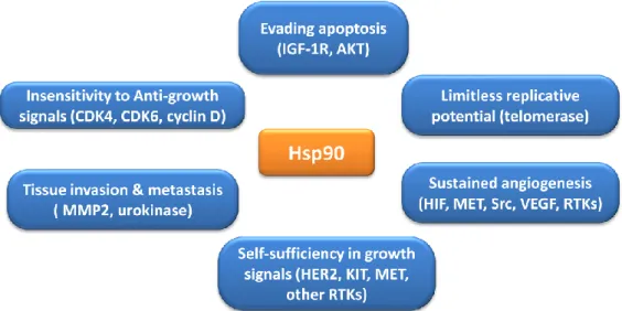
and resistance to chemotherapy. Moreover, in association with key apoptotic factors they are strong anti-apoptotic proteins, blocking the cell death process at different levels, for these reasons they are suitable targets for modulating the streets of cell death.57 Among the Hsps's family, heat shock protein 90 (Hsp90) has emerged as being of prime importance to the survival of cancer cells.58 Hsp90 is constitutively expressed at 1-2% of total cytosolic proteins and its level increases up to 10-fold in tumor cells, suggesting that it may be critically important for tumor cell growth and/or survival.59 Firstly, Hsp90 protein is involved in the maturation and stabilization of a wide range of ‘client proteins’ that plays a central pathogenic role in human diseases including cerebro- and cardiovascular diseases,60 autoimmune diseases,61 neurodegenerative diseases, viral infections and cancer.62 Regarding the latter, many Hsp90 client proteins are involved in critical cellular functions that promote cell growth, cell survival and proliferation as they are able to facilitate an escape from normal proteolytic turnover, thereby overall promoting an oncogenic transformation.63 Hsp90 client proteins comprise proteins that contribute to all of the six hallmarks of cancer, including the ability to produce growth factors, resistance to anticancer agents, avoidance of apoptosis, unlimited replicative potential, uninterrupted angiogenesis and invasiveness and metastasis.64-66 Hsp90 play a central role in acquisition and maintenance of each of six hallmarks of cancer (Figure 1.3). For example, Hsp90 influences angiogenesis by folding hypoxia-inducible factor‑1α (HIF‑1α) and vascular endothelial growth factor receptor (VEGFR) in addition to governing nitric oxide synthase upregulation. Many client proteins of Hsp90 are apoptotic mediators, including Bcl-2, Apaf-1, the serine-threonine protein kinase AKT/PKB and survivin.67 Also, Hsp90 may promote tissue invasion and metastasis through MMP-2 activation, or digesting extracellular matrix proteins.67 Other client proteins of Hsp90 that play a role in cell signaling processes include FAK (integrin pathway), IL6R
Introduction
- 15 -
(JAK/STAT3 pathway), IκB kinases (NFκB pathway), CDK 4, 6, 9, hTERT (cell cycle), p53 (tumor suppressor genes), and the steroid hormone receptors (estrogen receptor and androgen receptor).68
Figure 1.3 Hsp90 functions implicated in establishment of each of the hallmarks of cancer. Importantly, Hsp90 functions may also permit the genetic instability on which acquisition of
the six hallmarks depends.
In addiction, Hsp90 exists as a multi-chaperone complex with unusually high affinity for ATP and some substances in tumor cells, whereas in normal cells a latent form of Hsp90 protein is present. In the human proteome there are four isomers of Hsp90, the two major isoforms correspond to the inducible Hsp90α isoforms and the costitutive Hsp90β, that are located predominantly in the cytosol, whereas two non-cytosolic forms are known, the 94 kDa glucose-regulated protein (GRP94) and the Hsp75/tumor necrosis factor receptor associated protein 1 (TRAP-1), that are expressed in the endoplasmic reticulum and in the mitochondrial matrix respectively.69 Structurally, this enzyme exist as a homodimer and each monomer contains three flexibly linked regions.70 The amino (N)-terminal domain consists of a α- and β-sandwich motif, it is responsible for the protein‟s ATPase activity, since that contains an adenosine triphosphate (ATP)‑binding and hydrolyzing pocket.71
Introduction
- 16 -
Another intriguing characteristic of the N-terminal domain is that several conserved amino acid residues form a “lid‟ (residues 100–121) that closes over the nucleotide binding pocket in the ATP bound state but is open during the ADP-bound state.72 The middle domain consist of a large αβα segment connecting to a small αβα segment via several short α-helices,73
it is a linker region involved in co-chaperones and client proteins recognition/binding, also participates in forming the active ATPase.74 Finally, there is the carboxy (C)-terminal domain consist of a curved α-helix, a stranded β-sheet, a three-helix coil and an extended disordered arm, this region directs Hsp90 dimerization.73 This region contains another ATP-binding site, without hydrolases activity, this pocket is only available when the N-terminal domain ATP binding site is occupied.74-76 In particulary, the C-terminal domain is implicated into the functional switch between the closed and the open protein conformation,77 and there is a conserved MEEVD motif involved in the binding of co-chaperones which containing a tetratricopeptide repeat (TRP) motif.78,79 Hsp90 helps nascent proteins to assume their biologically active conformations, corrects the conformation of misfolded proteins, and helps incorrigibly misfolded proteins to be removed and degraded by the ubiquitin-proteosome system through the presence of co-chaperones, immunophilins, and partner proteins to form multiprotein complexes, thus facilitates the maturation, stability, activity of client proteins.80-83
ATP hydrolysis is necessary for Hsp90 activity.84 Structural studies showed that Hsp90 assumes several structurally distinct conformations. In the resting state, through the dimerization of its C-terminal domain, Hsp90 forms a V shape conformation which is defined as the “open conformation”.85 The ATP-binding N-terminal domain of Hsp90 induces a series of conformational changes including repositioning of the N-terminal lid region.86
Introduction
- 17 -
After lid closures, Hsp90 reaches a more compact conformation, termed “closed conformation” in which the N-domains are dimerized. After Hsp90 finishes its chaperoning tasks of assisting the proper folding, stabilization and activation of client proteins under the active state, the ATP molecule is hydrolyzed, so the N-domain release ADP, Pi and Hsp90 returns to the open conformation again.87
Figure 1.4 Conformational cycle and schematic domain organization of Hsp90 During different phases of the chaperone cycle Hsp90 binds more than 20 co-chaperones through both the N- and C-termini of the protein, some of which play the tuning roles by either activating or inhibiting the ATPase activity of Hsp90.88-90 Hsp90 contains several drugable sites at both its N- and C-terminal domains and for this reason, Hsp90 inhibitors are divided into several groups. Traditional Hsp90 inhibitors block the binding of the ATP at N-terminal domain, they include natural compounds, as geldanamycin (GDA) and radicicol (RDC),91,92 and synthetic molecules such as geldanamycin and
Introduction
- 18 -
radicicol derivatives,93-97, purines derivatives,98-100 pyrazoles derivatives,101 sulfonamide derivatives,102 C9-type iridoid (verminoside)103 and the three chimeric compounds of radicicol and geldanamycin radamide, radester and radanamycin.104-106 Even today, there are over 50 clinical trials testing different N-terminal Hsp90 inhibitors (ClinicalTrials.gov database) undergoing clinical evaluation.107,108 However, clinical trials have shown many limitations, including poor solubility and toxicity of these drugs that limit their use.109 The most well-known side effect is the induction of a cytoprotective mechanism termed as heat shock response (HSR),110,111 which induces drug-resistance and anti apoptotic mechanisms.112,113 A promising strategy to overcome these problems is to regulate Hsp90 function by targeting the C-terminal domain, as its inhibition does not induce the unsought HSR.114,115 Unfortunately, the few structural information on Hsp90 C-terminal domain currently obtainable have not provided the resolution necessary for structure-based drug design of new synthetic inhibitors. The development of more efficacious C-terminal inhibitors is desired to better understand the ramifications of C-terminal inhibition and to probe the mechanism by which Hsp90 interacts with client proteins.116 The first C-terminal inhibitor was discovered in 2000 by Neckers and co-workers, they showed the capabilities of the Hsp90 C-terminus to bind novobiocin.117 Novobiocin is not the only product which inhibits the Hsp90 activity through C-terminus modulation, but related family members such as chlorobiocin and coumermycin A1 were showed to bind this domain and induce a dose-dependent degradation of Hsp90 client proteins in a manner similar to Hsp90 N-terminal inhibitors.118 However, these inhibitors have micromolar and millimolar IC50 values (Novobiocin ≈ 700 µM in SKBr3 cells) and it is not sufficient for clinical application,115 but it give a starting point for the development of new more efficacious C-terminal inhibitors. Furthermore, structural optimization of
Introduction
- 19 -
novobiocin has led to analogues with more potent antiproliferative profiles than the starting compound.119-122 Many other inhibitors that bind C-terminal domain exist, they include epigallocatechin-3-gallate,123 taxol,124 cisplatin125 and sansalvamide A derivatives (Table 1).126,127
The latter are the allosteric modulators that bind the N-middle domain and block the binding of Hsp90 to client proteins and co-chaperones that bind to the C terminus of the protein.128 Until now, only natural and/or semi-synthetic compounds have been described. Recently, Strocchia et al. have identified the first synthetic compound able to inhibit C-terminal domain of the Hsp90 (Table 1).129 The principal benefit of C-terminal inhibitors is that they do not seem to be associated with the heat shock response than N-terminal inhibitors.130
Table 1. Hsp90 C-terminal inhibitors and their IC50 value.
Inhibitor IC50
700 µM In SKBr3
80 µM In SKBr3
Introduction - 20 - 70 µM In SKBr3 100.16 µM In SKBr3 - -
Introduction - 21 - 46.7 µM against an NCI 60 panel cell line 20.8 µM in Jurkat cell line
As described before, Hsp90 is the central point of the protein folding machine, because there are many binding sites for co-chaperones. So, each protein-protein interaction presents the opportunity for disruption the protein-protein folding process. Nowadays, only a few protein-protein interactions have been targeted such as interactions between Hsp90 and the co-chaperones Cdc37,131 p50,132 and the interactions between Hsp90 and the partner protein HOP.133However, the modulation of Hsp90 through the last two strategies (C-terminal inhibitors and disrupt protein-protein interaction), may allow to develop of new potential modulators of Hsp90 which could be used as anticancer agents and are expected to be free from side effects associated to the induction of heat shock response.
Introduction
- 22 -
1.6 Microsomal prostaglandin E2 synthase-1 (mPGES-1)
Prostaglandins (PGs) and thromboxane A2 (TXA2), collectively termed prostanoids, are a group of physiologically active lipid mediators derived enzymatically from fatty acids. This class of mediators are involved in homeostatic functions and mediate pathogenic mechanisms, including inflammatory response.
Inflammation is the body's natural response to infection and injury against an organ or tissue and has been implicated in the pathogeneses of arthritis, cancer and stroke, as well as in neurodegenerative and cardiovascular disease.134 Furthermore, inflammatory diseases increase the risk of developing many types of cancer such as bladder, cervical, gastric, intestinal, oesophageal, ovarian, prostate and thyroid cancer.135,136 There are four principal bioactive prostaglandins generated: prostaglandin E2 (PGE2), prostacyclin (PGI2), prostaglandin D2 (PGD2) and prostaglandin F2α (PGF2α).137 Biosynthesis of prostanoids starts by release of arachidonic acid (AA) from cell membrane phospholipids catalyzed by phospholipase A2 (PLA2). Subsequently AA isconverted to Prostaglandin H2 (PGH2) in two steps by cyclooxygenase 1/2 (COX-1, COX-2), bifunctional enzymes that contain cyclooxygenase and peroxidase activity, in the first step AA was oxidized to generate PGG2, then this intermediate is reduced into PGH2.138 The COX pathway produces PGG2 and PGH2, which then are converted into PGI2 by prostacyclin synthase, TXA2 by thromboxane synthase, and PGE2, PGD2 and PGF2α by their respective synthase enzymes (Figure 1.5).139
Among the prostaglandins, PGE2 is the most abundant in physiological conditions and plays a crucial role in several organs and tissues such as kidney and gastrointestinal tract.139,141 Further studies suggest that PGE2 had immunosuppressive effects by regulating the expression of EP2, EP3 and
Introduction
- 23 -
EP4,142 also, PGE2 protected the gastrointestinal mucosa and regulated the renal flow, natriuresis and blood pressure by modulating the expression of EP1 and EP2.143 Under physiological conditions, the level of PGE2 is regulated by three enzyme, the prostaglandin E2 synthases (PGEs), that including the cytosolic PGES (cPGES) and two membrane-bound proteins mPGES-1 and mPGES-2.144
cPGES and mPGES-2 are constitutively expressed in various cells and tissues, and their levels were not significantly increased under inflammatory conditions, but high levels of mPGES-2 was observed in human colorectal cancer, human gliomas and activated microglia.145 mPGES-1 is considered as an induced enzyme like COX-2, because, in normal conditions it is expressed at low level but under pathological conditions, this enzyme is significantly activated leading to over-production of PGE2.146
Introduction
- 24 -
A variety of signal transduction pathways are involved in the regulation of mPGES-1, its expressionincrease in response to various inflammatory stimuli and mediators, for example, cytokines (LPS, IL-1b and TNF-α).147,148
It is clear how mPGES-1 could represent a drug target associated with inflammation, fever and pain,149 so, the modulation of this enzyme could be a promising alternative in anti inflammatory therapy. mPGES-1 plays a key role, similar to COX-2, in the development of cancers. Several studies suggest that mPGES-1 was overexpressed in colon cancer,150,151 lung cancer,152 head and neck cancer, breast cancer, stomach cancer,153,154 and Alzheimer‟s disease tissues.155 Furthermore, several publications reported that mPGES-1 could cause lung metastatic tumor formation and growth, induce EGFR expression, and mediate EGFR-dependent tumor growth in prostate cancer.156 Moreover, mPGES-1 expression was hugely correlated with vascular invasion and worse prognosis in colorectal cancer.157158
Pharmacological inhibition of mPGES-1 reduced squamous carcinoma growth by suppression of PGE2 mediated-EGFR signalling and impairing tumor-associated angiogenesis. These findings highlight also the potential of mPGES-1 inhibitors as agents capable of reducing tumor growth.159
The discovery of mPGES-1 is attributed to Jackbsson in 1999160 who recognized it as a member of the Membrane-Associated Proteins in Eicosanoid and Glutathione Metabolism (MAPEG) superfamily.161 In this family, there are many other members including leukotriene C4 synthase (LTC4S), 5-lipoxygenase-activating protein (FLAP) and microsomal glutathione transferases (MGST) 1-3.162,163 The most closely related MAPEG member is the microsomal glutathione transferase-1 (MGST1), which shares 39% sequence identity with mPGES-1.164
Recently a study by Sjögren et al. showed that the accuracy of mPGES-1‟s structure reached a higher resolution of 1.2 Å x-ray in-plane via electron
Introduction
- 25 -
crystallography.165 Human mPGES-1 is a membrane homotrimer with four transmembrane α-helices (TM1-TM4) enclosing an inner core, which looked like a funnel-shaped opening towards the cytoplasm between TM1 and TM2 in each monomer (Figure 1.6A). Glutathione (GSH) is an essential co-factor for active binding in the U-shapes conformation at the interface between subunits in the proteins166-169 and attacked the peroxide group of PGH2 in the active site via its thiol group in the catalytic cycle process.164
GSH forms hydrogen bond with several amino acid residues such as, Arg73, Asn74, Glu77, His113, Tyr117, Arg126 and Ser127 from helices II and IV, and Arg38 from helix I. In addition, GSH forms a π-stacking interaction with Tyr130 due to its glutamate and its cysteine (Figure 1.6B).
The active site of mPGES-1 consists of three gatekeepers and a residue of Arg, all of that are located within the transmembrane-helix IV.166,170-172 In order to work its catalytic activity the active site of the mPGES-1 must be open to be able ensure the PGH2 access.168 The N-terminal region of helices II and IV jointly with the C-terminal portion of helix I and the C-cytolasmatic domain from neighboring molecule forms a deep cavity,173 researchers suggest that this is the active site based on the form, the size and the favorable interactions that this pocket has with PGH2.174,175
The proposed mechanism for PGH2 isomerization to PGE2 by GSH suggests that Ser127 activates the thiol of GSH to form a thiolate anion that acts as nucleophilie and directly attack on the endoperoxide oxygen atom at the C-9 carbon of PGH2 to produce an unstable intermediate. Deprotonation in 9-position and cleavage of S-O bond lead to a bidentate complex with Arg126. This results in the regeneration of the reactive thiolate anion (regeneration of GSH) and the formation of the product PGE2 (Figure 1.6C).165
Introduction
- 26 -
Figure 1.6 (A) Structure of the mPGES-1 trimer. (B) Interactions between
mPGES-1 and GSH. (C) Suggested mechanism of PGH2 isomerization to PGE2 by mPGES-1.
Inhibition of mPGES-1 offers many therapeutic opportunities in several pathologies, in fact, the suppression of massive PGE2 biosynthesis is a reasonable pharmacological strategy in many phatological conditions such as fever, inflammation, cardiovascular disease and cancer.176-178 Traditional anti-inflammatory agents are not selective towards this enzyme, drugs that target mPGES-1 are considered as valuable alternatives to NSAIDs/coxib and they may offer a better safety profile and, thus, lower risk of side effects that are usually associated with NSAIDs and coxib.179-182 NSAIDs not only inhibit the production of PGs but also annihilates the release of bradykinin, change lymphocyte reactions and decrease the migration of phagocytosis of
Introduction
- 27 -
granulocytes and monocyte in the inflammatory process. However, unselective NSAID are not able to differentiate between the two COXs, and this is the reason of their gastric side effects, because, PGE2 is clearly known to have the protective effects on the gastrointestinal mucosa.183 In order to overcome the side effects of NSAIDs selective inhibitors of the inducible COX-2 (COXibs) were discovered.184 These products modulate the COX-2 as well as the downstream PGEs enzymes. However, after the marketing of this class of medicine, they have proved to be associated with the increased cardiovascular risk in patients after long-term therapies, probably due to imbalance of PGI2 and TXA2 in the vasculature.185,186 In this field, mPGES-1 inhibitors constitute an attractive pharmacological approach which could overcome the classic side effects associated with COX-inhibitors, in fact, blocking the mPGES-1 enzyme we expect to selectively inhibit increased PGE2 production, without affecting other prostanoids of physiological importance. Despite that mPGES-1 was reported as a drug target already in mPGES-1999, progress in drug discovery of mPGES-1 has been very slow. Up to now, a number of compounds with different chemotypes have been identified, but none have entered clinical trials.187,188 Even today there are many problems to be studied and solved in developing real selective mPGES-1 modulators for humans, because, the phenotypic differences between human and murine enzyme have hampered research on the effects of mPGES-1 inhibition in cancer and in inflammatory diseases.189 For example, the variation of certain amino acids in the active site of mPGES-1 may lead to partial or complete loss the activity of potent inhibitors. Three amino acids located in transmembrane helix IV of mPGES-1 are not conserved between human and rat/mouse enzyme, these residues are the gatekeepers of the active site and the variation of these amino acids intercept the access of modulators for steric reasons.190 In the Table 2 were reported the most known mPGES-1 inhibitors and their IC50 valeus, grouped
Introduction
- 28 -
in three main ways: natural products and derivatives, synthetic compounds and dual inhibitors of mPGES-1 and 5-lipoxygenase (5-LO).
Table 2. Some mPGES-1 inhibitors
Introduction
- 29 -
Introduction
- 30 -
Dual inhibitors of mPGES-1 and 5-LO
a
In cell-free assay. bDocking scores. cPGE2 production inhibition ratio. d
In cell-based assay.
Despite good results have been obtained for some products with different chemical motifs, currently there is still no selective modulators acting on mPGES-1 in clinics. 217,218 Many industrial and academic groups have worked to develop mPGES-1 modulators. Their results show any excellent IC50 values in cell-free assay but they are not stable so they have a poor cell potency, another reason which blocks the drug development of mPGES-1 inhibitors, as
Introduction
- 31 -
previously mentioned, are the specific differences between the enzyme isoforms in various species.219 The rational inhibitors design of mPGES-1 was started in 2013 with the three-dimensional crystal structure of mGES-1 determined by Sjögren et al.165 The rational design through computational methods was complicated, because, the active site is shallow and located within the membrane-spanning region. In 2015, a crystal structures binding to four inhibitors of mPGES-1 including MF63 and MK886 (see Table 2: synthetic inhibitors) was reported by J.G. Luz et al., they clarified the binding mode between inhibitors and amino acid residues and explained the mechanism of isomerization from PGH2 to PGE2.220 However, the ultimate goal is to discover more effective, highly selective and highly safe drugs for the treatment of pain, inflammatory diseases and cancer, and in this context, the discovery of new mPGES-1 modulators is highly demanded, so, this crystal structure can expand our knowledge with regard to designing and pharmacologically validating inhibitors of mPGES-1.
1.7 How to proceed in the research project
My PhD program is aimed to the design, synthesis and biological evaluation of new chemical compounds able to modulate the activity of the two biological target described before. Therefore, the scientific activity carried out during the PhD was focused on the synthesis of small libraries of compounds in order to identify new products to guide the development of new modulators with inhibitory activity against the C-terminal domain of Hsp90 and mPGES-1. The research was focused on the analysis of data currently available in the literature, also taking advantage the information about Structure Activity Relationship (SAR) of known inhibitors of the two targets proteins.
Introduction
- 32 -
The general methodology used in this study can be described through these main steps:
1. Analysis of data currently available in the literature in order to identify some chemical motifs able to interact with the two biological targets; 2. Design and synthesis of the compounds through suitable synthetic
strategies;
3. Biological evaluation and individuation of new possible hits or lead compounds;
4. Rationalisation of ligand/protein interaction by computational analysis.
Concerning step 1, analysis data present in the literature was used for the identification of a few scaffolds poorly explored, able to interfere with the target of interest. In particular, the 3,4-dihydropyrimidin-2(1H)- one (DHPM) core for the Hsp90,126 while carbazole, dyaril and pyrazol-5-one scaffolds for mPGES-1.
These chemical moieties are seen as “privileged structures” in medicinal chemistry, because they possess significant biological activity, in fact, adequately decorated, they can selectively bind many enzymes, receptors or channels having different pharmacological effects.221-224
In respect to step 2, appropriate synthetic procedures were used and optimized in order to obtain the desired products in good yield. For the synthesis of DHPM core were used the well-known Biginelli condensation, a multiple-component chemical reaction that creates 3,4-dihydropyrimidin-2(1H)-ones core under acidic conditions from three component, aryl aldehydes, urea or its derivatives and β-ketoesters. This one-pot reaction was first reported by Pietro Biginelli in 1893 (Scheme 1.1).225
Introduction
- 33 -
Scheme 1.1 Biginelli condensation between urea derivatives, aryl aldehydes and ethyl acetoacetate
Although the original reaction conditions suffered from poor yields, in the last decades, the discovery of dihydropyrimidine biological activity has led to a renewed exploration of the reaction conditions, revealing a variety of compatible solvents, acid catalysts, and an expanded substrate scope.226 Some of this procedures have replaced the traditional Brønsted acids226-228 with different Lewis acids such as TMSCl,229 FeCl3,230 Yb(OTf)3,231 Cu(OTf)2.232 Moreover, different techniques have been described as the use of solvent-free conditions,233 solid-phase approaches,234 phase-transfer and polymer supported catalyst,235,236 ionic liquids,237 asymmetric methodology.238
Another optimization of the methodology concerned the usage of several high-seed microwave-assisted protocols in order to improve the yield of products and reduce the reaction time.239-241 For the synthesis of our compounds, DHPMs derivatives have been obtained through a protocol of Biginelli reaction supported by microwave-irradiation and chlorotrimethylsilane (TMSCl).242
Regarding the carbazoles derivatives has been used a synthetic strategy in two steps, the first step was performed through a classic Suzuki-Miyaura cross-coupling reaction in order to obtain 2-nitrobiaryl derivatives, while the second step was carried out via Cadogan cyclization protocol. This approach allows the reductive cyclization of 2-nitrobiphenyl compounds in the presence of suitable organophosphorus reagents.243,244
Introduction
- 34 -
The reaction of these compounds were conducted using an excess of triethyl phosphite (P(OEt)3), under solvent-free conditions and supported by microwave-irradiation.
This synthetic methodology, compared to others, provides a greater tolerance of functional groups on the substrate and more precise regiocontrol of functional group placement within the product. Since the biaryl compounds have been obtained in the same manner of the first step of carbazole compounds. Suzuki-Miyaura reaction is an organic reaction between organic boron compounds and organic halides catalyzed by a palladium(0) complex and it is widely used to synthesize substituted biphenyls.245
Cross-coupling reactions is one of the most straightforward methodologies for various carbon–carbon bond formations and it has many advantages, in fact, the reactants are readily available, nontoxic, and air- and water-stable. They react under mild conditions and are amenable to a variety of reaction conditions, including the use of aqueous solvents and substrate supports. The inorganic boron byproduct can be easily removed after the reaction.246 In our case, biaryl compounds were obtained using a classical Suzuki-Miyaura protocol promoted by Pd(PPh3)4, Tetrakis(triphenylphosphine)palladium(0) and potassium carbonate, K2CO3 as base.
Concerning the pyrazol-5-one derivatives has been used a synthetic strategy in two steps, in the first step there is the preparation of the pyrazol-5-one core using a tandem condensation and thermal cyclization between β-ketoesters and hydrazines. In the second step was performed through an aldol condensation, taking advantage of C4 (methylene group) reactivity with aldehydes (Scheme
Introduction
- 35 -
Scheme 1.2 Synthesis of pyrazol-5-one derivatives
Lastly concerning step 3, biological evaluation of the synthesized products were performed using appropriately assay for each of two investigated targets, e.g. Surface Plasmon Resonance (SPR), cytotoxicity, western blot assay and limited proteolysis assay in the case of Hsp90, a cell-free assay using the microsomal fraction of interleukin-1β-stimulated human A549 cells to evaluate the effect od several compound on mPGES-1 activity. Regarding step 4, the rationalization of ligand/protein interaction has been performed using docking studies.
___________________________________________Results And Discussion
- 36 -
___________________________________________Results And Discussion
- 37 -
- CHAPTER 2 -
Exploration of the structural elements of a
3,4-dihydropyrimidin-2(1H)-ones (DHPM) core, driving toward more potent Hsp90
C-terminal inhibitors.
Based on : Terracciano S., Foglia A., Chini M.G., Vaccaro M.C., Russo A., Dal Piaz
F., Saturnino C., Riccio R., Bifulco G., Bruno I. RSC Adv. (2016), 6, 82330-82340. And Terracciano S., Chini M.G., Vaccaro M.C., Strocchia M., Foglia A., Vassallo A., Saturnino C. Riccio R., Bifulco G., Bruno I. Chem. Commun. (2016), 52, 12857-12860.
Exploration of the structural element of 3,4-dihydropyrimidin-2(1H)-ones (DHPM) core, driving toward more potent Hsp90 C-terminal inhibitors
- 38 -
2.1 The need to develop Hsp90 C-terminal inhibitors
As outlined above, many Hsp90 N-terminal inhibitors have been shown excellent antitumor activity have some drawbacks in clinical application.249-252
In contrast to this strategy, an alternative approach to Hsp90 modulation for the treatment of cancer is represented by the C-terminal inhibitors, as they don't induce the heat shock response, a well-established compensatory mechanism that has likely limited the clinical impact of N-terminal Hsp90 inhibitors in cancers treatments.253-255
The heat shock response (HSR) is a cellular mechanism in response to stress, bacterial infections, heat, toxine and heavy metals.256
It is mediated by heat shock factor-1 (HSF-1), which activates transcription of heat shock genes.257 HSF-1 is ubiquitously expressed and has the principal role in the stress-induced expression of Hsp genes,258 this protein is a monomer in complex with Hsp90 and in this form it is unable to bind to DNA.259
In stress condition, HSF-1 is released from the chaperone complex then trimerized and it is transported into the nucleus where it undergoes hyperphosphorylation and binds to DNA containing heat shock elements sequence.260, 261 Herein it directs the trascription of Hsp genes which lead the increase of cellular resistance by elevating the expression of multiple heat shock proteins,254 including Hsp27, Hsp70 and Hsp90 (Figure 2.1).262 The N-terminal inhibitors of Hsp90 induce the dissociation of HSF-1 from Hsp90 and, with the same mechanism, trigger the pro-surival heat shock response that enables cancer cells to escape the cytotoxic effect, in fact, this cascade of events facilitates oncogenic growth and stops tumor cells from undergoing apoptosis.263
Exploration of the structural element of 3,4-dihydropyrimidin-2(1H)-ones (DHPM) core, driving toward more potent Hsp90 C-terminal inhibitors
- 39 -
Figure 2.1 Heat Shock Response
In addition, this attractive cytoprotective mechanism has driven researchers to find alternative molecules that repress Hsp90 activity without inducing HSR. The C-terminal modulation has an opposite effect, because C-terminal inhibitors block HSF-1 in its inactive form promoting its degradation via ubiquitin-proteasome pathway.263-265
In this context, the research is enormously engaged in the development and discovery of new molecules that bind the C-terminal domain of Hsp90, but the vast conformational space of this region is still a strong limitation for the rational design of a selective inhibitors.
2.2 Where do we start?
With the aim of identify new promising and more potent C-terminal inhibitors of Hsp90 published data were used. For the first time in 2015 Strocchia et al.129 reported a new 3,4-dihydropyrimidin-2(1H)-one derivative able to interact with C-terminal domain of Hsp90.
This compound was designed starting from one assumption, Csermely et al.78, 80
Exploration of the structural element of 3,4-dihydropyrimidin-2(1H)-ones (DHPM) core, driving toward more potent Hsp90 C-terminal inhibitors
- 40 -
pyrimidine nucleotides (GTP and UTP preferentially), so take in advantage the structural analogy between uridine triphosphate (UTP) and the DHPM core they synthesized seventeen DHPM derivatives through Biginelli multicomponent reaction.
Figure 2.2 First non-natural inspired modulator of C-terminal domain
Moreover, compound 1 (Figure 2.2) represents the first non-natural inspired product able to modulate the C-terminal domain of Hsp90 with a good antiproliferative profile (IC50 values 50.8 ± 0.2 and 20.8 ± 0.3 mM in A375 and Jurkat cell lines, respectively) whitout any apparent cytotoxic activity in non-tumor cells. Western blot analysis confirmed that the antitumor activity of this compound was provided by degradation of some Hsp90 client oncoproteins, moreover, they showed that the compound does not cause an increase of the levels of Hsp90 and Hsp70 demonstrating that the undesired HSR was not induced.
Finally, limited proteolisis essay showed that the compound 1 binds the C-terminal region of Hsp90.
Exploration of the structural element of 3,4-dihydropyrimidin-2(1H)-ones (DHPM) core, driving toward more potent Hsp90 C-terminal inhibitors
- 41 -
2.3 Influence of the chemical functionalization on the phenyl ring at C4 position of DHPM core based Hsp90 C-terminal inhibitors.
The great interest aroused by C-terminal Hsp90 inhibitors as potential therapeutic agent and the recent discovery of compound 1 as C-terminal Hsp90 inhibitor drove us to improve the exploration of this promising scaffold.
With the purpose to improve the information about the structure-activity relationship and to explore the influence of the chemical functionalization on the phenyl ring at C4 position of DHPM core a second collection of different decorated DHPM derivatives was synthesized (compounds 2-13, Figure 2.3), by a microwave-assisted Biginelli multicomponent reaction242 through the combination of the following synthons:
twelve different aldehydes (A-N);
N-phenylurea; Ethyl acetoacetate.
N-phenylurea and ethyl acetoacetate have been used to maintain the same functionalizations present on the other positions on DHPM core of compound
1. These compounds were obtained by using chlorotrimethylsilylane (TMSCl)
as the mediator of Biginelli reaction, especially for the synthesis of N1-substituted dihydropyrimidinones more difficult to obtain using a microwave-assisted Biginelli protocol (Scheme 2.1).222, 240
In particular, with the aim of shedding more light on the influence of chemical diversity around the phenyl ring at C4 position of this scaffold, diverse aryl aldehydes available in laboratory were chosen. These aldehydes have different substituents in the o-, m-, p- and o-, m- positions with either electron-withdrawing or electron-donating groups.
Exploration of the structural element of 3,4-dihydropyrimidin-2(1H)-ones (DHPM) core, driving toward more potent Hsp90 C-terminal inhibitors
- 42 -
Scheme 2.1 General synthetic procedure for the synthesis of compounds 2-13 and structures
of the aryl aldehydes used to generate the second collection of DHPMs derivatives.
Following the synthetic procedure described before compounds 2-13 were prepared in short reaction time (15-20 min) and with a good yields (55-80%).
Exploration of the structural element of 3,4-dihydropyrimidin-2(1H)-ones (DHPM) core, driving toward more potent Hsp90 C-terminal inhibitors
- 43 -
Figure 2.3 Structures of compounds 2-13
Biological evaluation has been conducted in collaboration with Professor Fabrizio Dal Piaz, Dr. Maria Carmela Vaccaro and Dr. Alessandra Russo of Salerno University.
2.3.1 Surface Plasmon Resonance assay
Once synthesized the compounds 2-13 were evaluated for their ability to interact with the recombinant Hsp90α by a Surface Plasmon Resonance (SPR) based binding assay.266, 267 On the basis of this screening, 7 out of 12 tested compounds efficiently interact with the immobilized protein with low kinetic dissociation constant (KD) values (Table 2.1). The lead compound 1 and the known Hsp90 inhibitor 17-N-allylamino-17-demethoxygeldanamycin
(17-AAG)268 were used as positive controls.Checking the results obtained compounds 2-6, 8 and 11 exhibit high affinity against the molecular chaperone with low KD values in the nM range, ranging from 2.9 to 76.2 nM. Analyzing the data obtained, all the compounds synthesized using p-substituted aromatic aldehydes were able to interact with the molecular chaperone, showing an increased affinity compared 1 against Hsp90 except for 2.