Scuola di Dottorato
“ARCHIMEDE” IN SCIENZE, COMUNICAZIONE E TECNOLOGIE Ciclo XXVII
Microbial mediation in the formation and alteration of
minerals in shallow marine environments
This work wants to investigate the role that microorganisms have in the alteration and precipitation of minerals, in particular Ca-carbonates, on natural and artefact rocks of archaeological interest in shallow marine environments. This finds numerous applications in the recognizing of the abiotic vs. biotic nature of neo-formed minerals, such as in the reconstruction of depositional palaeoenvironment, in astrobiology and in medicine. In addition, nanostructured microbially induced minerals have several technical application varying from bioconservation, bioremediation, biogrouting to biomedic.
Furthermore, the knowledge of such complex interaction between bacterial communities and hard substrates is essential for the preservation and the valorization in situ of underwater cultural heritage, which represents a particularly important element in the history of peoples.
For this purpose, several key-samples of natural and artifact rocks, have been collected in Santa Maria di Ricadi bay and in the archaeological submerged park of Monasterace, respectively sited on the Tyrrhenian and Ionian coast of Southern Calabria.
Optical and scanning electron microscopy (SEM) have been used for micro- and nano-scale investigation of the role of microorganisms in the precipitation and alteration of minerals. Observations showed that biofilms are characterized by: (1) Skeletal elements, such as red algae, bryozoan, polychaete, bivalves and diatoms; (2) bacteria such as cocci, bacilli, spirochaete, filamentous bacteria, often associated with organic matter remains which result sometimes mineralized; (3) Neo-formed Ca-carbonate and pyrite minerals; (4) bioerosion elements, such as grooves and boreholes.
Ricadi as aphanitic and peloidal deposits. All these deposits result made up by an assemblage of nanospheres, varying in diameter from 50 to 200 nm, tightly clotted together. The co-existence of degraded EPS and bacteria, strictly associated with Ca-carbonate nanospheres, implies that the organic matter and the microbial metabolism played a fundamental role in the precipitation of these minerals.
Moreover, in Monasterace biofilms, framboidal pyrite occur both in rock fracture up to 2 mm below rock surface and within cavities of encrusting red algae. The presence of framboidal pyrite and dumbbell- to spherical- shaped crystals imply they formed in anoxic environment by sulphate reducers bacteria.
As regards bioerosion products, these results formed by epilithic, endolithic and euendolithic organisms. In particular, in Ca-carbonate deposits forming part of Santa Maria di Ricadi rock samples, an intricate net of grooves is produced by cyanobacterium M. testarum.
All these elements suggest that biofilms are complex systems formed by different biotic processes occurred both in oxic and anoxic conditions. Epilithic and endolithic microorganisms resulted capable to modify chemical and physical micro-conditions leading to the precipitation and alteration of diverse rocky materials, in particular Calcium carbonate, in shallow marine environments.
Abstract
Chapter 1 - State of the art
1.1 Introduction………1
1.2 Microorganisms……….2
1.3 Microbial biofilm and mats………..….8
1.4 Extracellular Polymeric Substance………..………12
1.5 Biomediated mineral formation………...………16
1.5.1 Biologically controlled mineralization……….16
1.5.2 Biologically induced mineralization………17
1.6 Calcium carbonate precipitation by bacteria ………...20
1.7 Microbial bio- alteration………..…23
1.8 Application of nanostructured biologically induced minerals………….…29
Chapter 2 - Geographical setting and historical introduction
2.1 Geographical setting………...…..302.2 History of Monasterace………33
3.2 History of Santa Maria di Ricadi……….…………36
Chapter 3 – Methods
3.1 Sampling………..………393.2 Fixation and dehydratation………..…39
4.1 Introduction………..……....41
4.2 Monasterace samples………...42
4.2.1 Components of the biofilm……….…43
4.2.1.1 Skeletal organisms………..…….43
4.2.1.2 Bacteria and organic matter remains……….…..….50
4.2.1.3 Neo-formed minerals………..…..52
4.2.2 Biological weathering features………58
4.3 Santa Maria di Ricadi samples……….60
4.3.1 Components of the biofilm………...………..62
4.3.1.1 Skeletal organisms……….…………..62
4.3.1.2 Bacteria and organic matter remains………..……….63
4.3.1.3 Neo-formed minerals……….………..66
4.3.2 Biological weathering features………..……..69
Chapter 5 – Discussion
5.1 Biofilms formation………..….…725.2 Biologically mediated mineralization………..78
5.3 Bioerosion………86
Chapter 6 – Conclusions
………..…901
CHAPTER 1
STATE OF ART
1.1 Introduction
Since the early Precambrian (3.5 billion of years ago), microbial growth and metabolism have had a fundamental role in the geological and geochemical evolution of the Earth surface and atmosphere. Moreover, Bacteria of various kinds were the unique living forms on Earth during at least the first crucial 1.5 billion years. Obviously, how early microbes affected their habitat can be only supposed from knowledge about their activities in modern equivalent environments.
Microorganisms are ubiquitous settlers on every Earth solid surface and relatively shallow subsurface, and frequently their action significantly accelerates the effects of chemical and physical factors (Gaylarde et al., 2003).
The relationship between bacteria and the lithosphere can ranges from formation of minerals to microbial utilization of rocks and mineral as a nutrient source or as a habitat. Microbes may play a role in the authigenic and diagenetic formation of minerals.
The minerals may appear external to the microbial cell responsible for its genesis, on or in the cell envelope, or even within the cell. Some microbial mineral formation is active and may involve direct enzymatic intervention or metabolic production of specific chemical reactants that cause precipitates to form. Other microbial mineral formation is passive and may even be mediated by dead cells (Ehrlich, 1998).
Otherwise, alterations occur under the influence of physical, chemical and biological processes, though subaerial and subaqueous breakdown generally involves interaction between all three types (Brehm et al., 2004).
2
As a measure of the contribution of organic activity to rock weathering, it is estimated that 20–30% of stone weathering is the result of biological activity (Wakefield and Jones, 1998). Microbial marine communities represent key factors largely influencing minerals and global biogeochemical cycles of elements. So far, only a fraction of marine microbial organisms have been identified and it seems rather likely that numerous of unknown species survive performing chemical reactions that may either contribute precipitation and/or alteration of rocks and minerals (Ehrlich, 1998).
1.2 Microorganisms
A microorganism is, by definition, an organism that is unicellular or lives in a colony of cellular organisms.
This group encompasses Prokaryotes (Bacteria and Archea) and Eukaryotes (Fig. 1.1). Often, as well as in this study, bacteria is synonymous with the term prokaryote, not the domain.
3 Fig. 1.1 - The “universal” phylogenetic tree based on 16S/18S rRNA sequence comparisons, showing the
three domains of life: Bacteria, Archaea, and Eukarya. The Bacteria and Archaea are subdivided by representative phyla, and the Eukarya are outlined by familiar grouping for lack of general consensus among taxonomists. Note: all the branch lengths should be considered approximate (from Konhauser, 2007).
4
Most Bacteria and Archaea are between 500 nanometers (nm) and 2 micrometers (μm) in diameter, with a volume between 1 and 3 μm 3 and a wet weight approximating 10−12 grams. The only way to observe such small objects is by microscope, hence the term microorganisms or microbes. Individual cells occur basically occur as rods (known as bacilli), spheres (known as cocci), or helical shapes (known as spirilla, spirochete or vibrio) (Fig. 1.2). Individual cells are common, but they can form easily groups as clusters or filaments, when they grow end on end (Beveridge, 1988).
Fig. 1.2 – Basic bacterial shapes as seen under
SEM. (a) Bacillus methylotrophicus sp., with rod shape (Madhaiyan et al., 2009). (b)
Staphylococcus aureus, with sherical shape (Drake
et al.,2013). (c) Leptospira sp. , with helical shape (Konhauser, 2007).
A
B
C
5
All the Prokaryotes are characterized by the lacking of organelles, unlike the Eukaryotes.
Eukaryotes include microorganisms, such as protists and fungi, as well as multicellular life, such as the plants, algae and animals. Algae are a diverse group of photosynthetic organisms that produce O2 as a byproduct of their metabolism.
They are typically found in fresh- and sea-water, but they can also grow on rocks and trees, or in soils when water is available to them.
Algae are broadly classified into green, brown, golden, and red that are mainly filamentous forms. Small algae are usually associated with microbial mats, where they trap and stabilize sediment (Riding, 2000). Collectively they display a wide variety of morphologies, ranging from unicellular forms to macroscopic seaweeds that can grow tens of meters in length (van den Hoek et al., 1996; Konhauser, 2007). Others can grow symbiotically with animals, such as the dinoflagellates associated with coral reefs.
In contrast, diatoms are small unicellular algal forms, but they attach by mucilaginous stalks, secrete mucilage and, when abundant, are important sediment stabilizers in marine (Awramik and Riding, 1988) and non-marine (Winsborough, 2000) environments.
Fungi also have a range of morphologies, from unicellular yeasts, to filamentous molds that clump together to form what is known as mycelia, to macroscopic forms such as mushrooms.
They get many important roles, as decomposers of organic material, leading to nutrient cycling and spoilage of natural and synthetic materials (such as wood or food); as symbionts of algae and cyanobacteria (e.g. as lichens); and as producers of economically important substances, such as ethanol, citric acid, antibiotics, and vitamins (Moore-Landecker, 1996).
With regard to the study areas, cyanobacteria play an important role, contributing notably to biofilms formation and alteration.
6
Cyanobacteria, also known as Cyanophyta, are a phylum of bacteria. In the past, they were formerly known as "blue-green algae". The basic morphology comprises unicellular, colonial and multicellular filamentous forms. The majority of them are aerobic photoautotrophs, but certain cyanobacteria show a distinct ability for heterotrophic nutrition (Fay, 1965).
Two morphological types are distinguished: filamentous species that form elongated cell chains, called trichomes, often bundled together (multitrichomous species) and coccoid species that form spheroidal cells often arranged into cell clusters (Whitton and Potts, 2000).
Their life processes require only water, carbon dioxide, inorganic substances and light. Photosynthesis is their principal mode of energy metabolism. In the natural environment, however, it is known that some species are able to survive long periods in complete darkness. Furthermore, the prominent habitats of cyanobacteria are freshwater and marine environments. They flourish in water that is salty, brackish or fresh, in cold and hot springs, and in environments where no other “microalgae” can exist. Most marine forms grow along the shore as benthic vegetation in the zone between the high and low tide marks (Humm and Wicks, 1980).
The cyanobacteria comprise a large component of marine plankton with global distribution (e.g. Gallon et al., 1996). From the geological point of view, cyanobacteria are important because they colonize the interface between sediments and water and affect fluid flow dynamics and structure formation, enveloping the mineral grains forming biofilms (Noffke et al., 2003).
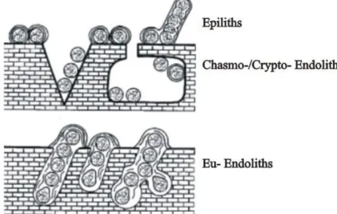
In the case of lithobiontic cyanobacteria we differentiate between epilithic, chasmo-/crypto-endolithic and euendolithic cyanobacteria (Golubic et al., 1981; Fig. 1.3).
Epilithic forms live on the surface of hard substrates, while endoliths reside inside hard substrates. Endoliths include organisms that colonize existing cracks and fissures (chasmo-endoliths) or cavities in porous and cavities of substrates
7
(cryptoendoliths), as well as those which actively penetrate carbonate substrates, such as euendoliths. These latter, actively excavate, bore tunnels or burrows within the mineral solid or a variety of calcareous substrates, such as shells, dead coral, etc. (Golubic et al., 1981).The first record of euendolithic cyanobacteria was found in 1.5 billion year old stromatolite rocks in China (Zhang and Golubic, 1987). As a result, they are believed to have played a major role in the production and destruction of carbonate, including reef frame builders and sediments, over long periods of geological time. The well fossilized microborings of euendoliths are used as proxies in paleoecological and paleobathymetrical studies (Glaub and Vogel 2004; Radtke and Golubic 2005) as their diversity, distribution and abundance depend on substrates and environmental conditions (Gektidis 1999; Vogel et al. 2000; Tribollet, 2008b).
They are also used for ichnological and ichnotaxonomic determinations. Those studies are based on comparisons between modern and fossilized microborings.
8
1.3 Microbial biofilms and mats
Several studies testify that solids added to aquatic systems are rapidly coated with an organic microbial accretion, forming a more or less thick layer. Such microbial communities are complex structures, ranging from micrometers to few centimeters in thickness, respectively called biofilms and mats.
In addition to thickness, another fundamental difference between them is that microbial mats are characterized by high population densities of photoautotrophic microorganisms that act as primary producers in the top millimeters and build up the mat matrix, while many biofilms are heterotrophic and depend on substrate supply from the surface or the surrounding water (De Beer & Külh, 2001).
Functionally, a modern living mat is a vertically compressed ecosystem that supports most of the major biogeochemical cycles within few millimeters. Due to differing growth requirements, it is common for mat microorganisms to orient themselves into vertically stratified subsystems that are manifested as distinct, and often, visible layers, where the lamination is determined by the light quantity and quality. The decreasing light regime that occurs with depth results in a cyanobacteria-dominated blue–green layer near the surface, often underlain by a reddish-pink layer of purple sulfur bacteria, a deeper layer of green sulfur bacteria. The deepest layers of the mat are typically black, with occasional gray bands (Fig. 1.4). Mats are largely self-sufficient, in that sunlight or chemical reductants provide the primary source of energy used to convert CO2 into biomass, while
heterotrophy recycles the carbon and reducing equivalents back to other mat microorganisms.
The most considerable components are the filamentous microorganisms, either photosynthetic or chemolithoautotrophic, which, associated with EPS, contribute notably to the cohesion of the mat (Konhauser, 2007).
9 Fig. 1.4 - Cross-section of mineralized microbial mat. Different colors of layers reflect the differentiated
microbial composition. Green layer on top is given by photosynthetic bacteria, followed by intervals of brownish layers, with heterotrophic bacteria; whereas red layers with purple sulphur bacteria and an underlying grey layer with sulphate reducers. White layers represent carbonate precipitates. (from Spadafora et al., 2010).
In a typical sedimentary system, the types of microbial metabolism found with depth exhibit a corresponding decrease in thermodynamic yields: aerobic respiration has the highest yield and is encountered at surface, while methanogenesis has the lowest energetic yield and occurs the deepest following the redox gradient (Fig. 1.5).
The activities of these microbes determine mat calcification and preservation (Canfield and Raiswell, 1991). In addition, algae add a further complexity to the bacterial surface community.
Biofilms are described as surface associated to bacterial communities, forming micro-colonies surrounded by a matrix of exopolymers and found in an extremely varied environment, from pure water systems to stream beds (Izano et al., 2007).
Biofilm formation occurs in response to a variety of environmental triggers including high cell density, nutrient deprivation and physical environmental stress
10
(Li et al., 2003). All these reasons lead to develop different biofilms structures expressed in various morphologies (Hermanowicz, 2001).
The primary stage for microbial communities formation is the attachment of bacteria (initially only one strain) to a surface followed by proliferation of attached cells which leads to the accumulation of multilayer clusters of cells and formation of EPS (Shakeri et al., 2007). Among the primary colonizers bacteria are usually dominant components owing to their high abundance in seawater (Dang and Lovell, 2000).
Microorganisms in biofilms display some particular features that are not shared with the same microorganisms in suspended form. Biofilms may contain mixed populations of bacteria, fungi, protozoa and if conditions allow, they can host even higher organisms in the food chain such as nematodes and larvae. Cyanobacteria, diatoms, and other micro-algae are dominant in the early stages of biofilm formation (Decho, 2000).
All bacteria within a biofilm live together and depend on other microorganisms for energy, carbon and other nutrients (Prakash et al., 2003).
11 Fig. 1.5 – Diel fluctuations of vertical geochemical gradients in a microbial mat and combined metabolic–
geochemical reactions leading to carbonate precipitation and dissolution. (A) Shows the variation in oxygen, sulfide and pH within a microbial mat over a 24 h period. Profiles I and II represent two geochemical “snapshots” taken at 2pm and 3am that show key differences in depth profiles between day and night. As soon as the dark period starts, the photosynthesis ceases and the mat turns completely anoxic because of rapid O2 consumption by aerobic heterotrophs. (B) The six major guilds of microorganisms that compose a typical microbial mat are arranged by their respective effects on the precipitation process (Dupraz et al., 2009).
12
1.4 Extracellular Polymeric Substance
Extracellular polymeric substance (EPS) is a biosynthetic polymeric compound formed by prokaryotic (bacteria, archaea) and eukaryotic (algae, fungi) microorganisms, which either form (loose or tight) slimes around the microbial cells or excreted as discrete gels to the surrounding environment. Typically, EPS are heterogeneous mixtures of polysaccharides, proteins, nucleic acids, lipids and other polymeric compounds. The highly diverse chemical composition of EPS is a result of the different processes related to their production and their immediate environment: active microbial secretion, shedding of cell surface materials, cell lysis and adsorption from the environment (Wingender et al., 1999).
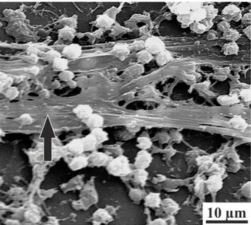
EPSs are often associated with the formation of biofilms and microbial aggregates, and some of them play a twofold role, either inhibiting or promoting carbonate formation, depending on the physicochemical characteristics (Decho, 2000) (Fig. 1.6). This work, in particular, refers to the above-mentioned kind of EPS.
Fig. 1.6 – Scanning electron micrograph of a
staphylococcal biofilm. Black arrow indicates EPS filaments (mod. from Donlan, 2002).
13
As reported by Dupraz et al. (2009), the EPS matrix represents an important component of marine biogeochemical processes (Decho, 1990; Bhaskar and Bhosle, 2005). The chemically-reactive EPS matrix is of considerable ecological importance because it is a physical barrier between the cell and organic and inorganic metabolic substrates, predators, antimicrobial agents, and other bacteria (Costerton et al., 1995). EPS may account for 50-90% of the total organic carbon of biofilms and can be considered as the primary matrix material of the biofilms (Flemming, 1998). The high molecular-weight (8 to N1000 kDa) matrix is composed of polysaccharides, and may include protein and peptides, nucleic acid, uronic acid, DNA, lipids and humid substances (Donlan, 2002; Cheng, 2007).
EPS is also highly hydrated because it can incorporate large amount of water into its structure by hydrogen bonding.
EPS can be produced by a wide array of microorganisms, both photoautotrophic and heterotrophic bacteria. In microbial mats, cyanobacteria are generally recognized as the most important EPS producers (Richert et al., 2005). Recently, Bosak and Newman (2005) and subsequently Braissant et al. (2007) demonstrated the potential role of heterotrophic bacteria, such as sulfate-reducing bacteria, in the production of the extracellular matrix.
Although some free-living bacteria can produce large amounts of extracellular polymers (Kives et al., 2006), EPS production is a seminal feature of benthic communities, enabling the formation of microbial mats and biofilms.
The EPS matrix fulfills many functions within microbial mats (Decho, 1990; Bhaskar and Bhosle, 2005):
1) It allows communities to attach to surfaces and create micro-domains, where various types of metabolism can coexist in microspatial proximity;
2) It physically stabilizes microbial cells under variable hydrodynamic regimes;
3) It may also help the microbial mat community to resist multiple stress conditions, such as nutrient shortages, UV exposure or desiccation.
14
EPS within a microbial mat can exist in a continuum of physical states, ranging from particulate to dissolved, or from a ‘cohesive gel’ to a ‘loose slime’ to a ‘dissolved solute’ state. The physical state is largely a function of the EPS concentration (or, the water activity), and the abundance and types of bonds or interactions among individual EPS molecules.
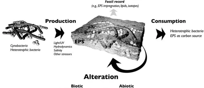
The EPS is a key player in organomineralization, having a distinct impact on the morphology and mineralogy of mineral products (Braissant et al., 2003). The physicochemical properties of the polymer matrix, such as the acidity or functional group composition, are important factors in the metal binding potential (initially inhibiting calcium carbonate mineral formation) and biotic and abiotic degradation or alteration of the EPS (favoring calcium carbonate precipitation).
Negatively-charged acidic groups within the EPS matrix can bind a large amount of mono- and divalent cations, which can help to maintain the structural integrity of EPS by promoting gel formation through bidentate bridge formation (Sutherland, 2001). The cation-binding capacity of EPS removes free Ca2+ ions from solution, inhibiting the precipitation of carbonate minerals by depleting them from the proximal surrounding environment. The role of acidic amino acids (e.g., aspartic or glutamic acid) and carboxylated polysaccharides (e.g., uronic acids) as strong inhibitors of calcium carbonate precipitation has been well documented (e.g. Gautret and Trichet, 2005). The functional group characteristics are therefore key in the mineral formation process. Although several functional groups contribute to the overall negative charge of the EPS and consequently to metal binding, carboxylic acids and sulfate groups are generally considered to be the most important ligands within EPS.
Differently, in order to precipitate calcium carbonate minerals within a microbial mat, the Ca-binding capacity of the EPS matrix has to be greatly reduced. This can be accomplished through via one of the two following
15
mechanisms 1) diagenetic alteration of EPS or 2A, B) supersaturation of the cation-binding sites, (Fig. 1.7).
Fig. 1.7 – Synthetic scheme representing the role of EPS in biological mineral precipitation (from Dupraz
16
1.5 Biomediated mineral formation
Biomineralization refers to processes in which organisms are involved in the formation of minerals. Most biominerals are calcium carbonates, silicates, iron oxides, and sulfides (Bazylinski, 2004).
In order to define the genesis of minerals produced dependently on biological activity, clear definitions of the relevant processes are required.
According to the definition proposed by Perry et al. (2007), organomineral includes any minerals precipitated by interaction with organopolymers, bioorganic, and/or non-biological organic compounds, without evidence of direct skeletal, intracellular or extracellular biological control. The term biomineral generally refers to a mineral that was produced by living organisms, forming both minerals and organic components (Weiner and Dove, 2003; Dupraz et al., 2009).
In particular, organomineralization process can be intrinsically (microbial metabolisms) or extrinsically driven (e.g., degassing, evaporation), therefore be respectively either active induced) or passive (biologically-influenced) process.
Two different modes of biomineral precipitation can be distinguished (for further details see Dupraz et. al., 2009): (1) biologically controlled mineralization and (2) biologically induced mineralization (Fig. 1.8).
17 Fig. 1.8 - Classification of mineralization processes showing the different types of biomineralization
(mod. after Dupraz, 2009).
1.5.1 Biologically controlled mineralization
In this kind of mineral precipitation, organisms exert a considerable control over all the aspects of the nucleation and mineral growth stages (Mann, 1988).
This kind of mineralization serves the function of specifically producing structural components, such as skeletons, bones, teeth, shells, etc. The molecular structure of nucleation sites are usually genetically controlled to ensure defined mineral precipitation in terms of shape and mineral composition. Minerals produced under biological control are usually well defined in terms of structure and chemical composition. Additionally, a high level of spatial organization is encountered, including complex morphologies and controlled aggregations and textures. Minerals produced in biologically controlled environments also exhibit a preferential crystallographic orientation.
Biologically controlled biomineralization is generally rare in the bacterial community. However, an example is given by magnetotactic bacteria (Komeili, 2004), which provide to the formation of intracellular magnetic crystals
18
(magnetosoma), allowing movement in marine sediments in response to the environmental magnetic field (Blakemore, 1975).
Others, or the same type of bacteria are capable to form greigite (Fe3S4),
mackinawite (tetragonal FeS) and/or a third phase cubic FeS with the sphalerite structure (Pósfai, 1998) by the same controlled intracellular mineralization process.
1.5.2 Biologically induced mineralization
Formally, two processes known as biologically induced and biologically influenced mineralization were distinguished from each other as microbially or environmentally driven (fig. 1.6).
However, as microbial activity and environmental conditions are close dependent associated, in this study the two processes will not be distinguished from each other and solely referred to as biologically induced mineralization, as suggested by Frankel and Bazylinski (2003) and Konhauser and Riding (2012).
Microorganisms interact in numerous ways with the surrounding environment, involving metabolic reactions and the mode of life. Re-mineralization is a primary ecological process where microbes degrade organic matter. This process is mandatory for element cycling. Microbial activity modifies spatial distribution of chemical components by liberation, production, transport, enrichment, and incorporation into biomass. The resulting alteration of geochemical properties as pH, Eh redox potential, and gas concentrations may induce dissolution and also precipitation of various minerals.In numerous instances, inorganic minerals are precipitated as the result of metabolically produced ions, released into the adjacent environment (Vasconcelos et al., 1995). Metabolic products are transported outside the cell passively via diffusion (e.g. inorganic carbonate ions) or actively via ion channels and excretion (Goodman, 2008). Continued supply of metabolic products to the environment surrounding the
19
cell may lead to supersaturation for one or several minerals, resulting in the subsequent precipitation and deposition of mineral particles.
Thus, biologically induced mineralization is considered an unintended and uncontrolled consequence of metabolic activities (Frankel and Bazylinski, 2003). As there is no cellular control, precipitates size, shape, structure, chemical composition, and organization are generally heterogeneous and often poorly defined (Weiner and Dove, 2003).
The minerals that form through this passive process have crystal habits and chemical compositions similar to those produced by precipitation under inorganic conditions (Konhauser and Riding, 2012).
Several biogenic minerals are passively formed. Some of them have been widely studied such as iron hydroxides, magnetite, manganese oxides, clays, amorphous silica, carbonate (calcium carbonate, dolomite, strontianite, siderite, etc.), phosphates, sulfates (gypsum, celestine and barite), sulfide minerals, etc. (Konhauser, 2007).
20
1.6 Calcium carbonate precipitation by bacteria
Bacterial carbonate deposits can be traced back for at least 2.6 billion years (Altermann, 2006). They are locally abundant sediments that record not just bacterial growth, but even environmental factors that promote calcite/aragonite (CaCO3) and dolomite Ca:MgCO3 precipitation in, on and around bacteria and the
organic matter that they produce. These sediments include carbonate mud, produced by whitings, and stromatolites (Konhauser and Riding, 2012).
Calcium carbonate precipitation by bacteria evidenced close dependence on environmental conditions, particularly the carbonate saturation state of ambient waters. It is, therefore, a good example of ‘induced’, as opposed to ‘controlled’ biomineralization.
Benthic cyanobacterial communities can form a wide variety of calcareous deposits. When cyanobacteria growing in biofilms calcify, they can form micritic coatings, crusts, and layers on submerged substrata.
Cyanobacteria can live as single cells (coccoid cyanobacteria) or several cells may be linked to form cell rows, called trichom. If the trichom is surrounded by EPS sheath, the structure is called a filament. The occurrence of cyanobacteria is frequently linked to the deposition of carbonate. By trapping sediment between the filaments, they are involved in the formation of stromatolites in both sea water and freshwater. Cyanobacteria can also calcify when carbonate is precipitated in association with the organism. The result of precipitation is often a micritic tube surrounding the filament or the trichom (Merz, 1992).
The role of cyanobacteria in carbonate precipitation is twofold: metabolic fixation of inorganic carbon tends to increase pH, leading to a state of supersaturation, while cation adsorption to the cell surface promotes heterogeneous nucleation (Fig. 1.9).
21 Fig. 1.9 – Schematic of metabolically-induced biomineralization in a cyanobacterium. The uptake of the bicarbonate anion leads to excretion of OH−, which in turn changes the alkalinity and inorganic carbon speciation proximal to the cell surface. The generation of carbonate anions and the pre-adsorption of calcium cations to the cell’s sheath can then induce calcification (mod. after Konhauser, 2007).
With respect to photosynthesis, in waters with neutral to slightly alkaline pH, cyanobacteria use HCO3− instead of, or in addition to, CO2 as a carbon source
(reaction 1.1). Hydroxyl ions are byproduct of this reaction, are then excreted into the external environment where they create localized alkalinization around the cell. This, in turn, induces a change in the carbonate speciation towards the carbonate (CO3 2− ) anion (reaction 1.2): HCO3− ←→ CO2 + OH − (1.1) HCO3− + OH− ←→ CO32− + H2O (1.2)
Cyanobacteria also provide reactive ligands towards metal cations and, once bound, they can react with the CO32− anions to form a number of carbonate phases,
such as aragonite or calcite (reaction 1.3):
CO3 2−
22
Extracellular layers are particularly favorable sites for nucleation, and cyanobacterial species that produce sheaths or EPS generally precipitate more calcium carbonate than those species without such structures (Pentecost, 1978).
When calcium carbonate nucleates on the sheath surface it grows radially upwards and, in some cases, this may lead to the complete encrustation of the cell. On the contrary, when Ca-carbonate nucleates within the intermolecular spaces of the sheath, this latter may become filled by mineral (Verrecchia et al., 1995).
EPS promotes carbonate precipitation by providing diffusion-limited sites that create localized alkalinity gradients in response to metabolic processes, while simultaneously attracting Ca2+ to its organic ligands (e.g., Pentecost, 1985).
Furthermore, the type of functional groups in EPS affects carbonate morphology and mineralogy, e.g., spherule vs. euhedral calcite or calcite vs. aragonite (Braissant et al., 2003). One line of thought suggests that the phase and morphology of calcium carbonate are bacterium or strain specific (Hammes et al., 2003). Cyanobacteria grown in the presence of various combinations of Sr2+, Mg2+, or Ca2+ can precipitate instead strontianite, magnesite, or mixed calcite-strontianite carbonates (Schultze-Lam and Beveridge, 1994). In general, cyanobacteria are equally capable of incorporating Ca2+ or Sr2+ during carbonate mineral formation, while magnesite is easily inhibited from forming by the preferential binding of the former two cations over Mg2+. Other studies have documented that cyanobacteria can partition of up to 1.0 wt% strontium in calcite (Ferris et al., 1995).
Filamentous cyanobacteria, such as Schizothrix species, have been heavily implicated in the growth process because they produce EPS that binds Ca2+ cations, and their metabolism changes the physicochemical properties at the ooid-water interface (Davaud and Girardclos, 2001).
Ca-carbonate precipitation, caused by microorganisms, may be exploitable in the preservation of monuments and statuary made from carbonate rock, and concrete as studied, for example, under laboratory conditions, respectively with
23 Bacillus subtilis (Barabesi et al., 2003), and with Bacillus sphaericus (De Muynck
et al., 2008) as inducers of CaCO3.
1.7 Mineral bio-alteration
Every rock and artifact presents in the uppermost lithosphere and hydrosphere is exposed to chemical and physical weathering. Physical erosion (e.g. storms) is temporary and localized, and chemical erosion is considered as negligible due to the actual seawater chemistry, so biological weathering operated by macro- and micro-organisms, including various kinds of bacteria, fungi, algae, and protozoa, have a primary role in both kind of weathering (Tribollet and Golubic, 2011).
Light availability in relation to bathymetry is one of the main factors influencing the distribution of macro- and micro-organisms and determining the composition of the borer communities.
Agents of bioerosion comprise internal and external bioeroders of different sizes and from a wide range of taxonomic affiliation. Internal bioeroders excavate carbonate substrates in search of shelter or food, while external bioeroders graze on both epilithic and endolithic organisms thereby abrading the substrates. Epiliths are attached to the surface of hard substrates, whereas endoliths reside inside hard substrates. Internal agents comprise micro-borers (<100 µm) and macro-borers (> 100 µm).
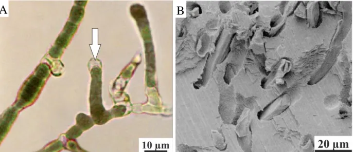
Microborers are phototrophic and organotrophic microorganisms (Tribollet, 2008). Microboring phototrophs are, for example, prokaryotic cyanobacteria (Fig. 1.10) and eukaryotic chlorophytes and rhodophytes. Organotrophs (heterotrophs) are fungi foraminifera, and other prokaryotic and eukaryotic light-independent microorganisms.
24 Fig. 1.10 – (A) Mastigocoleus testarum cultivated in marine water. White arrow indicates an heterocyst
cell (Montoya Terreros et al.,2006). (B) Details of Mastigocoleus sp. tunnels in a mussel shell (Webb and Korrûbel, 1994).
Macro-borers are comprised of various organisms (Hutchings, 1986 for review), including protists (foraminifera), sponges, bryozoans, polychaetes, sipunculids, bivalves, and crustaceans Organisms such as some foraminifera, serpulids (polychaetes) and bryozoans only etch the surface of the substrates to anchor themselves (Fig. 1.11).
Fig. 1.11 –Micrograph taken under SEM of the
shell etched by a bryozoan colony (Taylor et al.,1999).
25
In the shallow sub-littoral zone, biological corrosion and biological abrasion form bored limestones underneath the biogenic micro-reefs (Golubic, 1962; Schneider et al., 1983). Through the process of boring, euendoliths will leave clear traces of their activity in the substrates that they excavate, and these fossilize well. The fossil record presents evidence of euendoliths populating the Earth since the Precambrian. The oldest cyanobacterial euendolithic microfossil, Eohyella
campbellii, was recorded in carbonate rocks as old as 1.5 billion years (Zhang,
1987).
Other evidence of euendolithic fossils is found in microbial assemblages from the Neoproterozoic, some 700-800 million year ago, where the cells penetrated aragonitic ooid grains (Knoll, et al., 1989, Ramírez-Reinat and Garcia-Pichel, 2012).
Microorganisms attach exposed mineral surfaces, coat them with extracellular polymers substance (EPS) causing, subsequently, a physical disruption of the grains to gain access to nutrients and energy in the underlying substrata.
Endolithic cyanobacteria, algae, and fungi have been found to cause local dissolution of limestone, thereby forming tubular passages in which they can grow (Golubic et al., 1975). The kinds of limestone they attack in nature include coral reefs, beach rock, and other types. Active algae include some green, brown, and red algae (Golubic, 1969). The mechanism by which any of these organisms bore into limestone is not understood. Some filamentous boring cyanobacteria possess a terminal cell that is directly responsible for the boring action, presumably dissolution of the carbonate (Golubic, 1969). Different boring microorganisms form tunnels of characteristic morphology (Golubic et al., 1975). The depth to which cyanobacteria and algae bore into limestone is limited by light penetration in the rock, because they need light for photosynthesis. Boring cyanobacteria may have unusually high concentrations of phycocyanin, an accessory pigment of the photosynthetic apparatus, to compensate for the low light intensity in the limestone. In contrast, boring fungi are not limited by light penetration. Being
26
incapable of photosynthesis, they have no need for light (Ehrlich and Newman, 2009).
Moreover, they create a complex microenvironment at the mineral–water interface, where metabolic reactions and generation of acids lead to pH and concentration gradients markedly different from the bulk solution. This often promotes a state of thermodynamic disequilibrium that increases rates of chemical weathering (Konhauser, 2004).
Intended mineral weathering as a strategy for nutrient and trace element acquisition, as well as an energy source, has been documented on numerous different microbes (e.g. Shock, 2010; Edwards et al., 2005).
Acidification is of prime relevance for silicate and carbonate minerals, as their dissolution is positively correlated with decreasing pH. Once microorganisms are fixed to the substratum, they begin to produce organic acids, such as lactic, pyruvic, citric and oxalic acids, accelerating dissolution. The majority of organic acids are byproducts of fermentation and/or various intermediate steps of the aerobic respiration of glucose, but some microorganisms further excrete organic acids when growth is limited by the absence of an essential nutrient.
In aquatic systems, mineral dissolution can be attributed to the presence of carbonic acid, resulting from microbial respiration (Soetaert et al., 2007), and nitric and nitrous acid, produced by nitrifying bacteria. Also, the release of organic acids largely contributes to mineral dissolution (Mailloux et al., 2009). Even upon death, microorganisms are important agents in weathering because their decay, via the action of respiring heterotrophs, leads to elevated soil CO2 partial pressures, which, in turn, creates carbonic acid (Konhauser, 2004).
Silicate weathering normally undergo incongruent dissolution, in which most of the cations (as Ca2+, Mg2+, K+, Al2+and Na+) are leached out from the lattice, leaving a residual phase as clays or metal oxides.
In addition, cellular organic functional groups and chelating metabolites also induce mineral weathering via three simultaneously occurring mechanisms (Uroz
27
et al., 2009). Organic acids and chelating molecules adhere to mineral surfaces and liberate nutrients from mineral phases by electron transfer. These compounds also break the oxygen links and chelate dissolved ions in carboxyl and hydroxyl groups. Results from relatively recent laboratory experimentation on the effects that solutions of various organic acids reagent grade have on rock minerals, are still discussed as though, in nature, these reactions were purely abiotic (Ehrlich, 1998).
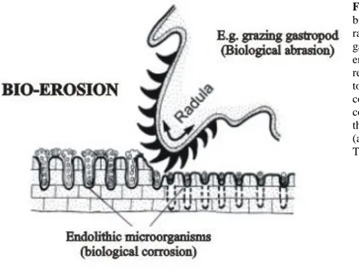
Furthermore, the dissolution of limestone by biological corrosion is promoted by the grazing activity of different organisms (nematodes, copepods, gastropods), which abrade the rock surface with the aim to remove endolithic microorganisms sited in (Fig. 1.12). This process of bioerosion develops gradually and accelerates between the calcified patch reefs where more light is available. In this way the biokarst relief of the furrowed limestones develops.
Fig. 1.12 – An example of
bioerosion scheme by the radula of the marine gastropod Patella. The endolithic cyanobacteria receive new stimuli to bore to the phototactically controlled light compensation depth after the surface is rasped away (after Schneider & Torunski, 1983).
28
The corrosive activity of epi- and endolithic cyanobacteria affects the substratum directly. On marine limestone coasts the carbonate rock surfaces are very densely populated with euendoliths (Schneider, 1976). More than half a million individuals per square centimeter are not unusual, as shown by the casting-embedding.
Tudhope and Risk (1985) showed that euendolithic autotrophs, present in Davies reef, Australia, penetrate rapidly into new available substrate by dissolving chemically its crystals. Crystals of substrates show a specific arrangement around microborings suggesting a precisely controlled excavating process (Golubic et al. 1975; Le Campion-Alsumard 1975, 1979, Tribollet et al., 2006).
Garcia-Pichel (2006) revisited mechanisms to explain dissolution of carbonate substrates by euendolithic cyanobacteria. Those mechanisms are (1) a temporal separation of photosynthetic and boring activities during the daily cycle (dissolution due to the CO2 produced during respiration at night), (2) a spatial
separation of photosynthetic and boring activities, and (3) the use of calcium pumps. A combination of those mechanisms is possible but the use of calcium pumps seems the most probable mechanism. Garcia-Pichel (2006) showed that an active transport of Ca2+ from the apical cell of euendolithic filaments to their trailing end would make dissolution thermodynamically favorable around the apical cell, while interstitial pH is high due to photosynthesis. This is consistent with the known range of bored substrates including aragonite, calcite, granite and hydroxylapatite, and with precipitation of micrite and brucite observed around euendolithic filaments at the surface of dead substrates. The reasons why several taxa of euendoliths cyanobacteria bore holes into minerals are not fully understood. However, advantageous aspects for boring include availability of nutrients, avoidance of competition, protection from extreme environments and prevention of detachment (Ramirez et al., 2010).
29
1.8 Application of nanostructured biologically induced minerals
The use of microbially induced carbonate biominerals, formed by primititve nanostructures, is becoming increasingly popular day by day.
Nano-structured biologically induced minerals have unique structural features; many physical and physicochemical properties render them different from usual nano-particulate materials and single-crystal materials. This provides better performance in numerous applications.
Bioremediation, the range of processes that use microorganisms and their products to return contaminated environments to their original condition, uses microorganisms, their enzymes and, increasingly, microbial induced Ca-carbonates to deal with contaminated soils, for example by lead (Pb), Cadmium (Cd), Copper (Cu), Arsenic (As), oil spills or chemical waste (Kang et al. 2014; Achal et al., 2011, 2012a, 2012b).
Nanostructured materials are widely used even as biomedical materials, such as tissue engineering scaffolds and drug carriers, and lightweight structural materials.
In construction materials, calcinogenic bacteria added to concrete and cement mortar improve compressive strength and the remediate cracks (e.g. Achal et al. 2010; De Muynck et al., 2010).
Biomineralization processes have been used also for the bioconservation of the built heritage and statuary made of stone. Structurally and mechanically coherent, and therefore durable, bacterial calcite cement is applied to calcitic stones such as limestone and marble consolidating their external structure, filling pores, in order to protect them from deterioration and alteration (e.g. González-Muñoz, 2003).
Recently, microbial induced carbonate precipitates are used for increase strengthening of sand and soil (so called biogrouting), for instance, at the base of
30
railroad embankments, dikes, etc. In addition, they are used as reducer of CO2
emission into the atmosphere, filler for rubber, plastics and ink (Dhami et al., 2013).
Furthermore, nano-scale study of the fabric and the morphology of neo-formed Calcium carbonate crystals results essential for recognition of the abiotic versus biotic nature of carbonate minerals in several fields such as in the reconstruction of depositional palaeoenvironments, in astrobiology (numerous similar features have been found on Mars and in meteorites; Folk and Lynch,
1997); in medicine, where different biologically induced minerals are formed by pathogenic bacteria (e.g. kidney stones, dental tartar; Kirkland et al., 1999).
31
CHAPTER 2
GEOGRAPHICAL SETTING
AND HISTORICAL INTRODUCTION
2.1 Geographical setting
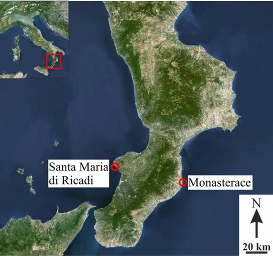
Samples used for this work have been collected in two different places, selected for their historical relevance: Monasterace, sited along the Calabrian Ionian coast, and Santa Maria di Ricadi, sited along the Calabrian Tyrrhenian coast (Fig. 2.1).
Fig. 2.1 – Maps
illustrate the position of the two different sites where samples have been collected, respectively Santa Maria di Ricadi, on the Tyrrhenian coast, and Monasterace, on the Ionian coast.
32
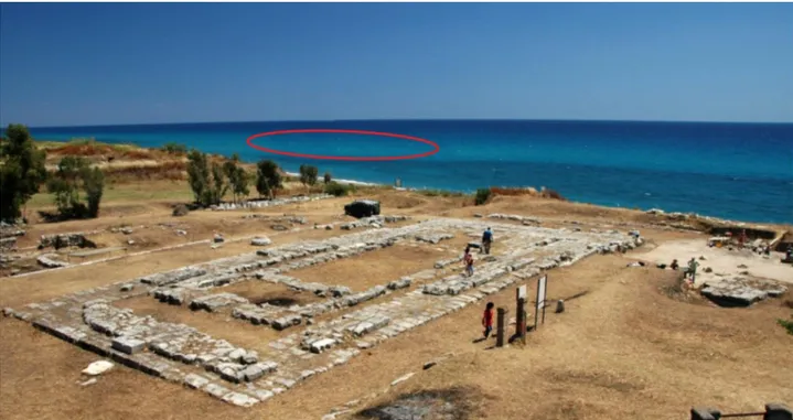
In Monasterace site, sampling occurred in the submerged archaeological park in front of the ruins of the ancient village of Kaulonia, between the actual Assi River and Doric temple (Fig. 2.2).
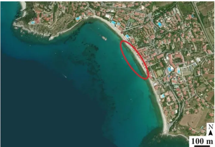
Differently, Santa Maria di Ricadi is a small village located along the Calabrian Tyrrhenian coast, on the southeast margin of the Capo Vaticano promontory (Fig. 2.3), facing the sea along more than 2 km of coastline.
Samples were collected from a stratified conglomerate which extends for about 1 km in the bay area, characterized by covering the seafloor for hundreds meters in length, parallel to the coastline, tilted about 10° seaward and cropping out from the medium sea level up to 50 cm (Fig. 2.4).
Fig. 2.2 - Part of the archaeological site of Kaulonia ancient village ruins. In close-up view, the remains
of the Doric temple of Kaulonia represented by the rocky basement. On the right side the votive area. Red circle indicates approximately the submerged archaeological area, where abundant archaeological material, including samples used in this study, has been found (Photo mod. after www.panoramio.com "ruins of Doric temple").
33 Fig. 2.3 - Planar view of Santa Maria di Ricadi bay that extends for about a 1 km. Red line indicates the
sampling area (mod. after www.viamichelin.it).
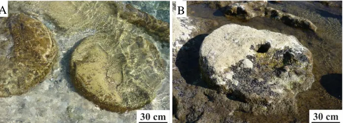
Fig. 2.4 - Part of Santa Maria di Ricadi bay (looking southward) is characterized by a stratified
conglomerate extended for hundreds meters in length. In particular, red arrow shows remains of a millstone, carved on edges and in the center and broken in the upper part.
34
2.2 History of Monasterace
The site of Monasterace represents the remains of Kaulonia, a rather modest-size Magna Graecia colony, founded at about 700 years BC by the Achaeans, a people from the Peloponnesus, and perhaps also by settlers from Kroton (Crotone), another Greek settlement located to the northeast. Kaulonia was conquered in 389 B.C. by Dionysius the 1st, tyrant of Syracuse, and its settlers deported to Locri, a colony positioned about 40 km to the SSW. A settlement was then rebuilt at the original Kaulonia site by the Brettii, a nomadic Indo-European tribe in the 3rd century BC, and was conquered once again, this time by the Romans in 205 BC (Stanley et al., 2007)
Archaeological excavations at Kaulonia are positioned immediately north of the small town of Monasterace Marina. The gentle arcuate headland immediately to the north of the site is known as Punta Stilo, or Capo Colonna (Fig. 2.6). It has had this name at least as far back as about 2000 years ago when the locality was cited by Pliny the Elder in his The Natural History.
This suggests that columns may still have been visible to the Romans in this sector to about 2000 years ago, i.e. perhaps as long as 3 to 4 centuries after exile of the Greek settlers in 389 BC (Stanley, 2007).
Monasterace shoreline varied in mid and late-Holocene. At the pre-Greek time, about 2500 years BC, was behind the present dunes, at Greek time (ca. 500 BC) advancing seaward of the present one by little more than 100 m; after migrating back landward during the post-Roman age and finally to present shoreline (Fig. 2.6). Actually, the coastline undergoes several storms that frequently, during the winter season, erode the sandy cliff behind which are settled the basement of the temple and a votive area.
35 Fig. 2.6 - Paleogeographic scheme of
the Kaulonia margin shows major Holocene coastal shifts: from former shoreline position (1), to seaward (2), and then return to the present coastline (3, 4). (After Stanley et al., 2007).
At 500 BC, sea level was only about 2 m or less below the present m.s.l., according to some presently used world curves (Fairbanks, 1989; Pirazzoli et al., 1997).
This eustatic rise, plus seafloor erosional downcutting phenomenon would thus account for total lowering of shoreface elevation to about 3 to 4 m below present m.s.l. at the time of Greek settlement (Stanley et al., 2007).
Abundant archaeological material as column sections, column bases, construction blocks, bollards, anchors associated with numerous potsherds, bronze, lead, and copper fragments (Lena and Medaglia, 2002) were discovered by divers in the mid- to late 1980’s at water depths ranging from 5 to 7 m up to 300 m offshore Monasterace (Fig. 2.7).
The columns are dated stylistically to about 480-470 BC. Archaeologists have determined that the large number of column sections (length to 113 cm, diameter
36
to 80 cm) and other materials on the seafloor were not distributed randomly as a result of shipwrecks (Lena and Medaglia, 2002). Rather, these and other archaeological items are positioned at, or proximal to, once exposed coastal near shore surfaces upon which they were worked and/or from which they were discharged from vessels.
Fig. 2.7 - Underwater photographs taken in the
1980s show examples of archaeological material off Kaulonia, including (A) fluted Ionic column section, (B) construction block, and (C) bollard (after Iannelli, 1997; Iannelli et al. 1993). Large, rounded boulders (some up to 50 cm diameter) of igneous and metamorphic material of Assi River derivation are widely distributed in this offshore sector (photos by S. Mariottini).
More recent geo-archaeological analysis, using data from geophysical and coring surveys, indicates that the seafloor on which the columns were discovered
A
B
37
was actually only a small part of a broad arcuate (not small hook-shaped) headland that subsided after time of Greek occupation. Moreover, other analyses show that a sector of the now-submerged surface, currently lying at a depth of 5 to 7 meters below m.s.l., had been partially covered by stratified sandstone originally formed along the beach-line to foreshore zone during the latter half of the Holocene (Stanley et al., 2007).
This surface seaward of Kaulonia, comprising sandstone slabs, cobbles and boulders and archaeological material, appears to have been submerged completely beneath sea level during approximately four to five hundred years from roughly 400 BC near the end of Greek occupation to the time of Roman rule in about the 1st century AD.
2.3 History of Santa Maria di Ricadi
The origin of the village of Santa Maria di Ricadi is not clear, and just few information are available but, according to Pantano (1972), the Ausones, an italic tribe, settled there about XII centuries B.P. After them, several populations colonized that area, even Greeks and Romans. In 1987, in Santa Maria di Ricadi, the excavations revealed a large settlement of the III-IV century BC with remains of Roman villas and a kiln with a wide deposit of cereal amphora. Jars have been recognized belonging to the same type, suggesting that there were used for shipping local alimentary products (mainly cereals).
Santa Maria di Ricadi bay was used for hundreds years as natural harbor. Last discoveries indicate that Romans built the first real port, known in the past as "portus Erculis" (port Hercules), cited in several ancient sources (Pliny the Elder and Strabo), but not yet identified (Pantano, 1972).
Rock formation, cropping out the medium sea level, is characterized by having many remains of millstones (Fig. 2.5). Millstones have been probably left in place
38
because broken during carving operations. Several unconfirmed sources indicate Romans as first people carving this tough rock were that used them for grinding cereals.
Various examples of well, or sometimes bad, preserved millstone quarries are found in the southern coasts of Italy, such as Palinuro in the region Campania; Crotone, Roccella Ionica, Capo dell’Armi in the region Calabria, Capo d’Orlando and Giardini-Naxos in the region Sicily.
Fig. 2.5 - Semi-manufactured millstone carved in place. They show a typical cylindrical shape and a
diameter up to 1,3 m.
This kind of cylindrical-shaped wheels with a diameter up to 1,30 m, are well documented since the beginning of the Hellenistic period in Israel and Cyprus, and widespread within the whole Mediterranean basin from the Roman to the modern age, until the XIX century (Amouretti and Brun, 1993).
According to Antonioli et al. (2006, 2009), the average uplift rate for the Capo Vaticano area is 1.5-1.6 mm/year. This means that the millstones whose actually settle at a maximum 60 cm below medium sea level, 2000 years ago where at least 2.7 m above m.s.l.
39
CHAPTER 3
METHODS
3.1 Sampling
Samples have been collected in collaboration with a diving team belonging to "Associazione culturale Kodros" and headed by Stefano Mariottini.
In Monasterace, sampling took place during the summer period at water temperature of 23°C and depths ranging from 5.8 to 6.7 m and about 250 m far away from the actual coastline. Among the various rests of artifacts present in the submerged archaeological area of Monasterace, ten key-samples that well represent the variety of natural rocks and artifacts used in that area by ancient people were collected.
Differently, in Santa Maria di Ricadi, two samples were extracted with chisel from the conglomerate, outcropping few centimeters up to the medium sea level. These samples result sufficient for well representing the conglomerate and its own biofilm.
3.2 Fixation and dehydration
Fixation is the first crucial step in the preparation of specimens. After a half hour from sampling, every specimen was quickly immersed in a solution composed of 1/4 of 4% paraformaldehyde and 3/4 marine water, with the purpose to preserve all the biological organisms and tissues.
40
Subsequently, samples were gradually dehydrated and dried in HMDS (hexamethyldisilazane; Arp et al., 1998; Fratesi et al., 2004). Dehydration process consists in the chemical removal of water from the specimen.
3.3 Optical microscope and SEM-EDS analyses
All the samples collected were prepared for optical and scanning electron microscope (SEM) analyses.
All 12 samples, respectively 10 of Monasterace outcrop and 2 of S. Maria di Ricadi, were worked in order to obtain for each sample a thin section and a fresh cutting. Cross-section cuttings were embedded in resin and cut to obtain thin sections of 30 μm in thickness and dimensioned 28 × 48 mm. As the same time, for every sample was extracted a piece, cut sub-perpendicularly to biofilm surface, carbon coated and fixed on stubs for SEM analyses.
Optical microscope analyses allowed evaluating physical, chemical and biological variations in the rock from the top, where biofilm is sited, to the inner 2 centimeters. Thin sections were analyzed in plane polarized light (PPL) and cross polarized light (CPL).
Scanning electron microscopy (SEM) has been finalized to micro and nano-scale observation of the biogenic elements forming biofilm and the recognition of the abiogenic or biogenic nature of the neo-formed precipitated minerals. Furthermore, mechanical/chemical, biological and weathering products were observed.SEM analyses occurred using FEI Quanta 200 F instrument, in high-vacuum condition with backscattered-electron-detention (BSE) and secondary-electron-detention (SE), even indicated as Everhart-Thornley Detector (ETD), imaging system at 20 kV, at an average working distance of 12 mm.
In addition, SEM instrument was equipped with an energy-dispersive X-ray spectrometer (EDS) for semi-quantitative analyses.
41
CHAPTER 4
RESULTS
4.1 Introduction
Among the several samples collected in the areas of Monasterace and Santa Maria di Ricadi, twelve key-samples were analyzed.
Foremost, every sample has been described at naked eye, in order to characterize the biofilms present on them.
Successively, further investigations were carried out with optical microscope and Scanning Electron Microscope with the purpose to observe micro-and nano-elements characterizing biofilms, such as skeletal organisms, bacteria, organic matter and neo-formed minerals.
42
4.2 Monasterace
Samples collected in the submerged archaeological area of Monasterace vary notably in composition.
Samples made of calcilutites, calcarenites and marble represent probably scraps produced during rock carving (Fig. 4.1 A, B, C).
Two samples of brick, probable fragments of tiles, have been found on the seabed isolated from the rests (Fig. 4.1 D).
Calcschist specimens resulted very tough, hardly breakable, but along cleavage planes (Fig. 4.1 E). These samples come from semi-manufactured slabs, widespread only in the submerged area.
Biofilms, observed on the surfaces of the samples collected in Monasterace, have variable thickness, from few microns to ≈ 1 cm, extension and are made up by numerous components varying from sample to sample.
43 Fig. 4.1 - Hand-samples in photos represent the
different categories of specimens collected in Monasterace. (A) Calcilutite; (B) calcarenite; (C) marble; (D) tile; (E) calcschist.
44
4.2.1 Components of the biofilm
4.2.1.1 Skeletal organisms
Skeletal organisms occupy great part of the exposed samples surfaces. At the sampling moment, many organisms resulted alive, but for many others, only the skeleton remained adhering on the rock surface.
Red algae
Highly calcified encrusting red algae (Rhodophyta), thick in average 1 mm (up to 5 mm), occur in many collected specimens. Some samples are characterized by the presence of a strong incrustation of red algae (Fig. 4.2 A, B), whereas other samples show just few patches of red algae appearing with reddish color (Fig. 4.2 C). Cellular structures vary from bad preserved (Fig. 4.2 D) to quite well preserved, especially in the upper part of the reticulate cellular structure, called
hypotallus, (Fig. 4.5 E, F). Moreover, EDS analyses indicates that, in average, red
45 Fig. 4.2 – (A) Marble sample strongly encrusted by thick overlapped layers of red algae. (B) Close up
view of the previous photo show a crust thick ≈ 6 mm. (C) Hand-sample surface shows several reddish patches (biggest indicated by white arrow) representing red algae. (D) PPL micrograph shows a bad preservation of the red algae skeleton. (E) SEM photomicrograph shows the upper part, called perithallus, of encrusting red algae. White dashed line indicates the limit algae/rock. (F) Close up view of perithallus shows its reticulate cells structure.
A
B
C
BE
F
D
B46 Bryozoans
Encrusting bryozoan are widespread as colonies, called zoarium, on many surface samples, appearing as a white patina, but most of the time showing different colors, since they are covered by cyanobacteria or others microorganisms that mask the original appearance (Fig. 4.3 A).
Every single element forming the zoarium is called zooid. This latter has sub-rounded to arcuate shape and diameter up to 300 μm, and resulted generally well preserved (Fig. 4.3 B, C, D). Zooids have a double-wall (zooecium) (Fig. 4.3 B).
Sometimes bryozoan results associated with filamentous bacteria (Fig. 4.3 C, D). The majority of the observed bryozoan lack in living organism, but in few cases its terminal part, called lophophore, is still visible with a ring of tentacles around the buccal opening (Fig.4.3 E).
Zoaria (pl. of zoarium) form widespread and patchy crusts, reaching 300 μm
of thickness, on the top of the samples.
Skeletal nanostructure is layered, made up by numerous thin isoriented lamellae, composed of Ca-carbonate with ca. 6 mole % of Mg (Fig. 4.6 F).
47 Fig. 4.3 – (A) Bryozoan are widespread on samples surfaces. They appear as a white patina, but often
show different colors, as green or black. (B)Transverse section of a colony fixed on rock surface. White arrows indicate the two walls forming zoecium. XPL. (C,D) SEM micrographs show a tangential section of a zoarium partially covered by cyanobacterial filaments. (E) Micrograph shows numerous tentacles around the buccal opening. (F) Thin, isoriented lamellae form the bryozoan skeletal structure.
A
B
C
BE
F
D
B48 Diatoms
Diatoms, unicellular photosynthetic algae, represent a diffuse component in analyzed biofilms, where they usually occur as individual organisms.
Occurring diatoms are benthic, belonging to the order Pennales (elongated form with bilateral symmetry in valve view) up to 40 μm in length.
Diatoms occur usually encrusted within biofilm among cyanobacterial filaments and neo-formational calcite (Fig. 4.4 A); rarely observed as fixed directly to rock substratum in poor biofilm colonized areas (Fig. 4.4 B).
Diatoms hard parts remains, termed frustules, are composed of amorphous silica.
Fig. 4.4 – (A) Photomicrograph shows a diatom encrusted by bacterial filaments and Ca-carbonate
crystals. (B) Diatom fixed on rock substratum, with its organic parts still in place.
49 Other skeletal microorganisms
Others less common skeletal microorganisms have been observed:
- Annelid worms, polychaete, with tubular shape and circular cross-section, encrusting hard surfaces are commonly associated with bryozoan (Fig. 4.5 A, B);
- Coccolithophores, are unicellular phytoplanktonic algae. They are formed by calcareous exoskeletal plates overlapped each other. Coccolithophores have a diameter of about 3 μm and occur trapped within the EPS of some bacterial colonies (fig. 4.5 C)
Fig. 4.5 - (A, B) Anellid worms associated with
bryozoan. (A) Hand-sample photo. (B) SEM micrograph. (C) Micrograph taken under SEM shows several coccolithophores. Black arrow indicates one in transverse view.