Alma Mater Studiorum – Università di Bologna
DOTTORATO DI RICERCA IN
SCIENZE BIOMEDICHE
Ciclo XXVII
Settore Concorsuale di afferenza: 06/A3Settore Scientifico disciplinare: MED/07
TITOLO TESI
CHARACTERIZATION OF WEST NILE VIRUS STRAINS ISOLATED IN
ITALY
Presentata da:
Dott.ssa Silvia Silenzi
Coordinatore Dottorato
Relatore
Prof. Lucio Cocco
Prof.ssa Maria Carla Re
Correlatore
Dott.ssa Giada Rossini
INDEX
ABSTRACT! ! ! ! ! ! ! ! ! ! ! 1!
INTRODUCTION! ! ! ! ! ! ! ! ! ! 2!!
1. INTRODUCTION TO THE ARBOVIRUSES 2
2. FLAVIVIRUS 5
3. WEST NILE VIRUS 8
3.1 Structure of WNV 9
3.2 Genome of WNV 10
3.2.1 Viral structural proteins 12
3.2.2 Viral non-structural proteins 13
3.3 WNV Replication cycle 17
3.4 Molecular Classification 20
3.4.1 Lineage 1 20
3.4.2 Lineage 2 21
3.4.3 Lineage 3 22
3.4.4 Additional proposed lineages 22
3.5 Transmission cycle 24
3.5.1 Vectors: mosquitoes and other arthropods 24
3.5.2 Birds 25
3.5.3 Humans, horses and other animals 26
3.5.4 Non-Vector-Borne Transmission 27
3.6 Epidemiology of WNV in humans 29
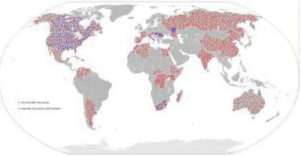
3.6.1 Worldwide WNV Epidemiology 29
3.6.2 Epidemiology of WNV in Italy 33
3.7 Pathogenesis 36
3.7.1 WNV propagation in the mosquito host 36
3.7.2 Initial infection, viral amplification and spread in humans 37
3.7.3 Neuroinvasion 40
3.8 Tropism 44
3.9 Clinical manifestation in humans 47
3.9.1 West Nile Fever (WNF) 47
3.9.2 West Nile Neuroinvasive Disease (WNND) 48
3.9.3 Host Risk factors 51
3.9.4 Viral Risk factors 55
3.10 Diagnosis 57
3.10.1 Nucleic acid based tests for WNV 58
3.10.2 Serologic diagnosis of WNV 58
3.11 Vaccines 59
4. INNATE IMMUNITY 61
4.1 Innate immunity to virus infection 61
4.1.1 RIG-I-like receptor signalling 62
4.1.2 Toll-like receptor signalling 64
4.1.3 NOD-like receptor signalling 67
4.1.4 Type I Interferon signalling 67
4.2 Innate immune evasion strategies of WNV 68
AIM! ! ! ! ! ! ! ! ! ! ! ! 71!
MATERIALS!AND!METHODS! ! ! ! ! ! ! ! 73!
1. Cells and Viruses 73
2. Virus Titrations 73
4. Virus strains susceptibility to Interferon-α (IFN-α) action 74
RESULTS 75
1. Growth properties of WNV strains on Vero cells 75 2. Growth properties of WNV strains on 1321N1 cells 77 3. WNV strains susceptibility to Interferon-α (IFN-α) action on Vero cells 80 4. WNV strains susceptibility to Interferon-α (IFN-α) action on 1321N1 cells 82
DISCUSSION 84
ABSTRACT
West Nile virus (WNV) is a neurotropic flavivirus that is maintained in an enzootic cycle between mosquitoes and birds, but can also infect and cause disease in humans and other vertebrate species. Most of WNV infections in humans are asymptomatic, but approximately 20% of infected people develop clinical symptoms, although severe neurological diseases are observed in less than 1% of them. WNV is the most widely distributed arbovirus in the world and has been recently associated with outbreaks of meningo-encephalitis in Europe, including Italy, caused by different viral strains belonging to distinct lineages 1 and 2. The hypothesis is that genetic divergence among viral strains currently circulating in Italy might reflect on their pathogenic potential and that the rapid spread of WNV with increased pathogenicity within naïve population suggest that epidemic forms of the virus may encode mechanisms to evade host immunity. Infection with WNV triggers a delayed host response that includes a delay in the production of interferon-α (IFN-α). IFNs are a family of immuno-modulatory cytokines that are produced in response to virus infection and serve as integral signal initiators of host intracellular defenses. The increased number of human cases and the lack of data about virulence of European WNV isolates highlight the importance to achieve a better knowledge on this emerging viral infection. In the present study, we investigate the phenotypic and IFN-α-regulatory properties of different WNV lineage 1 and 2 strains that are circulating in Europe/Italy in two cell lines: Vero and 1321N1. We demonstrate that: Vero and 1321N1 cells are capable of supporting WNV replication where different WNV strains show similar growth kinetics; WNV lineage 2 strain replicated in Vero and 1321N1 cells as efficiently as WNV lineage 1 strains; and both lineages 1 and 2 were highly susceptible to the antiviral actions of IFN-α.
INTRODUCTION
1. INTRODUCTION TO THE ARBOVIRUSES
The term arbovirus is an acronym for arthropod-borne virus (Hubalek et al., 2014). It has no taxonomic significance but rather is an ecologic term used to define viruses that require hematophagous (blood feeding) arthropod vectors such as mosquitoes and other biting flies, and ticks for transmission between hosts (WHO, 1985; Gubler DJ., 2001; Weaver SC. and Reisen WK., 2010).
Being, by definition, biologically transmitted, arboviruses must replicate in the arthropod vector prior to transmission, as opposed to being mechanically transmitted, without replication in the vector, through contaminated mouthparts (Weaver SC., 1997; Weaver SC. and Reisen WK., 2010). Biological transmission can be vertical, involving the passage of the virus from an infected female vector to both male and female offspring. Horizontal transmission can be venereal, from a vertically infected male directly to a female vector, as well as oral from a female vector to a vertebrate host via the saliva during blood feeding. The latter horizontal mode of transmission is most common for the majority tract following a viremic bloodmeal, dissemination of the virus in the vector, and eventual virus replication in the salivary glands, followed by the injections saliva during blood feeding (Weaver SC. and Reisen WK., 2010). Thus, in general arboviruses require a minimum of two hosts to complete their life cycle: a vertebrate and an arthropod (WHO 1985; Gubler DJ., 2002). For most arboviruses (e. g. Usutu virus, West Nile virus, Japanese encephalitis virus) humans are often dead-end hosts, as they do not develop the high viremias needed to infect the arthropod vectors (Filipe A., et al., 1985; Dobler G., 1996; Gubler DJ., 2001; Jones KE., et al., 2008; Cleton N., et al., 2012). Therefore, humans are not necessary for virus maintenance and they represent just an accident during the biological transmission among vectors and hosts (Diaz LA. et al., 2013). Only a few viruses like Yellow fever, Chikungunya and Dengue virus have expanded their host range to include humans as an amplifying host (Cleton N., et al., 2012).
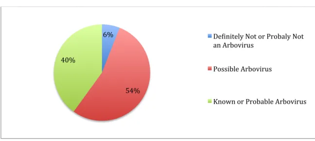
There are currently at least 530 viruses registered in the International Catalogue of Arboviruses: about 40% are known or probable arboviruses; another 54% are listed as possible arboviruses and about 6% are listed as definitely or probably not arboviruses (Fig. 1) (Karabatson N., 1985; Gubler DJ., 2002; Lequime S. and Lambrechts L., 2014). Most of the viruses listed in this catalogue are zoonoses or viruses that have vertebrate animals other than humans as their principal reservoir hosts (Gubler DJ., 2001) and of
the over 530 suspected arbovirus species more than 150 are documented to cause disease in humans (Karabatson N., 1985; Cleton N., et al., 2012; Lequime and Lambrechts, 2014). For most arboviruses (e. g. Usutu virus, West Nile virus, Japanese encephalitis virus) humans are often dead-end hosts, as they do not develop the high viremias needed to infect the arthropod vectors (Filipe A., et al., 1985; Dobler G., 1996; Gubler DJ., 2001; Jones KE., et al., 2008; Cleton N., et al., 2012). Therefore, humans are not necessary for virus maintenance and they represent just an accident during the biological transmission among vectors and hosts (Diaz LA. et al., 2013). Only a few viruses like Yellow fever, Chikungunya and Dengue virus have expanded their host range to include humans as an amplifying host (Cleton N., et al., 2012).
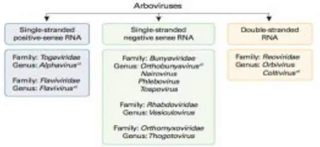
Arboviruses are included in different taxonomic families, the majority belonging to the Flaviviridae, Bunyaviridae or Togaviridae families, but a small number are member of the Rhabdoviridae, Reoviridae, and Orthomyxoviridae families (Fig. 2) (Dobler G., 1996; Claton N., et al., 2012; Go YY., 2014). Among them, four major viral genera account for the majority of arboviral disease: Flavivirus (e. g., Dengue, West Nile, Japanese encephalitis, and Yellow fever viruses), Alphavirus (e. g., Chikungunya, Eastern equine encephalomyelitis, Western equine encephalomyelitis and Venezuelan equine encephalitis viruses), Orthobunyavirus (e. g., California encephalitis and LaCrosse viruses) and Phlebovirus (e. g., Rift Valley fever and Sandfly fever viruses) (Lequime and Lambrechts, 2014).
!
Figure 1: Arboviral status of viruses registered in the arbovirus catalogue (Gubler DJ., 2001). ! 6%! 54%! 40%! De;initely!Not!or!Probaly!Not! an!Arbovirus! Possible!Arbovirus! Known!or!Probable!Arbovirus!
Figure 2: Classification of arboviruses. Arboviruses are included in six different taxonomic virus families. a) Arboviruses that cause human encephalitides belong to four
genera in four virus families (Go YY., et al., 2014).
Arboviruses as a group have a worldwide distribution and that of each arbovirus is restricted by the ecological parameters governing its transmission cycle (Gubler DJ., 2001; Gubler DJ., 2002). The majority of them were first isolated in tropical areas such as Africa, South America and in some Asian countries where climate conditions permit year-round transmission by cold-blooded arthropods (Karabatsos N., 1985; Gubler DJ., 1996; Gubler DJ. and Roehrig JT., 1998; Go YY., et al., 2014). However, the geographic distribution and frequency of epidemic outbreaks of arboviral diseases have expanded dramatically across the world in the past several decades and they are responsible for significant global public health problems (Gubler DJ., 1996; Gubler DJ., 2001). In general, several factors such as environmental disturbs from anthropogenic activities (Vasconcelos P. et al., 2001), climatic changes affecting vector and host population fluctuations (Weaver SC. and Reisen WK., 2010), human movements through airplanes, animal trade and migration (Pfetter M. and Dobler G., 2010), and changes in viral genetics (Go YY., et al., 2014) facilitated expansion and transmission of arboviruses resulting in emergence/reemergence of arboviral disease outbreaks in new regions in the world (Diaz LA., et al., 2013). Introduction of West Nile virus (WNV) into the New World and the emergence of Japanes encephalitis virus (JEV) in Australia are few prominent examples of recent unexpected emerging/reemerging zoonotic disease (Hanna JN., et al., 1996; Hanna JN., et al., 1999; Go YY., et al., 2014).
2. FLAVIVIRUS
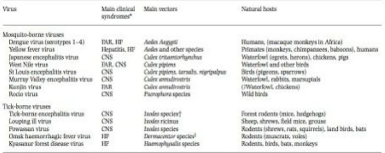
Flaviviruses are a group of arboviruses belonging to the family Flaviviridae (Pastorino B., et al., 2010). The genus Flavivirus consists of more than 70 positive-sense single-stranded RNA viruses (Tyler S., et al., 2011). Several members of this genus are the most clinically important arboviruses world-wide, that cause serious human and animal disease and constitute major international health problems. These include West Nile virus (WNV), Dengue virus (DENV), Japanese encephalitis virus (JEV), Yellow fever virus (YFV), tick-born encephalitis virus (TBEV), Murray Valley encephalitis virus (MVEV), and St. Louis encephalitis virus (SLEV) (Tab. 1) ( Mackenzie JS., et al., 2004; Gubler DJ., et al., 2007; Gould EA. and Solomon T., 2008; Cleton N., et al., 2012) and they are transmitted by mosquitoes (DENV, YFV, JEV, WNV) or ticks (TBEV) (Kuno G., et al., 1998; Gaunt MW. et al., 2001).
The Flaviviruses can be grouped by pathogenicity, geographic distribution, antigenic complex and subcomplexes based on classic serological criteria or into clusters, clades, and species, according to molecular phylogenetics (Calisher CH. and Gould EA., 2003; Lindenbach BD., et al., 2007; Ye J., et al., 2013). Generally, they can be divided in three distinct groups: mosquito-borne viruses, tick-borne viruses, and viruses with unknown vectors (Cook S., et al., 2012). Mosquito-borne viruses infect a variety of animal species and humans. They can be further subdivided into Culex and Aedes clades, which also differ in their vertebrate hosts and pathogenesis. Culex-clade viruses have bird reservoirs, are neurotropic, and frequently cause meningo-encephalitis, while Aedes-clade viruses have primate reservoirs, are non-neurotropic, and mainly result in hemorrhagic diseases (Solomon T., et al., 2000; Gaunt MW., et al., 2001; Beck C., et al., 2013). The tick-borne viruses also form two groups: one group circulates among seabirds, while the other, the tick-borne encephalitis group, is primarily associated with rodents. This latter group generally produces encephalitic disease, although Omsk Hemorrhagic Fever virus (OHFV) and Kyasanur Forest Disease virus (KFDV) also cause hemorrhagic deseases in humans (Beck C., et al., 2013). The mosquito-borne and tick-borne groups, although distinct, appear to have evolved via a common ancestral line that diverged from nonvector borne viruses (i.e., for which no arthropod vectors are known) (Lindenbach BD., et al., 2007). Moreover, tick-borne flaviviruses seems to evolve at slower rate than mosquito-borne flaviviruses, probably as a results of a slower virus replication rate in tick and longer generation times of their tick hosts (Gould EA., et al., 1997). The salient features of Flavivirus taxonomy are illustrated in Figure 3.
Because of their evolution and epidemiology is largely determined by ecological needs of their invertebrate and vertebrate hosts (Fig. 3), Flaviviruses have distinct geographical distributions. The viruses have evolved to use whichever animal host and insect vector are present in a particular area. In general, mosquito-borne viruses tend to occur in warm climates, whereas the tick-borne viruses are more important in cooler climates (Solomon T., and Mallewa M., 2001). For example, YFV is endemic in tropical and subtropical regions in Africa and South-America and the endemic regions of DENV, geographically, overlap with those of YFV in Africa and South-America. However, DENV extends not only to Middle America and southern parts of North America but also to large parts of South-East Asia, where YFV is not found (Vasilakis N., et al., 2011). In Europe, many Flaviviruses are endemic (West Nile, Usutu, tick-borne encephalitis viruses) or occasionally imported (dengue, yellow fever viruses) (Beck C., et al., 2013).
Figure 3: Phylogenetic tree showing the association of the groups of related viruses with their invertebrate vectors, vertebrate hosts, and geographic distribution.
ALF=Alfuy. MVE=Murray Valley encephalitis. JE=Japanese encephalitis. USU=Usutu. KOU=Koutango. KUN=Kunjin.WN=West Nile. YAO=Yaounde. CPC=Cacipacore. ARO=Aroa. IGU=Iguape. NJL=Naranjal. KOK=Kokobera. STR=Stratford. BAG=Bagaza. IT=Israel Turkey meningoencephalomyelitis virus. TMU=Tembusu. THCAr=strain of Tembusu. ILH=Ilheus. ROC=Rocio. SLE=St Louis encephalitis. DEN=dengue. SPO=Spondweni. ZIK=Zika forest. KED=Kedougou. UGS=Uganda S. JUG=Jugra. POT=Potiskum. SAB=Saboya. BOU=Bouboui. EH=Edge Hill. YF=yellow fever. SEP=Sepik. EB=Entebbe bat. SOK=Sokoluk. YOK=Yokose. GGY=Gadgets Gully. KFD=Kyasanur Forest disease. LGT=Langat. LI=Louping ill. NEG=Negishi. Sof=Sofj in. FETBE=far eastern TBE. Vs=Vasilchenko. OHF=Omsk haemorrhage fever. KSI=Karshi. RF=Royal Farm. POW=Powassan. KAD=Kadam. MEA=Meaban. SRE=Saumarez Reef. TYU=Tyuleniy. APOI=Apoi. BC=Batu Cave. PPB=Phnom Penh bat. CI=Carey Island. BB=Bukalasa bat. DB=Dakar bat. RB=Rio Bravo. MML=Montana myotis leucoencephalitis. CR=Cowbone Ridge. MOD=Modoc. SV=Sal Vieja. JUT=Jutiapa. SP=San Perlita. TBE=tick-borne encephalitis. WTBE=Western European TBE. RSSE=Russian spring and summer encephalitis. NKV refers to viruses with no known vector (Gould EA and Solomon T., 2008).
3. WEST NILE VIRUS
West Nile virus (WNV) is a mosquito-borne neurotropic pathogen, enveloped positive-strand RNA virus that belongs to the family Flaviviridae, genus Flavivirus (Andreson et al., 1999; Lanciotti et al., 1999; Cho H. and Diamond MS., 2012; Qian F., et al., 2014). Within the genus Flavivirus, WNV has been serologically classified within the Japanese encephalitis (JEV) antigenic complex, which includes the human pathogens JEV, Murray Valley encephalitis (MVE), St Louis encephalitis (SLE), and Kunjin (KUN) viruses. WNV is maintained in a mosquitoes-bird-mosquito transmission cycle (Work TH., et al., 1955), whereas humans and horses are considered dead-end hosts (De Filette M., et al., 2012). WNV is endemic in parts of Africa, Europe, the Middle East, and Asia (Dauphin G., et al., 2004), and since 1999 has spread to North America, Mexico, South America, and the Caribbean (Lim SM., et al., 2011).
The WNV has been reported in dead or dying birds of at least 326 species (CDC Database). In birds, the clinical outcome of infection is variable: some species are resistant to disease, while others are particularly susceptible (De Filette M., et al., 2012).
In humans, WNV was first isolated in 1937 from the blood of a woman with an undiagnosed febrile illness in the West Nile district of northern Uganda (Smithburn KC., et al., 1940). It was not observed again until the 1950s, when WNV was shown to be widespread in the Middle East and India and caused outbreaks of human disease in Israel. Moreover, sporadic epidemics were reported in southern France and Russia in the early 1960’s and in South Africa, Belarus, and Ukraine in the 1970’s. However, until the mid-1990’s, WNV was rarely seen and was considered as a minor importance to public health because it only appeared sporadically (Karabatsos N., 1985; Hayes C., 1989; Gubler DJ., 2002; De Filette M. et al., 2012). In the 1990’s, the epidemiology of infection apparently changed. Epizootic and epidemics of severe neurologic disease in horses, birds, and humans began to occur with increasing frequency and severity compared to previous outbreaks (Hubalek Z. and Halouzka J., 1999). The first human cases of WNV in its lethal encephalitis form were reported in Algeria in 1994. In 1996 severe outbreaks with a high incidence of neurological disease and death were reported in Marocco, Tunisia, Italy, Russia, Israel and France (Zeller HG. and Schuffenecker I., 2004). In the late 1990’s, the virus became more virulent and expanded its geographical range to the Western Hemisphere (Rossi SL. et al., 2010). Since its first incursion in New York city, in the 1999 (Hayes CG., 2001), it has rapidly spread throughout the continental United States where it has been estimated to cause more than 4 million infections, resulting in over 780.000 illnesses, 38.000 clinically confirmed cases, and
1.600 deaths between 1999 and 2014 (Petersen LR., et al., 2012; CDC 2013; Suthar MS. and Pulendran B., 2014) becoming a major public health in many parts of the world and veterinary concern (De Filette M. et al., 2012).
In humans, the clinical manifestations range from asymptomatic (approximately 80% of infections) to meningo-encephalitis/paralysis and death (less than 1% of infections) (Hayes EB. and Gubler DJ., 2006; Rossi SL., et al., 2010; Brandler S. and Tangy F., 2013). Despite the ongoing risk to public health, there are still no specific therapy or vaccine approved for use against WNV infection in humans (Lim SM., et al., 2011; Cho H. and Diamond MS., 2012).
3.1 STRUCTURE OF WNV
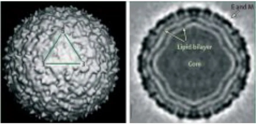
The structure of WNV particles, specifically New York 99, the strain responsible for the outbreak in the United States, have been elucidated by Mukhopadhay et al. in 2003 (Fig. 4) (Mukhopadhay S. et al., 2003; Kaufmann B., et al., 2010). Electron microscopy and image reconstruction techniques established that mature WNV virion is a small spherical icosahedral with a 50 nm diameter, with no surface projections or spikes. The outermost layer contains the highest density and corresponds to the viral envelope (E) and membrane (M) transmembrane proteins that are embedded in a lipid bilayer forming the envelope of the virion (Adams SC., et al., 1995; Berthet FX., et al., 1997; Mukhopadhay S. et al., 2003; Kramer LD., et al., 2007; Kramer LD., et al., 2007; Rossi SL., et al., 2010; Colpitts TM., et al., 2012; De Filette M., et al., 2012). This outer shell is constituted by 180 copies of M protein and an equal number of copies of the E glycoprotein disposed as 90 anti-parallel homodimers arranged in three distinct symmetry environments, thus resulting in a particle of icosahedral symmetry (Mukhopadhay S. et al., 2003; Kaufmann B., et al., 2010). Inside the envelope is the nucleocapsid core, which contains multiple copies of the capsid (C) protein and the genome RNA (Kramer LD., et al., 2007). The C proteins, located inside virions, have no discernible nucleocapsid symmetry and no contacts between C proteins and either E or M on the inner side of the virion envelope have been observed (Zhang W., et al., 2003). Although nucleocapsid particles consisting of multiple copies of the C protein and genome RNA are observed after removal of the virion envelop with nonionic detergent, capsid dimers can be dissociated from these structures by treatment with high salt (Kiermayr S., et al., 2004). C protein dimers have a very high charge, with half of the basic residues located on the face and conserved hydrophobic region that forms an apolar surface on the opposite face (Ma L., et al., 2004). It is thought that the apolar surface of the C dimer interacts with
the inner side of the virion envelop while the basic residue surface of the capsid dimer interacts with the genomic RNA (Brinton MA., 2014).
Figure 4: West Nile virion. The virus structure as reconstructed by cryo-electron microscopy. One asymmetric unit of the icosahedron is indicated by the triangle on the surface shaded view. The central section of the reconstruction shows the concentric layers of mass density. Reproduced with permission from the American Association for the Advancement of Science. (Mukhopadhyay S., et al., 2003; Kramer LD., et al., 2007).
3.2 GENOME OF WNV
The WNV genome is linear and is constituted by a single-stranded RNA molecule of positive polarity (Fig. 5). This RNA molecule of approximately 11.000 nucleotides (nts) in length, encodes a polyprotein in a single open reading frame (ORF) that is flanked by 5’ and 3’ untranslated regions (UTR). These form extensive secondary structures, which are important for replication, transcription, translation, and packaging (Shi PY., et al., 1996; Khromykh AA., et al., 2001; Friebe P. and Harris E., 2010; Martin-Acebes MA. and Saiz JC., 2012). The 5’ UTR of the WNV genome is 96 nts in length, while the length of the 3’ UTR varies from 337 to 649 nts. The 5’ end contains a type 1 cap structure (m7GpppAmp) that is added by NS5 during genome transcription (Brinton MA., 2014). The variable region of the 3’UTR is located just 3’ of the coding region stop codon (Beasly DW., et al., 2001). The 3’ end of the genome RNA does not contain a poly A tract but instead terminates with a conserved CUOH (Rice CM., et al., 1985; Brinton MA., et al., 1986; Wengler G., et al.,
1991). Proper methylations of the cap structure at guanine N-7 and ribose 2’-OH positions of the first transcribed adenine are necessary for optimal infectivity of WNV RNA. Viruses defective in the N7 methylation mechanism are non-replicative, and recently the 2’-OH
methylation has been related to evasion of innate immunity by evading certain components of interferon response, therefore WNV defective in this methylation mechanism can replicate but is attenuated in vivo (Dong H., et al., 2008; Daffis S., et al., 2010).
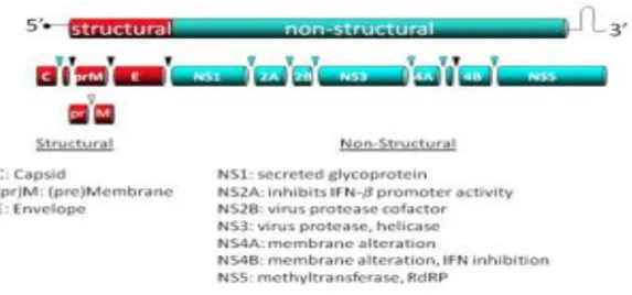
The single open reading frame (ORF) of 10.301 nts in most WNV isolated, is translated as a single polyprotein of approximately 3000 amino acids that is post- and co-translationally cleaved by cellular and viral proteases into ten proteins: three structural proteins (C, premembrane or membrane, and envelope) and seven non-structural proteins (NS1, NS2A, NS2B, NS3, NS4A, and NS5) (Fig. 5). The three viral structural proteins are encoded within the 5’ portion of the ORF and are mainly involved in viral particle formation, whereas non-structural proteins are encoded within the 3’ portion and their function consists in viral replication, virion assembly, and evasion of host innate response (Kramer LD., et al., 2007; Lindenbach BD., et al., 2007; Rossi SL., et al., 2010; Brinton MA., et al., 2014). The viral polyprotein contains multiple transmembrane domains that determine whether individual mature viral proteins are located on the cytoplasmic or luminal side of the endoplasmic reticular (ER) membrane after cleavage from the polyprotein (Lindenbach BD., et al., 2013). The C, NS3 and NS5 proteins are located on the cytoplasmic side while the PrM, E, and NS1 proteins are in the lumen and, with the exception of short regions between transmembrane domains, the NS2A, NS2B, NS4A and NS4B proteins are located within the ER membrane bilayer (Lindenbach BD., et al., 2013; Brinton MA., et al., 2014).
!
Figure 5: Schematic of WNV genome. A representation of the WNV genome including the 3 structural proteins that make up virion particle and the 7 non-structural proteins necessary for virus replication and immune evasion (Rossi SL., et al., 2010).
3.2.1 VIRAL STRUCTURAL PROTEINS
Capsid (C): The capsid, or core, (C) protein is a highly basic protein of ≈11 kd that contains a large number of scattered charged amino acids (Dokland T., et al., 2004; Lindenbach BD., et al., 2007) and is implicated in viral assembly and replication (Schrauf S., et al., 2009). The N- and C-termini parts of the protein are intrinsically disordered regions and may play a role in RNA folding during viral replication by conferring RNA chaperoning activity to the C protein (Ivanyi-Nagy R., et al., 2008). The central part of the C protein is a hydrophobic region that mediates membrane association (Ma L., et al., 2004; Lindenbach BD., et al., 2007). Nascent C (anchC) also contains a C-terminal hydrophobic anchor that serves as a signal peptide for ER translocation of prM. This hydrophobic domain is cleaved from mature C by the viral serine protease (Lobigs M. and Lee E., 1993). The protein dimerizes and tetramerizes to build the nucleocapsid that, together with viral RNA, forms the electron-dense core of the virion that is enveloped by the lipid bilayer. In WNV-infected cells, capsid protein can be detected in the cytoplasm, nuclei and the nucleolus of the cell, and it has been related to the induction of apoptosis (Yang MR., et al., 2008). Nuclear location of the C protein is mediated by a bipartite nuclear location signal and requires specific interaction with cellular importins (Bhuvanakantham R., et al., 2009). The capsid protein also interacts with other cellular factors, as the inhibitor of the serine/threonine phosphatase PP2A, I (2) (PP2A), Hsp70 and Jab1 (Oh WK. and Song J., 2006; Oh WK., et al., 2006; Hunt TA., et al., 2007). The phosphorilation status of the protein and Jab1 can regulate nuclear location and RNA binding activity (Oh WK., et al., 2006; Cheong YK. and Ng ML., 2011; Bhuvanakantham R., et al., 2010). The C protein has been also implicated in degradation of claudin proteins and disruption of epithelia barrier, thus helping to virus dissemination (Medigeshi GR., et al., 2009; Martin-Acebes MA., et al., 2012).
prM/M: The prM/M is a short transmembrane glycosylated protein associated to the lipid bilayer of the virion. The glycoprotein precursor of M protein, prM (≈26 kd), is translocated into ER by C-teminal hydrophobic domain of C. However, signal peptidase cleavage is delayed until the viral serine protease cleaves upstream of this region sequence to generate the mature form of C protein (Lobigs M. and Lee E., 1993; Amberg Sm., et al., 1994; Yamshchikov VF. and Compans RW., 1994). In addition, E protein expression influences the rate of this signalase cleavage (Lorenz IC., et al., 2002). The cleavage of this protein by a furin-like protease occurs within the trans-Golgi network and is necessary for particle maturation (Brinton MA., 2002). The conversion of immature virus particles to mature virions occurs in the secretory pathway and coincides with cleavage of prM into pr and M
fragments by the Golgi-resident protease furin enzyme (Stadler K., et al., 1997). This protein protects virions from fusion inside acidic vesicles of the Golgi complex (Martin-Acebes MA., et al., 2012). The furin-like protease cleaves the prM/M membrane protein, enabling a conformational rearrangement in the viral particle from immature particles (Zhang Y., et al., 2007) to mature ones (Mukhopadhyay S., et al., 2003). Modulation of the proportion of prM/M cleavage can also modulate the sensitivity of antibody-mediated neutralization (Nelson S., et al., 2008).
The envelope (E): The envelope (E) is a transmembrane protein anchored to the lipid envelope by a C-terminal α-helical hairpin. It is the most immunogenic protein of the virus and the target for most neutralizing antibodies. The protein is glycosylated on position 154 on most WNV strains (Beasley DW., 2005a). Glycosylation is important for efficient transmission in mosquito and birds (Moudy RM., et al., 2009; Murata R., et al., 2010) and may be related to neuroinvasiveness (Shitato K., et al., 2004). E protein contains 12 cysteines involved in the formation of intramolecular disulfide bonds and the production of homodimers. E glycoprotein is organized in three domains: DI, DII, and DIII. DI links domains II and III (Nybakken GE., et al., 2006). DII contains a conserved region of 13 hydrophobic amino acids that form an internal fusion loop necessary for virus fusion. DIII is an immunoglobulin-like domain that is thought to be involved in the interaction between virions and host cells to enable the virus entrance, moreover it contains multiple epitopes that are recognized by neutralizing antibodies. Upon acid exposure, the E glycoprotein undergoes conformational rearrangements and changes from dimers to trimers, exposing the fusion loop to enable viral fusion of the virion with cellular endosomal target membranes. For other flaviviruses, as tick-borne encephalitis virus, this process is triggered by protonation of an individual His residue on E glycoprotein (Fritz R., et al., 2008) that should act as a critical pH sensor. However, this hypothesis has not been validated for WNV (Nelson S., et al., 2009), although point mutations can modulate the fusion threshold (Martin-Acebes MA. and Saiz JC., 2011).
3.2.2 VIRAL NON-STRUCTURAL PROTEINS
Although the functions of the WNV non-structural proteins have not yet been completely characterized, all seven are directly or indirectly involved in viral RNA synthesis and additional functions for some of these proteins have been identified. Little is known about the interactions between the viral non-structural proteins or between viral non-structural proteins and cell proteins that are required for remodelling the cell environment and for appropriately regulating active viral RNA replication complexes at different phases of the
virus life cycle (Brinton MA., 2014).
NS1: This is a versatile non-structural viral glycoprotein that has a molecular weight of 46-56 kDa, depending on its glycosylation status. NS1 exists in multiple oligomeric forms: monomers, dimers (the primary form) and hexamers, and this seems to be related to its cellular retention or secretion stage (Brinton MA., 2002; Beasley DW., 2005a). NS1 is found at different cellular locations: either cell-membrane-associated (mNS1), in vesicular compartments within the cell or on the cell surface, and as a secreted lipid-rich, extracellular (nonvirion) species (sNS1) (Smith and Wright, 1985; Westaway and Goodman, 1987; Winkler et al., 1988; Mason, 1989; Gutsche et al., 2011). Intracellular NS1 acts as an essential cofactor for viral replication, it co-localizes with dsRNA and other components of replication complex (Mackenzie et al., 1996; Westaway et al., 1997), and inhibits Toll-like receptor 3 (TLR3) signalling (Wilson JR., et al., 2008). Whereas, cell surface and secreted NS1 antagonize complement activation, are highly immunogenic, and both the proteins and the antibodies it elicits have been implicated in disease pathogenesis (Chung KM., et al., 2006; Avirutnan P., et al., 2010; Muller DA. and Young PR., 2013). Recently, a larger NS1-related protein (termed NS1’), produced by a ribosomal frameshift near the beginning of the NS2A gene, has been detected in infected cells and related to neuroinvasiveness (Melian MB., et al., 2010).
NS2A: This is a small hydrophobic transmembrane protein involved in the biogenesis of virus-induced membranes, which have a vital role in virus assembly (Leung JY., et al., 2008). In fact, NS2A has been detected by immunogold labelling primarily within vesicle packets (VP), associated with labelled dsRNA (Mackenzie JM., et al., 1998). Moreover, NS2A has been reported to have an immunomodulatory role because it inhibits interferon-β promoter activation (Liu WJ., et al., 2004), and it has reported that mutations in this protein result in viral attenuation in vivo (Liu MJ., et al., 2006; Rossi SL., et al., 2007). Recently, an ER membrane topology model for flavivirus NS2A was reported (Xie X., et al., 2013): the N-terminal amino acids are located in the ER lumen while the C-terminal tail is in the cytosol. The first of five transmembrane regions located in the middle part of NS2A contains two helicase separated by the “helix-breaker” amino acids P85 and R84. Mutation of each of these amino acids in both a replicon and an infectious clone showed that R84 but not P85 was functionally important. Interestingly, an R48E mutation attenuated both viral RNA replication and virion production while an R84 mutation had no effect on viral RNA synthesis but inhibited the production of infectious virions (Brinton MA., 2014).
domain and functions as a protease co-factor (Erbel P., et al., 2006). Alanine scanning approaches of NS2B has revealed two sites critical for regulation of the proteolytic activity of NS2B-NS3 complex (Chappell KJ., et al., 2008). The interaction between NS2B and NS3 may also confer specificity for RNA unwinding of NS3 discriminating from DNA (Chernov AV., et al., 2008).
NS3: This is a highly conserved and multifunctional protein, consisting of the N-terminal serine protease domain localized to amino acids 1-169 and the C-terminal domain from residues 180-618, bearing helicase, nucleoside triphosphatase, and RNA triphosphatase activities, important for viral replication (Gorbalenya AE., et al., 1989; Wengler G. and Wengler G., 1991; Wengler G., et al., 1991a). The N- and C-terminal domains are linked via a flexible inter-domain, comprising residues 169-179 (Luo D., et al., 2008; Assenberg JM., et al., 2009). However, it is not active unless tethered to its cofactor, NS2B (Chappell KJ., et al., 2008a). This protease cleaves the viral polyprotein to release structural and non-structural proteins and, thus, disruption of its activity is lethal for virus replication. NS3 (and also its cofactor NS2B) has been localized within paracrystalline arrays (PC) or convoluted membranes (CM), suggesting that these membranes are the sites of proteolytic cleavage (Mackenzie JM., et al., 1998). Both the ATPase and helicase activity of NS3 have been shown to be regulated by NS4A (Shiryaev SA., et al., 2009), and the two activities can function independently of each other (Borowski P., et al., 2001). Within infected host cells, these functions appear to be regulated by their differential localization to separate virus-induced membranous compartments (Westaway EG., et al., 2001). All these properties of NS3 made of this protein and its active form, NS2B-NS3, a promising antiviral target (Martin-Acebes MA., et al., 2012)
NS4A: This is a small hydrophobic protein with several transmembrane domains that has been localized to the viral replication complex in virus induced membranes (VP, CM and PC) (Mackenzie JM., et al., 1998). The C-terminal region of NS4A can be cleaved by cell signalase generating the 2K fragment that may be responsible of membrane rearrangements in infected cells (Brinton MA., 2014). In addition, it has been reported that cleavage of NS4A C-terminal regional in DENV acts as a signal sequence for translocation into the ER of the adjacent NS4B protein (Miller S., et al., 2007). NS4A has been also related, together with NS2A, NS2B, and NS4B, to block type I interferon signalling in flavivirus infected cells (Martin-Acebes MA., et al., 2012; Brinton MA., 2014). Accumulation of NS4A (and also NS4B) into ER of infected cells seems to be involved in induction of the unfolded protein response upon WNV infection. Mutations in the 2K fragments have been related to
resistance against the antiviral action of the interferon-inducible 2’, 5’-oligoadenylate synthetase 1b protein, and also to resistance against the flavivirus inhibitor lycorine thanks to the enhancement of RNA replication. NS4A has been proposed to also act as a cofactor regulating ATPase activity of the NS3 helicase (Martin-Acebes MA., et al., 2012).
NS4B: This is a small hydrophobic non-structural protein that is hypothesized to participate in viral replication and inhibition of host interferon signalling (Munoz-Jordan JL., et al., 2005). Mutations in NS4B affect viral RNA replication (Wicker JA., et al., 2006; Puig-Basagoiti F., et al., 2007; Welte JA., et al., 2006), possibly through its interaction with NS3 helicase (Xie X., et al., 2011) and can result in attenuation of WNV in vivo (Wicker JA et al., 2006; Welte T., et al., 2011). NS4B has been localized in perinuclear membranes and in the nucleus of WNV infected cells (Westaway EG., et al., 1997a;Pheng S. and Pei-Yong S., 2013) where it may be involved in the formation of viral replication complex.
NS5: This is located at the C-terminus of the viral polyprotein and is the largest and most conserved protein amongst members of the genus Flavivirus. NS5 contains two domains that have different enzymatic activities. The N-terminal region contains an S-adenosyl methionine methyltransferase (MTase) domain that has N7 and 2’-O MTase activities and also acts as guanylyltransferase (Brinton MA., 2014). This domain is necessary for capping the 5’ end of the viral RNA that is performed by sequential methylation reactions. The C-terminal portion contains conserved sequence motifs characteristics of all RNA-dependent RNA polymerase for replication of viral genome (RdRp domain) (Martin-Acebes MA., et al., 2012). The methyltransferase activities together with the polymerase activities of NS5 are genetically validated to be essential for viral replication (Pheng S. and Pei-Yong S., 2013). This protein localizes to virus induced membranes in infected cells and colocalizes with dsRNA at viral replication complexes (Mackenzie JM., et al., 2007a). Due to the lack of proof-reading activity of NS5, WNV populations display a variable level of sequence diversity that favours selection of variants in response to selective pressures. NS5 is also a potent antagonist of interferon signalling to evade of host innate immune defences (Laurent-Rolle M., et al., 2010). Both the capping and RdRp activities made of NS5 also a promising antiviral target (Martin-Acebes MA., et al., 2012).
3.3 WNV REPLICATION CYCLE
The WNV life cycle consists of 4 principal stages: attachment/entry, translation, replication, and assembly/egress. WNV enters cells via receptor-mediated endocytosis after E protein interacts with one or more cell surface receptor(s). It is not completely clear which cellular receptors are involved in WNV binding, however DC-SIGN, several glycosaminoglycans, mannose receptor, c-type lectins and, although still controversial, integrin αvβ3 have been
proposed as potential receptors (Tassaneetrithep B., et al., 2003; Lee E., et al., 2004; Davis CW., et al., 2006; De Filette M., et al., 2012; Martin-Acebes MA., et al., 2012). After binding to the cell, the viral particles are internalized into host cells via a clathrin dependent mechanism. Rab 5 was reported to be required for the cellular WNV entry (Krishnan MN., et al., 2007). The virus-containing endosome matures during internalization from the cell surface, with the pH dropping from neutral to slightly acidic in the early endosome and becoming more acidic during maturation to the late endosome. Acidification inside the late endosome triggers rapid conformational changes on the E protein resulting in fusion between the viral and endosomal membranes, and release of the virus nucleocapsid into the cytoplasm for genome uncoating (Gollins SW., et al., 1986; Modis Y., et al., 2004; Mukhopadhyay S. et al., 2005; Martin-Acebes MA., et al., 2012). The optimal pH for conformational rearrangements and viral fusion is 6.3-6.4, and this fusion process is dependent on the presence of cholesterol in the target membrane (Moesker B., et al., 2010; Martin-Acebes MA., and Saiz JC., 2011). Once viral RNA genome reaches the host cell cytoplasm it is translated into a single polyprotein, which is proteolytically processed by viral and host proteases to generates structural and non-structural proteins involved in viral replication and viron assembly. Whereas the cleavages at the junction C-prM, prM-E, E-NS1, NS4A-NS4B (Nowak T., et al., 1989), and likely also NS1-NS2A (Falgout B., et al., 1995), are performed by the host signal peptidase located within the lumen of ER, the remaining peptide bonds are cleaved by the virus encoded NS3 protease. The structural proteins form the virion that encapsidates the viral RNA, and the non-structural proteins form the replication complex that is required for synthesis of viral RNA (Suthar MS., et al., 2013). The original viral RNA is replicated by viral and cellular proteins into multiple copies to be used in the production of new virions. WNV replication requires the viral protein NS5, which is an RNA-dependent RNA polymerase (Rice CM., et al., 1986; Poch O., et al., 1989). An “antisense” negative strand RNA is produced by this enzyme, which then serves as a template for the synthesis of many new copies of the infectious positive strand RNA genome (De Filette M., et al., 2012). WNV induces changes in the cellular environment in
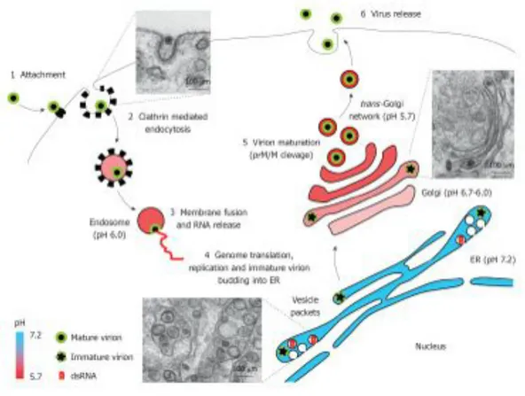
order to create conditions more appropriate for viral replication and undergo notable intracellular membrane remodelling. In particular, viral remodelling of ER (endoplasmic reticulum) membranes to form a network of replication complex provides a microenvironment required for productive viral replication (Bidet K., et al., 2014). These structures are important replication and virus protein processing and are termed vesicle packets (VP), paracrystalline arrays (PC) and convoluted membranes (CM) (Westaway EG., et al., 1997; Mackenzie JM., et al., 2001). Viral replication takes place at VPs, which are generated as invaginations of the membrane of ER and contact by pores with the cell cytoplasm. VPs contain dsRNA replication intermediates, and assembled virions bud into the ER (Gillespie LK., et al., 2010; Matin-Acebes MA., et al., 2012). A specific role of cholesterol and fatty acids in WNV-induced membrane structures has been proposed, and proteasome activity seems to be also important for viral replication (Mackenzie JM., et al., 2007; Gilfoy F., et al., 2009; Heaton NS., et al., 2010; Fernandez-Garcia MD., et al., 2011; Matin-Acebes MA., et al., 2012). Apart from providing the adequate platform for viral replication, these membrane rearrangements may also play a role for the evasion of innate immune response by interfering with the interferon signalling machinery (Hoenen A., et al., 2007; Mackenzie JM., et al., 2007). In addition, replication of WNV, accumulation of non-structural proteins at the ER induces ER stress activating the unfolded protein response and also induces apoptosis of infected cells (Parquet MC., et al., 2001; Medigeshi GR., et al., 2007; Ambrose RL., et al., 2011). Following replication and translation, genomes are packaged into virions, which travel to the cell surface in exocytic vesicles and mature through the ER-Golgi secretion pathway (Rice CM., 1996; Rossi SL., et al., 2010). This maturation process requires the cleavage of prM/M protein by a furin-like protease located at the acidic environment of the trans-Golgi network (Brinton MA., 2002). After maturation, viral particles are released by exocytosis from surface of infect cells (Rossi SL., et al., 2010).
Figure 6: West Nile virus replication cycle. Schematic view of West Nile virus replication cycle in an infected cell. Electron micrograph of West Nile virus-infected Vero cells illustrate distinct snapshots from infectious cycle. WNV infects a wide range of target cells. Virion entry is initiated after the envelope protein, E, engages an unknown cellular receptor (or receptors) (step 1), followed by receptor-mediated endocytosis of the virus (step 2). The low-pH environment within the endosomal vesicle triggers viral fusion with the endosomal membrane, leading to virion uncoating and release of the viral positive-sense single-stranded RNA ((+)ssRNA) genome into the cytoplasm (step 3). The viral (+)ssRNA is translated into a single polyprotein at the ER and cleaved into mature proteins by the viral serine protease non-structural protein 2B–3B (NS2B–NS3) and cellular proteases (step 4). The NS proteins, including the viral RNA-dependent RNA polymerase NS5, form the replication complex for the synthesis of full-length negative-sense ssRNA ((–)ssRNA) intermediates. These serve as templates for the synthesis of full-length (+)ssRNAs. The viral capsid protein, C, is responsible for encapsidating viral genomic RNA, with assembly occurring on rough ER membranes (step 8). Immature virions are transported through the host secretory pathway, resulting in glycosylation of the viral E protein and host cell furin mediated-cleavage of the protein prM to the mature membrane protein, M (step 5). Mature virions are transported to the plasma membrane and released by exocytosis (step 6) (Martin-Acebes MA., et al., 2012; Suthar MS. et al., 2013).
3.4 MOLECULAR CLASSIFICATION
First classifications of WNV were based on cross-neutralization reactions and revealed that WNV is a member of the Japanese encephalitis virus serocomplex. This complex includes also other neurovirulent viruses such as Murray Valley encephalitis virus, St. Louis encephalitis virus, or Usutu virus (Poidinger M., et al., 1996; Beasley DW., 2005). Even though WNV has a single serotype, it nonetheless exhibits considerable genetic variation (Bondre VP., et al., 2007). Phylogenetic classification of WNV remains dynamic, with the large increase in genome sequence and surveillance data in recent years. Present analysis support that WNV aligns into at least seven different lineages (Fig. 7), on the basis of nucleic acid homology, with the major lineages diverging by 25%-30% nucleotide differences (Hubalek Z., et al., 1998; Lanciotti RS., et al., 1999; Lvov DK., et al., 2004; Mackenzie JS., et al., 2009; May FJ., et al., 2011; Papa A., et al., 2011). WNV strains that cause disease in humans and horses belong into the major lineages 1 and 2 (Marka A., et al., 2013; Di Sabatino D., et al., 2014), while other lineages have been sporadically detected in mosquitoes and birds but not associated with human disease (Vazquez A., et al., 2010). The phylogenic classification does not consistently correlate with the geographical distribution of WNV, which may be attributed to the broad dissemination of the virus by migrating bird species (Gray TJ. and Webb CE., 2014).
3.4.1 LINEAGE 1
Lineage 1, the largest and the most widespread, contains WNV strains isolates from Europe, Africa, Australia, Asia, North and Central America, as well as the Middle East (Hosseini NS., et al., 2014; Gray TJ. and Webb CE., 2014; Lanciotti RS., et al., 1999). Lineage 1 can be further subdivided into three different clades: 1a, 1b and 1c. Clade 1a is the most widely distributed and contains strains from the Americas (including the NY99 strain), Europe, Africa, the Middle East and Israel. Until recently, clade 1a comprises most of the isolates associated with outbreaks of human encephalitis, including the ongoing epidemic in North America (Lanciotti RS., et al., 1999). Interestingly, this clade displays close genetic relationship between geographically distant areas which are supposed to be the result of WNV introductions via migratory birds (Martin-Acebes MA., et al., 2012) This clade can further be divided in six clusters with distinct evolutionary histories (May FJ., et al., 2011). Sublineage 1b, contains the Australian Kunjin virus, that is an uncommon cause of human disease endemic to Australia and it is probably found in South East Asia and Papua New Guinea (Hall RA., et al., 2001; Gray TJ., et al., 2011; Rossi SL., et al., 2012; Hosseini NS.,
et al., 2014). While few human cases were reported, a major epidemic of illness in horses was reported in southeast Australia in 2011 (Frost MJ., et al., 2012). Clade 1c is only found in India. It has been proposed that isolates previously classified as sublineage 1c be reassigned to a new lineage 5 (Lanciotti RS., et al., 2002; Beasley DW., 2005; Bronde VP., et al., 2007). The fact that only one endemic genotype has been detected in India (1c) and one in Australia (1b), suggests that WNV was successfully introduced into these locations only once, as well as it was the casa in the American continent, where WNV was introduced in 1999 in the East Cost of the US (Lanciotti RS., et al., 1999; May FJ., et al., 2011). The first North American WNV isolate was most closely related to a strain isolated from a dead goose in Israel (lineage 1) during the 1998 outbreak, suggesting that North American WNV was derived from this epidemic (Lanciotti RS., et al., 1999). However, recent data suggest that the epidemic in Israel in 1998 was not the direct progenitor of North American epidemics, but rather that both epidemics originated from the same (unknown) location (May FJ., et al., 2011).
3.4.2 LINEAGE 2
Lineage 2 WNV, until the mid-2000s, was predominantly limited to sub-Saharan Africa and Madagascar, where it has been a cause of mild febrile illness in humans, rarely progressing to severe disease and typically not associated with outbreaks (Lanciotti RS., et al., 2002). However, in 2004 and 2005, WNV belonging to lineage 2 was first identified in wild birds in Hungary, with subsequent rapid spread to central Europe (Bakonyi T., et al., 2005; Bakonyi T., et al., 2006). Since 2004, lineage 2 has been observed in central and Eastern Europe. In 2010 it caused outbreaks in Romania and Greece and in 2011 it was detected for the first time in Italy (Bakonyi T., et al., 2006; Platonov AE., et al., 2008; Sirbu A., et al., 2010; Papa A., et al., 2010; Bagnarelli P., et al., 2011; Papa A., et al., 2011). These lineage 2 viruses have been implicated in avian, equine, and human cases of neuroinvasive disease with associated deaths, including cases reported in Russia, Hungary, Italy and Greece (May FJ., et al., 2011; Papa A., et al., 2011; Barzon L., et al., 2013; Magurano F., et al., 2012). The Greek and Italian strains showed the highest homology to Hungarian and South African strains, differing from the Russian lineage 2 strains. This means that at least two lineage 2 strains are circulating in Europe causing severe neuroinvasive infections in birds, horses and humans (Papa A., 2012; Papa A., et al., 2012). Although there are exceptions, in general, lineage 1 viruses are considered to be more virulent than the lineage 2 viruses (De Filette MD., et al., 2012): lineage 1 (clade 1a) viruses can cause severe human neurologic disease
whereas lineage 2 viruses generally cause a mild, self-limiting disease. Both, lineage 1 and 2, are now considered endemic in southeastern Europe, with an over 700% increase in cases reported in the region since 2009 (European Centre for Disease Prevention and Control, 2013).
3.4.3 LINEAGE 3
Lineage 3 WNV was first isolated near the Austrian and Czech Republic border in 1997. WNV belonging to lineage 3 has also referred as Rabensburg virus 97-103, named after the nearby Austrian town where the first infected Culex pipiens mosquitoes were isolated (Hubalek Z., et al., 1998; Bakonyi T., et al., 2005; Hosseini NS., et al., 2014). On the basis of genomic and antigenic diversity, it has been suggested that Rabensburg virus be assigned a new species within the Japanese encephalitis virus group (Bakonyi T., et al., 2005). Lineage 3 strain has not been isolated from humans, and the pathogenic potential remains uncertain, particularly as Rabensburg virus has been shown not to infect mammalian or avian cell cultures, nor infect experimentally exposed birds (Aliota MT., et al., 2012).
3.4.4 ADDITIONAL PROPOSED LINEAGES
Additional lineage subdivisions have been proposed for novel flavivirus isolates, including lineage 4 that contains a new variant of WNV (strain LEIVKrnd88-190), which was isolated in 1998 from Dermacentor marginatus ticks in a valley in the northwestern Caucasus Mountains of Russia (Bakonyi T., et al., 2005; Hosseini NS., et al., 2014). Lineage 5 WNV has been proposed for a group of human and mosquito isolate from India as early as the 1950s and cluster to form sublineage 1c (strain 804994) (Bondre VP., et al., 2007; Botha EM., et al., 2008). Lineage 6 WNV has been proposed for virus isolated from C. pipiens mosquitoes in southern Spain in 2006, strain HU2925/06, and forms a common evolutionary branch with lineage 4 (Vazquez A., et al., 2010). In addition to these minor lineages, the African virus Koutango (KOUV), first isolated in Senegal, is currently recognized as a separate species but could be considered as a seventh WNV lineage (De Filette MD., et al., 2012; Pesko KN. and Ebel GD., 2012). The human pathogenicity of lineages 4, 6 and 7 WNV is poorly understood, with human infection not reported (Gray TJ. and Webb CE., 2014).
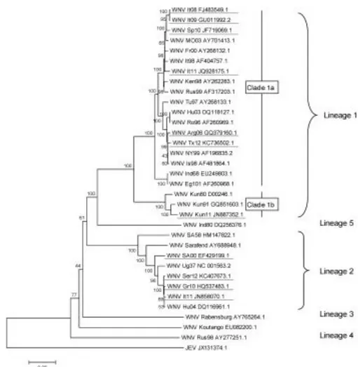
Figure 7. West Nile Virus (WNV) genetic diversity, evaluated using genetic alignment of complete genomic sequences. GenBank accession numbers are indicated on the tree branches of each virus; the first two or three letters stand for the country or the USA state reporting WNV (It = Italy, Sp = Spain, Mo = Morocco, Fr = France, Ken = Kenya, Rus = Russia, Tu = Tunisia, Hu = Hungary, Ro = Romania, Arg = Argentina, Tx = Texas, NY = New York, Is = Israel, Ind = India, Eg = Egypt, Kun = Kunjin Australia, SA = South Africa, Ug=Uganda, Ser = Serbia, and Gr = Greece) and the numbers indicate the year of isolation (96 = 1996, 10 = 2010). Japanese encephalitis virus (JEV), a closely related flavivirus, was used as an outgroup. The rooted phylogenetic tree was constructed using neighbor-joining with Jukes-Cantor parameter distances (scale bar) in MEGA (MEGA software, version 5.2) (Tamura K., et al., 2011). A bootstrapped confidence interval (1,000 replicates) and a confidence probability value based on the standard error test were also calculated using MEGA. The WNV strains responsible for recent human or equine outbreaks are underlined. The complete sequences of the most recent Romanian and Russian lineage 2 variants are not available, but at least two introduction events of lineage 2 strains have occurred in Europe: divergent lineage 2 strains have been observed in Romania/Russia and Hungary/Greece/Italy/Serbia/Austria (Ciccozzi M., et al., 2013; Donadieu E., et al., 2013).
3.5 TRANSMISSION CYCLE
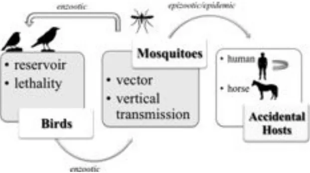
WNV is maintained in nature in an enzootic transmission cycle between avian hosts and ornithophilic mosquito vectors (Fig. 8). Mosquitoes become infected by feeding on birds that carry virus particles in sufficient concentrations in their blood (Marka A., et al., 2013). Apart birds, the virus can be transmitted to other animals including horses and humans as well (Hayes EB., et al., 2005). Humans and horses are considered incidental or “dead-end” hosts for WNV, as the low concentration of virus within the blood (viremia) in mammals is usually insufficient to infect a feeding naïve mosquito and maintaining the transmission cycle (Bowen RA., and Nemeth NM., 2007; Rossi SL., et al., 2010). Although human cases occur primary after mosquito inoculation, infection after blood transfusion, organ transplantation, and intrauterine transmission has reported (Hayes EB., et al., 2005).
3.5.1 VECTORS: MOSQUITOES AND OTHER ARTHROPODS
The ability of different mosquito species to acquire and transmit WNV is highly variable (Colpitts TM., et al., 2012). At least over 60 species of mosquitoes from 11 different genera have been described as competent vectors. Mosquitoes of the genus Culex are the predominant vectors in the enzootic cycle throughout the range of the virus distribution, although the particular species of Culex varies according to geographic locations (Martin-Acebes MA., et al., 2012). In North America Cx. pipiens, Cx. restuans, Cx. quinquefasciatus, Cx. salinarus, Cx. tarsalis, and Cx. nigripalpus have been described as the most efficient competent vectors; although other species such as Aedes albopictus, Aedes vexans, Ochlerotatus japonicus and Ochlerotatus triseriatus may also play role on viral transmission as bridging vectors that can transmit the virus to mammals (Brault AC., 2009). In Europe, the virus has been isolated from more than 40 different species, being again those of the Culex species the main vectors (Zeller HG. and Schuffenecker I., 2004). Several other species have been also implicated in the transmission cycle as competent vectors in other geographical areas, Cx. univittatus in Africa, Cx. annulirostris in Australia, and Cx. vishnui and Cx. tritaeniorhynchus in Asia (Hall RA., et al., 2002; Hayes EB., et al., 2005; Brault AC., 2009).
Vector competence varies between species and within populations of individual species. The C. pipiens complex contains two genetically distinct forms: pipiens and molestus that differ in physiology and behavior with obvious implications to their epidemiological importance. Form pipiens is thought to be exclusively ornithophilic, while the urban form molestus will feed on mammals. The two forms have been shown to not interbreed in the northern Europe,
in contrast to US and southern Europe population, which contain individuals with hybrid genetic signatures (pipiens x molestus) that may generate bridge vectors, disposed to feed on both birds and mammals. Indeed, US populations of C. pipiens, as well as C. nigripalpus and C. tarsalis, have been demonstrated to shift their feeding from birds to mammals in the late summer and early fall, and therefore may act as bridge vectors to infect equid and human hosts (Kramer LD., 2008).
Laboratory analyses have shown that C. tarsalis mosquitoes become infected after consumption of blood meals with viral concentrations over 107 PFU/mL, whilst only up to 30% do it if the concentration is in the 105 PFU/mL range (Goddard LB., et al., 2002). On the other hand, different species of mosquitoes inoculate quite variable doses of WNV (103.4 PFU to 106.1 PFU) into vertebrate hosts during natural feeding, of which around 102 PFU are directly inoculated into the blood (Styer LM., et al., 2007).
The mechanism(s) of WNV perpetuation overwintering and years may vary by region and country, but possible mechanisms include continuous low-level virus transmission, reinitiation after reintroduction of virus by migratory birds from locations where virus is active year-round, vertical transmission to females about to enter reproductive diapause in winter, and recrudescence of low levels of virus in chronically infected birds when mosquitoes are active (Anderson JF. and Main AJ., 2006; Nasci RS., et al., 2001).
Beside from mosquitoes, WNV has been sporadically isolated in other arthropods: WNV has been isolated repeatedly in Russia from soft ticks (Argasidae). In addition, soft ticks have been demonstrated to transmit virus in the laboratory, and nonviremic transmission has been demonstrated. Hard ticks (Ixodidae) allow the virus to pass transstadially, but are incompetent vectors. Moreover, other arthropods have been suggested as alternative vectors, including dermanyssoid mites, swallow bugs, and hippoboscid flies, but their role in the transmission cycle is not clear (Martin-Acebes MA., et al., 2012).
3.5.2 BIRDS
Birds are the natural reservoir of WNV. More than 300 avian species representing over 200 birds families from North America have been reported as susceptible to WNV infection after its first introduction in 1999, confirming their role as primary vertebrate in the enzootic cycle (Martin-Acebes MA., et al., 2010; Kramer LD., 2008). Many studies have been conducted to determine the precise role of birds in the transmission of the virus and have demonstrated that birds vary significantly in susceptibility and response to infection, with a great diversity in the profile of viremia among the different avian species. Various experimental studies
have estimated that the limit, for mosquitoes to become infected after consumption of blood meals, is of 105 plaque forming units (PFU) viral concentration and different birds species can develop sufficient viremia titres before the birds become moribund and die a few days after being infected in order to allow the transmission of the virus to the feeding mosquitoes. These birds belong to the orders of Passeriformes (corvids, sparrows, finches, etc.), Charadriiformes (woodcocks, gulls, etc.) Strigiformes (owls, eagle owls, etc.) (Komar N., et al., 2003; Beasley DW., 2005). In contrast, species of the order of Paciformes (woodpeckers), Columbiformes (doves, pigeons, etc.) and Anseriformes (ducks, geese, etc.) develop lower viremia titres, in many cases insufficient to transmit the virus in mosquitoes and they do not contribute in the epizootic cycle (Marka A., et al., 2013).
Feeding by infected mosquitoes is the most common route of infection, but transmission to birds also has been demonstrated by direct contact via the fecal-oral route: many avian species shed large quantities of virus in their feces or oral secretions when infected (Komar N., et al., 2003), allowing direct transmission from bird and even from bird-to-human. Experimental oral infection of birds has been demonstrated (McLean RG., et al., 2001) and prey-to-predator infection through ingestion of infected mosquitoes or of carrion by omnivorous birds such as corvids and raptors has been suggested (Garmendia AE., et al., 2000).
3.5.3 HUMANS, HORSES AND OTHER ANIMALS
Thirty species of mammals and occasionally other vertebrates including reptiles and amphibians have been found infected with WNV. Generally, humans, horses and other mammals infected in a spillover transmission are considered “dead end” hosts and their role in the transmission cycle is less significant than that of birds, because viral replication does not yield significant viremia to infect feeding mosquitoes. Enzootic in equines have occurred in the US, France, Italy, Marocco and in Israel. Unvaccinated equines develop infections ranging from asymptomatic to encephalitic disease, and demonstrate a case-fatality rate of about 25%. Because of their low viremias, they are considered incidental hosts in the transmission cycle. In experimental infections of horses with WNV, viremia levels are around 103 PFU/mL (Bunning ML., et al., 2002), thus being usually insufficient to sustain infectivity cycles (Martin-Acebes MA., et al., 2010). Several other animal species have been described as susceptible to WNV infection, with or without clear evidence of disease, including domestic and wild mammals such as: dogs, cats, pigs, cows, llamas, sheep, alpacas, deers, reinders, raccoons, bears, wolfs, squirrels, chipmunks, rabbits, and bats,
among others (Beasley DW., 2005; Blitvich BJ., 2008). As it has described for humans and horses, in most cases the viremia raised in these animals is low, only for rabbits and chipmunks, have been demonstrated in the laboratory to mount sufficiently high levels of virus in the blood to infect a small portion of feeding Culex spp. mosquitoes, but generally is probably not enough to initiate a new transmission cycle (Martin-Acebes., MA. et al., 2012). Apart from mammals, several reptiles and amphibians, such as snakes, crocodiles, alligators, iguanas and frogs (Kostiukov MA., et al., 1985; Steinman A., et al., 2003; Klenk K., et al., 2004; Steinman A., et al., 2006) have been also described as susceptible to WNV infection and some of them raise high viremia. In the US and Mexico, farmed alligators raised at high temperatures in crowded conditions demonstrated significant mortality and mount high viremia. Transmission appears to occur directly between alligators, as well as through ingestion of uncooked infected horse meat. However, the real contribution of animals other than birds and mosquitoes in maintaining WNV cycle in nature is still uncertain (Martin-Acebes MA., et al., 2010).
3.5.4 NON-VECTOR-BORNE TRANSMISSION
Even though the main mode of WNV transmission to vertebrate is via infected mosquito bite, it has been documented that alternative less common modes of non-vector-borne transmission in humans also exist: through solid organ transplantation from an infected donor to healthy recipient; the placenta from an infected mother to her fetus (vertical transmission), occupational infection concerning mainly laboratory professionals and through transfusion of infected blood and blood products (Martin-Acebes MA., et al., 2010; Marka A., et al., 2013). The first case of virus transmission through transfusion of red blood cells, platelets and fresh-frozen plasma has been reported in 2002 (Francis RO., et al., 2012) which drove, in 2003, to consequent screening of six million blood units with NAT test resulting in the removal of 818 positive for the virus units (Iwamoto M., et al., 2003; Pealer LN., et al., 2003; Hayes EB. and O’Leary DR., 2004; Paisley JE., et al., 2006). Routine testing of American Red Cross during 2003-2004 identified 540 donations that were WNV RNA positive but, although this technique is the one widely used for blood unit examination, a case of transmission followed by failure of NAT to detect units with a low viremia level was reported in Nebraska (De Oliveira AM., et al., 2004). In addition, in 2002 was reported the WNV transmission through solid organ transplantation from an organ donor, probably infected through blood transfusion, to four transplant recipients (CDC 2002; Iwamoto M., et al., 2003). Currently, there is not any national policy that requires organ donors screening,