Original Article / Orijinal Makale Radiology / Radyoloji
Value of split-bolus multidetector-row CT technique in the
quantitative assessment of pancreatic enhancement
Pankreatik tutulumun kantitatif değerlendirilmesinde bölünmüş-bolus
multidetektör-dizi BT tekniğinin değeri
Michele SCIalpI1, Valeria RoNdoNI1, Nicola ReCChIa1, Francesco BarBerini2, paolo Baccari2,
Maria antonietta MazzeI3, lucia MaNgaNaRo4, luca RoNCaTI5, Teresa puSIol6, Barbara PalumBo7
Received: 10.04.2017 accepted: 12.06.2017
1Department of Surgical and Biomedical Sciences, Division of Radiology and Nuclear Medicine, Perugia University, S.Maria della Misericordia Hospital,
Perugia, Italy
2Department of Surgical and Biomedical Sciences, S.Maria della Misericordia Hospital, Perugia, Italy 3Department of Radiology, University Hospital of Siena, Siena, Italy
4Department of Radiological, Oncological and Pathological Sciences, Sapienza University of Rome, Rome, Italy
5Department of Diagnostic and Clinical Medicine and of Public Health, Section of Pathology University of Modena and Reggio Emilia, Modena, Italy 6Institute of Anatomic Pathology, Santa Maria del Carmine Hospital, Rovereto, Italy
7Division of Nuclear Medicine, Department of Surgical and Biomedical Sciences, S.Maria della Misericordia Hospital, Perugia, Italy.
Yazışma adresi: Michele Scialpi, Department of Surgical and Biomedical Sciences, Division of Radiology and Nuclear Medicine, Perugia University, S.Maria
della Misericordia Hospital, S. Andrea delle Fratte, 06156 Perugia, Italy
e-mail: [email protected]; [email protected]
INTRoduCTIoN
Currently radiological techniques are the procedures of choice in the identification of malformative,
inf-lammatory and neoplastic diseases1-3;
histopatholo-gical analyses confirm the radiolohistopatholo-gical diagnosis4,5.
Multidetector-row Computed Tomography (MDCT) is
aBSTracT
The aim of this study is to quantitatively assess radiation com-ma, and pancreatic enhancement by split-bolus intravenous in-jection of contrast material using 64-slice CT. Single-pass split-bolus MDCT of the chest and abdomen was performed in 37 patients (female: 18, male: 9; mean age, 66.1±14.2 years; range 17-80 years) without pancreatic disease. Regions of interest in the pancreatic head, body and tail were drawn, and mean at-tenuation values for pancreatic parenchymal phase (PPP) of the standard MDCT protocol and split-bolus were calculated. P<0.05 was considered statistically significant. Mean effective dose by split-bolus was measured. In all MDCT examinations split-bolus protocol allowed acquisition of optimal images. Mean pancreatic enhancement was higher by split-bolus with respect to PPP of standard triphasic MDCT (131.35 HU±20.63 vs 126.1 HU±20.01). Reduction of dose using MDCT split-bolus was approximately 17%. In conclusion MDCT split-bolus protocol provides an optimal pancreatic enhancement, significantly greater than the enhance-ment of standard MDCT on PPP which confers an advantage for the detection and staging of pancreatic tumors.
Keywords: Pancreas, multidetector-row computed
tomog-raphy, radiation dose, oncology
Öz
Bu çalışmanın amacı, bölünmüş-bolus intravenöz kontrast mad-de kullanılarak yapılan 64 kesitli BT ile pankreatik artışı ve rad-yasyon dozunu kantitatif olarak değerlendirmektir. Pankreatik bir hastalığı olmayan 37 hastaya (kadın: 18, erkek: 9, ortalama yaş: 66,1±14,2 yaş, aralık 17-80 yaş) göğüs ve abdomen tek geçiş bölünmüş-bolus MDBT uygulandı. Pankreas başı, gövdesi ve kuy-ruğundaki ilgili bölgeler değerlendirildi ve standart MDBT proto-kolünün pankreatik parankimal faz (PPF) için ortalama zayıflama değerleri ve bölünmüş-bolus dozları ölçüldü. P<0.05 değeri ista-tistiksel olarak anlamlı kabul edildi. Bölünmüş-bolus uygulama-sının ortalama efektif dozu ölçüldü. Tüm MDBT incelemelerinde bölünmüş-bolus protokolü kaliteli görüntülere izin verdi. Ortala-ma panreatik artış PPF için bölünmüş-bolus uygulaOrtala-masında stan-dart trifazik MDBT e göre daha yüksekti (131,35 HU±20,63 vs 126.1 HU±20.01). bölünmüş bolus dozlu MDBT ‘de dozda azalma yaklaşık olarak %17 idi. Sonuç olarak, bölünmüş-bolus protokol-lü MDBT optimal pankreatik artış sağlar, pankreatik parankimal faz için özellikle standart MDBT değerlendirmesine göre önemli ölçüde üstünlük sağlar ve pankreas tümörlerinin belirlenmesi ve evrelemesi için avantajlıdır.
Anahtar kelimeler: Pankreas, multidetektör-dizi bilgisayarlı
the most widely used method for the evaluation of pancreatic and non-pancreatic neoplastic diseases6-8.
In this setting the knowledge of magnitude and con-sistency of the normal pancreatic enhancement and of optimal contrast differences between pancreas tumor and pancreas parenchyma is essential to en-sure detection of lesions and accurate evaluation of peripancreatic vessels or lymph nodes9,10.
In the literature a multitude of CT protocols for the assessment of pancreatic enhancement and staging of pancreatic tumors has been reported. Standard CT protocols involve bi- or triphasic techniques with dif-ferent results; one of the most relevant limitation of multiphasic CT technique is the patient’s exposure to increased radiation doses11-24.
Recently, instead of standard pancreatic multiphase or pancreatic CT, split-bolus MDCT (SB-MDCT) tech-nique for detection, staging and follow-up of
panc-reatic tumors has been proposed25. SB-MDCT,
com-bining arterial phase (AP) and portal venous phase (PVP), allows a better tumor conspicuity reducing radiation dose.
At our institution, we implemented the SB-MDCT protocol for oncologic patients that combines AP and PVP in a single-pass enabling, in addition to images of diagnostic quality, a relevant reduction in radiati-on exposure and in the number of images to be read and stored26.
Our aim was to quantitatively assess pancreatic en-hancement by split-bolus intravenous injection of contrast material using 64-section CT scanner, com-paring the results with those obtained during late ar-terial or pancreatic parenchymal phase (PPP) of the standard multiphase MDCT.
MaTeRIal and MeThodS
patients
For this retrospective study the institutional review board approval was obtained. We retrospectively re-viewed follow-up thoracic, and abdominal SB 64-slice CTs (Philips Healthcare, Best, The Netherlands) of 37 patients (female: 18, male: 9; mean age, 66.1±14.2 years; range 17-80 years) with normal pancreas per-formed at our institution between February 2012
Figure 1. Schematic view of split-bolus 64-detector row cT scanning of the chest and abdomen in a 75 Kg patient. First bolus [at the start of bolus injection (or time zero)]: 90 ml of contrast medium at 2.0 ml/sec, followed by 20 ml of saline solution at same flow rate, is in-jected to obtain adequate abdominal parenchymal and venous system enhancement; second bolus: 60 ml of contrast medium at 3.50 ml/s followed by 20 ml of saline solution at the same flow rate to obtain hepatic arterial phase. We used bolus tracking set (raising the threshold value at 500 Hu) with a circular region-of- interest (roi) placed in the descending aorta. approximately at the end of the second bolus injection of contrast medium, the scan started cranio-caudally after a delay of at least 6 sec from the arrival of the contrast medium in the aorta. using the scout film, a scan range from the pulmonary apex to the pubic symphysis was determined. a single acquisition from the pulmonary apex to the pubic symphysis was performed, resulting in a simultaneous contrast enhancement of the arterial and venous system.
and July 2014. These patients had undergone tripha-sic MDCT scanning for primary non-malignant panc-reatic tumors between January 2011 and April 2013. No patient had history of pancreatic or liver disease or marked atrophy of the pancreatic parenchyma. SB-MDCT protocol provided unenhanced scans of the upper abdomen and acquisition of the chest-abdomen-pelvis in a single-pass after intravenous in-jection of contrast material (Iopamidolo, Iopamiro® 370 mgI/mL; Bracco, Milano, Italy and Iopromide, Ultravist® 370 mgI/mL Schering AG, Berlin, Germany), splitted into two boluses (Figure 1) by a power injec-tor (Stelland CT; Medrad, Indianola, Pennsylvania). In the SB-MDCT protocol, by applying simultaneous injections of two contrast material boli, provided a late arterial (or PPP) and a PVP, in a single-pass. Volume of the contrast material was calculated as 2 mL/Kg, with a maximum dose of 150 mL. A schematic representation of SB-MDC single pass in a 75 Kg pati-ent is shown in Figure 1.
Flow rate of the contrast material, duration of injecti-on and injectiinjecti-on times were established according to the literature data24,26-28, and broad clinical
experien-ce. Hepatic enhancement during PVP occured about 80-85 seconds after the start of intravenous injection of a bolus of contrast material and the PPP occured about 35 seconds after the start of intravenous
injec-tion of a bolus of contrast material.
Thus, with the first dose of 90 mL of contrast materi-al, 20 mL of saline were delivered at a flow rate of 2 mL/sec within 55 seconds; the second dose of 60 mL of contrast material and 20 mL of saline was subse-quently injected, and scanning (from the pulmonary apex) was performed 30 seconds after the second bolus for PPP or 85 seconds after the first bolus for PVP.
We used manual bolus-tracking technique for the start of the scan. Threshold value at 500 Hounsfield Units (HU) was raised and a circular region of interest (ROI) in the descending aorta was determined. The scan was started manually 6 seconds after the arrival of the second bolus of contrast material in the aorta, and from the pulmonary apex to the pubic symphysis was scanned .
In a single-pass a synchronous optimal hepatic en-hancement during PVP and PPP was obtained. For SB-MDCT technique, the following acquisition parameters were set: slice thickness 2.5 mm; gantry rotation speed 0.75 seconds; pitch 0.935:1; recons-truction index 1.25; tube voltage 120 kVp; for the tube current was used the automatic milliampere setting, based on patient’s weight.
MDCT examinations were completed with
sagit-Table 1. mean attenuation values (in Hu) in 37 patients with normal pancreatic parenchyma at split-bolus and on pancreatic parench-ymal phase of standard mDcT in our experience and in literature.
Pancreas Celiac axis SMA SMV Portal vein Mean value 122 228 245 171 180 maximum mean value 165.80 474.81 420.10 269.5 240.22
*Data reported by Mc Nulty et al.9; SMA: superior mesenteric artery; SMV: superior mesenteric vein.
Minimum mean value 65.7 205.3 179 119.8 115.6 Mean value 126 324.3 314.39 180.79 190.48 maximum mean value 165.80 474.81 420.10 269.5 240.22 Minimum mean value 65.7 205.3 179 119.8 115.6 Mean value 126 324.3 314.39 180.79 190.48
mean attenuation values
on ppp mean attenuation values on split-bolus
Mean attenuation
values on ppp*
tal, coronal and curved multiplanar reconstructions (MPR).
analyses of images
All CT images were transferred from local digital me-dia to a viewing station (Advantage Workstation 4.2; Magicview Philips, Nederlands) and independently reviewed by two radiologists (M.S., A.D.A.) with at least 25, and 15 years of experience in interpreting body CT images, respectively.
Image quality was rated on the following 5-point scale:
1. Excellent (excellent delineation of the pancreas and of the surrounding structures);
2. Good (good pancreas delineation, low backgro-und noise);
3. Adequate (adequate delineation of the pancreas and of surrounding structures, mild background noise);
4. Poor (poor definition of pancreas, low attenuation and difficult delineation of the structures, increa-sed image noise, diagnostic confidence reduced); poor delineation of the pancreas and peripancre-atic structures, considerable background noise); 5. Very poor, not diagnostic.
The two radiologists also reported all factors influen-cing quality of images (obesity, metallic and motion artifacts, contrast timing and contrast material flow-related).
The attenuation values of the pancreas in HU, were measured positioning three circular ROIs of equal size, in the most homogeneous regions of the head, the body and the tail of the pancreas, respectively, and then the mean attenuation in HU was calcula-ted.
Mean attenuation values of the SB-MDCT were com-pared with the values of the PPP obtained in our ex-perience and reported in the literature9,25.
The statistical significance of the comparison was
as-sessed by Student’s t test. P value <0.05 was conside-red statistically significant.
Dose radiation analysis
The dosimetric evaluation was performed analyzing the MDCT examinations with SB protocol in the pati-ents enrolled in the study. Radiation dose measure-ment was based on volume CT dose index (CTDIvol) and dose-length-product (DLP). The effective dose (ED; Sievert, Sv) was calculated using the following equation:
ED= k x DLP k= 0.015 (conversion coefficient)30.
ReSulTS
SB- MDCT technique provided imaging of high qua-lity in all cases.
Mean attenuation values in HU, concentration and volume of contrast material in 37 patients with nor-mal pancreatic parenchyma who underwent SB-MDCT and those obtained on PPP in our experience and in the literature are shown in Table 1.
The mean enhancement value of normal pancreatic parenchyma (mean values in the head, body, and tail) by SB-MDCT was 131.35 HU±20.63 (ranged from 84.15 to 187.3 HU), and it was higher than that of the mean enhancement value on PPP of standard multiphase MDCT technique reported in the literatu-re (122 HU) and in our experience (126.1 HU±20.01, ranged from 65.7 to 165.80 HU). Difference between the mean attenuation values of SB-MDCT and PPP of standard MDCT technique in our experience was not statistically significant (P=0.27). These values were higher comparing with those reported on PPP in the literature (average 122 HU)9.
Maximal enhancement of the celiac axis observed on SB-MDCT and on PPP of standard triphasic MDCT in our experience (456.9 HU vs 474.81 HU) was subs-tantially similar, and statistically significant differen-ce was not detected between the mean attenuation
values of celiac axis at SB-MDCT and those of the standard multiphase protocol at PPP (310.6 HU vs 324.3 HU respectively; P=0.34).
Maximal enhancement of superior mesenteric artery observed during SB-MDCT and PPP of standard MDCT was 420 HU vs 420.1 HU respectively; any statistically significant difference was not observed between the mean attenuation values at SB-MDCT and those of the standard multiphase protocol at PPP (298.77 HU vs 314.39 HU respectively; P=0.27).
Maximal enhancement of the superior mesenteric vein and portal vein was observed during the SB-MDCT. A statistically significant difference was not observed between the mean attenuation values of
superior mesenteric vein at SB-MDCT and those of the standard multiphase protocol at PVP (195.85 HU vs 180.79 HU respectively; P =0.06).
The attenuation values of the main portal vein at SB-MDCT were significantly higher than those of the standard multiphase protocol at PVP (215.03 HU vs 190.48 HU, respectively; P=0.001).
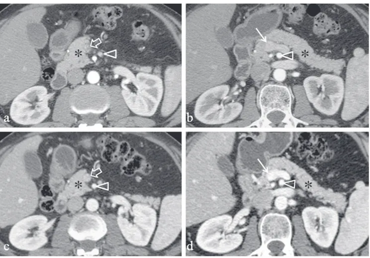
A representative case of pancreas with normal cont-rast enhancement at SB-MDCT is shown in Figure 2. In our 37 patients, whole-body SB-MDCT in a single-pass provided a radiation dose in mSv ranged from 12.14 to 27.01 mSv (average 19.3±6.3 mSv) with a reduction of approximately 17% with respect to
stan-Figure 2. 60-year-old man with normal pancreas. Pancreatic parenchymal phase during triphasic (a,b) mDcT and combined arterial-venous phase of the split-bolus (c,d) mDcT technique. Pancreatic parenchymal phase (a,b) shows pancreatic parenchyma (*) which is 114.6 Hu compared with 141.2 Hu of combined arterial-venous phase of the split-bolus (c,d). on combined arterial and venous phase of split bolus (c,d) mDcT, there is also greater enhancement of the superior mesenteric artery (arrow head in c,d) and mesenteric vein (arrow in c) and portal vein (thin arrow in d).
dard triphasic MDCT in our experience. A potentially dose reduction can be obtained when compared with an unenhanced acquisition, and SB of the upper abdomen. Our results were lower than those repor-ted in the literature25.
dISCuSSIoN
MCDT is the procedure of choice in the identifica-tion of pancreatic malignancy6 and other unusual
tumors7,8. CT, in particular, is the method of choice
in the assessment of normal pancreatic parench-yma and in the detection and staging of pancreatic adenocarcinoma9,12,20. The majority of standard CT
protocols9,11,12,31-34, entails an unenhanced
acquisiti-on followed by pancreatic parenchymal phase (PPP), portal venous phase (PVP) and delayed phase. This combination of contrast phases, although per-formed with different CT scanners, flow rates and vo-lumes of contrast material, provides perfect conspi-cuity of pancreatic tumor, its proximity to vessels and detection of metastases. PPP during the standard bi-or triphasic CT, is the most sensitive phase fbi-or the evaluation of pancreatic parenchyma9,20. An average
volume of 135 mL of contrast material delivered at a flow rate of 4 mL/sec, provides a pancreatic enhan-cement ranging from 82.2 HU to 122 HU (average 105.7 HU)9.
Nevertheless, multiphase CT also exposes the pati-ent to very high radiation doses.
Recently, to reduce the radiation exposure and to maintain diagnostic value of CT and its image quality, split-bolus (SB) contrast injection with spectral MDCT imaging of the normal pancreas and pancreatic ade-nocarcinoma has been proposed25.
As radiologists we need to reduce the radiation ex-posure, due to its known risks, without deterioration of the image quality and, most importantly, mainta-ining diagnostic efficacy of the CT scan. Among CT protocols for pancreas evaluation, the aim is to ob-tain an optimal pancreatic enhancement to identify
normal pancreatic parenchyma and to maximize the attenuation difference between the tumor and the surrounding pancreatic parenchyma.
In addition, assessment of peripancreatic vessels, lymph nodes and detection and characterization of focal liver lesion are needed.
Our aim is to evaluate the feasibility of SB 64-multidetector row CT compared to PPP of the stan-dard MDCT scan protocol in the evaluation of the en-hancement of the normal pancreatic parenchyma. In the study of Brook et al.25, using CT unit with
spect-ral imaging, SB spectspect-ral CT was combined with panc-reatic and portal venous phases in a single scan thro-ugh two contrast material injections separated by a short pause: 70 seconds before CT, 100 mL of cont-rast materials were injected for the PVP, followed by a second injection of 40 mL of contrast material app-roximately 35 seconds later, to boost the pancreatic phase. Using the bolus-tracking technique scanning started 15 seconds after aorta attenuation reached 280 HU. Mean values of enhancement of normal pancreatic parenchyma for SB and standard protocol in PPP were 212.2 HU±64.7 vs 105.1 HU±29.3 res-pectively and the mean effective dose by SB was 20 mSv25.
Our results, with respect to the normal pancreatic en-hancement, differ from those of Brook because our study included patients of different ages and weights. Besides, we did not use CT unit with spectral imaging and, finally, injections of two contrast materials were not separated by a short pause and flow rate and vo-lume of contrast materials were different.
These results have encouraged the use of SB-MDCT technique in the evaluation of the pancreas becau-se this improvement in enhancement can enable a greater conspicuity and visualization of the pancre-atic neoplasm and other pancrepancre-atic abnormalities. SB-MDCT also provides an accurate evaluation of the peripancreatic arterial and venous vessels and lymph nodes, that is essential to stage pancreatic cancer. In
addition synchronous phases (arterial and parench-ymal hepatic enhancement during PVP) provide an optimal evaluation of both pancreas and liver (ie de-tection of hypervascular and hypovascular metasta-ses). A further advantage of the SB-MDCT protocol is reduction of the effective dose of radiation compa-red to bi- or triphasic MDCT technique.
A potential reduction of radiation dose down to 60% should be obtained if the SB-MDCT protocol was used in the study of the upper abdomen. This is im-portant for example in the follow-up of the patients with necrotizing pancreatitis, in whom it is necessary to rescan them 7-10 days later, to evaluate size, ex-tension and characteristics of the postnecrotic fluid collections.
Study limitation: our study included a small number of patients with different ages and body weights wit-hout pancreatic abnormalities, who had undergone whole body CT in the follow-up for malignant tumor. In conclusion, these preliminary results demonstra-ted the effectiveness of SB 64-section MDCT scanner in the evaluation of the pancreas. The advantages of SB-MDCT protocol in the study of the pancreas are its ability to provide an optimal enhancement of the normal pancreatic parenchyma that allows detection of the tumor, in addition to the possibility to minimi-ze radiation dose and number of images and data to be stored.
ReFeReNCeS
1. Manganaro L, Scialpi M, Piscioli F, et al. MRI prenatal diagno-sis of genitourinary abnormalities in a case of inconclusive ultrasonography. J Obstet Gynaecol 2016;36(6):762-763. https://doi.org/10.3109/01443615.2016.1157154
2. Manganaro L, Vinci V, Giancotti A, et al. Bi-parametric mag-netic resonance imaging applied to obstetrics. J Obstet
Gyna-ecol 2017;17:1-3.
https://doi.org/10.1080/01443615.2017.1281237
3. Pusiol T, Scialpi M. Role of computed tomography in the pre-operative diagnosis of giant benign solitary fibrous tumor pleura. Lung India 2013;30(1):82-5.
https://doi.org/10.4103/0970-2113.106128
4. Pusiol T, Zorzi MG, Morichetti D, et al. Uselessness of per-cutaneous core needle renal biopsy in the management of small renal masses. Urol Int 2011;87(1):125-6.
https://doi.org/10.1159/000328195
5. Pusiol T, Zorzi MG, Morichetti D, et al. Uselessness of radi-ological differentiation of oncocytoma and renal cell carci-noma in management of small renal masses. World J Urol 2013;31(4):1013-4.
https://doi.org/10.1007/s00345-011-0693-0
6. Scialpi M, Cagini L, Pierotti L, et al. Detection of small (>2 cm) pancreatic adenocarcinoma and surrounding parench-yma: correlations between enhancement patterns at trip-hasic MDCT and histologic features. BMC Gastroenterol 2014;14:16.
https://doi.org/10.1186/1471-230X-14-16
7. Scialpi M, Schiavone R, Basilicata A, et al. Radiation dose reduction thanks to split-bolus multi-detector computer to-mography (MDCT) in children with non-thoracic neuroblas-toma. Pediatr Blood Cancer 2015;62(10):1865-6.
https://doi.org/10.1002/pbc.25567
8. Scialpi M, Pierotti L, Gravante S, et al. Split-bolus versus trip-hasic multidetector-row computed tomography technique in the diagnosis of hepatic focal nodular hyperplasia: a case report. J Med Case Rep 2014;8:425.
https://doi.org/10.1186/1752-1947-8-425
9. McNulty NJ, Francis IR, Platt JF, et al. Multi-detector row he-lical CT of the pancreas: effect of contrast-enhanced multip-hasic imaging on enhancement of the pancreas, peripancre-atic vasculature, and pancreperipancre-atic adenocarcinoma. Radiology 2001;220:97-102.
https://doi.org/10.1148/radiology.220.1.r01jl1897
10. Brennan DD, Zamboni GA, Raptopoulos VD, Kruskal JB. Comprehensive preoperative assessment of pancreatic ade-nocarcinoma with 64-section volumetric CT. Radiographics 2007;27(6):1653-66.
https://doi.org/10.1148/rg.276075034
11. Hollett MD, Jorgensen MJ, Jeffrey RB Jr. Quantitative evalu-ation of pancreatic enhancement during dual-phase helical CT. Radiology 1995;195:359-361.
https://doi.org/10.1148/radiology.195.2.7724753
12. Graf O, Boland GW, Warshaw AL, et al. Arterial versus portal venous helical CT for revealing pancreatic adenocarcinoma: conspicuity of tumor and critical vascular anatomy. AJR Am J
Roentgenol 1997;169:119-123.
https://doi.org/10.2214/ajr.169.1.9207510
13. Lu DSK, Vedantham S, Krasny RM, et al. Two-phase helical CT for pancreatic tumors: pancreatic versus hepatic phase enhancement of tumor, pancreas and vascular structures.
Radiology 1996;199:697-701.
https://doi.org/10.1148/radiology.199.3.8637990
14. Choi BI, Chung MJ, Han JK, et al. Detection of pancreatic ade-nocarcinoma: relative value of arterial and late phases of spi-ral CT. Abdom Imaging 1997;22:199-203.
https://doi.org/10.1007/s002619900172
15. Boland GW, O’Malley ME, Saez M, et al. Pancreatic-phase versus portal vein-phase helical CT of the pancreas: optimal temporal window for evaluation of pancreatic adenocarcino-ma. AJR Am J Roentgenol 1999;172:605-608.
https://doi.org/10.2214/ajr.172.3.10063844
16. Lu DSK, Reber HA, Krasny RM, et al. Local staging of pancatic cancer: criteria for unresectability of major vessels as re-vealed by pancreatic-phase, thin-section helical CT. AJR Am J
Roentgenol 1997;168:1439-1443.
https://doi.org/10.2214/ajr.168.6.9168704
17. Keogan MT, McDermott VG, Paulson EK, et al. Pancreatic ma-lignancy: effect of dual-phase helical CT in tumor detection and vascular opacification. Radiology 1997;205:513-518. https://doi.org/10.1148/radiology.205.2.9356637
18. Raptopoulos V, Steer ML, Sheiman RG, et al. The use of he-lical CT and CT angiography to predict vascular involvement from pancreatic cancer: correlation with findings at surgery.
AJR Am J Roentgenol 1997;168:971-977.
https://doi.org/10.2214/ajr.168.4.9124153
19. Tabuchi T, Itoh K, Ohshio G, et al. Tumor staging of pancreatic adenocarcinoma using early and late-phase helical CT. AJR
Am J Roentgenol 1999;173:375-380.
https://doi.org/10.2214/ajr.173.2.10430140
20. Fletcher JG, Wiersema MJ, Farrell MA, et al. Pancreatic malig-nancy: value of arterial, pancreatic, and hepatic phase ima-ging with multi-detector row CT. Radiology 2003;229:81-90. https://doi.org/10.1148/radiol.2291020582
21. Scaglione M, Pinto A, Romano S, et al. Using multidetector-row computed tomography to diagnose and stage panc-reatic carcinoma: the problems and the possibilities. JOP 2005;6:1-5.
22. Yamada Y, Mori H, Matsumoto S, et al. Pancreatic adeno-carcinoma versus chronic pancreatitis: differentiation with triple-phase helical CT. Abdom Imaging 2010;35:163-171. https://doi.org/10.1007/s00261-009-9579-7
23. Yoshikawa J, Matsui O, Kadoya M, et al. Delayed enhance-ment of fibrotic areas in hepatic masses: CT-pathologic cor-relation. J Comput Assist Tomogr 1992;16:206-211.
https://doi.org/10.1097/00004728-199203000-00006 24. Ichikawa T, Erturk SM, Sou H, et al. MDCT of pancreatic
ade-nocarcinoma: optimal imaging phases and multiplanar refor-matted imaging. AJR Am J Roentgenol 2006;187:1513-1520. https://doi.org/10.2214/AJR.05.1031
25. Brook OR, Gourtsoyianni S, Brook A, et al. Split-bolus spectral Multidetector CT of the Pancreas: Assessment of Radiation Dose and Tumor Conspicuity. Radiology 2013;269:139-148. https://doi.org/10.1148/radiol.13121409
26. Scialpi M, Palumbo B, Pierotti L, et al. Detection and charac-terization of focal liver lesions by split-bolus multidetector-row CT: diagnostic accuracy and radiation dose in oncologic patients. Anticancer Res 2014;34(8):4335-4344.
27. Bae KT. Intravenous Contrast Medium Administration and Scan Timing in CT: Considerations and Approaches. State-of-the-Art. Radiology 2010;256:32-61.
https://doi.org/10.1148/radiol.10090908
28. Erturk SM, Ichikawa T, Sou H, et al. Effect of duration of cont-rast material injection on peak enhancement times and valu-es of the aorta, main portal vein, and liver at dynamic MDCT with the dose of contrast medium tailored to patient weight.
Clin Radiol 2008;63(3):263-71.
https://doi.org/10.1016/j.crad.2007.02.024
29. Heiken JP, Brink JA, McClennan BL, et al. Dynamic incremen-tal CT: effect of volume and concentration of contrast mate-rial and patient weight on hepatic enhancement. Radiology 1995;195:353-7.
https://doi.org/10.1148/radiology.195.2.7724752
30. Deak PD, Smal Y, Kalender WA. Multisection CT Protocols: Sex- and Age-specifi c Conversion Factors Used to Deter-mine Effective Dose from Dose-Length Product. Radiology 2010;257(1):158-166.
https://doi.org/10.1148/radiol.10100047
31. Yanaga Y, Awai K, Nakayama Y, et al. Pancreas: Patient Body Weight tailored Contrast Material Injection Protocol versus Fixed Dose Protocol at Dynamic CT. Radiology 2007;245:475-482.
https://doi.org/10.1148/radiol.2452061749
32. Kondo H, Kanematsu M, Goshima S, et al. MDCT of the panc-reas: optimizing scanning delay with a bolus-tracking tech-nique for pancreatic, peripancreatic vascular, and hepatic contrast enhancement. AJR Am J Roentgenol 2007;188:751-756.
https://doi.org/10.2214/AJR.06.0372
33. Goshima S, Kanematsu M, Kondo H, et al. Pancreas: optimal scan delay for contrast-enhanced multi-detector row CT.
Ra-diology 2006;241:167-174.
https://doi.org/10.1148/radiol.2411051338
34. Schueller G, Schima W, Schueller-Weidekamm C, et al. Mul-tidetector CT of Pancreas: Effects of Contrast Material Flow Rate and Individualized Scan Delay on Enhancement of Panc-reas and Tumor Contrast. Radiology 2006;241:441-448. https://doi.org/10.1148/radiol.2412051107