DIPARTIMENTO DI SCIENZE ECOLOGICHE E BIOLOGICHE
DOTTORATO DI RICERCA IN GENETICA E BIOLOGIA CELLULARE
XXV CICLO
Red blood cell ageing in vivo and in vitro:
the Integrated omics perspective
Settore scientifico disciplinare: BIO/11
Candidato
Coordinatore del corso
Tutor
David Mitchell
I would like to dedicate this thesis to my friends and colleagues. I could not ever be able to even
scratch the surface of red blood cell biology without the invaluable contribution and the team
work of my friends and colleagues, Barbara Blasi, Federica Gevi, Gian Maria D’Amici, Maria
Giulia Egidi, Valentina Longo, Cristina Marrocco, Cristiana Mirasole, Leonardo Murgiano
Valeria Pallotta, Sara Rinalducci, Anna Maria Timperio, Valerio Zolla (strictly in alphabetical
order!!!). I love you, nakamas! With you I shared my hopes and my despair, my will and desires.
Wish you all the best!
I would also like to dedicate this thesis to Dr. Grazzini and the Italian National Blood Centre,
since they believed in me when I was but a M.Sc. graduate, full of hopes and void of the rest.
They filled that void with the passion for red blood cells, and that is why I owe you so much.
I would like to dedicate this thesis to my parents. In a country where social ladders have been
abated, a worker and a housewife never stopped fueling my hopes and pointing straight towards
the dream of a “three years old” boy, now closer to realization than ever before. I dedicate this to
their sacrifices and their silent suffering, constantly by my side.
I am grateful to destiny for allowing me to meet a handful of people that changed my life.
My friends, Marco, Giansante and Alessandro. You taught me that happiness can be found in the
smallest things, as Trilussa wrote: “C'è un'ape che si posa su un bottone di rosa: lo succhia e se
ne va. Tutto sommato, la felicità è una piccola cosa”
My “father in science”, Prof. Lello Zolla. I hope I repaid your bet on me. Together we traveled,
we invested, we planned, we worked hard. Very hard. You guided and protected me. Looking up
on you I found the example to follow. What I learned from you, paraphrasing Rabelais, could be
summarized as follows: “Science sans “passion”, n’est que ruine de l’
âme!”
Last but not least, I dedicate this thesis to the one who helped me the most, my soulmate. This is
for you:
“Souls cross ages like clouds cross skies, an' tho' a cloud's shape nor hue nor size don't stay the
same, it's still a cloud an' so is a soul. Who can say where the cloud's blowed from or who the
soul'll be 'morrow? … Our lives are not our own. We are bound to others, past and present, and
by each crime and every kindness, we birth our future.” David Mitchell
3
Chapter 1: Introduction
Contents
1.1 Introduction to Red Blood Cell storage: the clinical/biological question about storage quality and compromised safety and efficacy of long-stored erythrocyte concentrates
1.2 Introduction to the field of “Integrated Omics”: Proteomics, Metabolomics, Lipidomics and Interactomics and their application to Transfusion Medicine and Red Blood cell Biology relevant issues
The contents of this chapter represent a critical and updated re-elaboration of the following publications of the candidate:
1. D'Alessandro A, Liumbruno G, Grazzini G, Zolla L.
Red blood cell storage: the story so far. Blood Transfus. 2010;8(2):82-8.
2. Zolla L, D’Alessandro A. Proteomic Investigations on Stored Red Blood Cells. In Chemistry and Biochemistry of Oxygen Therapeutics: From Transfusion to Artificial Blood. 2010; Mozzarelli A. Editor; John Wiley and Sons Ltd The Atrium, Southern Gate Chichester, West Sussex, PO19 8SQ
3. Liumbruno G, D'Alessandro A, Grazzini G, Zolla L.
Blood-related proteomics. J Proteomics. 2010;73(3):483-507. 4. D'Alessandro A, Zolla L.
Proteomics for quality-control processes in transfusion medicine. Anal Bioanal Chem. 2010;398(1):111-24.
5. Liumbruno G, D'Alessandro A, Grazzini G, Zolla L.
How has proteomics informed transfusion biology so far? Crit Rev Oncol Hematol. 2010;76(3):153-72.
6. D'Alessandro A, Zolla L.
Pharmacoproteomics: a chess game on a protein field. Drug Discov Today. 2010;15(23-24):1015-23.
7. D'Alessandro A, Zolla L.
The SODyssey: superoxide dismutases from biochemistry, through proteomics, to oxidative stress, aging and nutraceuticals.
Expert Rev Proteomics. 2011;8(3):405-21. 8. Zolla L, D’Alessandro.
Shaking hands with the future through Omics application in Transfusion Medicine and Clinical Biochemistry. Preface
Blood Transfusion 2012; 10 Suppl 2:s1-3.
9. D’Alessandro A, Gevi F, Timperio AM, Giardina B, Zolla L.
Clinical Metabolomics: the next stage of clinical biochemistry Blood Transfusion 2012; 10 Suppl 2:s19-24.
10. D’Alessandro A, Zolla L.
Metabolomics and cancer drug discovery: let the cells do the talking Drug Discov Today 2012; 17(1-2):3-9.
11. Zolla L, D’Alessandro A.
Preface to the Special Issue Integrated Omics. J Proteomics 2012; doi: 10.1016/j.jprot.2012.10.007
4
1. Introduction to Red Blood Cell storage: the clinical/biological question about storage
quality and compromised safety and efficacy of long stored erythrocyte concentrates
Overview of this section
Keywords: red blood cell; storage lesion; blood transfusion; adverse effect; oxidative stress;
At the dawn of the RBC biopreservation-research era, the donor and the recipient were forced to lay side by side. A sequence of fundamental achievements, from storage solutions to plastic bags and additive solutions have lead to increase the shelf-life of stored RBCs. Cold liquid storage, enabling a 42 –day storage of RBCs, was early paralleled by long-term frozen storage, which potentially allows RBCs to be stored for more than three decades. Notwithstanding this, frozen storage has not hitherto found a broad diffusion due to its elevated costs for required facilities and trained personnel.Accumulating data over the years showed how the storage processes negatively affect the quality of RBCs, ultimately causing risks to the recipients. These risks are stressed on the critically ill patients.
Strikingly, one entire unit out of four meeting International criteria is rapidly removed from the circulation of the recipient only after 24 hours, owing to the senescent/apoptotic-like process which RBCs undergo during storage. Therefore, notwithstanding its long history, red blood cell storage is still a “work in progress”. Indeed, recent clinical retrospective non randomized trials have stressed the likely harmful potential and the reduced safety and effectiveness of long-stored red blood cells. However, conclusive data from prospective randomized studies are still missing and, although smoke could be seen on the horizons, it is still impossible to draw an unbiased conclusion on the presence of a burning fire nearby.
On the other hand, a growing body of molecular studies has been recently built which underlines the dramatic changes red blood cells undergo during prolonged storage. Although some are reversible (such as pH drop, 2,3-DPG and ATP consumption), other events such as fragmentation and aggregation occur, triggered by many factors, including oxidative damage. These changes irreversibly compromise the erythrocyte physiology and thus its functionality, survival and immunogenic/pro-inflammatory potential upon reinfusion to the recipients.
In this chapter, we summarize the recent past of the RBC biopreservation research, by focusing on a few milestones and pointing out future perspectives.
Moreover, RBC storage lesions will be briefly listed out. All these notions will contribute to depict a well-rounded portrait of the happenings at the molecular levels during RBC storage.In particular, reactive oxygen species seem to be the eligible trigger for these lesions and the main contributor to the final quality loss (both at the macroscopic and microscopic levels) of RBCs. Hereby we describe alternative storage protocols which have been proposed in order to overpass these hurdles, such as RBC anaerobic storage.
For the foreseeable future, the “quality issue” should become a top priority in the RBC biopreservation field and early attempts to prevent storage lesions appear to be a preferable option.Definitive clinical evidence is awaited to resume these whole observations under a unique question: the need for a new storage protocol. Whether this will become a priority, alternative storage strategies could represent a clue for a not-yet definitely posed question.
At the end of this thesis, upon evaluating currently allowed (either hypothermic and cryostorage) and recently proposed storage strategies (i.e. anaerobic storage), we will outline the necessity to wonder whether researchers should continue to pursue a longer storage or start focusing on a protocol to ensure a better one. In this view, we will also mention in this chapter the “antioxidant additive solution” perspective, which will be tested as well through Omics technologies within the framework of this PhD thesis project.
5
Red blood cell storage: the story so far
Red blood cells (RBCs) are still the most widely transfused blood component worldwide and their story is intimately intertwined with the history of transfusion medicine and the changes in the collection and storage of blood (Hess, 2006; Zimrin and Hess, 2009).
At present, the most widely used protocol for the storage of RBCs (for up to 42 days) is the collection of blood into anticoagulant solutions (typically citrate-dextrose-phosphate); red cell concentrates are prepared by the removal of plasma and, in some cases, also leukoreduction. The product is stored at 4 ± 2° C in a slightly hypertonic additive solution, generally SAGM (sodium, adenine, glucose, mannitol, 376 mOsm/L).
Despite this, a definitive protocol that reconciles long-term storage on the one hand and safety and efficacy of the transfusion therapy on the other is still the subject of intense debate and discussion. In fact, although the organisation of the blood system, through the achievement of self-sufficiency, currently enables ordinary requests of the transfusion 'market' to be met, in the case of a calamity, disaster, or emerging infections (Tinmouth and Chin-Yee, 2001; Liumbruno et al., 2008; Hess, 2009), or in particular periods of the year, local reserves can sometimes face a temporary shortage. There is still an underlying concern about the real need to store blood components for as long as possible in order to obtain a gradual increase in the interval between the donation and the transfusion, and how much this elastic time span can be prolonged without definitively compromising the quality of the product and, in the end, the recipients' health (Zimrin and Hess, 2009). Indeed, although the transfusion establishment initially pursued both objectives (product quality and prolongation of the storage period), recent retrospective studies (whose results are, therefore, weakened by all the statistical limitations of this type of analysis) (Tinmouth and Chin-Yee, 2001; Adamson, 2008; Steiner and Stowell, 2009; Zolla et al., 2009; Lelubre et al., 2009; Flegel, 2012; Grazzini and Vaglio, 2012;) have indicated the apparent irreconcilability of the two aims. These studies seem to suggest that the quality (in terms of safety and efficiency) of RBCs decreases in proportion to the time the storage period is prolonged. However, considerations about shortening the storage period have to be pondered in the light of the evident pitfalls on availability of erythrocyte concentrates (Flegel, 2012; Grazzini and Vaglio, 2012). Besides, it should be considered that although 92% of all RBC units that met release criteria actually find a recipient (Hess, 2006), the demand from organizations for longer and better storage has recently increased. Moreover, although the modern blood banking establishment keeps the pace with the current ordinary demand, it is nevertheless not tailored to meet the need for massive RBC supplies and rare blood-group units under extraordinary circumstances, such as in a calamity or a disaster (Tinmouth and Chin-Yee, 2001; Ramsey, 2008).
On the other hand, there is extremely convincing molecular evidence (Bennett-Guerrero et al., 2007; Bosman et al., 2008) which, together with the results of clinical studies (Wasser et al. 1989; Marik and Sibbald, 1993; Purdy et al., 1997; van de Edna and Bjerkeset, 1998; Zallen et al., 1999; Vamvakas et al., 1999; Vamvakas et al., 2000; Mynster and Nielsen, 2000; Mynster and Nielsen, 2001; Offner et al., 2002; Keller et al., 2002; Fernandes et al., 2001; Leal-Noval et al., 2003; Murrel et al., 2005; Hébert et al., 2005; Watering et al., 2006; Basran et al., 2006; Sakr et al., 2007; Koch et al., 2008; Yap et al., 2008; Leal-Noval et al., 2008; Weinberg et al., 2008), appears to confirm the preliminary conclusions regarding the likely poorer quality of red blood cells stored for a long time. However, the
6
statistical validity and methodological rigour, in terms of evidence-based medicine, of the clinical studies have recently been challenged, highlighting the need for prospective, double-blind, randomized studies, in like fashion to the one carried out by Walsh et al. (2004), which led the authors to conclude "the data did not support the hypothesis that transfusing red blood cells stored for a long time has detrimental effects on tissue oxygenation in critically ill, anaemic, euvolumaeic patients without active bleeding". The international scientific community now seems much more convinced of the need of prospective studies, since such studies, on large cohorts of subjects, are currently underway, including the Age of Blood Evaluation (ABLE) Study, the Age of Red Blood Cells in Premature Infants (ARIPI) Study, Red Cell Storage and Outcomes in Cardiac Surgery Trial, the Red Cell Storage and Duration Study (RECESS) (US Public Health Service, 2008; Lacroix, 2008; Koch, 2009; Bennet-Guerrero et al., 2009; Fergusson, 2010; Assmann, 2010).
The key point of the problem is probably the lack of universally accepted standard criteria that closely reflect the dramatic molecular changes that occur during prolonged storage of RBCs and which, simply put, would enable 'good' blood to be distinguished from 'no longer sufficiently good' blood (Cluitmans et al., 2012; Sparrow, 2012). The current standard requirements for patenting new additive solutions in the USA, and also suggested in the recommendations of the European Council, are essentially based on two parameters: the level of haemolysis (below the threshold of 0.8% at the end of the storage period, following the introduction of the "95/95" rule – Council of Europe, 2008; Hess et al., 2009) and a survival rate of the transfused cells of more than 75% at 24 hours after transfusion. This latter parameter can be assessed by measuring the half-life of RBCs labeled with 51chromium and/or 99technetium prior to transfusion (Peters et al., 1986). These parameters are, however, fairly general and easily affected by the considerable biological variability between donors, given that it is known that blood from some donors resists storage better than that from other donors (Moroff et al., 1984).
Haemolysis is an easier parameter to monitor. Typically, between 0.2 and 0.4% of RBCs stored in the presence of standard additive solutions are haemolysed after 5-6 weeks of storage, while prestorage leukoreduction halves the incidence of this phenomenon (Hess, 2002). These widely accepted and well established parameters do not, however, reflect the profound molecular changes that affect RBCs during their storage.
A brief list of the elements of the so-called "red blood cell storage lesion" includes (Bennet-Guerrero et al., 2007; D’Alessandro et al., 2010; Zolla and D’Alessandro, 2010):
(i) morphological changes,
(ii) slowed metabolism with a decrease in the concentration of adenosine triphosphate (ATP), (iii) acidosis,
(iv) decrease in the concentration of 2,3-diphosphoglycerate (2,3-DPG),
(v) loss of function (usually transient) of cation pumps and consequent loss of intracellular potassium and accumulation of sodium within the cytoplasm,
(vi) oxidative damage with changes to the structure of band 3 (Karon et al., 2009) (vii) and lipid peroxidation,
(viii) apoptotic changes with racemisation of membrane phospholipids and loss of parts of the membrane through vesiculation (Bosman et al., 2008).
7
Some of these changes occur within the first few hours of storage, for example, the decrease in pH or the increases in potassium and lactate; others, however, take days or weeks (Bennet-Guerrero et al., 2007). Together, these events risk compromising the safety and efficacy of long-stored RBCs, reducing their capacity to carry and release oxygen, promoting the release of potentially toxic intermediates (for example, free haemoglobin can act as a source of reactive oxygen species) and negatively influencing physiological rheology (through the increased capacity of the RBCs to adhere to the endothelium (Annis et al., 2005; Koshkaryev et al., 2009) or through their enhanced thrombogenic (Sweeney et al., 2009) or pro-inflammatory (McFaul et al., 2009) potential).
These observations at a molecular level were supported by the results of a series of clinical studies (albeit retrospective and not randomised). These studies appeared to show a relationship between the duration of storage and a proportional increase in adverse events in the transfused patients, although the data available are preliminary and the statistically more reliable studies that conform more closely with the gold standard criteria represented by evidence-based medicine are considered necessary by many (Hess, 2009) and are, indeed, underway (reviewed in Grazzini and Vaglio, 2012).
Clinical evidence of adverse effects following the transfusion of RBCs stored for prolonged
periods
Numerous clinical studies have been carried out throughout the world to identify a possible relationship between the duration of storage of RBCs, the changes observed at a molecular level and side effects in the transfused patients, in order to determine whether and, if so, to what extent RBCs stored for a long time lose safety and efficacy (Bennett-Guerrero et al., 2007; Bosman et al., 2008) which, together with the results of clinical studies (Wasser et al. 1989; Marik and Sibbald, 1993; Purdy et al., 1997; van de Edna and Bjerkeset, 1998; Zallen et al., 1999; Vamvakas et al., 1999; Vamvakas et al., 2000; Mynster and Nielsen, 2000; Mynster and Nielsen, 2001; Offner et al., 2002; Keller et al., 2002; Fernandes et al., 2001; Leal-Noval et al., 2003; Murrel et al., 2005; Hébert et al., 2005; Watering et al., 2006; Basran et al., 2006; Sakr et al., 2007; Koch et al., 2008; Yap et al., 2008; Leal-Noval et al., 2008; Weinberg et al., 2008). In 2009, Zimrin and Hess and Lelubre et al. conducted meticolous elaborations of the data from the studies published so far. Despite the intrinsic statistical limitations of retrospective, non-randomised studies, the results of such studies are undeniably useful if they are considered as a warning bell, albeit debatable, but not to be ignored, of a potential increase in the negative effects of the transfusion of RBCs in proportion to the duration of storage of the blood product in certain groups of patients such as those in intensive care (Marik et al., 1993; Purdy et al., 1997; Fernandes et al., 2001; Hébert et al., 2005; Taylor et al., 2006; Sakr et al., 2007), those undergoing cardiac Interventions (Wasser et al., 1989; Vamvakas and Carven, 1999; Vamvakas and Carven, 2000; Lel-Noval et al. 2003; van de Watering et al., 2006; Basran et al., 2006; Koch et al., 2008; Yap et al., 2008), those submitted to colorectal surgery (Edna and Bjerkeset, 1998; Mynster and Nielse, 2000; Mynster and Nielse, 2001), or traumatized patients (Zallen et al., 1999; Offner et al., 2002; Keller et al., 2002; Murrell et al., 2005; Leal-Noval et al., 2008; Weinberg et al., 2008).
8
The side effects described in these groups of patients following multiple transfusions of 'old' red cells are very varied, ranging from a decrease in gastric pH (Marik et al., 1993) to an increase in mortality rate (Purdy et al., 1997), from multiorgan failure (Zallen et al., 1999) to an increased incidence of pneumonia in patients transfused following aorto-coronary artery bypass (Vamvakas and Carven 2000 and 2001; van de Watering et al., 2006), from an increased susceptibility to infections (Offner et al., 2002) to major complications following heart surgery (Hébert et al., 2005; Basran et al., 2006; Koch et al., 2008), and from an increase in the duration of hospital admissions (Keller et al., 2002; Murrell et al., 2005) to the development of complications such as trasfusion-related acute lung injury (TRALI) (Silliman et al., 2005; Gajic et al., 2007).
It is, however, worth stating that given the current lack of irrefutable statistical proof, it cannot yet be concluded "there's no smoke without fire", to mention Steiner and Stowell (2009). In fact, it worthwhile to mention that a few years ago a series of retrospective, non-randomised clinical studies suggested a correlation between reduced efficacy of transfusions and lack of leukoreduction; the subsequent prospective, randomised studies did not, however, fully confirmed these observations (Vamvakas and Blacjchman, 2001 and 2007).
The storage of RBCs, however, until 2009 had not been the focus of prospective, randomised studies similar to those needed to market a new drug (Spiess, 2007). For this reason, although it has now been ascertained and widely accepted that something more or less irreparable occurs during prolonged storage of RBCs, it is currently impossible to conclude objectively and without preconceptions that these changes are accompanied by decreased efficacy and safety of the blood component.
Current research suggests that the RBC hypothermic storage lesions significantly influence the efficacy of transfusion since they are related to a worsened prognosis, increased oxygen affinity ultimately resulting in a reduced oxygen delivery capacity in tissues, proinflammatory and immunomodulatory effects, increased infections, multiple organ system failure, and ultimately, increased morbidity and mortality (Scott and Lecay, 2005). Indeed, while it is long known that storage has a negative effect on RBC oxygen delivery capacity (Valtis and Kennedy, 1954) there is a growing awareness around the potential hazards of allogenic RBC infusion, which may actually harm some recipients (Munoz et al., 2004; Rao et al., 2004; Rawn, 2008). On the oxygen delivery issue, a particularly telling experiment has been performed by Tsai and coworkers, who showed that when 25 percent of the circulating RBCs were replaced by RBCs stored for 28 days, microvascular flow was reduced by 63% and oxygen partial pressure in tissues was 3.5 mmHg against 14.4 mmHg when the RBCs were fresh (Tsai et al., 2004). Notably, systematic reviews of multiple randomized trials invited to reconsider and minimise the routine use of blood transfusion to maintain arbitrary hematocrit levels in stable patients with ischemic heart disease (Rao et al., 2004; Charles et al., 2006). The risk of serious complications dramatically rises when dealing with patients who are undergoing cardiac surgery (Koch et al., 2008).
Recent evidence supports an inflammatory mechanism in the development of atrial fibrillation which involves c-reactive protein inflammatory mediation (Anderson et al., 2004; Lo et al., 2005; Aviles et al., 2003). RBC transfusion modulates the inflammatory response to cardiac surgery by changing plasma concentrations of inflammatory mediators and augmenting the inflammatory response (Fransen et al., 1999).
9
One of the eligible criteria for a blood unit to be transfused is that RBC survival rate after re-infusion should be over the 75% threshold. This means that, at the end of the 42-day shelf life, a good-quality transfused RBC unit will contain up to 25% non-functional RBCs, whose removal by RES might be a basis for at least a transient immune depression (Kendall et al., 2000). It is striking that a patient receiving 4 full-term stored RBC units will benefit only of 3 of them while, statistically, an entire blood unit will end up to participate to the total reinfused volume though only providing inexorably and irreversibly damaged RBCs. Moreover, it is a fact that 70% of the re-infused RBCs are rapidly removed from circulation in the recipient after 3 days from the treatment, which definitely represents an alarming datum (Bratosin et al., 2002).
Contaminating WBCs and their by products present in the storage medium may affect and induce changes in RBCs, directly by consuming glucose needed by the RBC or indirectly by releasing bioreactive substances in the storage medium, which could endanger RBC integrity and functionality (Blajchman, 2006). Thus, besides clinical complication deriving from alterations on RBCs, many other clinical complications are related to bioreactive substances released by leukocytes in the storage medium of non-leukoreduced units, such as histamine, lipids, and cytokines, which may exert direct effects on the recipients. Cytokines known to increase during storage of RBC or platelets include interlukin (IL) 1 beta, tumor necrosis factor alpha (TNFα) and IL8. Both TNFα and IL8 are derived from WBCs and can potentially activate neutrophils. Several of the cytokines generated during storage including TNFα, IL1b, and IL8 have potent pyrogenic activity, can recruit neutrophils from the bone marrow and cause further release of cytokines 4. The effect of cytokines may account for some febrile transfusion reactions (Dwyre and Holland, 2008).
An increase in haemolysis and potassium leakage resulting from altered membrane permeability during storage has been also attributed to leukocyte enzymes such as elastase, collagenase and cathepsin G and/or activated neutrophils liberating toxic O2 species (Kriebardis et al., 2007). Enzymes and eicosanoids released by degenerating leukocytes
and platelets may be damaging to stored erythrocytes (Bell et al., 2000). Greenwalt et al. (1991) found that leukodepleted units of RBCs were significantly better preserved after 56 days of storage with a remarkable reduction of potassium, haemolysis and total vesicle membrane-protein shed and higher morphology scores in comparison to non-leukodepleted RBCs in the supernatant. Leukoreduction tends to reduce storage haemolysis by about 50% (Hess et al., 2002). In one situation where RBC recovery was compared directly between leukoreduced and non leukoreduced RBC stored in the same system, leukoreduction increased RBC recovery by 4% and reduced haemolysis from about 0.40% to 0.25% at 6 weeks (AuBuchon et al., 2006).
For these reasons, as previously mentioned, the removal of WBCs from RBC concentrates is now universally practiced in several European countries and Canada, and is widely used in the USA.
Until recently the clinical milieu and the academic setting have walked together through the trodden path of cold and frozen RBC storage, while few approaches have contemporary addressed the “quality deal”. It has been hitherto unclear whether what we could now do in terms of prolonged storage should be definitely done. In this chapter, we will try to shed light on this debated issue as well, by referring to the molecular changes at the membrane and metabolic level which RBCs undergo over storage duration (which will be further addressed through Omics
10
technologies in the present PhD thesis project. In order to better understand storage-associated changes to RBCs, it is worthwhile to describe the role and biochemistry of RBCs in vivo, a topic that will be indeed further addressed through Omics technologies inside this PhD thesis.
RBC ageing and metabolism in vivo
RBCs play a pivotal role in gas transport (i.e. oxygen and carbon dioxide) and a minor, but not less important, role in a range of other functions, such as transfer of GPI-linked proteins (Shichishima et al., 1993; Kooyman et al., 1995; Civenni et al., 1998) and transport of iC3b/C3b-carrying immune complexes (Schifferli et al., 1989).
In humans, the circulating mature RBC is the end stage of a developmental process which starts in the bone marrow, as hematopoietic stem cells differentiate to enucleated reticulocytes (Palis, 2008). After extrusion of nuclei and degradation of internal organelles and endoplasmic reticulum, reticulocytes emerge in the circulation, where they rapidly develop into mature RBCs (Koury et al., 2002; Pasini et al., 2006). Until the end of its life span of 120 ± 4 days, with 120 miles of travel and 1.7·105 circulatory cycles, the human RBC has successfully coped with a number of dangers, such as passages across narrow capillaries and splenic slits, periodic high turbulences and high shear stresses, along with extremely hypertonic conditions. Owing to its constant cytoskeleton rearrangement, RBCs are able to traverse passage ways as narrow as 1 µm in diameter, by changing their shape from a biconcave disc of 8 µm diameter to a cigar shape (Goodman et al., 2007). During the last decades, a plethora of studies sought to fathom the depths of the two dimensional meshwork of proteins called the spectrin membrane skeleton (Bennet, 1990; Bennet and Lambert, 1991; Lux, 1979; Marchesi, 1983; Agre, 1992; Agre and Cartron, 1991). These proteins lie on the cytoplasmic surface of the plasma membrane and give the RBC its properties of deformability (i.e. elasticity and flexibility) which represent the foundation of their successful journey. Spectrin, ankyrin, actin, band 4.1 and anion exchanger band 3 are the major protein actors of RBC deformability. The lack of internal organelles and nuclei intuitively eases protein complexity of RBCs (Goodman et al., 2007), making them an eligible target for early biochemical studies and for proteomic investigations, the latter recently gaining momentum (reviewed in D’Alessandro et al., 2010 and detailed in the next Chapter 2 of the Introduction).
Circulating RBCs undergo metabolic and physical changes associated with the process of senescence, viz membrane vesiculation, decrease in cell size, increase of cell density, alteration of cytoskeleton, enzymatic desilylation, and phosphatidylserine (PS) exposure just to mention few (Tannert et al., 1977; Clark and Shohet, 1985; Shinozuka, 1994). At the end of their life span, senescent RBCs are recognized and removed by the resident macrophages in the reticuloendothelial system (RES), mainly by Kupffer cells in the liver. It has been estimated that 5 million RBCs per second each day are endocytosed by RES macrophages (Bratosin et al., 1998).
More than one cause participates to the senescent/ageing process. Membrane and cytosolic proteins of RBCs are continuously stressed by oxygen radical attacks, which cause aminoacid modifications. Morphology, function and metabolism of RBCs suffer from continuous alterations matching with the cell winding its way through the circulatory system. Basically due to the lack of protein synthesis and inability to regenerate effete protein molecules, most notably enzymes, a multitude of alterations accumulate as the end of the RBC life span approaches. Neoantigens form from membrane proteins, especially through clustering of anion exchanger band 3 (Kay, 1993)
11
and haemoglobin (Hb) denaturation (Low et al., 1989). Both proteins are closely-related to gas transport (Hamasaki et al., 1996; De Rosa et al., 2007), cell homeostasis and shape (Jay, 1996), or to glycolitic metabolism (Low et al., 1993). Thus, physiologically fundamental proteins fail to fulfil their biological goal. Furthermore, these senescent antigens that appear on oxidatively-damaged old cells, radically accelerate RBC removal from blood flux through the activation of life-span immunoregulation mechanisms, via triggering macrophage erythrophagocytosis, complement deposition and Immunoglobulin G (Ig G) binding (Bosman and Kay, 1988; Turrini et al., 1991; Kay, 2005). Antibody binding induces major alterations in membrane organization as well as vesicle formation (Head et al., 2004; Head et al., 2005). Kupffer cells also remove RBC vesicles, with a major role for exposure of PS (Bosman et al., 2005). Taken together, all this data suggest more than a superficial resemblance between RBC ageing and apoptosis (Bosman et al., 2005). Particularly telling is the term “eryptosis”, coined by Lang’s group to identify that special form of apoptosis typical for the anucleated RBCs (Lang et al., 2008). Eryptosis is characterized by PS exposure, cellular shrinkage, membrane blebbing, ceramide formation, opening of cation channels, increase of intracellular Ca2+ activity, and activation of intracellular proteases such as μ-calpain, in the absence of hemolysis but ensuing into phagocytic recognition of exposed PS by a scavenger receptor on the macrophage (Lang et al., 2008). This mechanism is possibly a parallel pathway leading to RBC removal without passing through the Ig G mediation (Bosman and Kay, 1988; Turrini et al., 1991; Kay, 2005).
To counteract oxidative damages, the anucleated RBC, which is unable to synthesize new proteins, is equipped with protective enzymes fully adequate to sustain even excessive oxidative stress for limited time periods (D’Alessandro and Zolla, 2011). Indeed, it is not a coincidence that Goodman and Colleagues (2007), in their thorough review article mapping the RBC interactome, could point out a central core of proteins. They named it the Repair and Destroy Box after the activity of its protein constituents, which are involved in nascent protein folding or re-folding of denatured proteins (e.g. chaperonines, heat shock proteins, anti-oxidant proteins such as peroxiredoxins, catalases, glutathione peroxidases, superoxide dismutases).
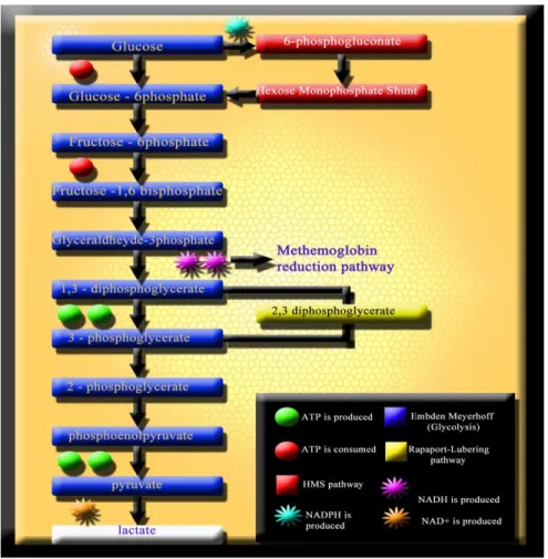
Together with membrane and cytoskeleton alterations, senescence also provokes metabolic anomalies in RBCs. Similarly, the energy-less RBC is inevitably lost (van Wijk and van Solinge, 2005). Because of the lack of nuclei and mitochondria, mature RBCs are incapable of generating energy via the (oxidative) Krebs cycle. Nonetheless, there are 4 RBC metabolic pathways (Figure 1): the Embden-Meyerhof pathway (glycolysis), in which most of the RBC adenosine triphosphate (ATP) is generated through the anaerobic breakdown of glucose; the hexose monophosphate shunt (HMS), which produces NADPH to protect RBCs from oxidative injury; the Rapoport-Lubering shunt, responsible for the production of 2,3-diphosphoglycerate (DPG) for the control of Hb oxygen affinity; and finally, the methemoglobin (met-Hb) reduction pathway, which reduces ferric heme iron to the ferrous form to prevent Hb denaturation (Wiback and Palson, 2002; Schmaier et al., 2003). Glucose, the only fuel utilized by mature RBCs, is primarily metabolized via anaerobic glycolysis. Following facilitated diffusion, glucose is immediately converted to glucose-6 phosphate. Glucose can be transformed to lactate via glycolysis, or to ribulose-5-phosphate via the oxidative section of HMS. Ribulose-ribulose-5-phosphate can re-enter glycolysis via the nonoxidative section of HMS. Under normal steady-state conditions, 92% of glucose is metabolized along glycolysis and 8% along HMS. Under oxidant conditions up to 90% of glucose can be metabolized along HMS. Net output of
12
glycolysis is 2 moles of ATP per mole of glucose metabolized. The main glycolytic pathway has two branching points: in the first one, the product of hexokinase, phosphate can be diverted to the HMS by glucose-6-phosphate dehydrogenase (G6PD). In the second branching point, 1,3-DPG can be diverted by DPG-mutase to produce 2,3-DPG.
Maintenance of the RBC membrane system and Hb function is dependent on energy generation through RBC metabolic pathways.
Five metabolic intermediates are particularly important in RBCs: ATP, DPG, NADH, NADPH and glutathione (GSH).
- ATP, the primary energy intermediate is essential to maintain electrolyte balance by powering sodium-potassium cationic pumps, which are necessary to preserve the cytoplasmic ionic milieu thus preventing colloidal osmotic lysis and, ultimately, conserving RBC shape and flexibility (Card, 1988); RBCs have an intrinsic program of cell death that is held in check by normal concentrations of RBC ATP. Normal ATP concentrations are necessary to prevent calcium-induced membrane loss by microvesiculation and for active transport of negatively charged phospholipids, specifically PS, from the outer to the inner leaflet of the RBC membrane to prevent RBC clearance from the circulation by macrophages (Kamp et al., 2001). ATP is an essential resource for other focal physiological activities: the synthesis of GSH and other metabolites; purine and pyrimidine metabolism; the maintenance of Hb iron in its functional, reduced, ferrous state; the protection of metabolic enzymes, such as Hb and membrane proteins, from oxidative denaturation; and the preservation of membrane phospholipid asymmetry (van Wijk et al., 2005).
- DPG, in association with pH and HCO3-/CO2 modulates position and shape of the oxygen dissociation curve
(Rouault, 1973; Salhany et al., 1973; meldon et al., 1983). When arterial blood arrives in peripheral capillaries, RBCs pass through the narrow capillaries one by one, CO2 is rapidly hydrated to H2CO3 inside RBCs by carbonic
anhydrase, and the H2CO3 promptly dissociates into H +
and HCO3 –
. Band 3 protein, the major integral membrane protein of RBCs, exchanges the cellular HCO3– with Cl– in plasma, a process that is conventionally known as the
‘chloride shift’. As the result of the anion exchange, the weak acid H2CO3 is converted to the strong acid HCl, and consequently the intracellular pH of RBCs is rendered acidic. This acidification is the trigger for the dissociation of O2 from oxyhemoglobin (HbO2), and the dissociated O2 is supplied to tissues that metabolically produce CO2. Protons formed in RBCs are accepted by the groups of deoxyhemoglobin (HbH+) participating in the ‘Bohr Effect’, and the pH within the RBCs is restored in order to prevent further dissociation of oxygen from HbO2. By means of the transient acidification triggered by the anion exchange activity, tissues producing more CO2 are supplied with more O2 from HbO2 (Hamasaki and Okubo, 1996). The rapid disappearance of 2,3-DPG from preserved blood has
not been of high concern because RBCs regain the ability to synthesize 2,3-DPG after transfusion. However, the restoration of 2,3-DPG in vivo requires up to 48 hours, and this period of altered oxygen affinity may be significant in certain clinical conditions (Valeri et al., 1971). Preserving RBC 2,3-DPG levels is therefore an essential element in maintaining the ex vivo quality of hypothermically stored RBCs. Inorganic phosphate is also added to the storage medium to act both as a buffer to the continuously decreasing pH and as a substrate for the synthesis of 2,3-DPG. - The ferrous iron of Hb is exposed continuously to high concentrations of oxygen and, thereby, is oxidized slowly to met-Hb, a protein unable to carry oxygen. To restore Hb function, met-Hb (methemoglobin also known as
13
ferrihemoglobin) must be reduced to Hb (ferrohemoglobin) (Salhany et al., 1973). Under physiological conditions, met-Hb reduction is accomplished mainly by red cell NADH-dependent cytochrome b5 reductase (NADH-methemoglobin reductase) so efficiently that there are insignificant amounts of (NADH-methemoglobin in the circulating blood (Abe et al., 1979; Borgese et al., 1993; Mansouri et al., 1993).
- Under oxidative stress, Hb could be oxidized to met-Hb (as it regards its heme iron) and to hemichromes, a variant of Hb in which cystein thiol groups have been dangerously oxidized to form denatured Hb-aggregates precipitating in inclusion bodies within RBCs, also known as “Heinz bodies”. GSH is the main protector of thiol groups, scavenger of oxides, peroxides, oxidant radicals and detoxicant of foreign compounds. Glutathione cycling from oxidized glutathione (GSSG) to the reduced form (GSH) is dependent upon NADPH generation, during the first two reactions of HMS, by via G6PD and 6-phosphogluconate dehydrogenase (6-PGD). NADPH is the substrate for GSH-reductase to regenerate glutathion after oxidant insults and protect catalase from inactivation (Untucht-Grau et al., 1981).
Oxidized glutathione (GSSG) + 2 NADPH + H+ → 2 GSH + 2 NADP+
- In concomitance with GSH-reductase, glutathione-S-Transferase, peroxidases (namely peroxiredoxins and glutathione peroxidases) and superoxide dismutase, catalase is believed to be very important in cellular antioxidant defence and therefore prolongs RBC life-span in mammals (Kurata et al., 1993). NADPH is also a protector of peroxiredoxin and catalase efficiency and an insufficient concentration of NADPH causes an impairement of the catalase-dependent detoxication route (Winterbourn, 1990).
FIGURE 1 RBC metabolism mainly gravitates towards four main pathways: the Embden-Mayerhoff glycolytic pathway (for ATP production), the methemoglobin reduction pathway, the Rapaport-Lubering pathway for 1,3-DPG conversion to 2,3-1,3-DPG and the Hexose Monophosphate Shunt (HMS) pathway (for NADPH generation). Whether NADH is not fully oxidized back to NAD+
through the methemoglobin reduction pathway, lactate is produced from pyruvate as a byproduct of anaerobic glycolysis.
14
Current storage protocolsErythrocyte biopreservation (Bp) is the ability to preserve the integrity of RBCs outside the native environment for extended periods. Its main end is to provide viable and functional RBCs for patients requiring a blood transfusion. The data provided in this section should contribute to help the readers glean insight of RBC physiology, which is essential to assess the effectiveness of a Bp approach, as well as the in vitro and in vivo quality of transfused RBCs. While at the dawn of the transfusion era donors and recipients were forced to lay side by side, owing to the practice of Bp in transfusion medicine they could be today separated in space and time (Hess, 2006). The development of effective RBC Bp-techniques that maintain ex vivo RBC viability and function has been experimented since the beginning of the 20th century (Rous and Turner, 1916) and paved the way for modern blood banking. Present approaches in RBC Bp will be described in this section, while their shortcomings and contraindications will be dealt with in the next one.
Current European and American guidelines for transfusable-RBC acceptability criteria (Council of Europe, 2008) primarily focus on the volume of blood collected and the proportion of viable RBCs present at the time of transfusion. These standards specify 450-mL collections and a mean 24-hour in vivo survival of at least 75% of the re-infused RBCs. In vivo survival of RBCs after reinfusion has been investigated since 1947, when radiolabeling experiments on erythrocytes were carried out for this purpose (Ross et al., 1947). However, it was only in 1985 that
51Chromium was introduced as a standard protocol for the follow-up of transfused RBCs (Ebaugh et al., 1985).
Other criteria include a threshold limit to the measured haemolysis value, which indicates the amount of free Hb, commonly not exceeding 1.0 percent of the RBC mass (Hogman and Meryman, 2006). Leukoreduction of white blood cells (WBCs) by centrifugation or filtration has become quite a routine practice in transfusion medicine almost worldwide [87-88]. In a comparative study between leukoreduced and non-leukoreduced RBC units, leukoreduction increased RBC recovery by 4% and reduced haemolysis from about 0.40% to 0.25% at 6 weeks (Heaton et al., 1994).
Leukodepleted RBCs should not contain more than 1 × 10⁶ WBCs per unit. A unit of RBCs should contain at least 45 g of RBC Hb and at least 40 g when leukoreduced. The European recommendations state that no more than 13% of a donor’s blood volume should be collected at one session.
Liquid storage of RBCs, usually referred to as cold or hypothermic storage, should be performed at approximately 4°C, a temperature definitely lower than the normal physiological temperature but higher than the freezing point of the storage solution (Council of Europe, 2008). Hypothermic preservation of RBCs is based on the principle that biochemical events and molecular reactions can be suppressed by a reduction in temperature (thermodynamically speaking, low temperature parallels a diminution of the free energy function). It has been estimated that storage temperatures ranging between 1 and 6°C minimize RBC degradation through an intense reduction of RBC metabolism, in the order of about 40 times (Hogman, 1998). Moreover, most bacterial organisms do not survive in the cold storage conditions, though a few such as Serratia marcesans, Yersinia enterocolitica, and Aeromonas species can grow at refrigerator temperatures (Brecher, 2005). Whereas they tend to grow slowly in cold blood, dividing about once a day, in approximately 27 days a single organism grows exponentially to generate up to 108 organisms, an overwhelming army at the cellular-scale which could be responsible of acute infections or endotoxic
15
shock. Many attempts have been performed to sterilely preserve RBCs, including heat sterilization, and plastic bags (Artz et al., 1954). The latter were seen as advantageous for military logistics because of their lighter weight and resistance to breakage. The ability to manufacture connected sets of bags enabled the design of a sterilized closed-collection system that decreased the rate of bacterial contamination from experimental surfaces. By the time they became approved commercial products in the 1960s, their vein-to-bag unitary construction, their ability to exclude air bubbles reducing the chance of air embolism during pressure infusion, small volume in refrigerator storage and optical clarity were all recognized as distinct advantages. In the field of plastic bags, the main improvement was the introduction of diethylhexyl phthalate (DEHP), the plasticizer used with polyvinyl chloride (PVC). DEHP plastic bags have been proven to reduce haemolysis by four fold at each weekly measurement (Hill et al., 2001), although the molecular mechanisms are yet to be uncovered.
Notably, the subsequent history of RBC storage has been characterized by slowly-progressing distinct cycles, each one needing roughly a decade to become well-rounded and finally encounter a widespread diffusion. Storage solutions and plastic bags are just two of the earlier but still most relevant steps (Hess, 2006).
Concerning the former, acid citrate dextrose (ACD) heat sterilizable solutions were introduced in the 1940s and enabled 3 weeks storage of RBCs (Loutit and Mollison, 1943). Citrate phosphate dextrose (CPD) solution, with 16 mM/l phosphate, increased the fraction of RBC recovered after 3 weeks of storage from ≈75% with ACD to > 79%, in the 1950s (Orlina and Josephson, 1969). In the first 2 weeks of storage, the 2,3-DPG is demolished to furnish the phosphate indispensable for the synthesis of ATP energetic tokens (Gibson et al., 1957; Hess et al., 2002). However, when all the 2,3-DPG is consumed, RBCs have no physiological way to contrast the energetic debacle. CPD solution was observed to replenish the phosphate supplies of stored RBCs.
In 1968 citrate phosphate dextrose adenine (CPDA-1) solution was developed and shown to permit a whole-blood extended storage for 5 weeks (Shields, 1969). CPDA solutions slowed adenine and adenosine lost due to deamination reactions while they improved osmotic fragility and recovery of RBCs (Simon et al., 1962). The main concern was about the safety of adenine, which was thought to lead to the formation of uric acid stones. That was the reason why CPDA solution licensure in the USA was delayed until 11 years later (Hess, 2006). Whole blood storage for 5 weeks yielded an average survival rate at 24h of ≈81%, while packed RBCs have a slight lower recovery percentage (72%) (Zuck et al., 1977)). It was concluded that the tighter the stored cells were packed, the more rapidly they ran out of glucose. However the addition of glucose dangerously raised the hematocrit and hampered a readily suitability of the unit for the administration to the recipient. Therefore, additive solutions (ASs) were engineered in order to provide additional volume and nutrients for longer storage and better flow of packed RBCs (Moore et al., 1980).
The first AS, saline, adenine and glucose (SAG) dramatically ameliorated and overpassed the high hematocrit and viscosity hurdles (Hogman et al., 1978)). SAG with the addition of mannitol (SAGM), chronologically the second AS, is now the standard AS used in Europe and in the USA, where two modestly differing variants are available (AS-1 and AS-5) (Hess, 2006). Mannitol works as a free radical scavenger, but also as a membrane stabilizer. In this respect, there is a significant likelihood that mannitol hampers haemolysis by preventing the osmotic swelling of RBCs that might otherwise increase their volume beyond their critical hemolytic volume (Beutler et al., 1988).
16
The third AS is AS-3. It is exclusively used in Canada, though being licensed in the USA. Again, it is based on SAG but also contains citrate and phosphate and a higher dose of dextrose. The citrate appears to serve the same membrane-protective and osmotic pressure-balancing function that mannitol serves in SAGM (Jarvis et al., 2003). AS-3 allows 6 weeks of storage and is associated with 78–84% recovery and 0.4% haemolysis.
Other ASs are adopted in some other countries (e.g. Circle Pack in Australia (Lovric, 1986) and MAP in Japan (Tanemoto et al., 1994)) and depend on a higher dextrose version of the primary and aforementioned CPD anticoagulant, thus they are called CP2D. Despite this, none of these ASs appears to markedly ameliorate the overall statistics regarding RBCs after storage when compared to the others (Hess, 2006).
All currently licensed ASs support the minimal 75%, 24-hour in vitro survival and 0.8% hemolysis standard criteria set by the American Association of Blood Banks and Council of Europe for up to 42 days of hypothermic storage at 1 to 6 °C (Hess et al., 2011). Notwithstanding this, current storage criteria are too general to depict a fully-detailed portrait of RBCs upon storage.
The pH conditions, which are also strongly associated with the RBC storage lesion, are affected by the volume and osmolality of the storage solution, as well as by the gas permeability of the storage container. Moreover, glycolysis slows as pH falls. As pH decreases and metabolism slows, RBC ATP concentrations reach a maximum higher and later than usual, resulting in a prolonged conservation of ATP levels exceeding the critical values that are necessary to suppress microvesiculation and PS exposure. The acidic pH of current ASs maintains ATP levels, but is detrimental to 2,3-DPG levels, which fall below 10% of the initial value by 3 weeks of storage (Valeri et al., 1971). Bicarbonate buffering is effective for the maintenance of acidic pH and ATP levels by driving the diffusion of carbon dioxide from PVC bags (Hogman, 1998). Some authors determined the effects on storage quality of ASs pH and volume, as well as of phosphate, sodium chloride and mannitol concentrations (Hess et al., 2001; Hess et al., 2003). The most complicated task is to find a delicate balance of the pH, which should not be too high in order to contrast 2,3-DPG generation with a consequent ATP depletion and, conversely, not too low, in order to prevent glycolysis to completely stop and hamper new ATP generation. ASs have an important part in this piece and ultimately contribute to the pH fine tuning.
For example, collecting whole blood into acidic CPD normally reduces its pH from ≈7.35 to ≈7.1 (Hess et al., 2002). Adding an acidic AS further reduces the pH to ≈ 7.0. However, if the pH of the AS is raised to 8.5 by adding disodium phosphate, then the resulting pH of the RBC suspending fluid can be raised to ≥ 7.2 at the beginning of storage. A pH of 7.2 at the beginning of storage means that the ATP production will suffice for several weeks (Hess, 2006). On the contrary, if the pH is raised above 7.2, then DPG is produced and consumes all of the intracellular phosphate, leading to a decrease in the ATP content that limits storage time and RBC quality.
Hogman and Meryman (Hogman and Meryman, 1999) proposed several practical procedures to extend the maintenance of RBC 2,3-DPG levels during hypothermic storage, including elevating the pH of ASs, increasing the volume of the additives, using hypotonic additives, and cooling the RBCs to room temperature after collection. Accordingly, several recent publications describe high pH storage solutions that preserve DPG content for many weeks (Hogman et al., 2002; Kurup et al., 2003; Murrell et al., 2005). Normally, the storage life of RBC is determined by the length of time it takes the cells to produce enough lactic acid and protons to reduce the pH to 6.5,
17
with a consequent low ATP production which no longer supports cell viability. This time is largely determined by the rate of production of protons and the buffer capacity of the suspension. Clearly, this buffer capacity can be increased by adding sodium bicarbonate to the additive solution. In solution, bicarbonate combines with a proton to produce carbonic acid, which is converted to CO2 and water by RBC carbonic anhydrase. The CO2 then diffuses out
through the plastic bag, effectively removing protons from the solution and slowing the rate of pH fall (Murrell et al., 2005). The combination of using alkaline additive solutions and bicarbonate buffering can effectively double the metabolic capacity of the storage system and allow RBC to be stored for longer and under better conditions (Hess et al., 2005).
ASs prospect a new deal in RBC storage and will hopefully integrate actual cold liquid storage protocols. Indeed, although hypothermic liquid storage drastically fades RBC metabolism, ageing and senescence seem to be exacerbated by the storage process, since a series of lesions accumulate as the erythrocytes sail across the cold and troublesome waters of their 42day hypothermic-storage odyssey. 4 RBC storage lesions
Storage lesions
While from a clinical standpoint there is only preliminary evidence, still to be confirmed, from the molecular point of view, the observations of changes that accumulate in red cells in proportion to the duration of their storage are numerous and indisputable, as described here. Although the average half-life of RBCs in the circulation is 120 ± 4 days (Palis, 2008), the standard maximum duration of storage of RCC is 42 days. This is because stored/transfused red cells seem to have a notably shorter half-life. In fact, 25% of the cell components are removed from the recipient's circulation within 24 hours of transfusion; in other words, of four units of red cells transfused, one is completely eliminated by the body already the day after the transfusion. There are probably two causes for this. The first, which is easily deducible, is that at the time of being donated, the unit of blood contains a percentage of already aged RBCs which, during storage, do nothing other than complete their aging process and are too old by the time of transfusion.
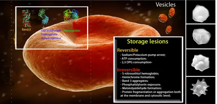
The second cause depends on the storage conditions, which are far from being normal, physiological conditions and which represent a greater and more long-lasting stress than the RBCs are able to counteract, despite their wellsupplied protein machinery ab origine. In fact, although RBCs are anucleated and, therefore, are devoid of an actual genome and consequently protein synthesis, they do have their own armamentarium of proteins devoted to protecting and maintaining pre-existing protein functions through a "central core" of chaperone proteins, heat shock proteins and proteins involved in the detoxification of free radicals (peroxiredoxins, catalases, glutathione peroxidases) whose role is critical in the economy of the RBC proteome (the protein complement of the genome) and interactome (the system of protein-protein interactions) (D’Alessandro et al., 2010).
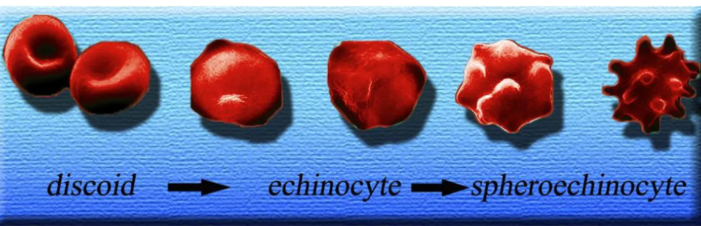
The most evident changes affecting RBCs during the storage period are alterations of the cell phenotype, which varies from a smooth discoid shape to a phenotype characterised by various membrane protrusions or spicula (echinocyte) and finally to a spheroid-shaped cell (spheroechinocyte) (Holme, 2005). The reversibility of these changes is inversely proportional to the duration of storage.
18
At the macroscopic level, RBC cold liquid storage induces a series of evident changes in the RBC shape (Berezina et al., 2007; Bosman et al., 2008) (Figure 2). During storage the erythrocyte shape visibly changes from a deformable discoid to a reversibly-deformed echinocyte to an irreversibly-deformed spheroechinocytes (Berezina et al., 2007). These changes are triggered by the irreversible loss of membrane through the formation of vesicles, which is the likely cause of an increased osmotic fragility, a reduced deformability and poor function after
FIGURE 2 Main phases of the macroscopic changes of RBC shape during storage: from a discoid shape to echinocyte and spheroechinocytes.
transfusion. Vesicles are commonly grouped under two distinct categories: microvesicles and nanovesicles. The former have a mean diameter of approximately 180 nm, while the latter have an approximate diameter of 80 nm (Salzer et al., 2002). Though deriving from the shedding membranes, microvesicles display a particular protein composition in that they contain a low number of membrane proteins, a high number of metabolic enzymes, and an elevated content of Hb (Bosman et al., 2008). These vesicles contain almost no integral membrane proteins or cytoskeletal components, with the exception of band 3 and actin. The protein composition of the nanovesicles is quite different from that of the microvesicles and of the RBC membrane, with a conspicuous large number of complement and immunoglobulin proteins (Rensing et al., 2001). Raft formation may be involved in vesicle formation (Salzer et al., 2002) and RBC storage has recently been shown to be associated with changes in the concentration of raft-associated proteins of the membrane fraction (Bosman et al., 2008). When examining the presence of the raft-associated proteins flotillin-1, flotillin-2 and stomatin Bosman et al. (2008) found out that, during storage, the RBC membrane content of flotillins and stomatin decreases, whereas the microvesicles become strongly (10-fold) enriched in stomatin, and that upon prolonged storage, the nanovesicles become 20-fold depleted of flotillins. These main changes are accompanied by a wide range of biochemical and molecular alterations. By a mere biochemical point of view, cooling below normal physiological temperatures inhibits metabolic processes and partially contrasts both the depletion of critical cellular metabolites and the accumulation of oxidative injuries. However, these benefits are counteracted by three effects: (I) the rate of met-Hb reduction by cytochrome b5 reductase is slowered; (II) met-Hb may be more prone to denaturation as suggested by lower thermodynamic stability of metmyoglobin at 4°C; and (III) the solubility of oxygen is doubled at 4oC. As a result, oxidative damage can accumulate with refrigerated red cell liquid storage (Zolla and D’Alessandro, 2011).
19
The storage lesion also involves the fluxes of sodium ions (massive entry into the cell) and potassium ions (exit from the cell), since the Na+/K+ pump is inactive at 4°C (Bennet-Guerrero et al., 2007). Although this is a reversible process (it takes 24 hours to restore the physiological gradients for sodium, and up to 4 days for potassium (Hogman and Meryman, 1999)), this phenomenon means that blood stored for a prolonged period should not be used for neonates or paediatric patients, unless first washed or the potassium removed from the storage medium (Klein et al., 2007).
Another biochemical effect is a clear decrease in the levels of 2,3-DPG (which is consumed already within the first week), translating into increased affinity of haemoglobin for oxygen and, consequently, decreased capacity of the RBCs to release oxygen according to local metabolic needs. The decrease in 2,3-DPG levels is also a reversible event, and completely normal levels can be restored within 3 days after the transfusion (Beutler et al., 1969). Contrary to senescent process, upon storage RBCs loose potassium, DPG, ATP and calcium stores. Moreover, RBCs undergo several changes including alterations in cellular membrane, shape changes, phospholipid content, phospholipid asymmetry and antigenic markers, while they become more rigid and demonstrate reduced oxygen off-loading (Rensing, 2001). RBCs become more acidotic and the suspending fluid has higher concentrations of free Hb and biologically active lipids and contains greater quantities of negatively charged microvesicles with pro-inflammatory and pro-thrombotic activity (Greenwalt et al., 1991; Ho et al., 2003; Bessos and Segatchian, 2005). The potassium loss is a consequence of the altered metabolic activity upon cooling, while the loss of DPG and reduced glycolytic activity provoke a decrease in pH levels. DPG is typically gone by the 10th day of RBC storage, whereas ATP concentrations initially increase, due to precox DPG breakdown, or are stable during the first 2 to 4 weeks of storage, with generally declining concentrations thereafter. New experimental solutions, such as CPD and other phosphate-containing ASs, are aimed to further delay the total ATP consumption (Hess, 2006). It is noteworthy that, at the end of the RBC life-span, its enzyme activities, ATP and other crucial metabolites are still present in sufficient amounts and do not justify RBC death. On the contrary, upon storage ATP and DPG are almost fully depleted and most of the membrane proteins are oxidized as well as lipids, while the cytosolic enzymes are only in part damaged.
The experimental evidence on the role of S-nitrosothiol-haemoglobin is, on the other hand, controversial. It was thought that reduced levels of this form of haemoglobin would be related to 'old' blood having a lesser vasodilatory effect in recipients (Bonaventura et al., 2007); however, recent molecular biology studies seem to suggest that this is not the case (Reynolds et al., 2007; Isbell et al., 2008).
Erythrocytes also undergo other irreversible damage, as exemplified by the haemolysis in the second half of the actual maximal blood bank storage period. Determination of the degree of haemolysis is currently based on the amount of extracellular Hb. Nonetheless, the detection of extracellular Hb is not the foremost and reliable criterion, since up to 50% of the extracellular Hb is contained within vesicles and could not be detected with routine approaches (Greenwalt et al., 1991).
HbO2 is potentially harmful as it promotes the generation of reactive oxygen species (ROS), putatively OH., after
Fenton’s reaction involving its haeme iron. During RBC storage, Hb becomes associated with the membrane fraction, mainly with the cytoplasmic domain of band 3 (Zhang et al., 2000), partially in a non-reducible,
cross-20
linked form (Wolfe et al., 1986). This association has been speculated to induce the generation of neoantigens that trigger immune recognition and removal of aged and/or damaged RBCs (Kriebardis et al., 2007).
Regarding membrane protein damages, it is well known that the etiology of lesions in RBC membranes is multifactorial, involving both ROS and proteolytic enzyme activity. Recently, in order to gather information on the time course of storage lesions, investigations on the relative contributions of oxidation and enzyme cleavage to this process and the fragmentation of RBC membranes have been documented by mapping the proteome changes over storage time.
Alongside these reversible changes, various irreversible events occur during the storage process, including fragmentation and aggregation of proteins and lipids, activated by radical species generated by prolonged, continuous oxidative stress (Wolfe, 1989; Racek et al., 1997; Sharifi et al., 2000). In this way oxygen constantly leaves one molecule of haemoglobin to bind to another. It is known that, occasionally, an oxygen leaving the haemoglobin molecule carries with it an electron, forming a superoxide ion (O2-) and (ferric) methaemoglobin. Normally, the methaemoglobin is reduced by cytochrome b5 reductase (Abe et al., 1979) and the superoxide is dismutated without consequences. However, during prolonged storage, the superoxide ion can interact with iron and water in a Fenton reaction, resulting in the formation of hydroxyl radicals capable of attacking and damaging both proteins and lipids, leading to their fragmentation and the formation of aggregates. For example, haemoglobin can be converted into hemichromes (haemoglobin whose cysteine residues have been oxidised, leading to the formation of aggregates). Eligible targets of the radical species generated in a cascade from the hydroxyl radical are membrane phospholipids (with the formation of lysophospholipids and malondialdehyde (Dumaswala et al., 1999)), and proteins within (or closely related to) the cell membrane, such as the band 3 ion exchanger (Kaon et al., 2009) (which plays a fundamental role in maintaining the oxygen transport function of RBCs (Hamasaki et al., 1996) and acts as an anchor for a series of key glycolytic enzymes (Low et al.., 1993; Weber et al., 2004)) and spectrin. These membrane alterations end up causing the previously-described echinocyte or spheroechinocyte phenotypes. Finally, it is known that the cell activates a process of vesiculation, in order to eliminate proteins and lipids that have been altered by oxidative stress, as to protect the cell from a further chain reaction of stress and consequent removal from the circulation (Willekenes et a., 2008). In fact, aggregates of band 3 appear at the membrane during both in vivo and in vitro aging (Willekenes et al., 2008; Karon et al., 2009), constituting membrane signals to "remove" the cell, through IgG- or complement-mediated phagocytosis by the recipients' Kuppfer cells. These membrane neoantigens, by stimulating the immune system, seem to be related to the onset of proinflammatory events, which are often harmful if not fatal in critically ill patients undergoing transfusion therapy (Tinmouth et al., 2001; Lelube et al., 2009). Alongside these signals, which are particular to red cell aging, a series of other markers appear; these markers are common in other physiological phenomena associated with programmed cell death or apoptosis, such as exposure of phosphatidylserine on the external leaflet of the lipid bilayer of the cell membranes, whose presence in microvesicles increases in proportion to the duration of storage (Bosman et al., 2005). This very same phenomenon of vesiculation through membrane protrusions (blebs) has contributed to strengthening the parallels between the processes of red blood cell aging and apoptosis (Bosman et al., 2008), leading Lang and colleagues (2006) to coin the term "eryptosis" to describe this physiological phenomenon, which is exacerbated during the