original research article
Introduction
The American Academy of Orofacial Pain de-fines “Craniomandibular Disorders” (CMD) as a heterogeneous group of clinical pathological conditions affecting the structures of temporo-mandibular joint, masticatory muscles and artic-ular disc (1).
CMD have a multifactorial etiology, with
differ-ent risk factors interacting at the individual level (2). CMD can be sometime associated with oral diseases (3-11).
The role of malocclusions as skeletal predisposi-tion to CMD disorders is a debated topic, still posing an unanswered question. Manfredini et al. in a systematic review of the literature on tem-poromandibular disorders and facial morphology (12) reported that only a single research group assessed the relation of specific cranio-facial
C
ORRELATION BETWEEN DENTO
-
SKELETAL
CHARACTERISTICS AND CRANIOMANDIBULAR
DISORDERS IN GROWING CHILDREN AND
ADOLESCENT ORTHODONTIC PATIENTS
:
RETROSPECTIVE CASE
-
CONTROL STUDY
D. DI VENERE
1, R. M. GAUDIO
2, A. LAFORGIA
1, G. STEFANACHI
1,
S. TAFURI
1, F. PETTINI
1, F. SILVESTRE
3, M. PETRUZZI
1, M. CORSALINI
11Interdisciplinary Department of Medicine (DIM) - Section of Dentistry, University “Aldo Moro” of Bari, Bari, Italy 2Department of Medical Sciences, University of Ferrara, Ferrara, Italy
3Departimento de Estomatologia, University of Valencia, Valencia, Spain
SUMMARY
Purpose. The aim of this retrospective case-control study was to identify, in a group of growing children and adolescents
affected by malocclusion, specific dento-skeletal characteristics which could be correlated to the onset, in the above-mentioned subjects, of craniomandibular disorders (CMD).
Materials and methods. Among the patients treated at the Paedodontics and Orthodontics department of Bari Dental School,
we recruited a group of patients with malocclusion and symptoms of temporomandibular disorders, as an experimental group. We considered as controls those patients who, match-paired to their skeletal class depending on the ANB angle, did not show any CMD sign or symptom.
Results. Of the 128 examined patients, 15 showed signs and/or symptoms of CMD (11.7%). When compared to 15
pa-tients non-affected by CMD, we could not detect statistically significant differences in both skeletal and occlusal charac-teristics. It is still interesting to notice how in CMD patients, characteristics of skeletal hyperdivergence are often to be found.
Conclusions. The present study seems to confirm that in growing children and adolescents, the presence of signs and/or
symptoms of CMD is not associable to a specific vertical skeletal growth pattern or to other specific occlusal character-istics.
original
research
article
characteristics with CMD in adolescents.
In young patients there are some difficult situa-tions (13-15) which require imaging investiga-tion (16).
The objective of the present retrospective case-control study was to identify, in a group of grow-ing children and adolescents affected by maloc-clusion, specific dento-skeletal characteristics we could correlate to the onset of craniomandibular disorders (CMD).
Materials and methods
The sample of this retrospective study included 128 patients (mean age: 12,7 years; 65 females, 63 males) treated at the Paedodontics and Or-thognatodontics department of University of Bari Dental School. Among these patients we selected as experimental group 15 patients with type I, II, III skeletal class malocclusions (measured by the ANB angle), who showed, after a gnathological evaluation, signs or symptoms of temporo-mandibular disorders according with the RDC/TMD (Reaserch Diagnostic Criteria for TemporoMandibular Disorders) (17). We consid-ered as controls 15 patients who did not manifest any signs and/or symptoms of craniomandibular disorders, and affected by the same skeletal sagittal malocclusion, in accordance with the matching of the ANB angle (considered as a matching criteria).
All the data and information relative to each re-cruited patient were inferred from their clinical history, examining the patient’s clinical records, radiographs and dental casts.
The inclusion criteria were as follows: age under 18; no history of orthodontic treatment; diagnosis of CMD; lateral X-rays with excellent contrast. The exclusion criteria were: incomplete clinical documentation; presence of systemic pathologies and conditions characterised by facial and/or cer-vical algia different from CMD (for example, fi-bromyalgia); previous gnathological and/or or-thodontic treatments and/or prosthetic rehabilita-tions; previous traumas and/or fractures.
We performed a clinical evaluation of cran-iomandibular disorders, according with the Euro-pean Academy of Craniomandibular Disorders (E.A.C.D.) protocol, consisting in:
• an anamnestic questionnaire;
• a clinical examination recording the subjec-tive perception of the pain with a Visual Ana-logue Scale (VAS) adequate to the patient’s age. The presence of vicious habits was also investigated;
• extent of the maximum opening. The presence of a limitation was recorded for maximum opening below 40 mm;
• with the same method (see above) we meas-ured the distance between the upper and low-er median line in latlow-erality and protrusion movements. For laterality the normal value ranges from 8 and 12 mm, while for protru-sion it is higher or equalling 6 mm;
• the opening deviation was evaluated consider-ing the path of the median line of the mandible during maximum opening, it was re-ported as present or absent;
• evaluation of “endfeel”;
• execution of the “joint muscle test”; • execution of the “joint play test”; • execution of the “compression test”;
• the masseter, the anterior, median and posteri-or region of the tempposteri-oral bone, the lateral and median pterygoid muscle, the muscles of the submandibular region, the neck and shoulders were digitally palpated, detecting the absence/ presence of pain;
• spontaneous or induced pain in the ATM was determined with the digital palpation of the joint, both at rest and in dynamic phase, dur-ing mandibular movements (18).
Lateral cephalograms for the craniofacial skeletal evaluation were traced on acetate sheets. The lin-ear and angular measures analysed are shown in Figure 1.
On dental casts we evaluated the extent of over-jet and overbite, with a millimetre callipers, and the presence/absence of anterior and/or posterior crossbite (unilateral or bilateral) and scissor-bite. For both overbite and overjet we considered pathologic values below 0 and over 2 mm.
original research article
For each recruited patient we collected data, in anonymous form, on an Excel sheet; these data were analysed with Stata/MP11 software. For dichotomous and category variables we eval-uated the absolute frequency, the proportions were compared with the chi-square test. In order to evaluate the association between variables, we calculated the odds ratio values, indicating the confidence intervals and using the z-score test. Continuous variables were described as mean, in-dicating the standard deviation and the median; medians were compared with the Wilcoxon-Mann-Whitney rank-sum test, analogue non-parametric of the t-student test for non-matched samples.
Results
Of the 128 examined patients, 15 subjects (10 boys and 5 girls) showed signs and/or symptoms of CMD (articular and/or myofascial unilateral or bilateral pain, click, reduced mouth opening -locking or functional limitation - and deviation of
the median line during mandibular movements); a percentage of 11.72%.
We selected our experimental sample after a gnathological evaluation, then we subdivided it according to the typology of symptoms and signs. Of the 15 patients, 10 complained myofas-cial and/or articular pain (at rest or during palpa-tion), 7 showed articular noises (click) and 3 had limited mouth opening. Of these, 1 showed pain and click simultaneously, 2 had pain associated with reduced opening and 1 had all 3 symptoms; these data are shown in Figure 2. Furthermore, 3 subjects had deflexion during opening and 2 re-ported two accessory symptoms like cephalea and otalgia.
The control group consisted of 15 patients (8 girls and 7 boys) chosen among those who did not show any sign and/or symptom of CMD, in accordance with the inclusion/exclusion crite-ria, were match-paired with the subjects of study for gender and age (approximately), and for skeletal class (precisely), depending on the correspondence of the cephalometric value of the ANB angle.
With the cephalometric measurement of the ANB
Figure 1
Angular and linear measure-ments used in the study. An-gular measures: SNA angle, SNB angle, ANB angle, SNP, SNA-GoGn, SN-GoGn, Ba-S-N, Pc-Go-Gn. Linear meas-ures: S-N, Ba-S, Ba-N, Sor-SNA, SNA-Me, Pc-Go.
original
research
article
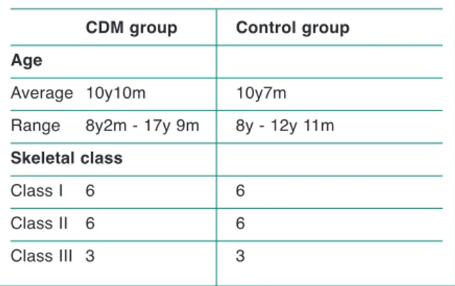
angle, our matching factor, the sample was divid-ed into the three skeletal classes: 6 class I sub-jects, 6 class II subjects and 3 class III subjects for each group, as shown in Table 1.
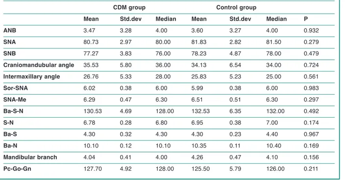
We radiographically analysed the skeletal charac-teristics of the entire sample and we evaluated the differences between the two groups calculat-ing the non-parametric correlation coefficient (Table 2).
We measured the angular and linear measures of the cranial base (Ba-S-N, S-N, Ba-S Ba-N), as
well as the gonial angle and the length of the mandibular branch and then we matched the two groups with the same methods, obtaining the same, non-significant, results.
In the same way we then matched the values of the craniomandibular and intermaxillary angle and the anterior vertical dimensions.
After this, we evaluated the divergence measuring the intermaxillary angle, observing that none of the patients showed the distinctive characteristics of hypodivergence, while 13 subjects resulted mesodivergent and 17 hyperdivergent; 9 of them belonged to the experimental group (Table 3). The ratio between the anterior vertical dimen-sions (Sor-SNA e SNA-Me) was evaluated in re-lation to the age of each subject. It emerged that only 3 subjects out of 30 showed a skeletal nor-movertibite, while 14 had a open bite ratio and 13 a deepbite one (Table 4).
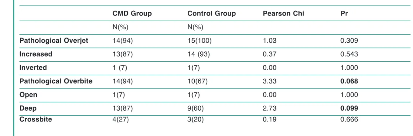
The dental patterns were evaluated on casts, cal-culating the extent of the overjet and the overbite and the potential presence crossbite and its typol-ogy (anterior, unilateral posterior, bilateral poste-rior, scissors-bite). Of 30 subjects, 7 had cross-bite, of whom 4 unilateral posterior crosscross-bite, 2
Figure 2
Distribution of symptoms in the experimental group.
Table 1 - Descriptive statistic of age and skeletal class
of study groups.
CDM group Control group Age
Average 10y10m 10y7m Range 8y2m - 17y 9m 8y - 12y 11m
Skeletal class
Class I 6 6 Class II 6 6 Class III 3 3
original research article
scissors-bite and 1 anterior crossbite, with simi-lar distribution in both groups. The overjet re-sulted to be normal in just one subject, whereas the vast majority showed an increased ratio (27 individuals); 2 patients had inverted overjet. In this latter case we could not detect significant
differences among the two samples.
A more interesting result, although just below the significance threshold, was the one relative to pathological overbite: this characteristic was in fact found with a higher frequency in the study sample (P=0.068), with a prevalence of the deep
Table 2 - Mean, standard deviation, median and coefficient of correlation between the two groups. CDM group Control group
Mean Std.dev Median Mean Std.dev Median P ANB 3.47 3.28 4.00 3.60 3.27 4.00 0.932 SNA 80.73 2.97 80.00 81.83 2.82 81.50 0.279 SNB 77.27 3.83 76.00 78.23 4.87 78.00 0.479 Craniomandubular angle 35.53 5.80 36.00 34.13 6.54 34.00 0.724 Intermaxillary angle 26.76 5.33 28.00 25.83 5.23 25.00 0.561 Sor-SNA 6.02 0.38 6.00 5.99 0.38 6.00 0.983 SNA-Me 6.29 0.47 6.30 6.51 0.51 6.30 0.297 Ba-S-N 130.53 4.69 128.00 132.53 6.35 132.00 0.492 S-N 6.78 0.28 6.80 6.95 0.38 7.00 0.174 Ba-S 4.30 0.32 4.30 4.30 0.23 4.40 0.967 Ba-N 10.10 0.12 10.10 10.35 0.11 10.40 0.169 Mandibular branch 4.04 0.41 4.00 4.26 0.47 4.10 0.156 Pc-Go-Gn 127.70 4.92 128.00 125.50 5.79 126.00 0.211
Table 3 - Test results of the chi-square relative to the divergence.
CMD group Control group TOT P Odds ratio 95% IC Divergence N(%) N(%) N(%)
Mesodivergence 5 (33.3) 8 (53.3) 13 (43.3) 0.269 2.286 0.42-12.95 Huperdivergent 10 (66.7) 7 (46.7) 17 (56.7)
Table 4 - Test results of the chi-square relative to the anterior vertical dimensions.
CMD group Control group TOT P Odds ratio 95% IC Anterior vertica
dimensions N(%) N(%) N(%)
Normovertibite 1 (6.7) 2 (13.3) 3 (10) 0.543 2.154 0.098-135.985 Openbite 7 (46.6) 7 (46.7) 14 (46.7) 0.599 2 1.081-134.579 Deepbite 7 (46.6) 6 (40) 13 (43.3) 0.522 2.33 0.093-156.989
original
research
article
typology (P=0.099), than in the control group (Table 5).
Discussion
The percentage of individuals affected by cran-iomandibular disorders resulted very low in our study, even when compared to the young age of the subjects: only 15 of the 128 patients (11.27%) had signs and/or symptoms of CMD. Interesting-ly, this value is comparable or even lower than what reported in literature in epidemiological studies carried out on subjects of the same age range, randomly selected in the population. Mag-nusson (19) estimated the prevalence of CMD in a population of children and adolescents around 14.10% between 12 and 15 years of age, and 5.11% between 5 and 11 years of age.
In our sample the presence of malocclusions does not seem to be a predisposing factor for the onset of temporomandibular disorders, contrary to what was strongly held in the past, at least in the age range we considered. The reason of this uni-formity of prevalence between CMD in patients with malocclusions and healthy subjects, could be explained by the young age of the selected subjects, thus confirming the theory according to which CMD symptoms emerge when the articu-lar structure adaptation threshold is overcome (12). The temporomandibular joint has in fact a
significant capacity for adaptation to masticatory micro-traumas (20); however, this capacity wears off in time since some predisposing factors like ligament laxity, incorrect posture, extreme psy-chic irritability or sensitivity, parafunctions and, of course, malocclusions or traumas concur to al-ter the existing balance and engender the pathol-ogy at issue. The adaptation mechanisms are more efficient in young subjects because of a higher elasticity of articular structures able to better stand mechanical stress. These mecha-nisms reduce their efficiency in time, because of modifications affecting structural components caused by ageing. It is in fact to be seen a de-crease in blood perfusion and, consequently, in the nutrient supply, together with an increase in rigidity and, in predisposed subjects, progressive tissue modifications due to chronic inflammation mediated by enzymes, bone morphogenetic pro-teins, free radicals and proinflammatory and no-ciceptive neuropeptides.
In our study, we evaluated the skeletal and dental characteristics of the examined subjects. Several recent studies (21-25), investigated the relation between dento-skeletal characteristics and tem-poromandibular disorders, based on the hypothe-sis that some morphostructural variables can pre-dispose or increase the risk to develop articular disorders.
For what concerns the cephalometric data, we could not find any significant difference between the two samples. These results are consistent
Table 5 - Test results of the chi-square relative to the pathological dental relations.
CMD Group Control Group Pearson Chi Pr
N(%) N(%) Pathological Overjet 14(94) 15(100) 1.03 0.309 Increased 13(87) 14 (93) 0.37 0.543 Inverted 1 (7) 1(7) 0.00 1.000 Pathological Overbite 14(94) 10(67) 3.33 0.068 Open 1(7) 1(7) 0.00 1.000 Deep 13(87) 9(60) 2.73 0.099 Crossbite 4(27) 3(20) 0.19 0.666
original research article
with those obtained by Gidarakou et al. (22). These Authors compared two groups of patients with normal articular morphology and correct position of the disc, having pain as the only dis-criminating factor. The fact that in this analysis there were not any discrepancies between the dento-skeletal characteristics of symptomatic and asymptomatic subjects, suggests that the algic symptomatology of CMD can onset independent-ly from ATM pathological alterations, which was confirmed by our study. This confers a higher im-portance to other variables composing the multi-factorial etiopathogenesis of these disorders, like those related to the subjective perception of pain (genetic, hormonal, central mediation mecha-nisms).
Different results were obtained by the same Au-thors (21, 23, 25) when the examined samples were composed of subjects affected by unilateral or bilateral disc dislocation, with or without re-duction, compared to healthy subjects. In these studies significant differences were found in the dimensions of the cranial base; linear dimensions were reduced in relation to controls, while the cranial base angle (Ba-S-N) was more obtuse. The experimental group also showed complete maxillary retrognathism (evidenced by SNA and SNB values lower the norm). The analysis of these results led us to ask if retromaxillism and retromandibulism, perhaps due to alterations in the cranial base, could predispose to the develop-ment of CMD or if, vice versa, these morpholog-ical modifications could be caused by a disc dis-location affecting the skeletal growth direction and type. The last hypothesis seems to be the most accredited, although the mechanisms pro-ducing the aforementioned modifications have not been clarified yet.
A difference was found in relation to the cran-iomandibular angle (between S-N and Go-Gn), being higher in subjects with disc dislocation, to-gether with a more obtuse gonial angle and a shorter mandibular branch, with consequent in-crease in the vertical dimension of the face third inferior. These data are consistent with the stud-ies reported in literature in the last years: the most recent tendency describes in fact the
hyper-divergent facial typology as the most frequently associated with articular pathologies, perhaps be-cause the retruded and post-rotated position of mandible could contribute to the determination of disc dislocation (26). Our study is consistent with this assumption since, although any signifi-cant difference between the experimental group and the control one were to be seen, it is still in-teresting to point out that any patient affected by CMD had hypodivergence, while 10 subject out of 15 (66.67%) had an increased intermaxillary angle and the typical facial features of hyperdi-vergent subjects.
These observations are in contrast with the past when the decrease in inferior vertical dimension and deepbite were believed to be the main factors responsible for craniomandibular disorders (27). Once again, it is evident that opinions vary much in time and in the type of study and depending on the samples; our knowledge in these matters is not sufficient yet, and it should born in mind that there are several unclear aspects about the rela-tion between occlusion, skeletal characteristics and CMD.
For what concerns the dental patterns, in our study the overjet resulted increased in nearly all patients, in fact the 86.67% of the experimental group (13 subjects out of 15), showed such sagit-tal discrepancy between the superior fronsagit-tal and inferior group. It is possible to correlate this data with the loss of the anterior guide, a factor which was reported by several authors in association with CMD as a cause of occlusal instability with consequent functional overload on articular structures. However, it is true that that this char-acteristics were also common in controls, evi-dence explained by the fact that our investigation was done on patients with orthodontic problems who could therefore be more likely to show al-teration of dental relations.
Although the role of malocclusions in the ae-tiopathogenesis of craniomandibular disorders has been considerably scaled down, the evidence persists, confirmed by several studies, that a cor-relation (even slight) between these two condi-tions exists. In recent literature, this fact is ex-plained with the development of an articular
or-original
research
article
temporomandibular disorders. The concurrence of these two factors could be determined by al-terations in mandibular morphology, potentially predisposing the development of an orthopaedic instability of the articular structures. This fact should be evaluated more in depth with further testing, nevertheless it suggests the need for fur-ther researches on ampler samples in order to in-dentify other possible associations.
The limit of our study was the limited sample size which prevented us from obtaining more sig-nificant results. This problem is due to the low incidence of craniomandibular disorders in young children and adolescent population and, consequently, to the limited number of affected subjects we could study.
Our future aim will be to widen the sample to ex-amine and to study the simultaneous presence of different morphological characteristics, whose association may be more significantly correlated to CMD.
References
1. De Leeuw R. Temporomandibular disorders. In: (eds. Quintessence). American Academy of Orofacial Pain. Orofacial pain: guidelines for assessment, diagnosis and management. Chicago: 2008.
2. Greene CS. The etiology of temporomandibular disor-ders: implications for treatment. J Orofac Pain. 2001;15(2):93-105.
3. Corsalini M, Di Venere D, Pettini F, Lauritano D, Petruzzi M. Temporomandibular disorders in burning mouth syndrome patients: an observational study. Int J Med Sci. 2013;10(12):1784-9.
4. Petruzzi M, Lucchese A, Lajolo C, Campus G, Lauri-tano D, Serpico R. Topical retinoids in oral lichen planus treatment: an overview. Dermatology. 2013;226 (1):61-7.
5. Petruzzi M, Campus G, Paparusso F, Lucchese A, Lau-ritano D, De Benedittis M, Serpico R. Analysis of plasma fibronectin levels in patients affected by oral lichen planus. European Journal of Inflammation. 2012;10(1):45-50.
6. Lauritano D, Bussolati A, Baldoni M, Leonida A. Scle-roderma and CREST syndrome: a case report in den-tistry. Minerva Stomatol. 2011;60(9):443-65. 7. Petruzzi M, Lucchese A, Campus G, Crincoli V,
Lau-ritano D, Baldoni E. Oral stigmatic lesions of
gastroe-thopaedic instability secondary tothe occlusal in-stability due to peculiar alterations in dental pat-terns. This is believed to be a risk factor for the anterior disc dislocation, as claimed by Manfre-dini (28, 29). It has been recently assumed that in predisposed subjects a “occlusal hypervigilance” mechanism is present, making these subjects more sensitive to occlusal modifications and in-evitably leading them to discomfort, pain and dysfunction. The occlusal hypervigilance theory can be envisaged in the wider hypothesis of the General Hypervigilance, according to which it is the perceptive habit that determines the subjec-tive amplification of a variety of unpleasant sen-sations, not just the painful ones, thus suggesting the involvement of central sensitisation mecha-nisms responsible for hyperalgesia (30).
The occlusal factor which is currently mostly as-sociated with the onset of temporomandibular disorders is the unilateral posterior crossbite, consistently with the theories of Michelotti and Tecco (31, 32). This type of crossbite seems to al-ter the occlusal balance necessary to keep a phys-iological relation between the different articular structures, this justifies the orthodontic correc-tion of this malocclusion to prevent the onset of CMD (33). In our sample, however, crossbite was equally distributed in both groups.
The only factor which resulted poorly correlated, even though it did not overcome the significance threshold, was the pathologic overbite, found with higher frequency in the study sample (P=0.068), with prevalence of the deep typology (P=0.099), compared to the control group. This could suggest a mild association between this characteristic and CMD, highlighting the need for studies on larger samples, as to increase the test sensitivity; this could show a clearer correla-tion between dental deepbite and articular disor-ders.
Interestingly, in our samples we often found the morphological characteristics of hyperdiver-gence in parallel with dental deep-bite. The si-multaneous presence of these two requisites in the same subject is not common. We could there-fore assume that the very association between these two elements could be the risk factor for
original research article
sophageal reflux disease (GERD). Rev Med Chil. 2012;140(7):915-8.
8. Petruzzi M, Lucchese A, Nardi GM, Lauritano D, Favia G, Serpico R, Grassi, FR. Evaluation of autofluores-cence and toluidine blue in the differentiation of oral dysplastic and neoplastic lesions from non dysplastic and neoplastic lesions: a cross-sectional study. J Bio-med Opt. 2014;19(7):76003.
9. Lauritano D, Petruzzi M, Di Stasio D, Lucchese A. Clinical effectiveness of palifermin in prevention and treatment of oral mucositis in children with acute lym-phoblastic leukaemia: a case-control study. Int J Oral Sci. 2014;6(1):27-30.
10. Lauritano D, Petruzzi M. Decayed, missing and filled teeth index and dental anomalies in long-term sur-vivors leukaemic children: a prospective controlled study. Med Oral Patol Oral Cir Bucal. 2012;17(6):e977-80.
11. Lucchese A, Guida A, Capone G, Petruzzi M, Lauritano D, Serpico R. Designing a peptide-based vaccine against Porphyromonas gingivalis. Front Biosci (Schol Ed). 2013;5(631-7).
12. Manfredini D, Segu M, Arveda N, Lombardo L, Sicil-iani G, Alessandro R, Guarda-Nardini L. Temporo-mandibular Joint Disorders in Patients With Different Facial Morphology. A Systematic Review of the Liter-ature. J Oral Maxillofac Surg. 2016;74(1):29-46. 13. Clementini M, Ottria L, Pandolfi C, Agrestini C,
Bar-lattani A. Four impacted fourth molars in a young pa-tient: a case report. Oral Implantol (Rome). 2012;5 (4):100-3.
14. Inchingolo F, Marrelli M, Annibali S, Cristalli MP, Di-palma G, Inchingolo AD, Palladino A, Inchingolo AM, Gargari M, Tatullo M. Influence of endodontic treat-ment on systemic oxidative stress. Int J Med Sci. 2014;11(1):1-6.
15. Gargari M, Lore B, Ceruso FM. Esthetic and function rehabilitation of severely worn dentition with pros-thetic-restorative approach and VDO increase. Case re-port. Oral Implantol (Rome). 2014;7(2):40-5. 16. Lione R, Pavoni C, Lagana G, Fanucci E, Ottria L,
Cozza P. Rapid maxillary expansion: effects on palatal area investigated by computed tomography in growing subjects. Eur J Paediatr Dent. 2012;13(3):215-8. 17. Dworkin SF. Research Diagnostic criteria for
Tem-poromandibular Disorders: current status & future rel-evance. J Oral Rehabil. 2010;37(10):734-43.
18. Dworkin SF, LeResche L. Research diagnostic criteria for temporomandibular disorders: review, criteria, ex-aminations and specifications, critique. J Craniomandib Disord. 1992;6(4):301-55.
19. Magnusson T, Egermarki I, Carlsson GE. A prospective investigation over two decades on signs and symp-toms of temporomandibular disorders and associated variables. A final summary. Acta Odontol Scand. 2005;63(2):99-109.
20. Favia G, Corsalini M, Di Venere D, Pettini F, Favia G,
Capodiferro S, Maiorano E. Immunohistochemical evaluation of neuroreceptors in healthy and patholog-ical temporo-mandibular joint. Int J Med Sci. 2013;10(12):1698-701.
21. Gidarakou IK, Tallents RH, Kyrkanides S, Stein S, Moss ME. Comparison of skeletal and dental mor-phology in asymptomatic volunteers and symptomatic patients with bilateral disk displacement with reduction. Angle Orthod. 2002;72(6):541-6.
22. Gidarakou IK, Tallents RH, Kyrkanides S, Stein S, Moss ME. Comparison of skeletal and dental mor-phology in asymptomatic volunteers and symptomatic patients with normal temporomandibular joints. Angle Orthod. 2003;73(2):116-20.
23. Gidarakou IK, Tallents RH, Kyrkanides S, Stein S, Moss ME. Comparison of skeletal and dental mor-phology in asymptomatic volunteers and symptomatic patients with unilateral disk displacement without re-duction. Angle Orthod. 2003;73(2):121-7.
24. Gidarakou IK, Tallents RH, Kyrkanides S, Stein S, Moss M. Comparison of skeletal and dental morphol-ogy in asymptomatic volunteers and symptomatic pa-tients with bilateral degenerative joint disease. Angle Orthod. 2003;73(1):71-8.
25. Gidarakou IK, Tallents RH, Stein S, Kyrkanides S, Moss ME. Comparison of skeletal and dental mor-phology in asymptomatic volunteers and symptomatic patients with unilateral disk displacement with reduc-tion. Angle Orthod. 2004;74(2):212-9.
26. Tallents RH, Stein S, Macher DJ, Katzberg RW, Mur-phy W. Predisposing and precipitating factors in tem-poromandibular disorders. Semin Orthod. 2012;18(10-29).
27. Lieberman MA, Gazit E, Fuchs C, Lilos P. Mandibu-lar dysfunction in 10-18 year old school children as re-lated to morphological malocclusion. J Oral Rehabil. 1985;12(3):209-14.
28. Manfredini D, Vano M, Peretta R, Guarda-Nardini L. Jaw clenching effects in relation to two extreme oc-clusal features: patterns of diagnoses in a TMD patient population. Cranio. 2014;32(1):45-50.
29. Manfredini D, Perinetti G, Stellini E, Di Leonardo B, Guarda-Nardini L. Prevalence of static and dynamic dental malocclusion features in subgroups of tem-poromandibular disorder patients: Implications for the epidemiology of the TMD-occlusion association. Quin-tessence Int. 2015;46(4):341-9.
30. McDermid AJ, Rollman GB, McCain GA. Generalized hypervigilance in fibromyalgia: evidence of perceptual amplification. Pain. 1996;66(2-3):133-44.
31. Michelotti A, Iodice G. The role of orthodontics in temporomandibular disorders. J Oral Rehabil. 2010;37 (6):411-29.
32. Tecco S, Festa F. Prevalence of signs and symptoms of temporomandibular disorders in children and adoles-cents with and without crossbites. World J Orthod. 2010;11(1):37-42.
original
research
article
33. Iodice G, Danzi G, Cimino R, Paduano S, Michelotti A. Association between posterior crossbite, masticatory muscle pain, and disc displacement: a systematic re-view. Eur J Orthod. 2013;35(6):737-44.
Correspondence to:
Dr. Rosa Maria Gaudio
Section of Legal Medicine and Public Health Department of Medical Sciences
University of Ferrara Via Fossato di Mortara 64B 44121 Ferrara, Italy