Università degli Studi di Ferrara
DOTTORATO DI RICERCA IN
"Biochimica, Biologia Molecolare e Biotecnologie"
CICLO XXVI
COORDINATORE Prof. Francesco Bernardi
Cellular activity of microRNAs dysregulated in breast cancer
Settore Scientifico Disciplinare: BIO/11
Dottorando Tutore Co-Tutore
Dott. Carlotta Zerbinati Prof. Giovanna Marchetti Dott. Stefano Volinia
1 Contents: Page: Abstract ………...………..4 1.Introduction ………...……6 1.1 Cancer ………..……..6 1.2 Breast Cancer ………..……...7
1.2.1 Breast Cancer development……….……..………..9
1.3 Classification of Breast Cancer ……….…….…..10
1.3.1 Immunohistochemical classification of breast cancer ……….……….10
1.3.2 Molecular Classification of breast cancer: intrinsic subtypes ………....……....11
1.3.3 Luminal A and Luminal B Breast Cancers ……….12
1.3.4 HER2+ Enriched ………12
1.3.5 Basal-like and Claudin-Low ……….13
1.3.6. Normal-like ………..13
1.4. Estrogen and estrogen receptor ……….14
1.5. Therapy for the different subtypes of breast cancer ……….16
1.5.1. ER-positive breast cancer: Endocrine Therapy ………17
1.5.2. Anti EGFR therapy ………..19
1.5.3. Anti HER2 positive therapy ……….19
1.6. Triple negative breast cancer : TNBC ………20
1.7. Breast cancer in men ……….20
1.8. Classification of non-coding RNAs ………..21
1.8.1. Introduction to miRNAs ……….22
1.8.2. Genomic organization of miRNAs ……….23
1.9. Current Model of Mammalian miRNA Maturation and Processing ………24
1.10. Molecular mechanisms of miR action ………26
1.11. MiRNA Classification ………..27
1.12. Experimental techniques for miRNA analysis ………28
1.12.1. Microarray ………..28
1.12.2. Next generation sequencing techniques ………29
1.12.3. Real time Quantitative PCR ……….30
1.13. MicroRNAs in Tumorigenesis ………31
1.13.1. Deregulation of miRNAs in cancer ……….31
1.14. MiRNAs as tumor suppressor ……….31
1.15. MiRNAs as oncogenes ………32
2
2. Materials and methods ……….35
2.1. Cell culture ………...35
2.2. Analysis of mutation in breast cancer cell lines ……….35
2.3. MicroRNA microarrays ……….36
2.4. Transient miRNA transfection ……….36
2.5. Proliferation assay ……….37
2.5.1.MTS assay ………37
2.5.2.Analysis of data ………37
2.5.3. Real-time cell proliferation assay ………..38
2.5.4. Real Time cell invasion assay ………38
2.6. Scratch wound healing assay ………39
2.7. Class comparison ………40
2.8. Total RNA isolation ………..40
2.8.1 Quantification of RNA ………41
2.9. Statistical analysis ………..41
Rationale ………42
3. Results and discussion ……….42
3.1. Genomic characterization of cell lines ………..42
3.2. Curves of growth for MDA-MB-231 and for MCF7 ………48
3.3. Efficiency of transfection ………..49
3.4. Viability regulation by miRNAs ………50
3.5 Effect of co-trasfection of selected miRNAs ………54
3.6. Viability regulation by prognosis-related miRNAs ...55
3.7. Wound healing assay ……….58
3.8. Real time measurement of cell invasiveness and cell proliferation ………60
3.9. mRNA profiling after miRNA treatment ……….63
3.10. Cross validation on breast cancer tumors from TCGA cohort ……….67
4. Discussion ……….68
5. Conclusion ………76
References ……….77
4
Abstract
Breast cancer is one of the major health problems worldwide and it is the second cause of cancer-related in women. Patients often develop resistance to the current therapies. For this reason, the identification of new specific clinical molecular markers and pharmacologic targets in cancer research is an ongoing challenge. Over the last years, microRNAs (miRNAs) have become one of the main subjects of study in the area of cancer genomics. They negatively regulate gene expression post-transcriptional by inhibiting translation and causing degradation of target mRNA. More than a thousand miRNAs exist in the human genome and each one can potentially regulate hundreds of mRNAs. By regulating the expression of target genes, miRNAs can have a tumor suppressor or oncogenic role. Therefore, miRNAs can play an important role in all the phases of cancer, like as initiation, progression, growth, apoptosis, invasion and metastasis.
In a previous study based on miRNA profiling, in different solid tumors, comprised breast cancer, and normal tissues, several miRNA were reported to be over- or down-regulated in solid tumors in comparison to normal tissues (Volinia et al. Genome Res. 2010). In other scientific reports other miRNAs were positively or negatively correlated with tumor progression (Volinia S. and Croce CM. PNAS 2013).
Starting from these literature data, we decided to analyze the in vitro effect of the administration of this tumor-related miRNAs in order to verify their effect on the viability and transcriptional regulation in breast cancer cell line, in order to gain experimental evidences on their actual involvement in the tumor illness.
Firstly, we have chosen 10 different cell lines of breast cancer origin (MCF-7, MDA-MB-231, MDA-MB-468, MDA-MB-361, SKBR3, T47D, BT474, ZR75.1, MDA-MB-453, HBL-100) and on 2 cell lines of breast normal epithelium (MCF10A and 184A1). Genomic analysis revealed presence of a complex panel of cancer-related mutations, and the cell lines were different in term of cell growth. We checked also the miRNA levels inside cell lines.
Two groups of primary solid tumor-related (23 miRNAs: miR-126*, let-7d*, 326, 320c , 302a , 222, 218, 210, hsa-miR-206, hsa-miR-203, hsa-miR-202, hsa-miR-181a , hsa-miR-142, hsa-miR-145, hsa-miR-143, hsa-miR-138, hsa-miR-130b , hsa-miR-126, hsa-miR-99a , hsa-miR-28-5p , hsa-miR-33b , hsa-miR-26b , hsa-miR-21) or progression-related (15 miRNAs: hsa-miR-9, hsa-miR-10a, 25, 27, 30a, 93, 103, 148b, hsa-miR-151, hsa-miR-301a, hsa-miR-328, hsa-miR-484, hsa-miR-615, hsa-miR-874, hsa-miR-1307) miRNAs were transiently transfected into cells, and viability was assessed. We were able to
5
experimentally identify two groups of miRNAs which were able to significantly increase or decrease cell viability. The miRNAs that showed to increase cellular viability in five out twelve cell lines were: miR-130b, miR-138, miR-210, miR-148b and miR-1307. On the other hand, mirR-93, miR-126 and miR-145 displayed a significant inhibitory effect on cell viability in five out twelve cell lines. These miRNAs were further investigated for their capacity to affect cell migration, cell invasion, and genome profiling.
The main outcome of our work has been the identification from a wide list of cancer-related miRNAs of few of them involved in the in vitro regulation of cell growth and invasion. As a first attempt to identify target genes commonly regulated in vivo and in vitro, we have bioinformatically identified, in a first not exhaustive screening, PTEN and DICER1 genes, which in vivo and in vitro negatively correlated with miR-210 and miR-130b, respectively.
6
1. Introduction
1.1. Cancer
Cancer is the result of many complex changes occurring in a “normal” cell, progressing through to malignant and potentially metastatic. The 6 hallmarks of cancer as outlined in Hanahan and Weinburg’s review (1) are shown below (Figure 1). Cells commonly become cancerous when they acquire irreparable DNA damage, changing the sequence of genes which code for important regulatory proteins. After replication the mutations are passed down to the next generation of cells which can lead to deregulated growth, tumor progression and invasion through the basement membrane.
Figure 1. The 6 hallmarks of cancer.
Accumulations of successive DNA mutations arise over the course of a person’s life time (somatic gene changes) which is why age is often a risk factor in cancer. Also, lifestyle factors such as diet, alcohol, stress and tobacco, have all been implicated in either causing or increasing the risk of cancer. The environmental exposure to chemical carcinogens and the ultra-violet rays of the sun have also been shown to result in DNA damage and result in cancer. There are examples of gene mutations that are hereditary for instance BRCA1 and BRCA2 which are tumor suppressor genes involved in DNA repair of double stranded breaks. Mutations in these genes can cause instability of the human genome. Women that have heterozygous germ-line mutations in BRCA1 or BRCA2 have a substantially increased risk of highly penetrative breast cancer and ovarian cancer. Patients that are positive for this mutation can chose to have pre-emptive mastectomies or preventative Tamoxifen treatment (2).
7
1.2. Breast Cancer
Breast cancer (BC) is a complex and heterogeneous disease, characterized by variant genetic alterations and distinct morphologic and molecular features. Despite common histopathological features at diagnosis, BC is noted for disparate clinical behaviors and patient outcomes. The heterogeneity observed among breast cancer reflects the now well accepted notion that is not just one disease, but that instead represents a collection of distinct neoplastic diseases of the breast and the cells composing the breast (3).The character and nature of this diseases can be made through traditional pathological and morphological examination, but only through molecular analyses can be appreciated the extent of diversity among breast cancer. Breast carcinomas can be divided into two major groups:
In situ Carcinomas - the tumor cells remain confined to the ducts or lobules and show no evidence of microscopic invasion into the surrounding breast stroma. There are two types of in-situ carcinoma; ductal and lobular, named according to the predominant cell type from which the tumour arises.
Invasive carcinoma - the tumor cells invade the breast stroma and have the potential to metastasize to distant sites. The invasive breast carcinomas consist of several histological subtypes; the commonest being infiltrating ductal adenocarcinoma (75-80%), followed in frequency by invasive lobular (10-15%), Mixed ductal-lobular (<5%), Inflammatory (2-3%), Colloid (2-3%), Tubular (<2%), Medullary (<2%), and Papillary (1%) 5. Rarer subtypes, including metaplastic breast cancer and invasive micropapillary breast cancer, all account for less than 5 percent of cases overall.
Breast Cancer is a major health problem in the United States and worldwide, it is now the second cause of cancer-related in women (second only to lung cancer) with approximately 40,030 death expected in 2013 for both men and women (or 39,620 deaths among women, representing 14% of all cancer-related deaths among women) and the vast majority of breast cancer-related deaths involve metastatic disease. Metastasis is a multistep process which consists of a cell: - detaching ang migrating out of the primary tumor site; - invading the basement membrane to enter the circulatory system (intravasation); - surviving cell detachment-mediated apoptosis (anoikis); - exiting circulatory vessels at the metastatic site (extravasation); and – adapt into a new environment and soil a metastatic tumor. Breast cancer is about 100 times more common in women than in men although males tend to have poorer outcomes due to delays in diagnosis. The American Cancer Society estimates that 232,340
8
new cases of invasive ductal breast cancer (IDC) and 64,640 new cases of ductal carcinoma in situ (DCIS) will be diagnosed among women in the United States in 2013. Invasive breast cancer accounts for 29% of all cancer diagnoses among women in the United States and 23% of all cancer diagnoses among women worldwide. In the worldwide there is an estimated 1,383,500 new cases of BC will be diagnosed in women in 2013 (4).As such, this disease represents one of the most serious and costly health issues. The incidence of breast cancer increases with age (around 80% of diagnosed cases are in women over the age of 50) and there are other risk factors; such as obesity, high socio-economic group, alcohol use, expression of BRCA1 or BRCA2, ethnicity, early menarche, and childbirth late in life. A family history of breast cancer in female relatives has shown to be an important predisposing factor. In women, the breast cancer susceptibility genes BRCA1 and BRCA2 are thought to account for most hereditary breast cancers. Mutations in these genes confer a 40% to 70% risk for developing breast cancer by age 70 years. Other molecular markers are important in the pathogenesis and prognosis of breast cancer in women, as C-erbB-2, p53, Bcl-2, cyclin D1, and epidermal growth factor receptor (EGFR). The c-erbB-2 protooncogene encodes for a transmembrane receptor of the tyrosine kinase family, which is closely related to EGFR. This protein is expressed in 20% to 30% of breast cancers and may be associated with a poor prognosis. P53 is a tumor suppressor gene that controls cell growth by inducing cell cycle blockade, apoptosis, and cell differentiation. P53 gene alterations are the most common single genetic abnormality in breast cancer and are present in approximately 30% of cases. Bcl-2 is a protooncogene that inhibits apoptosis and thereby promotes cell growth. In breast cancer, expression of Bcl-2 has been associated with favorable prognostic features. Cyclin D1 is involved in cell-cycle regulation and helps control the cell’s entry into S phase. In breast cancer this gene is oncogenic but appears to be associated with a favorable prognosis. Epidermal growth factor receptor is a transmembrane glycoprotein that is present in low levels in normal breast tissue and is overexpressed in 35% to 60% of BC. Over expression of EGFR is inversely correlated with estrogen receptor expression and may be a negative prognostic factor. Significant prognostic factors are also axillary lymph node status, tumor size, histological grade and hormone receptor status, as estrogen receptor, progesterone receptor, and ErbB2, these are used to decide the treatment options for the patient. The patient can be classed as “triple negative” where they do not express any of these 3 receptors.
9
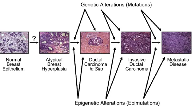
1.2.1. Breast Cancer development
Breast Cancer develops over a long period of time and requires multiple molecular alterations. The time required for the process of carcinogenesis is not well established for any human cancer, estimates suggest that this multistep process unfolds over many years and possibly several decades. Generally, sporadic breast cancer, in which there is no recognizable strong genetic component, emerge later in life (reflecting mostly postmenopausal breast cancer), whereas hereditary breast cancers occur earlier in life (reflecting the contribution of genetic predisposition) (5). The main hypothesis for the natural history of breast cancer development is stepwise progression from atypical ductal hyperplasia to ductal carcinoma in situ (DCIS), followed by evolution of this pre-invasive lesion to invasive ductal breast cancer (IDC) (Figure 2). DCIS is by definition noninvasive, but can vary from low-grade to high-grade lesions that may contain invasive elements. For this reason, DCIS, especially if high grade, is a risk factor for development of IDC. Many IDC are associated with adjacent DCIS lesions. It is not entirely clear if DCIS is a required precursor for development of IDC, but many IDC at the time of diagnosis are accompanied by DCIS, and there is consensus that DCIS in the absence of intervention will progress to invasive disease. In the progression of the disease might occur genetic and epigenetic alterations between the altered cells of the different morphological stages that contributed at the development of the disease (6). Since the late 1990s, invasive breast cancer have been characterized using gene expression analysis and classified on that basis into several molecular subtypes. Analyses of gene expression patterns between DCIS and IDC, have identified one correspondence in this two subtypes, this suggest that DCIS lesions are likely the direct precursors of invasive cancers. However, some recent molecular analysis suggest that the diversity of subtypes observed in invasive breast cancers derives from an evolution of low-grade to high-grade DCIS lesions.
10
Figure 2. Natural history of breast cancer development. Breast cancer develops from normal breast epithelial cells that evolve through atypical hyperplasia (and eventually dysplasia), DCIS, and invasive breast cancer. Multiple molecular alterations occur during this process, involving genetic and epigenetic alterations in precursor and neoplastic cells. Genetic predisposition can contribute to this process, but early molecular alterations (preceding DCIS) have not been well characterized. Original magnification, 20x. Modified from Rivenbark and Coleman (6),with permission from Elsevier.
1.3 Classification of Breast Cancer
1.3.1. Immunohistochemical classification of breast cancer
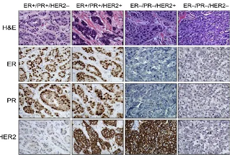
The morphological subclassification of invasive ductal carcinomas is accomplished by histopathological immunostaining to detect the differential expression of protein biomarkers to provide a clinical classification for breast cancer and dictates therapeutic approaches for treatment. Routine histopathological subclassification is made for detect the expression of estrogen receptor (ER), progesterone receptor (PR), and human epidermal growth receptor 2 (HER2; alias c-ErbB-2 or, in rodents, Neu), as well as HER1 and various cytokeratins (eg, CK5/6) (7). Approximately 70% of invasive breast cancers express ER, and the majority of ER+ cancers also express PR. Although the presence of normal PR levels suggests an intact ER signal transduction pathway in the breast cancer cells, discrepant ER and PR expression patterns (ER+/PR- and ER-/PR+) are sometimes observed, this may be attributable to false positive or false negative results, although technical improvements have reduced errors significantly. Collectively, the ER+ malignant neoplasms are classified as luminal cancers. These cancers are further subclassified based on their HER2 status and proliferation rate, giving rise to the ER+/PR+/HER2+ and ER+/PR+/HER2- subtypes. The ER- breast cancer are subclassified as HER2+ and triple negative based on HER2 overexpression or gene
11
amplification status, basal cytokeratin expression, and EGFR (HER1) expression, giving rise to ER-/PR-/HER2+ (HER2-enriched) and ER-/PR-/HER2- (triple negative) subtypes (Figure 3).
Figure 3. Clinical classification of invasive breast cancer based on expression of ER, PR, and HER2. Representative examples of invasive breast cancers that correspond to the general clinical classifications are shown. Cancer histology is depicted using H&E staining; expression of ER, PR, and HER2 is visualized using immunohistochemistry. Breast cancers are generally classified as positive or negative for hormone receptors ER and PR and for HER2, resulting in four major clinical groupings: ER+/PR+/HER2-, ER+/PR+/HER2+, ER-/PR
-/HER2+, and the triple-negative ER-/PR-/HER2-. Original magnification, 40x.
1.3.2. Molecular Classification of breast cancer: intrinsic subtypes
Perou and Sorlie and colleagues (8) studied gene expression profiling using DNA microarrays have grouped breast cancer into 5 distinct molecular classes or intrinsic subtypes named:
- Luminal A (ER+)
- Luminal B (ER+/HER2-enriched) - Basal-like
- HER2+-enriched - Normal-like
More recently, a further subtypes have been identified: - Claudin-low
Significantly, the molecular subtypes of breast cancer revealed by transcriptomic analysis are associated with different clinical outcomes. Although breast cancer classification methods
12
show good reproducibility, suggesting that these are robust biological subtypes, breast cancers that are not classifiable are identified with regular frequency (8).
1.3.3. Luminal A and Luminal B Breast Cancers
ER+ breast cancers occur most frequently and comprise two major molecular classifications: Luminal A and Luminal B.
Luminal A is the more abundant subtypes and accounts for 40-60% of breast cancer. Predominantly is ER+/PR+/HER2-, with low histological grade and low expression of proliferative genes. This subtype is associated with lower relapse rate and longer survival, compared with the other subtypes. The TP53 pathway is conserved and the management of this tumors is centered on antiendocrine therapies because this subtype is poor responsive to chemotherapy.
Luminal B subtype accounts for 15% of breast cancers, as luminal A is predominantly ER+ although it has lower expression of ER-related genes and it is histological high grade with variable PR and HER2 status. Luminal B tumors in contrast with luminal A are more aggressive, with poorer prognosis. The TP53 pathway is frequently inactivated. Although luminal B tumors are ER+, a significant number do not respond to antiestrogen therapy but show greater response to chemotherapy.
In general, the two ER+ breast cancer subtypes, Luminal A and Luminal B, are associated
with a good prognosis and excellent long-term survival, whereas the ER- subtypes (HER2+ and basal-like) are difficult to treat and are associated with poor prognosis.
1.3.4. HER2
+Enriched
The HER2 gene, also known as HER2/neu or c-erbB2, is located on chromosome 17q. It is a proto-oncogene, a normal gene with the potential to become an oncogene as a result of molecular alterations like mutation, amplification or overexpression of its protein product. HER2 is a member of the human epidermal growth factor receptor family, a family of tyrosine kinases, which normally regulate a series of cellular processes, such as proliferation and growth. HER2 is notable for its role in the pathogenesis of breast cancer and as a target of treatment. It is a cell membrane surface-bound receptor tyrosine kinase and is normally involved in the signal transduction pathways leading to cell growth and differentiation. HER2 is thought to be an orphan receptor, with none of the EGF family of ligands able to activate it. However, ErbB receptors dimerise on ligand binding, and HER2 is the preferential dimerization partner of other members of the ErbB family (9).
13
The HER2+-enriched subtype represent approximately 17% of all breast cancers. It is characterized by high expression of HER2 and related genes and it has high genomic instability. HER2 overexpression in breast cancer is associated with poor clinical outcomes, but it is also predictive of positive therapeutic responses to anti-HER2 drugs (eg, monoclonal antibody trastuzumab). HER2+ breast cancer are typically ER-, so the treatment for these cancers does not include anti-estrogenic hormonal therapies.
1.3.5. Basal-like and Claudin-Low
Basal-like and Claudin-low together represent subsets of triple negative breast cancers, lacking expression of ER and PR and also lacking amplification of HER2. These tumors represent 10% to 25% of breast cancers, they are highly proliferative and express genes characteristic of normal breast myoepithelial cells, such as cytokeratins 5, 6, and 17, and epidermal growth factor receptor (EGFR). They have a high rate of p53 mutations and are associated with increased genetic complexity. Not all tumors with basal-like gene expression profile are triple negative; 15% to 45% express ER and HER2 or both. The basal-like breast cancer have high rates of cell proliferation and extremely poor clinical outcomes. These cancers are associated with distinct risk factors, including early-onset menarche, younger age at first full-term pregnancy, and abdominal adiposity. Basal-like breast cancers have been shown to be over-represented in patients of certain age and ethnic groups, specifically young Black women (10). Claudin-low subtype breast cancer is enriched for markers of epithelial-to-mesenchymal transition (EMT) and stem cell-like and/or tumor-initiating cell features. Although these tumors are chemo responsive, they have shown a poor prognosis across several studies.
1.3.6. Normal-like
The normal-like represents 3% to 10% of breast cancers, it is frequently ER+ and has low levels of proliferative genes and low tumor cellular. The normal-like breast cancers are so designated because they tend to cluster closely with normal breast epithelium in microarray studies. It is not yet clear whether this is a distinct molecular subtype of breast cancer or simply a grouping of breast cancers that are not otherwise classifiable because of contaminating normal epithelium. Nevertheless, the prognosis of this group is reported to be intermediate, with better survival than all but luminal A breast cancers.
The identification of these molecular signatures has helped to understanding breast cancer and informed the search for novel therapies. However, this classification is a work in progress,
14
requiring refinement and standardization before it can be incorporated into clinical practice and decision making. Therefore, despite the progress and better understanding of the drivers of this disease, from a clinical management perspective, breast cancer remains divided into 3 therapeutic categories:
1. ER+ disease, which is targeted with antiendocrine strategies
2. HER2+ disease, which is treated with HER2-targeted agents
3. Triple negative breast cancer, which lacks validated targeted therapy options and is treated with traditional cytotoxic therapy
1.4. Estrogen and estrogen receptor
Estrogen is an important regulator in the development and progression of breast cancer and also in the development of normal breast. More than a century ago, Scottish surgeon George Beatson performed an oophrectomy and noted this procedure induced regression of breast cancer which was later proven to be due to a reduction in systemic estrogen levels (11). Estrogen mainly originates in the ovaries in pre-menopausal women, whereas in post menopausal women the main source is in the aromatization of androgens in adipose tissue. This accounts for the difference in therapeutic regimen between the two groups of women. Estrogen functions by activating two nuclear steroid receptors: ERα and ERβ. Both receptors bind estrogen and initiate gene transcription through ERE (estrogen response elements) in estrogen target tissues but have distinct functions and tissue distribution (12). In most breast carcinomas (~70%) ERα is highly expressed which results in the increased rate of proliferation without differentiation or apoptosis. ERα is exclusively epithelial (13, 14) whereas the distribution of ERβ in breast cancer is exclusively nuclear but expressed in multiple cell types (stromal fibroblast, endothelial and immune infiltrating cells) its expression has also been shown to correlate with an increase in aggression of the tumor (15). ERβ expression has been reported to range from 26% to 94%, the lowest of which was in a Japanese study, this implicates that the expression could be related to ethnicity. When Caucasian cohorts of patients were compared, the range was less extensive at 74%-94% (14). The expression of ERβ in the breast has been well described although its usual function, clinical value in carcinogenesis, and its relevance to the pathological diagnosis of breast cancer, is yet to be determined (14). Estrogen receptors that are not bound to ligand are inactive and usually sequestered in multi-meric protein complexes organized around the molecular chaperone heat shock protein 90 (HSP90). ER signaling pathways can be classed into 4 main mechanisms of action: Classical, ERE independent, ligand independent and
non-15
genomic (Figure 4). Classically, the cytoplasmic ER translocates into the nucleus upon ligand
binding. In the cytoplasm, the receptor dimerizes, transcription factors and co-activation proteins are recruited, and the target genes are then activated through an estrogen response element (ERE). Estrogen activates genes that are involved in survival and cell proliferation amongst other actions (16, 17). Estrogen bound ER interaction with Fos and Jun, which dimerize and become part of the activator protein-1 (AP-1) complex modulating gene expression. This ligand bound ER gene modulation can also occur with GC box bound specificity protein -1 (SP- 1) this is ERE independent modulation. ER can act independently of estrogen by being phosphorylated on multiple residues within the receptor after growth factor activation. The phosphorylation of ER leads to the dimerization of the receptor, DNA binding and ultimately activation of transcription (18). Estrogen can also activate membrane bound ER and cause a rapid estrogen signaling response through non-genomic activation (19). The non-genomic simulation of the estrogen receptor is a rapid response to the ligand binding and its response in the cell is independent of the gene transcription. Estrogen receptors act through a complex interplay of signaling cascades; such as insulin-like growth factor 1 receptor (IGF-1 R) (20), EGFR (21), G- proteins, Src, and PI3K.
16
Figure 4. A simplified view of the 4 mechanisms by which estrogen can exert its action upon a cell (roles and names of co-factors have been omitted). 1, Classical pathway: This is a genomic, ligand dependent mechanism; where estradiol (E2) bound dimers bind to the EREs in target promoter genes; this leads to an up regulation or down regulation of gene transcription and resulting in the estrogenic effects being seen in the cell. 2, ERE independent pathway: this is also a genomic, ligand dependent mechanism where estrogen bound ER dimers bind to alternative gene response elements such as AP-1 through Fos and Jun association and thus lead to gene transcription. 3, Ligand independent mechanism: this is a genomic ligand independent mechanism where the phosphorylation and subsequent activation of the ER, following growth factors and/or signalling molecules stimulation leads to target gene transcription. 4, the non-genomic ligand dependent mechanism; this is where the estradiol binds to a membrane associated receptor activating intracellular signalling generating rapid estrogenic effects in the cell.
1.5. Therapy for the different subtypes of breast cancer
Development of new therapeutics for breast cancer has resulted in new agents directed against certain molecular targets such as c-met (22), HSP90 (23), or angiogenesis related targets such as VEGFR2 (24), signalling pathways such as mTOR (25) all of which could prove to aid in treatment of the heterogeneous disease and a more unique patient treatment regimen. Presently, the majority of these new therapeutic agents are not clinically available and most are at preliminary laboratory stage of investigation.
17
1.5.1. ER-positive breast cancer: Endocrine Therapy
Patients with high levels of ER are treated with endocrine therapy (26). Endocrine therapy for breast cancer involves Selective ER Modulators (SERMS) which act as ER antagonists in breast tissue or aromatase inhibitors (AI) which work by inhibiting the action of the enzyme aromatase which converts androgens into estrogens (27).
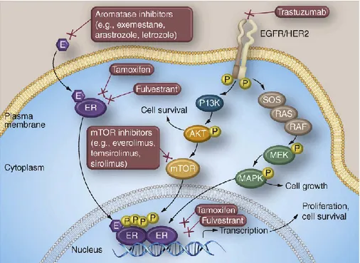
Targeting the ER is well established in the treatment of early and advanced ER+ breast cancer. Tamoxifen, a selective ER modulator, was approved in the late 1970 for the treatment of postmenopausal women with breast cancer. Later, its role in preventing disease recurrence was established, and tamoxifen was incorporated into adjuvant therapy, for all women with ER+ breast cancer (28). In the mid-1990, anastrozole was the first aromatase inhibitor (AI) developed for postmenopausal women with ER+. Other AIs, letrozole and exemestane, are applied in both the metastatic and adjuvant settings. The AIs have shown increased efficacy compared with tamoxifen, but their benefit is limited to postmenopausal women based on their mechanism of action, which causes a paradoxic estrogen surge in women with functioning ovaries. Fulvestrant, a selective ER downregulator, was subsequently developed for postmenopausal women with ER+ following antiestrogen therapy. In premenopausal women, tamoxifen remains the first antiendocrine strategy in both the adjuvant and metastatic setting. The role of ovarian suppression has been explored and in premenopausal women with metastatic breast cancer , the combination of tamoxifen with ovarian suppression improves survival (29). Despite these effective antiendocrine strategies, several patients develop treatment resistance. This may be caused by de novo resistance or acquired resistance shown by refractory disease following an initial period of response. Extensive efforts have been made to better elucidate mechanisms of resistance to identify potential therapeutic targets that may overcome this resistance. Signaling downstream of ER involves multiple pathways, including mitogen-activated protein kinase (MAPK), phosphatidylinositide 3-kinase (PI3K)/AKT/mammalian target of rapamycin (mTOR), epidermal growth factor receptor (EGFR), insulin like growth factor receptor (IGF-IR), fibroblast growth factor (FGF) receptor. Inibition of ER may cause up regulation of these alternative pathways, driving resistance to therapy (Figure 5).
18
Figure 5. Intracellular ER bound with their ligand estrogen, leads to cell survival and proliferation. Therapy with tamoxifen, AIs and fulvestrant blocks this signaling. However, there is a crosstalk between the ER pathway and other growth pathway as AKT, MAPK, EGFR, and activation of these pathways are implicated in resistance to antiendocrine therapy.
Endocrine resistance in breast cancer cells is associated with increased activity of PI3K/AKT (30). There are significant interest in targeting mTOR, a downstream mediator of this pathway. Inibition of mTOR using inhibitors developed for transplant immunotherapy, restored sensitivity to endocrine therapy in breast cancer cells. The combination of everolimus (an mTOR inhibitor) and exemestane (an AI) improved progression-free survival (PFS). There is also interest in targeting PI3K, the upstream driver of the PIK3/AKT/mTOR pathway. PI3K is commonly mutated in breast cancer and activation of this pathway has been implicated in acquired and de novo resistance to endocrine therapy. There is a crosstalk between ER and PI3K/AKT pathways; however they also signal independently, suggesting that dual pathway targeting may be required to optimize outcomes. There are trial that are studying the role of PI3K inhibitor BKM120 with fulvestrant in women who have progressive ER+ metastasis breast cancer (MBC) folloving an AI (NCT01610284). An ongoing trial is studying fulvestrant with or without GDC0941, an alternative PI3K inhibitor in a similar population. The outcome of these studies will further inform the role of targeting this pathway.
In addition to mTOR, over expression of the HER2 proto-oncogene has been clinically validated as a mediator of resistance to endocrine therapy. But this applies only to a small subgroup because only 10% of ER+ are also HER2+. HER2-driven resistance to antiendocrine
19
therapy is mediated via decreased ER level and increased ER phosphorylation, altered ER transcription, and activated downstream PI3K/AKT and MAPK pathways. Dual targeting of both ER and HER2 overcomes this resistance in preclinical models. Crosstalk between the HER2 and ER pathways is thought to drive endocrine therapy resistance. In ER+ HER2- cell lines with acquired resistance, lapatinib, an oral tyrosine kinase inhibitor of HER2, was able to restore sensitivity (31). In a study, women with ER+ HER2+ and metastatic breast cancer, the combination of anastrozole (AI) and trastuzumab, a monoclonal antibody to HER2, improved response rate (RR) and progression free survival (PFS) (32).
1.5.2. Anti EGFR therapy
EGFR is a member of the HER family of receptors, along with HER2.
Lapatinib is an oral small-molecule dual inhibitor of both EGFR (HER1) and HER2 kinases. Lapatinib exerts its antitumor effects by inducing growth arrest and or apoptosis, as well as by blocking downstream MAPK and AKT signaling pathways. Cetuximab is a monoclonal antibody that binds to the EGFR with high specificity blocking ligand-induced phosphorilation and activation of EGFR.
In preclinical models, EGFR contributes to endocrine resistance; but clinical trials targeting this pathway have had only moderate success. For metastatic breast cancer (MBC) ER+, the addition of gefitinib to anastrozole as first line therapy, improved PFS. However, gefitinib monotherapy show a clinical benefit rate of 54% in tamoxifen resistant MBC (33).
1.5.3. Anti HER2 positive therapy
The HER2 protein is over expressed and/or its gene is amplified in approximately 20% of invasive breast cancers, and it is associated with more aggressive biology, increased risk for progression of disease, and decreased overall survival (OS). The HER2 receptor is composed of an extracellular ligand binding domain, a single transmembrane domain and an intracellular domain with tyrosine kinase activity. Advances in translational science have led to the development of several therapies that target HER2, including the monoclonal antibody trastuzumab. This antibody selectively binds to the external ligand-binding domain, downregulates the ligand-independent HER2 dimerization and growth factor signaling cascades downstream of HER2, including the PI3K/AKT/mTOR pathway, thereby suppressing HER2 activity. In 2001 was first shown that trastuzumab have clinical benefit for patients that are HER2 positive, amplified or overexpressed, in a study of randomized trial the addition of trastuzumab to chemotherapy was associated with higher overall RR and
20
improved PFS and OS, in patients with MBC. Another therapeutic strategy for suppression of HER2 activity, is represents by inhibition of the tyrosine kinase domain with low-molecular weight tyrosine kinase inhibitors (TKI). Examples include lapatinib, gefitinib, erlotinib; however, many tumors either exibit de novo resistance to anti-HER2 therapy or acquire resistance over time, leading to disease progression and shortened survival for patients. More recently, novel agents with varying mechanisms of action have been described, and emerging data indicate that combinations of anti-her2 agents may overcome resistance (34).
1.6. Triple negative breast cancer : TNBC
Triple negative breast cancers represents the most problematic subtype with regard to effective management because there are no effective treatment targets. This type of cancer accounts for nearly 20% of all breast cancers. TNBC are tumors that do not express ER, PR, and HER2. TNBC is associated with younger age and more aggressive tumor type. In this subgroup the antiendocrine and anti-HER2 targeted therapies are ineffective, and traditional cytotoxic chemotherapy seems to be insufficient. Approximately 19.5% of triple negative patients carry BRCA mutations (35). Preclinical evidence suggests platinum-based therapy for TNBC and BRCA-1 associated malignancy is of benefit because it causes DNA cross-link strand breaks. In cells that lack homologous repair (ie, BRCA1 associated breast cancer), carboplatin or cisplatin have been hypothesized to have particular anticancer activity by leveraging the vulnerability of the cancer cell to DNA damage. In patients with triple negative breast cancers, the use of PARP inhibitors (polyadenosine diphosphate ADP-ribose polymerase) are novel strategy employed in clinical trials.
1.7. Breast cancer in men
Carcinoma of the male breast is a relative rare disease that accounts for less than 1% of all cases of cancer in men. The median age at diagnosis is 68 years compared with 63 years in women (36). The risk factors for breast cancer in men involve abnormalities in estrogen and androgen balance, which indicates that breast cancer in men, as in women, may be hormonally driven. An elevated risk has been seen in patients with undescended testes, congenital inguinal hernia, orchitis, testicular injury, infertility, and the Klinefelter syndrome, which is characterized by a 47, XXY karyotype, small testes, azospermia, and gynecomastia. Other possible risk factors that relate to hormonal levels include obesity, which causes increased peripheral aromatization of estrogens, and cirrhosis, which results in a hyperestrogenic state (37). Approximately 15% to 20% of male patients with breast cancer
21
have a positive family history (38). In women, the breast cancer susceptibility genes BRCA1 and BRCA2 are thought to account for most hereditary breast cancers (40% to 70% risk for developing breast cancer). In men, BRCA1 does not appear to be associated with a significantly increased risk for breast cancer, however, men with BRCA2 mutations are predisposed to breast cancer. All of the histological subtypes of breast cancer that have been described in women, have also been reported in men. Approximately 90% of all breast tumors in men are invasive carcinomas; the remaining 10% are noninvasive. Almost all of the noninvasive cancers are ductal carcinoma in situ. Carcinomas of the male breast have a higher rate of hormone receptor positivity than do carcinomas of the female breast when matched for tumor stage, grade, and patient age. 81% of breast cancers in men are ER+, and 74% are PR+. In contrast to women, men do not have a higher incidence of estrogen receptor positive tumors with advancing age (39). As in women, lymph node status, tumor size, histological grade, and hormone receptor status have been shown to be significant prognostic factors in men with breast cancer.
1.8. Classification of non coding RNAs
Non-coding RNAs (ncRNAs) are RNA molecules that do not function by encoding for proteins. They are loosely grouped into two major classes based on their size: small ncRNAs less than 200 nucleotides (nt), and long ncRNAs (lncRNAs) longer than 200 nt. LncRNAs are mRNA-like transcripts ranging in length from 200 nt to ~ 100 kilobases (kb) lacking significant open reading frames. Many identified lncRNAs are transcribed by RNA polymerase II (RNA pol II) and are polyadenylated. Although only a minority have been characterized in detail, lncRNAs participate in diverse biological processes through distinct mechanisms. LncRNAs have been implicated in chromosome dosage-compensation, imprinting, epigenetic regulation, cell cycle control, nuclear and cytoplasmic traffing, transcription, translation, splicing, cell differentiation and others (40). A lncRNA could act as a scaffold that keeps proteins together, or as a guide that helps recruit proteins to specific genomic DNA sequences. For instance, several well characterized lncRNAs, including AIR, HOTAIR, and XIST, interact with chromatin-remodeling complexes and target them to specific genes, thereby affecting the ability of these complexes to regulate gene transcription. Small ncRNAs, ranging from 20 to approximately 300 nt in size, include a broad range of known RNA species that are involved in the most basic cellular mechanisms, such as tRNA and rRNA which are essential for fundamental cellular functions, splicing RNAs (snRNAs) that regulates mRNA splicing, site specific RNA modification, telomere synthesis,
22
transcription, modulation of protein function and regulation of gene expression. In some cases, the molecular mechanisms are well understood, whereas in others they are completely unknown. Transfer RNAs (tRNAs) serve as key molecules to decode the genetic information stored in mRNAs by base pairing with cognate codons on the mRNA and delivering amino acids to the translation machinery. However, recent research implied tRNAs also in signal transduction pathways responding to nutrient deprivation, regulation of apoptosis and in the retroviral life cycle. Small nucleolar RNAs (snoRNAs) are involved in ribosomal RNA (rRNA) processing and are responsible for the 2’O-methylation or pseudouridylation of rRNAs, snRNAs and tRNAs. Novel reports furthermore show that smaller processed forms of snoRNAs are able to act as miRNAs or regulate alternative splicing of the serotonin receptor 2C. Small nuclear RNAs (snRNAs) are part of the spliceosome complex directing the accurate removal of intronic sequences of pre-mRNAs (41).
However, the most extensively studied small RNAs in cancer are microRNAs (miRNAs).
1.8.1. Introduction to miRNAs
The first miRNA was discovered in 1993 by Victor Ambros and colleagues Rosalind Lee and Rhonda Feinbaum. They reported that the Caenorhabditis elegans gene lin-4 coded for a small antisense RNA complementary to a developmentally regulated protein-coding gene
lin-14. The second miRNA, let-7, was found 7 years later, also through forward genetics
approaches in worms.
After the initial discovery, microRNAs have undergone a long period of silence. It took indeed several more years to realize that these small RNA molecules are actually expressed in several organisms, including Homo sapiens, and are highly conserved across different species, highly specific for tissue and developmental stage. MicroRNAs play crucial functions in the regulation of important processes, such as development, proliferation, differentiation, apoptosis, metabolism and stress response, as well as human diseases, such as diabetes, immune or neurodegenerative disorders, heart disease, vascular diseases, viral infection and cancer (42). In the last few years, microRNAs have indeed took their place in the complex circuitry of cell biology, revealing a key role as regulators of gene expression. To date, more than 1,500 miRNAs (www.mirbase.org) and over 8,000 long non-coding RNAs (lncRNAs) are known to be encoded by the human genome. It is estimated that more than 30% of the human genome is targeted by miRNA (43). Half of the known miRNAs are located inside or close to fragile sites and in minimal regions of loss of heterozygosity, minimal region of
23
amplifications, and common breakpoints associated with cancer, suggesting that microRNA abnormalities play a broad role in cancer development (44). For example, the miRNA cluster 17-92 is located at 13q31, a region commonly amplified in lymphomas; 143 and miR-145 are located at 5q33, which is frequently deleted in myelodysplastic syndromes; and a rearrangement of miR-125b-1, juxtaposed to the immunoglobulin heavy chain locus, was described in a patient with B cell acute lymphocytic leukemia. Several groups, including our own, have systematically analyzed miRNA expression in cancer samples and their corresponding normal tissues (45). In both hematological malignancies and solid tumors, including breast cancer, miRNA were discovered aberrantly express, creating a “signatures” that distinguish between tumoral and normal cells.
1.8.2. Genomic organization of miRNAs
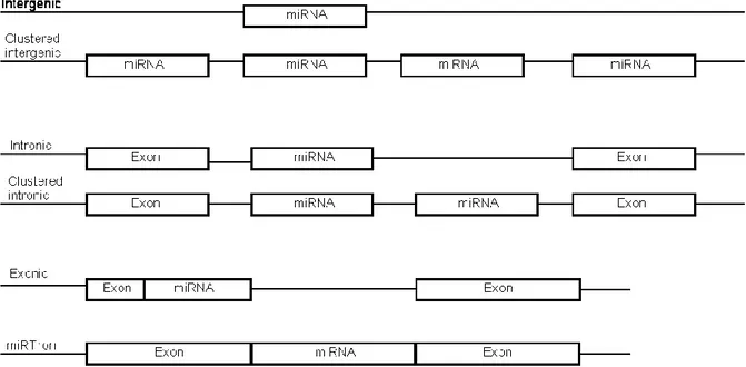
MiRNAs are not randomly distributed throughout the genome. It was found early on that miRNAs can form polycistronic transcripts consisting of clearly defined clusters within the genome (46). It is often found that clusters were formed by local duplication of an existing miRNA locus. Nevertheless, there are also many cases of miRNA families with paralogues at different genomic locations, and also miRNA clusters containing a wide variety of miRNA families. These loci can be found in several different patterns of genomic organization (Figure 6). MiRNA loci can be intergenic, encoded in monocystronic or polycistronic transcripts. They are also frequently found in the introns of protein-coding genes. In rare circumstances, miRNAs can also be found in the exons of protein coding genes (47), or be derived from other classes of non-coding RNAs. It is important to note that what we consider to be exonic miRNAs is dependent on our knowledge of precise gene splicing patterns. It has also been found that miRNAs can form a whole intron by themselves, thus bypassing the requirement of Drosha for their processing. Genomic miRNA clusters tend to be relatively small, rarely containing more than five or six distinct loci. Nevertheless there are exceptions. Human chromosome 14 contains the largest known cluster of miRNA loci that is conserved among many species, containing 37 miRNA loci, belonging to 6 distinct miRNA families. Other large clusters have been described, namely the cluster that is present on Human chromosome 19, and is conserved in most other primates that have been sequenced to date. Repeat derived miRNAs can be located in locally duplicated clusters along the genome (e.g. miR-427 and miR-430) or be spread in an almost random fashion throughout the genome (e.g. miR-548).
24
Figure 6.Possible genomic organization of miRNA loci. In rare cases, miRNA loci can be contained inside an exon, or be the exclusive feature within an intron, which allows its maturation using the splicing machinery instead of requiring Drosha (miRTron).
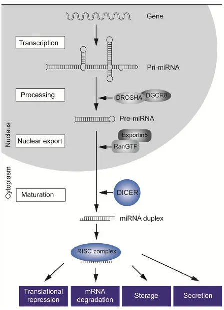
1.9. Current Model of Mammalian miRNA Maturation and Processing
MicroRNA genes represent approximately 1% of the genome of different species, and each of them has hundreds of different conserved or nonconserved targets: it has been estimated that approximately 30% of the genes are regulated by at least one microRNA. MiRNAs may be located either within the introns (mirtron) or exons of protein-coding genes (70%) or in intergenic areas (48) (30%). Mirtrons representing a subspecies of miRNAs, were firstly discovered in D. Melanogaster (49). Mirtrons constitute 5-10% of miRNA genes in invertebrates and vertebrates, they are expressed at much lower levels than typical canonical microRNA (50). Canonical miRNA are processed by the endoribonuclease Drosha; mirtrons are instead processed by the spliceosome. MiRNAs are higly conserved, small, noncoding RNA molecules, 19-22 nucleotides in lengh, which control the expression of genes on the post-transcripional level. MicroRNAs are transcribed for the most part by RNA polymerase II as long primary transcripts, up to several kilobases, characterized by hairpin structures (pri-microRNAs) 5’ capped, spliced and polyadenylated, and then processed into the nucleus by the enzyme RNAse III Drosha and its cofactor DGCR8 (DiGeorge syndrome critical region gene 8), into 70- to 100-nts long pre-microRNAs. These precursor molecules are exported through the nuclear pores by Exportin 5-mediated mechanism to the cytoplasm, where an additional step mediated by the RNAse III Dicer generates a dsRNA of approximately 22 nts, that consists of the leading-strand miRNA and its complementary miRNA sequence, named
25
miR/miR*(miRNA star). This duplex RNA is unwound by a helicase into a single-stranded miRNA. The mature single stranded microRNA product is then incorporated in the complex known as microRNA-containing ribonucleoprotein complex (miRNP), miRgonaute, or microRNA-containing RNA-induced silencing complex (miRISC), whereas the other strand is likely subjected to degradation (Figure 7). However there are also reports of functional miRNA* sequences, especially under distinct cellular conditions and in different tissues. For example, in the case of miR-126/miR-126*, both miRNAs are stable and mediate characteristic functions (51).
Figure 7 Biogenesis of miRNAs. miRNAs are transcribed as RNA precursor molecules (pri-miRNA), which are processed by Drosha and its cofactor DGCR8 into short hairpin structures (pre-miRNA). These are exported into the cytoplasm, where they are further processed into mature single-stranded miRNAs by the endonuclease Dicer. Those mature miRNAs are incorporated into the RISC complex and regulate posttranscriptional gene expression through various mechanisms
26
1.10. Molecular mechanisms of miR action
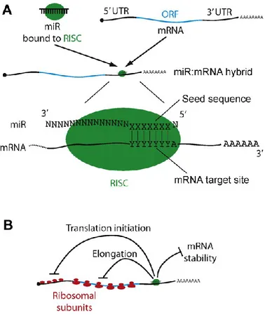
The activity of mature miRs is dependent upon the recognition of a target sequence within mRNAs. For the most part, the mature 22-nt strand recognizes complementary sequences in the 3’ untranslated region (3’UTR) of target mRNAs, and are short and often imperfectly complementary.
Typically, functional target sites consist of a 6-7 nt sequence complementary to the “seed sequence” of the miR, followed by an A residue, but may contain further regions of complementarities further upstream. These rules permit miR targets to be predicted from mRNA sequences; however such prediction algorithms are imperfectly specific and sensitive, and need to be experimentally validated in each case. Perfect miRNA:mRNA complementarity leads to cleavage of the mRNA by AGO2; this is the small interfering RNA (siRNA) pathway, which while important experimentally, is not thought to occur with endogenous mammalian miRs. Instead, the imperfect pairing with mRNA causes a down-regulation of translation. The mechanism by which this occurs is still not clear; there is evidence supportive of models that translation is affected at initiation (52), or during elongation (53) (Fig. 8). A third possibility is that different mechanisms are used at different times, with the intriguing observation that in one system at least, the nature of the promoter driving mRNA synthesis can determine at which stage down-regulation occurs (54).
27
Figure 8. Mechanisms of miR action. (A) A stereotypical miR: mRNA hybrid. (B) Modes of miR-mediated down-regulation of gene
expression.
1.11. MiRNA Classification
The discovery of miRNAs caused a new surge of research in the field of molecular biology. To ensure the identification of novel miRNAs is properly regulated, in 2003, V. Ambros established universal guidelines for proper miRNA annotation (55). Briefly, legitimate miRNA identification must follow expression and biogenesis criteria, in which evidence is required for the expression of a distinct ~22 nt RNA transcript, together with a miRNA precursor that contains the miRNA sequence in one arm of the stem-loop structure. Additionally, phylogenetic conservation of the suspected miRNA as well as the predicted precursor must also exist, in addition to conservation of the 5’ terminus containing the seed region. Furthermore, when Dicer function is purposely disabled, evidence must support increased accumulation of the predicted precursor to ensure that the processing pathway of the miRNA involves Dicer. A single one of these conditions is insufficient on its own to annotate a novel miRNA, and evidence of both expression and biogenesis must be included (56).
28
Mature miRNAs are named using a “miR” prefix, followed by an identification number (ie. miR-31), however if only the miRNA precursor has been determined then the miRNA will be annotated using “mir.” Additionally, the gene encoding the miRNA uses the same prefix, although it is italicized (ie. mir-31) (56, 57). Letters preceding the “miR” prefix refer to the organism that the miRNA is found in (ie. hsa-miR-31 refers to Homo sapien or mmu-miR-31 refers to Mus musculus). A letter occurring after the identification number refers to its relation to another miRNA, which may only differ from another by 1 or 2 nt (ie. miR-200a is related to miR-200b). The designation of “3p” or “5p” following the identification number refers to the arm from which the miRNA is contained in the pre-miRNA, while a “*” in the miRNA name refers to the passenger strand miRNA, which appears as a minor product in some cell types. However, the most recent version of miRBase (version 17.0) released in 2011 states that miR and miR* annotations will be changing to miR-3p and miR-5p, since accumulating evidence shows that the passenger strand may actually dominate over the supposed mature miRNA in some tissue or cell types (55).
1.12. Experimental techniques for miRNA analysis
The explosion of interest in miRNAs over the past few years necessitates effective tools for detecting their presence, quantification, and functional analysis. Isolation of miRNAs from specimens required modification of existing RNA extraction protocols, to take into account their tiny size and unique structure. Column based approaches were adapted to selectively capture and retain both the large and small RNA fractions e.g. using Qiagen RNeasy kits. Co purification methods have also been developed to isolate total RNA, inclusive of the small RNA fraction. MiRNA expression profiling has been facilitated by the advent of high-throughput profiling techniques such as miRNA microarrays and bead-based miRNA profiling. These methods are far superior to existing low through-put techniques, such as Northern blotting and cloning.
1.12.1. Microarray
Microarray technology has also advanced to facilitate miRNA expression profiling. Labelling and probe design have improved to address the poor specificity initially observed when array technology could not distinguish between signals from premiRNA, pri-miR and mature miRNA sequences. Castoldi et al described a novel miRNA microarray platform using locked nucleic acid modified capture probes (58, 59). Locked nucleic acid modification improved probe thermostability and increased specificity, thus enabling miRNAs with single nucleotide differences to be discriminated—an important consideration as sequence-related family
29
members may be involved in different physiologic functions (60). An alternative high-throughput miRNA profiling technique is the bead-based flow cytometric approach developed by Lu et al (61), a method which offers high specificity for closely related miRNAs because hybridisation occurs in solution. The high throughput capability of array based platforms make them an attractive option for miRNA studies compared with lower throughput techniques such as Northern blotting and cloning, which remain essential for the validation of microarray data.
1.12.2. Next generation sequencing techniques
One of the limitations of microarray expression profiling is the requirement of prior sequence information to be used for probe design. Until recently, this sequence information has been limited mostly to that found in public databases (e.g. miRBase). These data have been gathered mainly through a combination of bioinformatics, and extensive cloning experiments. In contrast, deep sequencing is not dependent on any prior sequence information. Instead it provides unbiased information about all RNA species in a given sample, thus allowing for discovery of novel miRNAs or other types of small RNAs that have eluded previous cloning and standard sequencing efforts. Next generation sequencing utilizes massively parallel sequencing, generating millions of small RNA sequence reads from a sample (62). This provides an excellent tool for those studying species where limited sequence information is currently available. Additionally, new sequence information generated using these techniques can be used to design improved microarray platforms for future large scale expression studies (63). Currently available deep sequencing technologies include the Roche 454 and Illumina’s Solexa platforms. Roche 454’s platform utilizes emulsion PCR for template amplification, and pyrosequencing technology on a high well-density picotiter plate. Illumina’s Solexa platform uses bridge amplification on glass surface for template preparation and reverse terminator technology for sequencing. Both platforms provide high throughput and high quality sequencing production at low cost. In conjunction with the evolution of next generation sequencing technologies, which generate massive amounts of data, bioinformatic tools have had to evolve in concert. Several bioinformatics analysis programmes have been developed specifically to interpret and interrogate deep sequencing data. Examples include miRDeep, deepBase, miRExpress, and miRanalyzer. As these highly sophisticated techniques continue to develop, the extent and significance of miRNA regulation of gene expression will become even more evident.
30
1.12.4. Real time Quantitative PCR
Quantitative real-time PCR (RQ-PCR) methodologies are considered the gold standard for quantification of nucleic acid levels (64) and have been widely applied to miRNA investigations. This technique is based on the quantitative relationship between the amount of starting target sample and the amount of PCR product at any given cycle number. The RQ-PCR process involves a reverse transcription (RT) reaction to convert isolated RNA into complementary DNA (cDNA), amplification of the cDNA using PCR, and quantification of the amplicons in real-time. Whilst the steps involved in RQ-PCR have largely remained constant since its conception (65), the technology has evolved to become increasingly sensitive, specific and versatile. The short length of mature miRNAs posed difficulties initially, but with the design of effective primers and probes with adequate specificity, this was overcome. Random primers and poly-T oligonucleotide primers are not suitable for RT of miRNA given their short length and the absence of a polyadenylated 3’ tail. To overcome this, the extracted small RNA may be polyadenylated using a poly (A) polymerase followed by a RT reaction using a poly-T oligonucleotide primer. Alternatively, a gene-specific, stem-loop RT primer may be used (66). When compared to linear RT primers, the stem-loop RT primers are far superior in discriminating between miRNA sequences that differ slightly and are at least 100 times better at discriminating between the mature miRNA and its longer precursor. The stem-loop creates steric hindrance that prevents priming of the precursor miRNA. To date, the most successful approach in terms of specificity and sensitivity is a two-step approach using looped miRNA-specific reverse transcription primers and TaqMan probes from Applied Biosystems, (67). The stem-loop structure is specific to the 3’end of the mature miRNA. It extends the short mature miRNA and adds a universal 3’ priming site for real-time PCR. To correct for variables such as the amount of starting template and enzymatic efficiencies, RQ-PCR data is routinely normalized using endogenous control genes (‘house-keeping genes’) which are stably expressed across a sample set. The appropriate choice of endogenous control is critical to ensure validity and accuracy of the results generated. Evidence exists to support particular miRNAs as appropriate normalizers for given datasets, depending on the tissue of origin. The many advantages of RQ-PCR in miRNA analysis include its efficiency, relatively low cost, low starting miRNA requirement, and the fact that both high and low abundance miRNAs can be detected. It is therefore particularly useful for validating the data obtained from miRNA microarray expression profiling.
31
1.13. MicroRNAs in Tumorigenesis
1.13.1. Deregulation of miRNAs in cancer
Since miRNAs participate in a vast array of normal functions, it seems logical that they would also be associated with abnormalities in disease states. Indeed, investigations based on this idea have revealed that alterations in miRNA genes have a significant involvement in cancer initiation and progression (68). MiRNAs expression is influenced by genomic abnormalities such as chromosomal amplifications or deletions, mutations, and rearrangements, which cause aberrant gene expression when compared to normal tissues. Calin et al. (2004) found that over 50% of miRNAs are over-represented at fragile chromosomal regions prone to breakage or rearrangement, and are frequently altered in cancer. For example, two known tumour suppressor miRNAs that are clustered together, miR-15a and miR-16-1, are down-regulated in over 70% of human chronic lymphocytic leukemia (CLL) and their genes are situated at the 13q14.3 loci, which is the region of deletion in human CLL. Abnormal miRNA expression is also caused by impairments of the miRNA processing machinery (69). Drosha up-regulation is apparent in approximately half of all cases of cervical squamous cell carcinoma (SCC), likely caused by an amplification mutation at chromosome 5p where the Drosha gene is located (70, 71). Additionally, Melo et al. (2009) found that mutations in TRPB led to Dicer destabilization in colorectal and endometrial cell lines, resulting in the global downregulation of miRNAs. This widespread miRNA downregulation has also been determined in other studies. Lu et al. (2005) did a systematic analysis on 217 different miRNAs in various tumours samples such as brain, breast, lung, colon, stomach, pancreas, kidney, and found that 129 of them were downregulated in the tumours compared to normal tissues. Other miRNA profiling studies such as that performed by Volinia et al. (2006), which looked at characterization of six different tumour types (prostate, lung, breast, colon, pancreas, and stomach) have revealed that an abundance of miRNAs are both upregulated and downregulated in cancer cells compared to normal tissue (68). Moreover, many studies have been performed such as that by Liu et al. (2004) on human and mouse RNA profiling, which have revealed a specific pattern of miRNA expression in numerous tumour types. For example, Volinia et al. (2006) found that miR-21 was over-expressed in all six tumors types analyzed, while miR-191 was expressed in five.
1.14. MiRNAs as tumor suppressor
A miRNA, like a protein coding-gene, can act as a tumor suppressor when its function loss can initiate or contribute to the malignant transformation of a normal cell. The loss of function
32
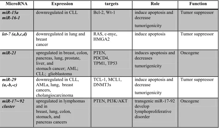
of a miRNA can be attributed to several mechanism. For example to genomic deletion, mutation, epigenetic silencing and miRNA processing alteration. The family of let-7 (let-7a,b,c,d) of miRNAs is down regulated in many tumors including breast cancer (72).
Many members of the let-7 family are located in fragile genomic areas associated with lung, breast and cervical cancer. Functionally, let-7 family members inhibit the mRNA of well-characterized oncogenes, such as the RAS family, HMGA2 and c-myc, and induce apoptosis and cell cycle arrest when overexpressed in lung and colon cancer and in Burkitt lymphoma cell lines.
MiR- 29 family members have been shown to be downregulated in CLL, lung cancer, invasive breast cancer, AML, and cholangiocarcinoma. The enforced expression of miR-29b induced apoptosis in cholangiocarcinoma and lung cancer cell lines and reduced tumorigenicity in a xenograft model of lung cancer (73). The tumor suppressor effects of the miR-29 family can be explained in part by the direct targeting of the antiapoptotic protein MCL-1 and the oncogene TCL-1.
1.15. MiRNAs as oncogenes
The amplification or over-expression of miRNAs which target tumour suppressors can lead to significant down-regulation of these tumour suppressors, or of genes involved in cell differentiation. This may incite uncontrolled proliferation, loss of apoptotic activity, promote angiogenesis and/or invasion, thus contributing to tumour formation. In this way, miRNAs can act as oncogenes. The list of miRNAs that function as oncogenes is short, but the evidence of their role is strong. One such oncomiR is miR-21. This miRNA is up-regulated in a variety of malignancies, including AML, CLL, cancers of the pancreas, prostate, stomach, colon, lung, breast and liver. Over-expression of miR-21 in glioma blocks apoptosis (74) whereas silencing its expression inhibits cell growth and increases apoptotic cell death by targeting genes such as PTEN, PDCD4 (programmed cell death 4), or TPM1 (tropomyosin 1). The polycistronic miR-17/92 cluster represents another miRNA with oncogenic function. Over-expression of the seven miRNAs in this cluster (miR-17- 5p, miR-17-3p, miR-18a, miR-19a, miR-20a, miR-19b, and miR-92–1) have been associated with a variety of malignancies (colon, lymphoma, breast, lung, pancreas, prostate and stomach. The mechanism by which this cluster acts is likely to be due to suppression of PTEN, a tumour suppressor gene and negative regulator of the highly oncogenic prosurvival PI3K/AKT signalling pathway.