BIOCH
G
EL
A
S
D DottUn
HIMICA
LATINA
S
B
IOM
Dottoran t. Trentininiver
DOT
A, BIOL
COORDIASES
,
N
MARKE
Setto do Alessandrrsità
TTORAT
LOGIA M
C NATORE PN
EURO
ASPA
ERS
I
N
ore Scient ro Anndegl
TO DI R
MOLECO
CICLO XXI Prof. FRANOFILAM
ARTIC
A
D
IFFE
tifico Disci ni 2008/20i Stud
RICERC
OLARE
II NCESCO BEMENTS
A
CID
ERENT
plinare BI 010di di
A IN
E BIOT
ERNARDIS AND
N
P
ATHO
O/10 Tu Prof. BelFerr
TECNOL
N-A
CE
OLOGI
utore lini Tizianaara
LOGIE
ETYL
IES
aINDEX
INTRODUCTION ... 1
Biomarker Concept ... 1
Gelatinases in inflammatory conditions ... 3
Structure of Gelatinases ... 5
Propeptide ... 6
Catalytic Domain ... 7
Hemopexin-like domain ... 8
Fibronectin II-like domain ... 10
Hinge-region and collagen V-like domain ... 10
Substrate specificity ... 11 Regulation of Gelatinases ... 14 Transcription ... 14 Glycosylation ... 17 Pro-enzyme activation ... 19 Activation of proMMP-9 ... 22 Activation of proMMP-2 ... 24
Receptors that mediate the catabolism of MMP-2 and MMP-9 ... 26
Inhibition ... 27
Mechanism of inhibition of TIMP-1 and TIMP-2 ... 30
Different forms of gelatinases ... 34
Inflammatory conditions ... 36
Multiple Sclerosis ... 37
Spontaneous Intra Cerebral Hemorrhage (SICH) ... 42
Neurofilament subunits and N-Acetyl aspartic Acid (NAA) as markers
of axonal damage in progressive multiple sclerosis ... 53
Neurofilaments ... 54
NAA ... 58
AIMS OF THE THESIS ... 61
MATERIAL AND METHODS ... 62
Active MMP-2 and TIMP-2 in Multiple Sclerosis Patients ... 62
Patient selection ... 62
Cerebrospinal fluid and serum sampling ... 63
Magnetic resonance imaging examination ... 64
MMP-2 activity assay ... 65
TIMP-2 ELISA assay ... 66
Determination of total and active MMP-2 and TIMP-2 Indices ... 66
Statistical analysis ... 66
Active gelatinases and TIMPs in SICH ... 68
Patient selection ... 68
CT examination ... 68
Serum sampling ... 69
MMPs activity assays and TIMPs assays ... 69
Statistical analysis. ... 70
Detection of 66 kDa active MMP-9 in Serum ... 71
Blood specimen collection and storage ... 71
Enzymes, antibodies and reagents ... 71
Gelatin Zymography ... 72
Densitometric analysis of the zymographic patterns ... 72
MMP-9 activity assay ... 73
MMP-9 inhibition by TIMP-1 ... 74
Activation of pro-MMP-9 ... 74
Immunoprecipitation ... 75
Gelatinases separation by Concanavalin-A chromatography ... 75
MMP-9 released from neutrophils ... 77
Separation of Neutrophils from leukocytes-enriched Buffy Coats .. 77
Stimulation of neutrophils with LPS ... 78
Stimulation of neutrophils with IL-8 ... 78
Total MMP-9 Assay (ELISA) ... 78
Statistics ... 79
Neurofilament subunits and NAA in progressive multiple sclerosis .. 80
Patients and controls ... 80
MR methods ... 81
Preparation of cell-free CSF ... 82
CSF NAA measurements ... 82
CSF Neurofilament measurements ... 82
Nf light ELISA ... 82
Nf heavy Luminex assay ... 82
Statistics ... 83
RESULTS ... 85
Active MMP-2 and TIMP-2 in Multiple Sclerosis Patients ... 85
Cerebrospinal fluid and serum levels and intrathecal synthesis of active MMP-2 and TIMP-2 in MS patients and controls ... 85
Cerebrospinal fluid and serum levels and intrathecal synthesis of active MMP-2 and TIMP-2 in MS patients categorized according to clinical and MRI activity ... 87
Correlation between CSF and serum levels and intrathecal synthesis of active MMP-2 and TIMP-2 and MS clinical features ... 88
MMPs and TIMPs time course ... 90
Hematoma and edema volumes time course ... 91
Relationships between MMPs and TIMPs and CT and clinical findings ... 92
Detection of 66 kDa active MMP-9 in Serum ... 94
Inhibition of MMP-9 by TIMP-1 ... 94
Identification of 66 kDa MMP-9 form in serum ... 95
Quantification of 66 kDa MMP-9 form in serum of healthy donors 97 MMP-9 released from neutrophils ... 99
Levels of MMP-9 released by not stimulated neutrophils ... 99
Levels of MMP-9 released by neutrophils stimulated with LPS ... 100
Levels of MMP-9 released by neutrophils stimulated with IL-8 ... 102
Neurofilament subunits and NAA in progressive multiple sclerosis 105 CSF NAA, Nf light and Nf heavy levels in patients with progressive MS and controls with OND ... 105
Correlation between CSF biomarkers for axonal damage and clinical and radiological parameters ... 107
Combination of CSF biomarkers for axonal damage improves discrimination between MS patients and controls ... 108
DISCUSSION ... 110
I
NDEX OFF
IGURESFigure 1. Domain structure of gelatinases. ... 5
Figure 2. Tridimensional structure of the MMP-9and MMP-2 propeptide. ... 6
Figure 3. Catalytic domains of MMP-9 and MMP-2. ... 8
Figure 4. 3-D structure of the hemopexin-like domain. ... 9
Figure 5. Placement of the fibronectin II-like domain respect to the catalytic domain. ... 10
Figure 6. Main regulatory cis elementsin the promoter/enhancers region of MMP-2 and MMP-9 genes. ... 15
Figure 7. Glycosylation of MMP-9. ... 17
Figure 8. Possible 3D structure of glycosilated MMP-9. ... 19
Figure 9. Activation cascade of MMP-2 and MMP-9 (gelatinase B). ... 20
Figure 10. Cysteine switch mechanism for the MMPs activation. ... 21
Figure 11. Cleavage of MMP-9 propeptide by MMP-3. ... 23
Figure 12. Activation mechanism of proMMP-2. ... 24
Figure 13. The two steps in the activation process of proMMP-2. ... 25
Figure 14. Primary structure of TIMP-1and TIMP-2. ... 29
Figure 15. Interaction of TIMP-1 with active MMP-9 and proMMP-9 during the formation of the complex. ... 31
Figure 16.Mechanism of interaction of the N-terminal domains of TIMP-1 and MMP-9. ... 33
Figure 17. Representation of two hemopexin-like domains involved in the dimerization. ... 35
Figure 18. Clinical subtypes of MS. ... 39
Figure 19. Demyelinating lesion evidenced by MRI: the first is hyperintense, the second is after the administration of gadolinium. ... 39
Figure 20. Spontaneous intraparenchymal hematoma. ... 43
Figure 21. Hemorrhagic core and peri-hematomal area evidenced by Computed Tomography. ... 44
Figure 22. Constitution of the Blood Brain Barrier. ... 46
Figure 23. Granules of the human neutrophils. ... 50
Figure 24. Neurofilaments subunits. ... 55
Figure 25. Neurofilaments release in the extracellular fluid following axonal injury. ... 56
Figure 26. CSF active MMP-2 levels, serum TIMP-2 levels, and CSF and serum values of active MMP-2/TIMP-2 ratio, and percentages of intrathecal MMP-2 synthesis in MRI active (Gd+) and MRI inactive (Gd−) RRMS patients. ... 88
Figure 27. Correlation between serum mean values of MMP-2/TIMP-2 ratio and disease duration. ... 89
Figure 28. Temporal profile of serum active MMP-9 and MMP-2 levels in SICH patients. ... 91
Figure 29. Perihematomal edema volume in SICH patients. ... 92
Figure 30. Time course activation of proMMP-9. ... 95
Figure 31. MMP-9 and MMP-2 activity before and after concanavalin-A-sepharose chromatography. ... 96
Figure 32. MMP-9 activity before and after two-step immunoprecipitation. ... 97
Figure 33. Total active MMP-9 and TIMP-1 not inhibited active MMP-9 in sera of healthy donors. ... 98
Figure 34. Mean levels of MMP-9 released by not stimulated neutrophils in medium at different pH. ... 100
Figure 35. Mean levels of MMP-9 released by LPS stimulated neutrophils in medium at different pH. ... 102
Figure 36. Mean levels of MMP-9 released by IL-8 stimulated
neutrophils in medium at different pH. ... 103 Figure 37. Concentrations of the analyzed biomarkers in CSF of controls and MS patients. ... 106 Figure 38. Correlation between biomarkers and clinical and radiological parameters. ... 107
I
NDEX OFT
ABLESTable 1. ECM and non-ECM substrates for MMP-9and MMP-2. ... 13 Table 2. Some inducing factors and suppressing factors of MMP-9 synthesis. ... 16 Table 3. Dissociation constants for active and pro MMP-9. ... 30 Table 4. Demographic and baseline clinical characteristics of
progressive MS patients and OND controls. ... 81 Table 5. CSF and serum levels of active MMP-2,TIMP-2, MMP-2/TIMP-2 ratios and intrathecal synthesis in patients with MS, OIND and NIND controls. ... 86 Table 6. Significant correlations between serum mean levels of MMP-9 and MMP-2 and hematoma and perihematomal edema volumes in 28 patients with spontaneous intracerebral hemorrhage (SICH). ... 93 Table 7. Levels of total MMP-9 (ng/ml) released at different pH by not stimulated neutrophils. ... 99 Table 8. Levels of total MMP-9 (ng/ml) released by neutrophils
stimulated with LPS. ... 101 Table 9. Levels of total MMP-9 (ng/ml) released by neutrophils
stimulated with IL-8. ... 103 Table 10. One way ANOVA followed by Bonferroni post-hoc test. ... 104 Table 11. CSF levels of NAA, Nf light and Nf heavy in progressive MS patients and OND controls. ... 106 Table 12. Percentage of progressive MS patients and OND controls with abnormal biomarker values. ... 109
I
NTRODUCTIONBiomarker Concept
A biomarker is defined as a quality objectively measured and evaluated as indicator of physiological and pathological processes or pharmacologic responses to a therapy (Biomarkers Definitions Working Group, 2001). In the past, biomarkers were primarily physiological indicators, for example body temperature rise is a biomarker for fever or blood pressure is used to determine the risk of stroke. More recently, biomarker is becoming a synonym for molecular marker, namely chemical molecules, genes, proteins and enzymes that can be measured in body fluids and are related to a specific pathology (i.e. transaminases levels as markers for liver functions). Also, biomarkers can be specific cells or complex organ functions and characteristic changes in biological structures.
We can distinguish between two strictly-related kinds of biomarkers:
disease-related and drug-related biomarkers. Disease-related biomarkers
give an indication for a threat of disease (risk indicator or predictive biomarkers i.e. Anti-citrullinated protein/peptide antibody as marker for Rheumatoid Arthritis), if a disease already exists (diagnostic biomarkers i.e. prostate specific antigens for prostate cancer), or how a disease may develop in a patient (prognostic biomarker i.e. cancer biomarkers). In contrast, drug-related biomarkers indicate whether a drug will be effective in a specific patient and therefore, they should vary with a drug treatment.
A number of diseases, such as Alzheimer’s disease, rheumatoid arthritis or Multiple Sclerosis, often begin with an early, symptom-free phase. In
these cases, biomarkers may help identifying high-risk individuals to develop a disease, so they can be treated before onset of the disease or either as soon as possible thereafter.
In order to use a biomarker for diagnostic purposes, it should have some well-defined characteristics:
the sample material to use for the biomarker detection must be as easy to obtain as possible, with a low invasiveness. For example, this may be a blood sample, a urine or saliva sample, or even a drop of blood;
the assay for a biomarker should be as quick as possible. In fact, for rapid initiation of treatment, the speed with which a result is obtained from the biomarker test is critical. A test which delivers results only in few minutes is optimal.
the detection method for a biomarker must be accurate and as easy to carry out as possible. The results from different laboratories may not differ significantly from each other, and the biomarker must have proven its effectiveness for the diagnosis, prognosis and risk assessment of the disease in independent studies.
Together with molecular biomarker assayed in biological fluids there are also other kinds of biomarkers referred as imaging biomarkers, developed using imaging technologies like coronary angiography, computed tomography, magnetic resonance imaging, positron emission tomography.
Imaging biomarkers have many advantages: usually they are non-invasive and produce intuitive, multidimensional results. Yielding both qualitative and quantitative data, they are usually relatively comfortable for patients and combined with other sources of information, they can be
very useful to help clinicians in a diagnosis. In contrast to these advantages, often the required analyses are expensive and may require the use of radiations or contrast liquids which can lead to allergies. For these reasons, molecular biomarkers are essential to develop large scale screenings on patient populations and can give, together with imaging biomarkers, more complete information.
In the present study, I aimed to evaluate the possible role of active Matrix Metalloproteinase-9 (MMP-9) and Matrix Metalloproteinase-2 (MMP-2) as biomakers in inflammatory pathologies, since their involvement in inflammation is well documented but is still unclear (Opdenakker G et al., 2003; Cunningham et al., 2005; Yong VW, 2005; Fainardi et al., 2006). Moreover, I also investigated the role of Neurofilament light and heavy subunits and N-Acetyl aspartic Acid as biomarkers of axonal damage in a neurodegenerative disorder like Primary Progressive Multiple Sclerosis (PPMS), as a lack of reliable data remains on the role of these markers in such MS subtype (Norgren N et al., 2003; Jasperse B et al., 2007; Teunissen CE et al., 2009).
Gelatinases in inflammatory conditions
Matrix Metalloproteases (MMPs) were discovered in 1962, and since then more than twenty five members of this family have been identified, cloned and sequenced (Lijnen HR. et al., 2001).
The MMPs are endopeptidases zinc and calcium dependent which are classified on the basis of the recognized substrates and sequence homology in: collagenases, gelatinases, stromelysins, membrane-type metalloproteases (MT-MMPs) and matrilysins.
Collectively, they are capable of degrading essentially all the Extracellular Matrix (ECM) components, playing an important role in
ECM remodeling during both physiological and pathological conditions. In fact they are expressed during embryonal development, tissue regeneration and wound repair (Van den Steen PE et al., 2002). Also, they play a role in pathological conditions such as rheumatoid arthritis, autoimmune disorders of the skin, chronic ulcerations, malignant tumors and metastasis (Van den Steen PE et al., 2002).
One of the most complex MMPs family is the gelatinases family, that includes MMP-9 (Gelatinase B) and MMP-2 (Gelatinase A). Among these, MMP-9 has been associated with a large number of pathological conditions, such as acute respiratory distress syndrome, destructive lung disease, Sjögren’s syndrome, peripheral nerve injury, Guillain-Barré syndrome, blood-brain-barrier damage, Multiple Sclerosis, Alzheimer’s diseases, cancers and brain ischemia (Lacraz S et al., 1995). On the contrary, MMP-2 has been associated to both tissue repair and destruction in inflammatory pathologies (Yong VW, 2005).
The objective of my study was to analyse in detail the role of gelatinases in pathological inflammatory conditions such as Multiple Sclerosis or Spontaneous Intra Cerebral Haemorrhage (SICH). Moreover, to better understand the real involvement of MMP-9 in inflammatory processes, I tried to assess in vitro the release of MMP-9 from neutrophils during environmental pH changes because an acidic pH in tissues is related to inflammatory conditions.
Struct
Gelatina organize detail be 1. P c 2. Z c t 3. H i t 4. H f h 5. F r b s 6. p h h Figure 1. C5, Coll peptide; (ture of Ge
ases (gelati ed into six elow: Propeptide coordinatio Catalytic d Zinc Bindin calcium io tridimensio Hemopexin important fo tissue speci Hinge regio flexible str hemopexin-Fibronectin repeats, it i binding dom specificity f Collagen V present on hemopexin-high glycos . Domain stru lagen V-like (Parks W.C. eelatinases
inase A or main doma : it maintain on of a cyste domain: it c ng Domain on which nal structur -like domaifor the bindi ific inhibito on: a prolin ructure, co -like domai n II-like do is placed in main and th for gelatin, V-like doma nly in MM -like doma sylation con ucture of gela domain; Fn, et al., 2004). MMP-2 an ains (Figur ns the enzym eine residue ontains the with an hi is essent re; in: placed in ing of substr rs TIMPs; e rich regio onnecting t n; omain: com
nto the cata he active s laminin, typ ain: Rich i MP-9 betwe in and it i nfers to this tinases. fibronectin nd gelatinas re 1) which me into ina e with the ca catalytic s igh conserv tial to m n the Carbo trates and fo on with a va the catalyti mposed by alytic doma site, and it pe I and IV in serine/pr een the hin
is heavily region a rig repeat; Pro, se B or MM will be ex active form atalytic zinc ite with the ved Methion maintain th oxy-termina or the intera ariable lengt ic domain three fibro ain between confers the collagens; roline/threon nge region O-glycosyl gid structure propeptide; MP-9) are plained in due to the c ion; e zinc, the nine and a e correct l end, it is action with th, it has a with the onectin II n the zinc e substrate nine, it is n and the lated. The e. SP, signal
Propep The pro amino a MMP-9 zinc ato sequenc al., 199 enzyme proteoly family a “Cystein Figure 2. In yellow N-Glycos The sec organize which in ptide opeptide (Fi acids in MM 9 and the C om via its s ce (…PRCG 98). The int e into the in ysis results are produce ne Switch” . Tridimensio w is marked the sylation site. condary str ed in three n the MMP igure 2) co MP-2, cont ys102 in the side chain t GXPD…) i teraction of nactive form in zymoge ed in a laten (Massova I onal structure e cysteine coo ructure of t e α-helices -9 has a N-g onsist of 86 aining a cy MMP-2, w thiol group. is present in f the cystei m (zymogen n activation nt form. Thi I. et al., 199 of the MMP-ordinated to th the propept linked by glycosylatio 6 amino acid ysteine resid which intera . A highly n the prope ine with th n). Removal n, as all me s activation 98). -9and MMP-2 he catalytic zin tide in MM several fle on site (Asn ds in MMP due, the Cy acts with the conserved eptide (Mas he zinc mai l of the prop embers of t n mechanism 2 propeptide. nc. In grey is m MP-9 and M exible loop n38). P-9 and 80 ys99 in the e catalytic consensus ssova I. et ntains the peptide by the MMPs m is called marked the MMP-2 is ps, one of
Catalytic Domain
The catalytic domain consist in 190 amino acids in MMP-9 and 205 amino acids in MMP-2, and contains an highly conserved domain called Zinc Binding Domain, which coordinates a zinc ion through three histidines and is essential for the enzymatic activity
(…AH401EXGH405XXGXXH411… in MMP-9 and
…AH403EXGH407XXGXXH413… in MMP-2) (Van den Steen PE. et al.,
2002). Within the catalytic domain there are also one other zinc, coordinated by three histidines (His175, la His190 and His203 in MMP-9;
His178, His180 and His193 in MMP-2), and two calcium ions; all these ions
are essential to maintain the correct spatial structure for the interaction of the substrates with the active site (Massova I. et al., 1998). The secondary structure of the catalytic domain (Figure 3) consists in five β-sheets that wrap around three α-helices. One important amino acid close to the catalytic zinc is the Glu402 in MMP-9 and Glu402 in MMP-2, which
binds water molecules and is essential for the hydrolysis of the substrates. In addition, there are also two N-glycosylation sites in the Asn120 and Asn127 residues only in the catalytic domain of MMP-9.
Figure 3. In differe Yellow, c ions. Hemop The hem (from th 660 in M and sho This dom b i a a r The sec barrel su . Catalytic dom ent colors are catalytic zinc; pexin‐like mopexin-lik he amino ac MMP-2), is ows a sequen main has se binds the su interacts wi and TIMP-2 an elevated respective g condary stru uperstructur mains of MM showed the m Green, struct domain ke domain, cid 513 to 7 s present in nce similar everal funct ubstrates;
ith the spec 2 for MMP d affinity gelatinase (M ucture of th re compose MP-9 and MM metal ions and ural zinc; Pin
placed at th 707 in MMP n all the MM to the hemo ions: cific inhibito P-2): the C-t for the h Massova I. his domain ed of four bl MP-2. the residues w nk, Orange an he C-termin P-9 and fro MPs (excep opexin of th ors TIMPs terminal en hemopexin-l et al., 1998 is organize lades, each which coordin d Light Green nal end of th om amino a pt in the m he plasma. (TIMP-1 fo d of the inh like domai ). ed in a β-pr blade consi nate them. n, calcium he protein cid 466 to matrilysins) or MMP-9 hibitor has in of the ropeller: a ists of one
α-helix axis (Fi The firs two bla interact 2002). O propelle Figure 4. From I to Another to the L hepatoc leads to causing and four an igure 4). st blade is b ades are rela
with the C-Only in MM er structure. . 3-D structur o IV are marke r important LPR-1 rece cytes and m o the interna a down-reg nti-parallel ound to the atively awa -terminal en MP-2 there . re of the hemo ed the four β-s function of eptor (LDL acrophages alization of gulation of t β-strands; a e fourth thro ay from the nd of the inh is one calc opexin-like do sheets that com f the hemop Receptor-r . The bindin the enzyme the enzyma all is organ ough a disul e catalytic d nhibitor (Van cium ion in omain. mpose the β-p pexin-like d related Pro ng of gelati e and the su atic levels. ized around lfide bridge domain and n den Steen the middle propeller struc domain is th tein-1), exp inases to thi ubsequent de d a central ; the other d probably n PE et al., e of the β- cture. he binding pressed in is receptor egradation
Fibron This do repeats, II. Thes external Figure 5. In pink, g the cataly This do V collag The typ depend Hinge‐ The hin whereas catalytic nectin II‐lik omain is ch each of on se three repe l position (F . Placement of grey and purp ytic domain. omain confe gens, elastin pical collag exclusively region and nge-region i s in MMP-2 c domain an ke domain haracteristic ne of 49 am eats belong Figure 5). of the fibronec ple are marke
ers the speci n, and nativ genolytic an y on the pres d collagen s a sequenc 2 it has a len nd the hem n c of gelatin mino acids le to the catal ctin II-like do d the three fib
ificity for ty ve type I co nd elastino sence of thi V‐like dom ce compose ngth of 20 r mopexin-like nases and is ength, hom lytic domain omain respect bronectin II r ype I, type ollagen (Ma olytic activi is domain. main d by 16 am residues. It i e domain. O s composed mologue to f n and are pl t to the catalyt repeats, in blu IV (gelatin assova I. et ities of thi mino acids in is placed be Only in MM d by three fibronectin aced in an tic domain. ue is marked ) and type al., 1998). s enzyme n MMP-9, etween the MP-9, after
this sequence there is another region of 54 amino acids called collagen V-like domain. It is rich in Ser/Thr/Pro and is heavily O-glycosylated: as the O-glycosylation confers to this region a rigid structure, probably the role of this sequence is to maintain the correct distance between the hemopexin-like domain and the other domains.
Substrate specificity
The specificity depends on primary sequence of the substrate: the endoproteases prefer the sequences that bind to the pocket of the catalytic site. However, also the three dimensional conformation of the substrate and the accessibility of the cleavage sequence are important features. Finally, the presence of exosites in the catalytic domain which can bind distant residues on the substrate can have a key role promoting the hydrolysis.
Probably, the fibronectin II-like domain of gelatinases plays a role as exosite that binds collagens increasing the hydrolysis efficiency.
The substrate specificity for MMP-9 and MMP-2 has been analyzed using synthetic short peptides as substrates, resembling the sequence Gly-Pro-Gln-Gly-Ile-Ala-Gly-Gln which correspond to the P4-P3-P2-P1-P1’-P2’-P3’-P4’ sites in the substrate. Mutagenesis studies showed that MMP-9 has a preference for small amino acids (Gly, Ala, Pro) in the P1 site, and hydrophobic residues (Ile, Leu, Tyr, Met) in the P1’ and P2 sites. On the contrary, MMP-2 cleaves the substrate between the Glycine in the P1 site and a hydrophobic amino acid in the P1’ site, although in this last site hydroxylated and acid residues are tolerated (Van den Steen PE et al., 2002).
Up to now, only a small number of physiologic substrates belonging to the extracellular matrix and cleaved by MMP-9 were described; the most
known are denatured collagens or gelatins. Moreover, also the type II and type V collagens can be cleaved by this enzyme. Other substrates that belong to the ECM include aggrecans, the link protein and elastin (Van den Steen PE et al., 2002) (Table 1).
MMP-2 has a similar substrate specificity as MMP-9 for ECM compounds, although differences were observed in substrate type, catalytic efficiency and cleavage pattern of the same substrate for both gelatinases (i.e. gelatin). Among the ECM substrates, there are collagens I, IV, V, VII, X, XI, XIV, gelatin, elastin, fibronectin, laminin-1, laminin-5, galectin-3, aggrecans, decorin, proteoglycan link protein and osteonectin (Van den Steen PE et al., 2002).
There are also several non-matrix proteins degraded by MMP-9 and MMP-2. MMP-9 is able to degrade myelin basic protein (which degradation can lead to the release of encephalitogenic peptides), α1-antitrypsin and α1-antichimotrypsin, P substance, IL-2 receptor- α, TGF-β, β-amiloyd peptide (Aβ1-40), IL-1TGF-β, proTNF- α. Recently, has been observed that MMP-9 can cleave six N-terminal amino acids from IL-8 (1-77), generating another IL-8 form (7-77) which is more bioactive. Moreover, it can generate a positive feedback signal as IL-8 can stimulate the release of MMP-9 from neutrophils (Van den Steen PE et al., 2002). The non-ECM substrates degraded by MMP-2 include IL-1β, α1-PI, prolysyl oxidase fusion protein, MMP-1, MMP-9, MMP-13.
Table 1. ECM and non-ECM substrates for MMP-9and MMP-2. (Chakraborti S. et al., 2003)
Protease ECM Substrates Non-ECM substrates
MMP-9
Collagens (IV, V, VII, X, XIV), gelatin, elastin, galectin-3, aggrecans, fibronectin, proteoglican link protein, entactin, osteonectin. Plasminogen, IL-1β, MBP, ET-1 (endotelin-1), amyloid peptide Aβ1-40,
IL-8, GRO- α (growth-related oncogene-α), SDF-1 (stromal-cell derived factor-1), α1-antitrypsin,
α1-antichimotrypsin, P
substance, IL-2 receptor- α, TGF-β, proTNF- α.
MMP-2
Collagens (I, IV, V, VII, X, XI, XIV), gelatin, elastin, fibronectin, laminin-1, laminin-5, galectin-3, aggrecans, decorin, proteoglican link protein, osteonectin.
IL-1β, α1-PI, prolysyl
oxidase fusion protein, 1, 9, MMP-13.
Regulation of Gelatinases
The regulation of gelatinases occurs on different levels: Transcription;
Glycosylation;
Activation of the pro-enzyme;
Inhibition by specific inhibitor s (TIMP-1 and TIMP-2); Uptake and internalization by membrane receptors.
Transcription
Usually, MMPs expression in tissues is low whereas it increases when ECM remodeling is required.
The different response of the genes of MMP-2 and MMP-9 is due to differences in the promoter region (Figure 6). In fact, the promoter region of MMP-9 gene contains regulatory elements recognized by transcription factor induced by growth factors and cytokines, whereas MMP-2 has not. For this reason, the MMP-2 gene is considered more refractory to modulation by growth factors and cytokines produced during inflammation, while MMP-9 can be up-regulated.
Figure 6 and MMP (Van den All the regulati low lev the tran Morisak the pro-with va Steen et not syn stimulat Instead, in certai 6. Main regul MP-9 genes. Steen et al., 2 elements p on of MMP els while it nscription ca ki., 1990). I -enzyme is ariable amo t al., 2002) nthesized: it tion with di , MMP-2 is in neoplasti
latory cis elem 2002) present in P-9 expressi can also in an be regula In the cells released aft ounts of the ). Neutroph t is stored ifferent agon expressed c ic cells its e mentsin the p the promot ion, as in m ncrease 100 ated in a tis where MM fter stimulat e inhibitor ils are the within terti nists. constitutive xpression m promoter/enha ter region many cell ty times when ssue specific MP-9 synthe tion: often i TIMP-1 an only cell ty iary granule ely by sever may increase ancers region are essenti ypes it is ex n induced; c manner (F esis can be it is secrete nd MMP-2 ype where M es and rele ral cell type
e. n of MMP-2 al for the xpressed at moreover, Frisch and regulated, d together (Van den MMP-9 is ased after s and only
There are several factors that control the transcription of MMP-9: we can distinguish between suppressing factors and inducing factors of the enzyme synthesis and they can vary depending on the examined cell type (Table 2).
Table 2. Some inducing factors and suppressing factors of MMP-9 synthesis. (Van den Steen PE et al., 2002)
Inducing factors Suppressing factors
EGF (epidermal growth factor), PDGF (Platelet derived growth factor), bFGF (basic fibroblast growth factor), TGF-, amphiregulin, TNF-, 1, IL-1, IFN- (interferon-), IFN-, TGF-, phorbolic esters.
IFN-, IFN-, IL-4, IL-10.
In some cell types also IFN-, IL-1, IL-13, EGF, PMA (phorbol 12-myristate 13-acetate) and TGF-.
While there are numerous cytokines that can induce MMP-9 synthesis, only a small number of them (IL-4, IL-10, IFN- and IFN-) have an inhibitory effect on basal or stimulated expression of MMP-9 (Van den Steen PE et al., 2002).
Generally, IFN- and IFN- have an inhibitory effect on MMP-9 expression, although it has been shown that the treatment of melanoma metastatic cells with these cytokines cause an up-regulation of MMP-9 (Hujanen et al., 1994; Wu et al., 1997).
Concern they hav macroph activatio MMP-9 Furtherm of mon factors o In addi powerfu decrease pro-coll Finally, acting o synthesi Glycosy MMP-9 glycosy Three p N-termi while th Tyr-Ser Figure 7. The N-lin with ver fibronecti hemopexi ning IL-4 a ve an inhib hages at pre on of Prote 9 synthesis ( more, IL-4 nocytes and of MMP-9. ition to the ul regulator e directly t lagen mRNA also the o on MMP-9 is (Siwik A ylation 9 is a hea ylated. possible N-g inal end of he others ar r) (Figure 7 . Glycosylatio nked glycans rtical lines. P in domain; Z in-like domain and IL-10, bitory effect e-translation ein Kinase (Van den St and IL-10 c d macropha e modulati s of collage the collagen A (Siwik A oxidative st activation/ AD et al., 20 avily glyco glycosylatio the protein re in the ca 7). on of MMP-9. are indicated Pro, prodoma Zn2+, Zn2+-bin
n (Van den Ste
cytokines p t on MMP-nal level: p A (PKA) teen PE et a
can act indi ages, such on of MM en metaboli n synthesis AD et al., 20 tress can re /transcriptio 04). osilated pro on sites in M : one is in t atalytic dom d with Y, whi ain; Act, ac nding domain; een PE et al., produced b -9 productio probably thi which nor al., 2002). irectly inhib as the pro MP-9 expre ism: IL-1, s varying th 04). egulate the on and dec otein, wher MMP-9 hav the prodom main (Asn12 ile O-glycosyl ctive site dom
; OG, Collag 2006). by lymphoc on by mono s is due to rmally prom biting other oduction of ession, cyto TNF- and he expressi extracellul creasing the reas MMP ve been loca main (Asn38 -0-Ile-Thr an lation sites ar main; FN, th gen V-like do cytes Th2, ocytes and the down-motes the r functions f inducing okines are d IL-6 can ion of the lar matrix e collagen -2 is not ated in the -Leu-Thr), nd Asn127 - re indicated hree repeat main; Hem,
The N-linked glycans in the MMP-9 have a variable composition and can have different functions. For example, during the proteolytic cascade that leads to MMP-9 activation, the glycosylation is important for the specific activity of t-PA and plasminogen. Furthermore, N-linked glycans could influence the MMP-9 stability, the resistance to proteases or they could modulate the interaction with substrates, inhibitors or with enzymatic complexes that degrade the ECM. However, it has been shown that the enzymatic activity of MMP-9 does not change after the removal of N-linked glycans (Rudd PM et al., 1999).
The majority of the carbohydrates are O-linked (about 85%), and are located especially in the collagen V-like domain, typical of this metalloproteinase (Figure 8). The analysis of these glycans showed that a big variability in its composition exists (Rudd PM et al., 1999).
Different functions have been hypothesized for these O-linked glycans, including protection from proteolysis and increase of the protein stability. Moreover, knowing that O-linked glycan clusters increase the extension and the rigidity of the polypeptide chain, probably they could contribute maintaining the hemopexin-like domain in the correct distance from the other domains and favoring the interaction with its specific inhibitor (Rudd PM et al., 1999).
In fact, it has been shown that the deletion of the collagen V-like domain decrease 4 times the affinity for TIMP-1, suggesting that this domain is essential to maintain the correct space between the C-terminal domain and the catalytic one (Van den Steen PE et al., 2006).
Figure 8. Yellow – – fibrone domain ( Pro‐en Both g enzyme The rem activatio membra Plasmin activato can acti . Possible 3D N-linked glyc ectin domain; (Van den Steen
nzyme activ
elatinases es, which rem moval of the on system ane type MM nogen can b or (t-PA) or ivate other structure of g cans; Orange ; Cyan – coll n PE et al., 20 vation are synthes main inactiv e propeptide of plasmin MPs (MT-M be converted the urokina MMPs. Th glycosilated M – O-linked gly lagen V-like 006). sized and ve until pro e can occur nogen/plasm MMPs). d to plasmin ase plasmin hese MMPs MMP-9. lycans; Green domain; Ligh secreted as opeptide rem by other pr min and ot n through th nogen activa s can also b n – catalytic do ht pink – hem s zymogen moval. roteases inc ther MMPs he tissue pla ator (u-PA) be activator omain; Blue mopexin-like s or pro-luding the s, such as asminogen , and then rs of other
metallop to the fi Another furin-se the intra plasma 13, whi Therefo are MM Figure 9. (Chakrab The pro Switch” catalytic 1990). W cysteine proteases g inal convers r activation ensible moti acellular sp membrane, ich both ca ore, two imp MP-2 and MM . Activation ca borti S. et al., oposed mech ”, from the c zinc whic When the p e from the generating a sion of proM n way involv if located in pace. After , they can ac an activate portant mem MP-9. ascade of MM 2003). hanism for e cysteine ch maintains propeptide zinc occurs an activatin MMP-9 to a ves the MT n the pro-do their expos ct on other the MMP-9 mbers in thi MP-2 and MM the activati of the pr s inactive th has been r s, with the ng protease active MMP T-MMPs: th omain and sure on the proteases li 9 (Van den is proteolyt MP-9 (gelatin ion of MMP ropeptide c he enzyme ( removed th exposure o cascade wh P-9 (Figure hese proteas can be acti extern surf ike MMP-2 n Steen et a tic activatio nase B). Ps is called coordinated (Van Wart he dissociati of the activ hich leads 9). ses have a vated into face of the or MMP-al., 2002). on cascade “Cysteine with the HE. et al., ion of the e site that
become for the c Figure 10 Therefo activity seen as In addit activate the cata of the sulphidr modifie Among aminoph es accessible catalysis (F 0. Cysteine sw ore, when t is virtually the switch t tion to prot e the MMPs alytic zinc. T conformat ryl group ers of the su all, the mo henylmercu e to the sub igure 10). witch mechan the cysteine y switched o that turns on teases, there s disrupting These can b tion (SDS, (Hg (II), o ulphidryl gro ost widely u uric acetat bstrate and t
ism for the M
e is coordi off; thus, th n the activit e are severa g the intera e grouped i , NaSCN), organomerc oup (oxidan used are the
e, APMA to the water MMPs activati inated to th he dissociat ty of the enz al chemical action betw in different , reversible curial agen nts and alkyl e organome A); the tre
r molecules ion. he zinc, th ion of the c zyme. compound ween the cys
categories: e modifier nts), and ir lating agent ercurial agen eatment w necessary e enzyme cysteine is ds that can steine and disrupters rs of the rreversible ts). nts (i.e. 4-with these
compounds cause the displacement of the sulphidryl group from the zinc leading to an early non-proteolytic activation of the enzyme (Van Wart et al., 1990). Afterwards, it takes place an auto-proteolytic cleavage of the propeptide which leads to the final activation of the enzyme. Furthermore, in the MMP-9 has been observed the loss of the hemopexin-like domain.
In addition, MMP-9 can be activated by reactive oxygen species (ROS) including hypoclorous acid: this is interesting since neutrophils can produce ROS after the respiratory burst activation and they also release MMP-9 (Siwik AD et al., 2004).
Activation of proMMP-9
MMP-9 is synthesized as a 92 kDa proenzyme. As said before, one activation way is through the plasminogen/plasmin system, which converts the proMMP-3 (or pro-stromelysin-1) to active MMP-3, a physiologic activator of proMMP-9 (Ogata Y et al., 1992).
The activation of MMP-9 by MMP-3 occurs in two subsequent steps: the first step is the cleavage of a first portion of the propeptide between the residues Glu59-Met60, whereas the second step results in the complete
removal of the propeptide with a cleavage between the residues Arg106
-Phe107 (Figure 11). The first cut is necessary because the second takes
place: this suggests that probably the residues are not accessible to MMP-3 when MMP-9 is still zymogen.
Figure 11 MMP-3 c scissors coordinat The firs inactive nonspec cleavag enzyme hemope residues molecul proMM it may s in this w Finally, 50 kDa active: enzyma see a s organom forms b 1. Cleavage of cleaves the p mark the cl ted with the ca st cleavage e, since the cific inhibi e gives a 82 e. In fact, exin-like d s Pro447-Glu lar weight i MP-9 not bou
skip the nor way it may c there is als due to the this could atic activity imilar patte mercurial co before produ of MMP-9 pro propeptide of leavage sites atalytic zinc ( gives a tr e inhibitor itor doesn’ 2 kDa MMP there is a omain, pro u448 (Okada is 66 kDa a und to TIM rmal regula cause incon so another f degradation be a phys of the 66 k ern of proM ompounds, ucing the ac opeptide by MM f proMMP-9 ; with a red (adapted from ansient 86 α2-macrogl ’t bind ina P-9 which i also anothe oduced aft a Y et al., and it might MP-1. This f atory mecha ntrolled dam form of MM n of the 66 k siologic me kDa MMP-9 MMP-9 act generating ctive 82 kDa MMP-3. in through a d rectangle Ogata Y et al kDa form lobulin doe active prot is one of th er active f ter the cle 1992; Saph t be produc form is real anism from mages to the MP-9 with a kDa MMP-echanism t 9 (Saphiro tivation wh various no a and 66 kD a two-steps p is marked t l., 1992). of MMP-9 esn’t bind t teases). Th e active for form that eavage bet hiro et al., ed only star lly importan its specific ECM. a molecular 9, which is o down-reg et al., 1995 hen it is tre n-active int Da forms. process. The the cysteine 9 which is to it (this he second rms of this lacks the ween the 1995): its rting from nt because inhibitor: weight of no longer gulate the ). We can eated with termediate
Activat MMP-2 weight o The acti enzyme (figure Figure 12 High leve of MT1-M 2003, Ca The MT binds to The form domain proenzy cut the (figure tion of proM 2 is synthes of 72 kDa. ivation mec e with its sp 12). 2. Activation els of TIMP-2 MMP occurs (p mbrige Unive T1-MMP on o the active med comple of TIMP-2 yme. One fr propeptide 13) formin MMP-2 sized as ina chanism is c pecific tissu mechanism of (panel b) pre (picture adapt ersity Press). n the cell su e site of M ex acts as r 2 interactin ree active m of proMM ng an interm active form complex an ue inhibitor of proMMP-2. vent the activa ed from Expor urface acts a MT1-MMP t eceptor for ng with the molecule of MP-2 betwee mediate spe m (proenzym nd involves r, TIMP-2, . vation of proM rt Reviews in as a recepto through the proMMP-2 hemopexin MT1-MMP en the resid ecies which me) with a the interact and the M MMP-2, as the Molecular M or for TIMP e N-termina 2, with the C n-like doma P next to the dues Asn66 is released molecular tion of the MT1-MMP inhibition Medicine, P-2, which al domain. C-terminal ain of the e complex and Leu67 d from the
“recepto residues the pro (Morgu Figure 13 The acti TIMP-2 High co complex also link 2. ProMM TIMP-4 observe MT1-M The bio known y or”. A seco s Asn109 an opeptide w unova E et a
3. The two ste
ivation mec 2 concentrat oncentration x MT1-MM ked to the f MP-2 can al 4 with a sim ed that the MMP withou ological sig yet. ond autopro d Tyr110 (fi with the fo al., 1999).
eps in the acti
chanism pro tions, since ns of TIMP MP/TIMP-2/ free MT1-M lso combin milar mecha proMMP-2 ut producin gnificance o oteolytic cle igure 13) le rmation of ivation proces ovided by th it can also P-2 allow t /proMMP-2 MMP preven ne with two nism descri 2-TIMP-4 c ng active M of the comp eavage of M eads to the f the activ ss of proMMP his model is inhibit MT the formati 2, but the T nting the ac o other inh ibed for the complex is MMP-2 (Nag plex proMM MMP-2 be complete r ve 67 kDa P-2. s possible o T1-MMP (fi ion of the TIMP-2 in " ctivation of hibitors, TIM e TIMP-2. I able to inte gase H et a MP-2-TIMP tween the emoval of a MMP-2 nly at low igure 12). molecular excess" is proMMP-MP-3 and t has been eract with al., 2006). P-3 is not
Receptors that mediate the catabolism of MMP‐2 and MMP‐9
One mechanism that could be important in the regulation of the levels of these two proteases is the binding to specific receptors, the low density Lipoprotein Receptor-related Protein-1 (LRP-1) and low density Lipoprotein Receptor-related Protein-2/megalin (LRP-2/megalin). These receptors are abundant in hepatic cells and macrophages: these last are one of the major sources of MMP-9 and MMP-2.
It has been shown that these receptors can bind the MMP-2 in complex with trombospondin-2 or TIMP-2, but are not able to bind the free enzyme (Hahn-Dantona E. et al., 2001). On the contrary, the LRP-1 and LRP-2 receptors can bind the free MMP-9 and its complex with TIMP-1, without changing the affinity: this suggests that the site recognized by the receptor is on the MMP-9 molecule (Hahn-Dantona E. et al., 2001; Van den Steen PE. et al., 2006). Mutagenesis experiments showed that the collagen V-like domain is important for the binding to these receptors: its deletion decrease 10 times the efficiency of the binding, suggesting that it is essential for the correct orientation of the molecule. Moreover, these experiments identified in the hemopexin-like domain the binding site to the receptors (Van den Steen PE. et al., 2006). Once internalized, the protease is degraded within the lysosomes.
It is clear that this mechanism of MMP-2 and MMP-9 uptake is important to regulate the extracellular levels of these two enzymes. In fact, it has been observed that many tumours are associated to high levels of gelatinases correlating with a low expression of the LRP-1 and LRP-2 receptors, conferring to the cell a more aggressive behaviour (Hahn-Dantona E. et al., 2001).
Inhibition
Once MMPs have been secreted and activated into the extracellular space, one important mechanism of control on their activity is the inhibition. It can take place through non-specific inhibitors, such as α2
-macroglobulina, or by the interaction with specific inhibitors namely TIMPs (Tissue Inhibitor of Matrix Metalloproteinase). Up to now, four members of TIMPs family have been identified: TIMP-1, TIMP-2, TIMP-3 and TIMP-4. All these inhibitors share a high structure homology.
In the primary structure there are 12 conserved cysteines forming 6 disulfide bridges (Figure 14) essential for the correct folding and inhibitory activity of the protein (Murphy G and Willenbrock F, 1995). These disulfide bridges define 6 loops in the secondary structure of the protein: the first halves are in the N-terminal domain and the second halves in the C-terminal domain. These two domains follow an independent folding: it has been shown that keeping only the N-terminal domain the resulted molecule still has inhibitory activity (Van den Steen et al., 2002). Moreover, this suggests that the inhibition is due to the N-terminal domain.
However, also the C-terminal domain plays important roles: in fact, it is involved in the interaction between the gelatinases and their inhibitors. TIMP-1 is a glycoprotein of 28.5 kDa composed by 184 amino acids: the glycosylation sites are placed in the first and second loops (Figure 14, CHO mark). Moreover, its content in sialic acid is extremely variable conferring a heterogeneous charge to the molecule.
In addition to the inhibitory role on MMP-9 activity, TIMP-1 can also act on the MMP-3-mediated proMMP-9 activation: when MMP-3 concentration is less than TIMP-1, the protease is inhibited and cannot
activate proMMP-9. On the contrary, if MMP-3 concentration is greater than TIMP-1, proMMP-9 can be activated (Bode et al., 1999).
TIMP-2 is a protein of 194 amino acids with a molecular weight of 21 kDa, differing from TIMP-1 for the lack of glycosylation and the different sequence alignment of the loop 1. Moreover, TIMP-2 has a more extended C-terminal end negatively charged. The presence of this negative charge in the C-terminal end seems to be essential for the not-inhibitory interaction with MMP-2 leading to its activation by MT1-MMP (Bode et al., 1999).
Beside the inhibitory effect, TIMPs have also other physiological functions like stimulating the growth of keratinocytes and fibroblasts: this effect could be due to the inhibition of growth factors degradation mediated by MMPs (Van den Steel PE. et al., 2002).
Figure 14 The loop contribut lines) (M 4. Primary str s are determi te to the form Murphy G. and ructure of TIM ined by six d ation of the s Willenbrock F MP-1and TIM disulfide bridg six β-sheets (a F, 1995). MP-2. ges. Here are
arrows) and te shown the rthe two α-heli
esidues that ices (dashed
Mechanism of inhibition of TIMP-1 and TIMP-2
The mechanism of interaction of TIMP-1 and TIMP-2 with the corresponding MMPs (MMP-9 and MMP-2) follows a general pattern, with little variations.
For both gelatinases there are two sites of interaction with the inhibitor: one at high affinity, represented by the hemopexin-like domain with a nanomolar Kd, and one at low affinity in the active site of the enzyme, with a micromolar Kd. As a general mechanism of interaction, the C-terminal domain of the inhibitor binds to the C-C-terminal domain of the enzyme, while the N-terminal domains of both molecules interact together; this so-defined mechanism provides a 1:1 stoichiometry (Bode et al., 1999).
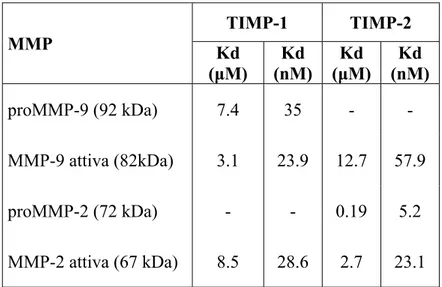
The forms of MMP-9 and MMP-2 have different dissociation constants from the inhibitors, due to different ways of interaction with them (Table
3; Figure 15).
Table 3. Dissociation constants for active and pro MMP-9. (Olson MW et al., 1997). MMP TIMP-1 TIMP-2 Kd (μM) Kd (nM) Kd (μM) Kd (nM) proMMP-9 (92 kDa) 7.4 35 - - MMP-9 attiva (82kDa) 3.1 23.9 12.7 57.9 proMMP-2 (72 kDa) - - 0.19 5.2 MMP-2 attiva (67 kDa) 8.5 28.6 2.7 23.1
Figure 15 formation Adapted f Althoug has bee TIMP-2 Regardi higher a negative the C-te are also interacti proMM 1995). Even in affinity 5. Interaction n of the comp from Murphy gh TIMP-1 en observed 2 can bind M ing to the ac affinity than ely charged erminal dom o responsibl ion betwee MP-2 still h n the case o to the TIM n of TIMP-1 w plex. G. and Willen binds speci d that it is MMP-9, but ctive MMP-n the biMMP-ndiMMP-n d C-terminal main of MM le for the b en the N-te has the pro
of MMP-9, MP-1 compar with active MM nbrock F, 199 ifically and also able to t with low a -2, the bind ng of TIMP l tail of TIM MP-2. Thes inding of T erminal dom opeptide (M the enzym red with TI MP-9 and pro 95. with high a o bind MM affinity. ding of TIM P-1. This is MP-2 which se interacti TIMP-2 to p mains are Murphy G. me binds fas IMP-2, and oMMP-9 duri affinity to M MP-2; more MP-2 is faste s probably d h can specifi ons betwee proMMP-2, not possib and Willen ster and wi this seems ing the MMP-9, it over, also r and with due to the ically bind en charges , since the ble as the nbrock F, ith greater due to the
interaction between the C-terminal domains of the two proteins. In fact, it has been shown that the TIMP-2 binds only to the catalytic site (with low affinity) of MMP-9, without any interaction between the C-terminal domains (figure 15).
As for the association between proMMP-2/2, the ability of TIMP-1 to bind to proMMP-9 is due to the interaction between the C-terminal domains of the two proteins.
The inhibitors can bind both sites on the active form of the enzymes except for the TIMP-2/active MMP-9 interaction, where only the N-terminal domains are involved (figure 15).
The inhibitory activity of TIMP-1 and TIMP-2 is due to the interaction of the first five amino acids (from Cys1 to Pro5) and the strand C of the
inhibitor with the enzyme (Bode et al., 1999).
The first five residues of TIMP bind the active site of MMP mimicking the P1-P1’-P2’-P3’-P4’ sites of a possible substrate.
The Ser68/Val69 residues of TIMP-1 and Ala70/Val71 of TIMP-2 from the
strand C interact with the S2 and S3 sites on the enzyme in a reverse way respect to a substrate (Bode et al., 1999) (Figure 16).
Figure 1 MMP-9. The TIMP From a catalytic Cys1 oc a hydro in MM experim inhibitio The Thr with the bond wi The Cy S2’, S3’ 16.Mechanism P-2 follows a all these in c zinc, avoi ccupies roug gen bond w MP-2, avoid ments have b on (Bode et r2 and Ser2 e site S1’ o ith the catal ys3, Val4 (T ’ and S4’ si m of interact similar mecha nteractions, iding the en ghly the site with the cata
ding any c been shown t al., 1999). residues re of the MMP lytic residue TIMP-1) or tes as a sub tion of the N anism, only th the Cys1 o nzyme to b e of water bi alytic residu catalytic fu n that this in espectively P-9; the Ser e Glu404 of M Ser4 (TIM strate shoul N-terminal d he interacting of the TIM be active. T inding in th ues Glu402 i unction. Ho nteraction is of TIMP-1 r2 of TIMP MMP-2. MP-2) and P ld do (figur domains of T residues chan MP is place he α-amino he active site in MMP-9 a owever, mu s not essent and TIMP-P-2 forms a Pro5 interact re 16). TIMP-1 and nge. ed on the o group of e, forming and Glu404 utagenesis tial for the -2 interact hydrogen t with the
Different forms of gelatinases
We can distinguish different forms of gelatinases, both not active or active.
MMP-2 exists only as monomeric form: a proenzyme of 72 kDa, which is not glycosylated; an active form of 62 kDa generated after plasma membrane activation with the N-terminal sequence starting at Tyr81.
Moreover, an additional active species of about 45 kDa was reported, with high specific activity (Fridman R et al., 1995). This last form lacks the C-terminal domain, which is important for the interaction with its inhibitor TIMP-2. Thus, the nature of the active species of MMP-2, formed after plasma membrane activation, may regulate enzyme activity and inhibition by TIMP-2 (Fridman R et al., 1995).
Also MMP-9 exists in different forms: a monomer of 92 kDa, corresponding to the inactive enzyme, an homodimer of ~220 kDa and an heterodimer of ~120-130 kDa bound to the lipocalin-2 or NGAL (Neutrophil Gelatinase-Associated Lipocalin; Olson et al., 2000). All these forms were identified in different MMP-9-producing cell types, both normal or pathological, and in various biological fluids and tissues, confirming that both dimers and monomers are physiologically present in the human organism.
Concerning dimerization, it takes place within the cell (Dufour A. et al., 2010) involving the formation of a disulfide bridge between two cysteines of the blade IV of the hemopexin-like domain (Dufour A. et al., 2010).
Figure 1 dimerizat The het been fo in the e with th although MMP-9 2001). In addit forms a produce these tw which is 1. In fac decrease 7. Representa tion. erodimeric und within extracellula he monome h it has b 9 can preve tion to dime as previous ed by MM wo forms is s necessary ct, it has be es the rate ation of two h form of MM the tertiary ar space aft r. However been shown
ent the deg ers and pro sly reported P-3 and A the lack of y to bind wit een propose of format emopexin-like MP-9 coval y granules o er different r, its physi n that the a gradation of oenzyme for d: an 82 k APMA activ f C-terminal th high affin ed that the C ion of the ke domains inv lently assoc of neutroph t degranula iological ro association f the gelat rm, MMP-9 kDa and a vation. The l domain in nity the spe C-terminal t enzyme-in volved in the iated with N hils, and it i ation stimul ole is not between N inase (Yan 9 exists in t 66 kDa fo e difference n the 66 kDa ecific inhibit truncation o nhibitor com NGAL has is released li together yet know NGAL and n L et al., two active orm, both e between a MMP-9, tor TIMP-of MMP-9 mplex (O’
Connell JP et al., 1994) and might reduce the TIMP-1 binding ability (Roeb E et al., 2000). However, up to now this potentially uncontrolled MMP-9 active form is not yet evidenced in body fluids.
Inflammatory conditions
Several evidences exist on the role of MMP-9 in pathological conditions. When the failure of regulatory mechanisms occurs, it might lead to diminished or excessive production of MMP-9 and subsequently to restricted, extensive, or improperly timed degradation of ECM (Van den Steen PE et al., 2002). Examples of this pathway are premature rupture of amniotic membrane and pathologic bone resorption. Other mechanisms of disease induction are inflammation, infection, vascular pathology, degeneration and malignancy.
In particular, MMP-9 has been associated to inflammatory conditions playing a dual role: direct, by tissue destruction when its activity is not controlled by TIMP-1, or indirect, by generation of an inflammatory signal or recruitment of inflammatory cells (Delclaux et al., 1996).
Its role is well-accepted in neuroinflammatory diseases where the impairment of the Blood Brain Barrier (BBB) occurs, such as Multiple Sclerosis (MS), Intra Cerebral Hemorrhage (ICH), brain ischemia, as the up-regulation of the total enzyme levels were detected, but there are not information on the levels of the active form of MMP-9.
On the other hand, the role of MMP-2 in inflammatory conditions is controversial. In fact, MMP-2 has been associated to both tissue damage and repair (Itoh T et al., 2002; Yong VW, 2005).
One evidence of the role of MMP-2 in the repair of injured tissue after the onset of inflammation is its ability to process a chemokine, the
MCP-3 (Monocyte Chemoattractant Protein-MCP-3), resulting in a shorter form with antagonist activity on receptors that bind the entire MCP-3 or other chemokines (McQuibban GA et al., 2000).
The production of this form causes a variation in the chemotactic gradient with a corresponding decrease in the recruitment of other inflammatory cells that may produce lytic enzymes such as MMP-9 (i.e. monocytes and macrophages). Thus, MMP-2 seems to contribute to the resolution of the inflammation, a very important aspect of tissue repair. In addition, the TGF-β1, a growth factor that starts the tissue repair, stimulates the MMP-2 production but inhibits other MMPs: this could contribute to the attenuation of the inflammatory response by decreasing pro-inflammatory stimuli (McQuibban GA et al., 2000).
Therefore, I aimed to evaluate the levels of active 9, active MMP-2, and their inhibitors TIMP-1 and TIMP-MMP-2, in inflammatory pathologies in order to clarify the role of the active enzymatic forms as biomarkers of inflammation and resolution of the inflammatory process. Furthermore, I evaluated the influence of pH environmental changes on MMP-9 release from neutrophils, as different pH conditions in tissues are associated with different inflammatory status (Trevani AS et al., 1999; Kellum JA et al., 2004).
Multiple Sclerosis
Multiple Sclerosis (MS) is a chronic demyelinating inflammatory disease which affects the Central Nervous System (CNS), in particular the white matter; it is characterized by a progressive loss of myelin and oligodendrocytes and axonal damage (Noseworthy JH et al., 2000). The etiology of MS is still unknown, although it is thought to be on autoimmune base, where the loss of tolerance to one or more unidentified
myelin and oligodendrocytes antigens occurs, starting an immune response (Noseworthy JH et al., 2000).
The progressive myelin degradation in several points of the CNS expresses as clinical variable symptoms, as the destruction of myelin sheaths slows or blocks the transmission of nerve impulses along the fibres of the brain and spinal cord (Noseworthy JH et al., 2000). The areas of the white matter interested in the removal of the myelin are known as demyelination plaques.
Different clinical subtypes of MS are defined on the basis of the disease course (Figure 18):
o Relapsing-Remitting (RR), which affects the 80 % of the MS patients, characterized by the abrupt start of symptoms and acute episodes of worsening (exacerbations or relapses), with complete or partial recovery. Between these episodes, patients may be clinically stable, may experience gradual progression or disability, or may undergo to a combination of both (Hauser S et al., 2006);
o Secondary Progressive (SP), in which the RR subtype converts in most cases, characterized by gradual progression of disability with or without superimposed relapses;
o Primary Progressive (PP), that accounts for 10-15 % of all the MS clinical subtypes and it is characterized by gradual progression of disability from disease onset, without superimposed relapses (Hauser S et al., 2006).
Figure 18 The dia characte clinical MRI is active paramag Figure 19 second is 8. Clinical su gnosis of M erize the di examinatio a powerfu lesions en gnetic contr 9. Demyelina s after the adm
btypes of MS. MS is based sease (Nose on and the M ul instrumen nhanced aft rast agent (M ting lesion ev ministration of . on the pres eworthy JH Magnetic Re ntal tool th ter the adm Miller DH e videnced by M of gadolinium ence of sym H et al., 200 esonance Im hat can evi dministration
et al., 1998)
MRI: the first i m. mptoms and 00), detected maging (MR idence dem n of gado ) (Figure 19 is hyperintens signs that d with the RI). myelinating linium, a 9). se, the
As the assumption of gadolinium reflects an alteration of the Blood Brain Barrier (BBB) due to the ongoing inflammation, the foci with contrast enhancement are considered plaques of recent onset where the inflammatory process is still active. On the contrary, the lesions without contrast enhancement are defined as early onset where the inflammatory process is idle (Hawkins et al., 1990). Therefore, MRI is able to detect disease activity as clinical examination do but with more sensitivity, because the appearance of a enhanced lesion may also occur in the absence of relapses or clinical disease activity. This could be due to the appearance of the lesion in a brain area “silent” form a functional point of view, without causing symptoms.
Currently, it is thought that MS is caused by the infiltration into the CNS of autoreactive CD4+ T helper 1 (Th1) lymphocytes, characterized by the production of pro-inflammatory cytokines and directed against myelin components (Prat et al., 2005). In normal conditions, these lymphocytes encounter a hostile and inhospitable anti-inflammatory microenvironment that prevents the starting of any immune reaction. In MS, the presence of a chronic inflammation into the CNS triggered by the tissue damage converts the microenvironment into a pro-inflammatory type, characterized by the massive recruitment of inflammatory cells and blood proteins into the CNS through the BBB. It causes the accumulation of chemical mediators and amplification of the tissue damage (Prat et al., 2005). Such mediators are proinflammatory cytokines, chemokines and metalloproteases, that facilitate the infiltration of other cells like macrophages, CD4+ Th1 and Th2 lymphocytes, B lymphocytes and CD8+ T lymphocytes (Opdenakker et al, 2003). These cells can increase the tissue damage directly through the cytotoxic action, as well as indirectly through the release of soluble products toxic for the
myelin and neurons (Opdenakker et al., 2003). Among these molecules, MMPs may act directly causing damage to the myelin and axons and indirectly by favouring the infiltration of immune cells into the CNS through the BBB. Of all the MMPs, an over-expression of MMP-9 not counterbalanced by the action of TIMP-1 seems to be a crucial step involved in the modulation of the immune and inflammatory responses in MS. In fact, an impairment in the MMP-9/TIMP-1 balance towards uncontrolled proteolytic activity of MMP-9 may lead to promotion of the BBB degradation and to the myelin damage (Opdenakker et al., 2003). Thus, the MMP-9 proteolytic activity can favour the recruitment of inflammatory cells into the CNS enhancing the ongoing inflammation (Opdenakker et al., 2003). Furthermore, the importance of this enzyme in MS pathogenesis was confirmed by neuropathological studies observing that MMP-9 is expressed by all the cells into the demyelinating lesions typical of MS (Lindberg et al., 2001).
Recently, it has been shown that the variation of active MMP-9, which is the only form that exerts the catalytic activity, rather than the total MMP-9 may be a more appropriate biomarker to monitor the status and course of the disease (Fainardi E et al., 2006). In fact, it has been observed that the levels of active MMP-9 in serum and cerebrospinal fluid (CSF) of MS patients were increased during active state of disease evidenced by both clinical examination and MRI investigation (Fainardi E et al., 2006). Furthermore, it has been found a decrease in TIMP-1 levels in patients with clinical and MRI active state of disease: this is in line with the hypothesis of an imbalance towards the proteolytic activity of MMP-9. Therefore, the variation in the active MMP-9/TIMP-1 ratio might be used as biomarker of disease activity, to monitor the disease course even during pharmacological therapy (Fainardi E et al., 2006).
Unlike MMP-9 that is predominantly up-regulated during inflammatory conditions, MMP-2 is constitutively expressed in the brain (Rosenberg GA, 2002). An over-expression of MMP-2 could be both protective and harmful in MS (Yong VW, 2005; Yong VW et al., 2007) since MMP-2 seems to promote tissue repair as well as penetration of CD4+ Th1 cells and macrophages across the BBB and myelin and/or axonal injury. Contradictory findings have also been found in previous neuropathological, CSF, and blood studies in which MMP-2 levels in acute and chronic demyelinated lesions as well as in CSF, in serum and in peripheral blood mononuclear cells (PBMCs) were increased, decreased, or represented in equivalent amounts in MS patients when compared to controls (Anthony DC et al., 1997; Lindberg LR et al., 2001; Avolio C et al., 2003; Kouwenhoven M et al.,2003).
Spontaneous Intra Cerebral Hemorrhage (SICH)
Within the various forms of stroke, spontaneous cerebral intraparenchymal hematoma (Spontaneous Intra Cerebral Hemorrhage, SICH) is a relatively common disease which accounts for 10-15% of all cerebrovascular strokes, with a worldwide incidence from 10 to 20 cases each 100.000 inhabitants increasing with age (Qureshi et al., 2001; Xi et al., 2006). Currently, the spontaneous intraparenchymal hematoma is the main cause of death and disability worldwide (Qureshi et al., 2001). Spontaneous intraparenchymal hematoma is a leakage of blood into the brain parenchyma due to the rupture of small penetrating arteries that originate from the major intracranial arteries (Figure 20).
Figure 20 Intrapar phenom (TIA), quickly or hours The wo hematom bleeding the mos the hem hemorrh The intr center o and the edemato al., 200 0. Spontaneou renchymal mena as hap but begins and contin s (Qureshi e orsening of ma may be g that incre st common matoma, wh hage onset ( raparenchym of the lesion periphery ous area sur
1). us intraparen hemorrhag ppens in br s with acut nuously, rea et al., 2001) f the sympt primarily eases the po cause of n hich usually (Mayer SA mal hemato n (hemorrh of the lesio rrounding t nchymal hema ge is no rain stroke te focal ne aching the h ). toms in th caused by t ossibility of neurologic d y occurs wi et al., 1994 oma is comp agic core) t on (peri-he the central c atoma. ot precede with transi eurological highest leve he hours fo the extensio f a negative deterioration ithin the fir 4). posed by tw that represe matomal a core (Figur ed by pr ient ischem defects tha el within few ollowing the on of intrav e outcome. n is the exp rst 3 hours wo different
ents the blee
area), consi re 19) (Pari emonitory mic attacks at worsen w minutes e cerebral ventricular However, pansion of from the areas: the eding area, isting of a izel PM et