REVIEW
Guillain- Barré syndrome in SARS- CoV-2 infection: an
instant systematic review of the first six months
of pandemic
Antonino Uncini ,
1Jean- Michel Vallat,
2Bart C Jacobs
3To cite: Uncini A, Vallat J- M, Jacobs BC. J Neurol Neurosurg Psychiatry 2020;91:1105–1110. 1Department of Neuroscience, Imaging and Clinical Sciences, University “G. d’Annunzio” Chieti- Pescara, Chieti, Italy 2Department of Neurology, National Reference Center for “Rare Peripheral Neuropathies”, CHU Dupuytren, Limoges, France
3Departments of Neurology and Immunology, Erasmus MC, Rotterdam, The Netherlands Correspondence to Professor Antonino Uncini, Department of Neuroscience, Imaging and Clinical Sciences, University “G. d’Annunzio” Chieti- Pescar, Chieti, Italy; uncini@ unich. it Received 11 July 2020 Revised 10 August 2020 Accepted 10 August 2020 Published Online First 27 August 2020
© Author(s) (or their employer(s)) 2020. No commercial re- use. See rights and permissions. Published by BMJ.
ABSTRACT
A systematic review from 1 January to 30 June 2020 revealed 42 patients with Guillain- Barré syndrome (GBS) associated with SARS- CoV-2 infection. Single cases and small series were reported from 13 countries, the majority from Europe (79.4%) and especially from Italy (30.9%). SARS- CoV-2 infection was demonstrated by nasopharyngeal swab (85.7%) and serology (14.3%). Median time between COVID-19 and GBS onset in 36 patients was 11.5 days (IQR: 7.7–16). The most common clinical features were: limb weakness (76.2%), hypoareflexia (80.9 %), sensory disturbances (66.7 %) and facial palsy (38.1%). Dysautonomia occurred in 19%, respiratory failure in 33.3% and 40.5% of patients were admitted in intensive care unit. Most patients (71.4%) had the classical clinical presentation but virtually all GBS variants and subtypes were reported. Cerebrospinal fluid (CSF) albumin- cytological dissociation was found in 28/36 (77.8%) and PCR for SARS- CoV-2 was negative in 25/25 patients. Electrodiagnosis was demyelinating in 80.5% and levels 1 and 2 of Brighton criteria of diagnostic certainty, when applicable, were fulfilled in 94.5% patients. Antiganglioside antibodies were positive in only 1/22 patients. Treatments were intravenous immunoglobulin and/or plasma exchange (92.8%) with, at short- time follow- up, definite improvement or recovery in 62.1% of patients. One patient died. In conclusion, the most frequent phenotype of GBS in SARS- CoV-2 infection is the classical
sensorimotor demyelinating GBS responding to the usual treatments. The time interval between infectious and neuropathic symptoms, absence of CSF pleocytosis and negative PCR support a postinfectious mechanism. The abundance of reports suggests a pathogenic link between SARS- CoV-2 infection and GBS but a case- control study is greatly needed.
INTRODUCTION
SARS- CoV-2 and its associated COVID-19 were reported to originate in December 2019 in Wuhan (China) spreading rapidly around the world. On 30 January 2020, the WHO declared the SARS- CoV-2 a public health emergency of international concern and by 6 July, when this review was completed, there have been 11. 327 .790 confirmed cases of COVID-19 including 532. 340 deaths.
COVID-19 is a systemic disorder presenting typically with fever and respiratory symptoms but neurological manifestations such as acute cere-brovascular diseases, seizures, ageusia, anosmia
meningitis, encephalitis and skeletal muscle involve-ment were soon reported.1 More recently,
progres-sively increasing case reports of Guillain- Barré syndrome (GBS) in SARS- CoV-2 infection raised the concern over a possible association.
This may not surprise as approximately 70% of patients with GBS have a preceding illness and infectious agents such as Campylobacter jejuni, Influenza virus, Cytomegalovirus and recently Zika virus have been demonstrated to trigger GBS.2–5
Aim of this study is, by a systematically review of the reported cases of GBS in SARS- CoV-2 infec-tion, to clarify the clinical and electrophysiological phenotype, to discuss, on the basis of the available data, whether the disease mechanism could be para-infective or postpara-infective and to speculate on the possible pathogenesis.
METHODS
A PubMed search was completed on 6 July to identify papers reporting patients with GBS with SARS- CoV-2 infection and COVID-19 from 1 January to 30 June 2020 using the following terms: “Guillain- Barré syndrome”, “Miller Fisher syndrome”, “cranial polyneuritis”, “facial diplegia”, “Acute sensory ataxia”, “Bickerstaff encephalitis” “acute inflammatory demyelinating polyneurop-athy”, “acute motor axonal neuroppolyneurop-athy”, “acute motor and sensory axonal neuropathy” combined with “SARS- CoV-2” and “COVID-19”. Refer-ence lists of articles were also examined. Full- text papers in English were analysed and those reporting sufficiently detailed information, according to a predefined list of 12 items (the headings of online supplementary table 1), were selected. Duplicated reports were searched by author names and patient’s characteristics. Data were extracted from reports according to a template. Clinical characteristics were retrieved as the number of patients in whom the variable was present in the numerator, and the total number of reported cases in the denom-inator: n/N (%). If clinical features were reported at multiple time points, data representing the full disease course were presented. Continuous vari-ables (age, time between infectious and neuropathic symptoms) were expressed as medians. Certainty of GBS and Miller Fisher syndrome (MFS) diagnosis was assessed, on the basis of the reported findings, by the Brighton Collaboration GBS Working Group criteria.6 Level 1 of Brighton criteria indicate the
highest degree of diagnostic certainty supported
G.D'Annunzio. Protected by copyright.
on March 29, 2021 at Universita degli Studi
http://jnnp.bmj.com/
1106 Uncini A, et al. J Neurol Neurosurg Psychiatry 2020;91:1105–1110. doi:10.1136/jnnp-2020-324491
by nerve conduction studies and the presence of albuminocyto-logical dissociation in cerebrospinal fluid (CSF). A level 2 diag-nosis is supported by either a CSF white- cell count of less than 50 cells/µl (with or without an elevated protein level) or nerve conduction studies consistent with the polyneuropathy patterns described for GBS and MFS (if the CSF is unavailable). A level 3 diagnosis is based on clinical features without support from nerve conduction or CSF studies. A diagnostic classification was also employed to categorise the different GBS and MFS presen-tations.7 This systematic review was made following, when
applicable, the Preferred Reporting Items for Systematic Reviews and Meta- Analyses statement.8
RESULTS
A total of 33 papers (28 single case reports) describing GBS vari-ants and subtypes in patients with SARS- CoV-2 infection were found. No duplicated reports were identified and overall 42 patients were in included the systematic review.9–41
The demographic data, the clinical, laboratory and imaging findings of the 42 patients, are detailed in the online supple-mental table 1 and summarised in tables 1 and 2. Because of the characteristics of the reports, the low number of patients and the variability in the reported features, we described the studies and summarised their results qualitatively and quantitatively rather than by a meta- analysis approach. The median age was 57.5 years, and the majority of patients were men (64.3%). The first patient, a woman who returned from Wuhan to Shanghai, was reported online on 1 April 2020.9 Overall, patients were
reported from 13 countries but mostly from Europe (79.4%) and especially from Italy (30.9%).
Six (14.3%) patients were admitted to the hospital because of COVID-19 symptoms and developed GBS during hospital-isation; 4 (9.5%) were admitted for COVID-19, discharged and then readmitted because of the onset of neuropathic symptoms; 32 (76.2%) patients presented to the hospital because of neuro-pathic symptoms. The diagnosis of SARS- CoV-2 infection was made by positive RT- PCR of nasopharyngeal swab in 36 (85.7%) patients (sometimes after repeated tests) and when negative by serology in 6 (14.3%) patient (online supplementary table). Diagnosis of SARS- CoV-2 infection was made before the onset of GBS in 16 (38.1%) patients, in 21 (50%) patients during the hospitalisation for GBS and in 2 (4.8%) patients retrospectively by serology. In three patients, no information on the timing of diagnosis was given.
Other preceding infections related to GBS such as C. jejuni,
Cytomegalovirus, hepatitis E, Mycoplasma pneumoniae and
Epstein- Barr virus, varicella zoster virus, HIV were variably searched for and excluded in seven patients.
Most frequent presenting symptoms of COVID-19 were fever (73.8%) and cough (66.7%). Hypoageusia and hypoanosmia were reported in 38% and 26.2% of patients. One patient did not present any sign of and four were oligosymptomatic showing only mild loss of smell and taste, or dry cough and low- grade fever that resolved spontaneously in few days.16 25 30 32 36
Inter-stitial pneumonia was documented by chest RX or CT in 61.9% of patients. Specific information on treatment of COVID-19 was reported in 20 (47.6%) patients. Thirteen patient were treated with hydroxychloroquine and antiretroviral drugs (lopinavir and ritonavir) alone or in combination. Seven patients received azith-romycin alone or in combination with hydroxychloroquine. In two patients, additional treatment with steroids or tocilizumab was reported.
The temporal relationship between onset of COVID-19 symptoms and GBS was not reported or not calculable in four (9.5%) patients. In the very first reported, Chinese patient, the onset of neuropathic symptoms preceded by 8 days fever, cough, pneumonia and positive nasopharyngeal swab. However, at admission, the patient presented lymphocytopenia and throm-bocytopenia suggesting an antecedent SARS- CoV-2 infec-tion.9 In another patient, GBS was the presenting feature and
COVID-19 symptoms never developed although chest CT revealed ground glass opacities.30 The mean interval between
onset of COVID-19 and GBS symptoms in the remaining 36 patients was 11.5 days (IQR: 7.7–16; range: 3–28 days). In 11/42 (26.2%) patients, the COVID-19 symptomatology clini-cally resolved before the onset of GBS. In the remaining patients, the neuropathic symptomatology started while COVID-19 was ongoing. The most commonly reported clinical features of GBS were: limb weakness (64.3% tetraparesis, 11.9% lower limbs
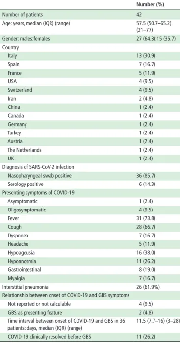
Table 1 Demographic, geographical, clinical and laboratory features of patients with SARS- CoV-2 and Guillain- Barré syndrome (GBS)
Number (%)
Number of patients 42
Age: years, median (IQR) (range) 57.5 (50.7–65.2) (21–77) Gender: males:females 27 (64.3):15 (35.7) Country Italy 13 (30.9) Spain 7 (16.7) France 5 (11.9) USA 4 (9.5) Switzerland 4 (9.5) Iran 2 (4.8) China 1 (2.4) Canada 1 (2.4) Germany 1 (2.4) Turkey 1 (2.4) Austria 1 (2.4) The Netherlands 1 (2.4) UK 1 (2.4)
Diagnosis of SARS- CoV-2 infection
Nasopharyngeal swab positive 36 (85.7) Serology positive 6 (14.3) Presenting symptoms of COVID-19
Asymptomatic 1 (2.4) Oligosymptomatic 4 (9.5) Fever 31 (73.8) Cough 28 (66.7) Dyspnoea 7 (16.7) Headache 5 (11.9) Hypoageusia 16 (38.0) Hypoanosmia 11 (26.2) Gastrointestinal 8 (19.0) Myalgia 7 (16.7) Interstitial pneumonia 26 (61.9%) Relationship between onset of COVID-19 and GBS symptoms
Not reported or not calculable 4 (9.5) GBS as presenting feature 2 (4.8) Time interval between onset of COVID-19 and GBS in 36
patients: days, median (IQR) (range)
11.5 (7.7–16) (3–28) COVID-19 clinically resolved before GBS 11 (26.2)
G.D'Annunzio. Protected by copyright.
on March 29, 2021 at Universita degli Studi
http://jnnp.bmj.com/
paraparesis), hypoareflexia (80.9 %), sensory disturbances (66.7 %) and facial palsy (38.1%, in 81.2% bilateral). Dysautonomia occurred in 19.5% of patients. Three patients had urinary reten-tion,14 18 38 three unstable blood pressure,31 40 one severe drug-
resistant hypertension,19 one gastroplegia, paralytic ileus and
loss of blood pressure control.28 One patient showed transient
episodes of confusion and agitation possibly imputable to the severity of COVID-19.21 According to the clinical diagnostic
classification, most patients (71.4%) had the classic form of GBS characterised by symmetrical weakness of the limbs, sensory symptoms and reduced or absent tendon reflexes. Two patients (4.8%) had the paraparetic form, three (7.1%) facial diplegia with or without paraesthesia, two (4.8%) cranial polyneuritis, three (7.1%) MFS and two (4.8%) could be classified as an acute ataxic neuropathy. Most of patients (80.5%) had electrodiag-nostic features of acute inflammatory demyelinating polyradic-uloneuropathy (AIDP). Five patients (13.9%) were reported to have the acute motor and sensory axonal subtype; however, the examination of the electrophysiological results, when available, showed in at least one case increase distal motor latencies, slow conduction velocities and prolonged F wave latencies well in the demyelinating range.5 11 CSF examination was performed in 36
(85.7%) patients and showed albuminocytological dissociation in 28 (77.8%). The search for the viral RNA in CSF was nega-tive in all 25 cases in whom was done. Antigangliosides anti-bodies were searched in 22 (52.4%) patients and IgG to GD1b was found in only one (4.5%) patient with MFS.13 MRI of head,
spine and nerves was performed in 25 (59.5%) patients and showed thickening or contrast enhancement of cranial nerves and of nerve roots in 10 (40%) patients. A leptomeningeal brain-stem and spine enhancement was described in one patient.33
The Brighton criteria for diagnostic certainty of GBS and MFS were not applicable in the seven patients diagnosed as facial diplegia, cranial polyneuritis and acute ataxic neuropathy. In the remaining 35 patients: 54.3% reached the level 1 (the highest) of certainty, 40% the level 2 and 5.7% the level 3. Respiratory failure occurred in 14 (33.3%) patients and required invasive ventilation in 12 (85.7%). Intensive care unit (ICU) admission was required in 40.5% of patients. In 12 patients, respiratory failure developed after the diagnosis of GBS; on the other hand, two patients were already on invasive ventilation for COVID-19 pneumonia when neuropathic symptoms started.28 It was often
difficult to dissect in the individual subject the relative contribu-tion of interstitial pneumonia and GBS in inducing respiratory failure. However, the presence of hypercapnia, paradox respira-tion, acidosis (suggesting a neuromuscular respiratory failure) or
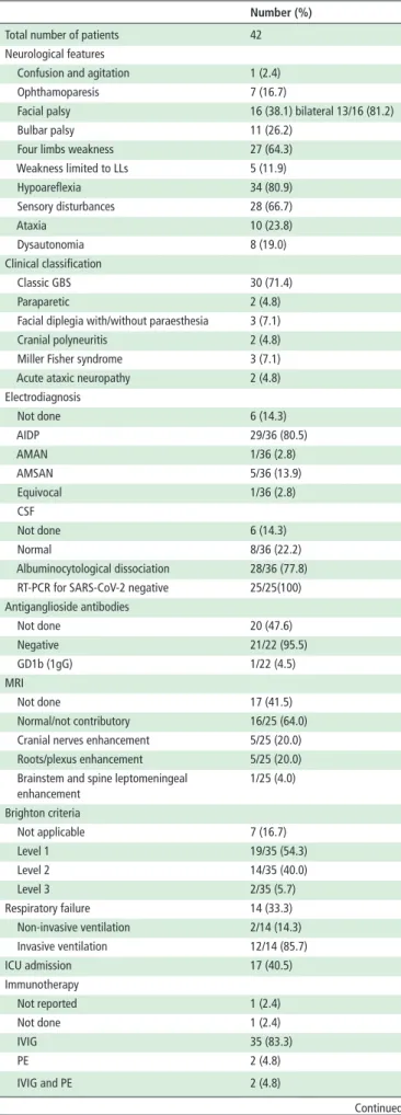
Table 2 Clinical and laboratory features of patients with Guillain- Barré syndrome (GBS) associated with SARS- CoV-2 infection
Number (%) Total number of patients 42 Neurological features
Confusion and agitation 1 (2.4) Ophthamoparesis 7 (16.7)
Facial palsy 16 (38.1) bilateral 13/16 (81.2) Bulbar palsy 11 (26.2)
Four limbs weakness 27 (64.3) Weakness limited to LLs 5 (11.9) Hypoareflexia 34 (80.9) Sensory disturbances 28 (66.7) Ataxia 10 (23.8) Dysautonomia 8 (19.0) Clinical classification Classic GBS 30 (71.4) Paraparetic 2 (4.8) Facial diplegia with/without paraesthesia 3 (7.1) Cranial polyneuritis 2 (4.8) Miller Fisher syndrome 3 (7.1) Acute ataxic neuropathy 2 (4.8) Electrodiagnosis Not done 6 (14.3) AIDP 29/36 (80.5) AMAN 1/36 (2.8) AMSAN 5/36 (13.9) Equivocal 1/36 (2.8) CSF Not done 6 (14.3) Normal 8/36 (22.2) Albuminocytological dissociation 28/36 (77.8) RT- PCR for SARS- CoV-2 negative 25/25(100) Antiganglioside antibodies Not done 20 (47.6) Negative 21/22 (95.5) GD1b (1gG) 1/22 (4.5) MRI Not done 17 (41.5) Normal/not contributory 16/25 (64.0) Cranial nerves enhancement 5/25 (20.0) Roots/plexus enhancement 5/25 (20.0) Brainstem and spine leptomeningeal
enhancement 1/25 (4.0) Brighton criteria Not applicable 7 (16.7) Level 1 19/35 (54.3) Level 2 14/35 (40.0) Level 3 2/35 (5.7) Respiratory failure 14 (33.3) Non- invasive ventilation 2/14 (14.3) Invasive ventilation 12/14 (85.7) ICU admission 17 (40.5) Immunotherapy Not reported 1 (2.4) Not done 1 (2.4) IVIG 35 (83.3) PE 2 (4.8) IVIG and PE 2 (4.8) Continued Number (%) Oral steroids 1 (2.4) Follow- up Not reported 5 (12.0) Death 1/37 (2.7) No improvement 4/37 (10.8) Minimal/slight improvement 7/37 (18.9) Definite improvement 16/37 (43.2) Recovery 7/37 (18.9)
AIDP, acute inflammatory demyelinating polyneuropathy; AMAN, acute motor axonal neuropathy; AMSAN, acute motor and sensory axonal neuropathy; CSF, cerebrospinal fluid examination; ICU, intensive care unit; IVIG, intravenous immunoglobulin; LLs, lower limbs; PE, plasma exchange; RT- PCR, reverse transcriptase PCR.
Table 2 Continued
G.D'Annunzio. Protected by copyright.
on March 29, 2021 at Universita degli Studi
http://jnnp.bmj.com/
1108 Uncini A, et al. J Neurol Neurosurg Psychiatry 2020;91:1105–1110. doi:10.1136/jnnp-2020-324491
a picture of mild interstitial pneumonia not compatible with the respiratory status could be identified in 7/14 (50%) patients indi-cating that in these cases GBS was the main cause for respiratory failure.10 12 17 27 35 37 38
Thirty- five (87.5%) patients were treated with intravenous immunoglobulins (IVIG) and two (5%) with plasma exchange. Two (5%) patients received both. In one patient, low dosage of oral steroids was administered.22 In one patient, treatment
was not reported and another with bilateral VI nerve palsy and areflexia was treated symptomatically with acetaminophen and spontaneous recovery occurred in 2 weeks.13
Follow- up was not reported in 5/42 (12%) patients; follow- up interval was specified in 15 patients and the median time was 14 days (IQR 10–30, range 1–60 days). No improvement was described in 4/37 (10.8%) patient, minimal or slight improve-ment in 7 (18.9%), definite improveimprove-ment in 16 (43.2%) and recovery in 7 (18.9%). Only one patient died.19
DISCUSSION
Starting from 1 April and until 30 June 2020, 42 patients with SARS- CoV-2 infection and GBS have been reported mostly from Europe and particularly from Italy. Curiously, looking at the chronology of the online publications (online supplementary table), it seems that case reports have followed, with some delay, the virus trail: first from China, then Iran, France, Italy, Spain and USA. The classical clinical sensorimotor presentation with hypoareflexia, with or without cranial nerve involvement, was most frequent but virtually all GBS variants and subtypes were described. According to the Brighton criteria, 94% of patients fulfilled the level 1 or 2 indicating a high diagnostic certainty for GBS or MFS. Respiratory failure occurred in about one- third and ICU admission was required in at least 40% of patients. Interstitial pneumonia due to COVID-19 was reported in about two- third of patients. In some patients, it may be difficult to assess the relative role of COVID-19 and GBS in determining the disease severity and the cause of respiratory failure, above all in those in whom GBS developed during ICU stay. Some features as hypercapnia, paradox respiration, acidosis or a discrepancy with the severity of interstitial pneumonia indicated a neuromuscular respiratory failure. The great majority of patients showed the AIDP electrophysiological subtype and CSF albuminocytological dissociation. These features together with cranial nerve involve-ment can help to distinguish in the ICU setting GBS from critical illness neuropathy and/or myopathy that is expected to happen in patients with severe COVID-19. Patients were treated mainly with IVIG and, at a short time follow- up, 62% showed defi-nite improvement or recovered. This information is important as suggests that GBS in SARS- CoV-2 infection should be treated in the usual way. The abundance of GBS cases reported during the COVID-19 outbreak worldwide may suggest a possible pathogenic link between SARS- CoV-2 and GBS. To date, the incidence of GBS in the ‘COVID-19 era’ has been analysed only in Friuli Venezia- Giulia, Italy, where an unusual cluster (eight cases) occurred in the months of March and April 2020.23
Monthly incidence was 0.65/100.000 versus 0.12/10.000 in the same months of previous years with 5.41- fold increase. However, only one patients (twice negative at swab test) had positive serology for SARS- CoV-2 and chest CT scan showing interstitial pneumonia. SARS- CoV-2 may damage the nerves directly by invadingthe nerves, or by triggering a postinfectious immune response, either via systemic effects as a cytokine storm, or specific attack to neural targets.42 43 The frequent early, and
often persistent, anosmia and ageusia reported in large cohorts,1
and the combination with other cranial neuropathies in the cases we analysed, may suggest a neurotropism of SARS- CoV-2. Two similar coronaviruses SARS- CoV and MERS- CoV can enter the CNS via olfactory nerves or retrograde axonal trans-port through other cranial nerves.44 The reported MRI studies
showing enhanced oculomotor, trigeminal, facial, nerves roots and plexuses may corroborate this hypothesis but could also simply reflect a postinfectious nerve inflammation. On the other hand, the SARS- CoV-2 spike protein interacts with the GalNAc residue of GM1 and ganglioside dimers for anchoring to cell surface and an immune cross reaction between epitopes within the spike- bearing gangliosides and the sugar residues of surface peripheral nerve glycolipids is possible.45 How SARS- CoV-2 may
trigger GBS is currently debated, especially if there is a parain-fectious or postinparain-fectious (immune- mediated) pathogenesis. GBS is considered the prototype of postinfectious neuropathy usually developing 1–3 weeks after an acute infection.3 Parainfectious
neuropathies, as those caused by Borrelia Burgdorferi, Brucella and West Nile virus, develop during or within a few days after the infection by the direct effect of the agent or an hyperimmune response.46 However, the distinction between parainfective and
postinfective disorders solely on the basis of the time interval seems simplistic when considering the time interval between SARS- CoV-2 infection and GBS for several reasons. COVID-19 may be oligosymptomatic or even asymptomatic and the incu-bation period is up to 14 days making difficult the calculations of time interval between the infection and the development of GBS.47 SARS- CoV-2 infection, in its most severe form, has been
thought to evolve through a continuum of three stages: early infection, pneumonia and hyperinflammatory response.48 The
active viraemia occurs in the first two stages while the immu-nological and inflammatory complications were observed in the hyperinflammatory phase. In ICU patients, it is difficult to tell when and in which phase GBS develops. Moreover, respiratory symptoms, as well as lung CT scans abnormalities, may persist beyond the acute infection phase. In the cases we reviewed, the median interval between the onset of COVID-19 and GBS symp-toms, when calculable, was 11.5 days and in 26.2% of patients GBS started when COVID-19 was clinically resolved. In all tested cases, CSF pleocytosis was absent and PCR for SARS- CoV-2 in CSF negative. Overall, the above findings support more a post-infective than a parapost-infective mechanism. Regarding a cross- reactive autoimmune pathogenesis involving the gangliosides, antibodies antigangliosides were found only in only one patient making this hypothesis unlikely. Moreover, antiganglioside anti-bodies are associated with axonal GBS subtypes whereas most of reported cases with SARS- CoV-2 infection were classified as AIDP. The combination of sensorimotor signs with facial palsy, respiratory insufficiency and the demyelinating electrophysio-logical subtype has been described in patients with GBS with other preceding virus infections, such as Cytomegalovirus and more recently Zika virus, suggesting that such clinical and elec-trophysiological profile may be the signature of an antecedent viral infection, in contrast to a bacterial infection as
Campylo-bacter jejuni, that is associated with primary axonal subtypes.3 5 49
At last, hydroxychloroquine and antiretroviral drugs, employed in COVID-19 treatment, may rarely cause or increase the risk for peripheral neuropathy.50 51 Considered the duration of
treat-ment in patients with COVID-19 infection, we think that the likelihood of inducing a neuropathy is low. Regarding differen-tial diagnosis it should be underlined that these toxic neuropa-thies are axonal. Limitations of this review are the restriction to English- language publications and the not uniformity, in the considered studies, of the reported features and findings that
G.D'Annunzio. Protected by copyright.
on March 29, 2021 at Universita degli Studi
http://jnnp.bmj.com/
made possible to evaluate some features only in subgroups of patients. A general limitations of the existing literature is that the cases, understandably, were investigated on an ad hoc and retro-spective basis. In addition, other preceding infections related to GBS were excluded in only few patients and most reports have a short clinical follow- up and limited description of clin-ical outcome. Not overlooking the difficulties due to a severe, sometimes overwhelming health emergency, to establish if a true association exists between SARS- CoV-2 and GBS a prospec-tive standardised cohort study with a case- control design, with predefined case definition for GBS diagnosis and follow- up, and uniformity in the electrodiagnostic criteria applied is greatly necessary.
CONCLUSIONS
► Starting from 1 April until 30 June, 42 patients with SARS- CoV-2 infection and GBS have been reported.
► Most patients had the classical GBS presentation but virtu-ally all variants and subtypes were reported.
► The great majority of patients showed the AIDP electrophys-iological subtype and CSF albuminocytological dissociation.
► Levels 1 and 2 of Brighton diagnostic criteria of certainty were fulfilled in a very high percentage of patients.
► Respiratory failure occurred in one- third and ICU admission was in required in 40% of patients.
► Patients seemed to respond to the usual treatment.
► The median time interval between COVID-19 symptoms and GBS onset, normal CSF cell count and PCR for SARS- CoV-2 negative support a postinfective autoimmune disorder but antiganglioside antibodies are likely not involved.
► The abundance of GBS cases reported during the COVID-19 outbreak suggests a pathogenic link but to establish a true association a case- controlled study is greatly needed.
Contributors AU conceived the review, searched the literature and wrote the first draft. J- MV and BCJ checked the data and integrated the manuscript. All authors critically revised all the versions of the manuscript.
Funding The authors have not declared a specific grant for this research from any funding agency in the public, commercial or not- for- profit sectors.
Competing interests None declared. Patient consent for publication Not required.
Provenance and peer review Commissioned; externally peer reviewed. This article is made freely available for use in accordance with BMJ’s website terms and conditions for the duration of the covid-19 pandemic or until otherwise determined by BMJ. You may use, download and print the article for any lawful, non- commercial purpose (including text and data mining) provided that all copyright notices and trade marks are retained.
ORCID iD
Antonino Uncini http:// orcid. org/ 0000- 0002- 8131- 8912
REFERENCES
1 Mao L, Jin H, Wang M, et al. Neurologic manifestations of hospitalized patients with coronavirus disease 2019 in Wuhan, China. JAMA Neurol 2020;77:1–9.
2 Orlikowski D, Porcher R, Sivadon- Tardy V, et al. Guillain- Barré syndrome following primary cytomegalovirus infection: a prospective cohort study. Clin Infect Dis
2011;52:837–44.
3 Wakerley BR, Yuki N. Infectious and noninfectious triggers in Guillain- Barré syndrome.
Expert Rev Clin Immunol 2013;9:627–39.
4 Cao- Lormeau V- M, Blake A, Mons S, et al. Guillain- Barré syndrome outbreak associated with Zika virus infection in French Polynesia: a case- control study. The Lancet 2016;387:1531–9.
5 Uncini A, González- Bravo DC, Acosta- Ampudia YY, et al. Clinical and nerve conduction features in Guillain- Barré syndrome associated with Zika virus infection in Cúcuta, Colombia. Eur J Neurol 2018;25:644–50.
6 Sejvar JJ, Kohl KS, Gidudu J, et al. Guillain- Barré syndrome and Fisher syndrome: case definitions and guidelines for collection, analysis, and presentation of immunization safety data. Vaccine 2011;29:599–612.
7 Wakerley BR, Uncini A, Yuki N, et al. Guillain- Barré and Miller Fisher syndromes--new diagnostic classification. Nat Rev Neurol 2014;10:537–44.
8 Moher D, Liberati A, Tetzlaff J, et al. Preferred reporting items for systematic reviews and meta- analyses: the PRISMA statement. PLoS Med 2009;6:e1000097. 9 Zhao H, Shen D, Zhou H, et al. Guillain- Barré syndrome associated with SARS- CoV-2
infection: causality or coincidence? Lancet Neurol 2020;19:383–4.
10 Camdessanche J- P, Morel J, Pozzetto B, et al. COVID-19 may induce Guillain- Barré syndrome. Rev Neurol 2020;176:516–8.
11 Sedaghat Z, Karimi N. Guillain Barre syndrome associated with COVID-19 infection: a case report. J Clin Neurosci 2020;76:233–5.
12 Toscano G, Palmerini F, Ravaglia S, et al. Guillain- Barré syndrome associated with SARS- CoV-2. N Engl J Med 2020;382:2574–6. 25.
13 Gutiérrez- Ortiz C, Méndez- Guerrero A, Rodrigo- Rey S, et al. Miller Fisher syndrome and polyneuritis cranialis in COVID-19. Neurology 2020;95:e601–5.
14 Virani A, Rabold E, Hanson T, et al. Guillain- Barré syndrome associated with SARS- CoV-2 infection. IDCases2020;20:e00771.
15 Velayos Galán A, Del Saz Saucedo P, Peinado Postigo F, et al. Guillain- Barré syndrome associated with SARS- CoV-2 infection. Neurologia 2020;35:268–9.
16 El Otmani H, El Moutawakil B, Rafai M- A, et al. Covid-19 and Guillain- Barré syndrome: more than a coincidence! Rev Neurol 2020;176:518–9. 17 Padroni M, Mastrangelo V, Asioli GM, et al. Guillain- Barré syndrome following
COVID-19: new infection, old complication? J Neurol 2020;267:1877–9. 18 Coen M, Jeanson G, Culebras Almeida LA, et al. Guillain- Barré syndrome as a
complication of SARS- CoV-2 infection. Brain Behav Immun 2020;87:111–2. 19 Alberti P, Beretta S, Piatti M, et al. Guillain- Barré syndrome related to COVID-19
infection. Neurol Neuroimmunol Neuroinflamm 2020;7:e741.
20 Arnaud S, Budowski C, Ng Wing Tin S, et al. Post SARS- CoV-2 Guillain- Barré syndrome. Clin Neurophysiol 2020;131:1652–4.
21 Ottaviani D, Boso F, Tranquillini E, et al. Early Guillain- Barré syndrome in coronavirus disease 2019 (COVID-19): a case report from an Italian COVID- hospital. Neurol Sci
2020;41:1351–4.
22 Juliao Caamaño DS, Alonso Beato R. Facial diplegia, a possible atypical variant of Guillain- Barré syndrome as a rare neurological complication of SARS- CoV-2. J Clin Neurosci 2020;77:230–2.
23 Gigli GL, Bax F, Marini A, et al. Guillain- Barré syndrome in the COVID-19 era: just an occasional cluster? J Neurol 2020:1–3.
24 Riva N, Russo T, Falzone YM, et al. Post- Infectious Guillain- Barré syndrome related to SARS- CoV-2 infection: a case report. J Neurol 2020. [Epub ahead of print: 26 May 2020].
25 Scheidl E, Canseco DD, Hadji- Naumov A, et al. Guillain- Barré syndrome during SARS- CoV-2 pandemic: a case report and review of recent literature. J Peripher Nerv Syst
2020;25:204–7.
26 Fernández- Domínguez J, Ameijide- Sanluis E, García- Cabo C, et al. Miller- Fisher- like syndrome related to SARS- CoV-2 infection (COVID 19). J Neurol 2020:1–2. 27 Bigaut K, Mallaret M, Baloglu S, et al. Guillain- Barré syndrome related to SARS- CoV-2
infection. Neurol Neuroimmunol Neuroinflamm 2020;7:e785.
28 Assini A, Benedetti L, Di Maio S, et al. New clinical manifestation of COVID-19 related Guillain- Barrè syndrome highly responsive to intravenous immunoglobulins: two Italian cases. Neurol Sci 2020;41:1657–8.
29 Lantos JE, Strauss SB, Lin E. COVID-19- Associated Miller Fisher syndrome: MRI findings. AJNR Am J Neuroradiol 2020;41:1184–6.
30 Chan JL, Ebadi H, Sarna JR. Guillain- Barré syndrome with facial diplegia related to SARS- CoV-2 infection. Can J Neurol Sci 2020:1–3.
31 Lascano AM, Epiney J- B, Coen M, et al. SARS- CoV-2 and Guillain- Barré syndrome: AIDP variant with favorable outcome. Eur J Neurol 2020. doi:10.1111/ene.14368. [Epub ahead of print: 01 Jun 2020].
32 Oguz- Akarsu E, Ozpar R, Mirzayev H, et al. Guillain- Barré syndrome in a patient with minimal symptoms of COVID-19 infection. Muscle Nerve 2020. [Epub ahead of print: 4 Jun 2020].
33 Sancho- Saldaña A, Lambea- Gil Álvaro, Liesa JLC, et al. Guillain–Barré syndrome associated with leptomeningeal enhancement following SARS- CoV-2 infection. Clin Med 2020;20:e93–4.
34 Manganotti P, Pesavento V, Buoite Stella A, et al. Miller Fisher syndrome diagnosis and treatment in a patient with SARS- CoV-2. J Neurovirol 2020:1–2.
35 Helbok R, Beer R, Loscher W, et al. Guillain- Barré syndrome in a patient with antibodies against SARS- CoV-2. Eur J Neurol 2020.
36 Kilinc D, van de Pasch S, Doets AY, et al. Guillain- Barré syndrome after SARS- CoV-2 infection. Eur J Neurol 2020. doi:10.1111/ene.14398. [Epub ahead of print: 13 Jun 2020].
37 Webb S, Wallace VC, Martin- Lopez D, et al. Guillain- Barré syndrome following COVID-19: a newly emerging post- infectious complication. BMJ Case Rep
2020;13:e236182.
38 Rana S, Lima AA, Chandra R, et al. Novel Coronavirus (COVID-19)- Associated Guillain- Barré Syndrome: Case Report. J Clin Neuromuscul Dis 2020;21:240–2. 39 Farzi MA, Ayromlou H, Jahanbakhsh N, et al. Guillain- Barré syndrome in a
patient infected with SARS- CoV-2, a case report. J Neuroimmunol
2020;346:577294.
G.D'Annunzio. Protected by copyright.
on March 29, 2021 at Universita degli Studi
http://jnnp.bmj.com/
1110 Uncini A, et al. J Neurol Neurosurg Psychiatry 2020;91:1105–1110. doi:10.1136/jnnp-2020-324491 40 Reyes- Bueno JA, García- Trujillo L, Urbaneja P, et al. Miller- Fisher syndrome after SARS-
CoV-2 infection. Eur J Neurol 2020. doi:10.1111/ene.14383. [Epub ahead of print: 05 Jun 2020].
41 Hutchins KL, Jansen JH, Comer AD, et al. Covid-19- associated bifacial weakness with paresthesia subtype of Guillain- Barré syndrome. AJNR Am J Neuroradiol 2020. doi:10.3174/ajnr.A6654. [Epub ahead of print: 25 Jun 2020].
42 Li Y- C, Bai W- Z, Hashikawa T. The neuroinvasive potential of SARS- CoV2 may play a role in the respiratory failure of COVID-19 patients. J Med Virol
2020;92:552–5.
43 Qin C, Zhou L, Hu Z, et al. Dysregulation of immune response in patients with coronavirus 2019 (COVID-19) in Wuhan, China. Clin Infect Dis 2020;71:762–8. 44 Desforges M, Le Coupanec A, Dubeau P, et al. Human coronaviruses and other
respiratory viruses: underestimated opportunistic pathogens of the central nervous system? Viruses 2019;12:14.
45 Fantini J, Di Scala C, Chahinian H, et al. Structural and molecular modelling studies reveal a new mechanism of action of chloroquine and hydroxychloroquine against SARS- CoV-2 infection. Int J Antimicrob Agents 2020;55:105960.
46 Brizzi KT, Lyons JL. Peripheral nervous system manifestations of infectious diseases.
Neurohospitalist 2014;4:230–40.
47 Qin J, You C, Lin Q, et al. Estimation of incubation period distribution of COVID-19 using disease onset forward time: a novel cross- sectional and forward follow- up study. medRxiv 2020. doi:10.1101/2020.03.06.20032417. [Epub ahead of print: 10 Mar 2020].
48 Siddiqi HK, Mehra MR. COVID-19 illness in native and immunosuppressed states: a clinical- therapeutic staging proposal. J Heart Lung Transplant 2020;39:405–7. 49 Leonhard SE, Bresani- Salvi CC, Lyra Batista JD, et al. Guillain- Barré syndrome
related to Zika virus infection: A systematic review and meta- analysis of the clinical and electrophysiological phenotype. PLoS Negl Trop Dis
2020;14:e0008264.
50 Wasay M, Wolfe GI, Herrold JM, et al. Chloroquine myopathy and neuropathy with elevated CSF protein. Neurology 1998;51:1226–7.
51 Ellis RJ, Marquie- Beck J, Delaney P, et al. Human immunodeficiency virus protease inhibitors and risk for peripheral neuropathy. Ann Neurol 2008;64:566–72.
G.D'Annunzio. Protected by copyright.
on March 29, 2021 at Universita degli Studi
http://jnnp.bmj.com/