1 23
Hormones
International Journal of Endocrinology
and Metabolism
ISSN 1109-3099
Hormones
DOI 10.1007/s42000-018-0082-9
Transsphenoidal surgery for pituitary
adenomas: early results from a single center
I. Karamouzis, M. Caputo, C. Mele,
A. Nuzzo, M. Zavattaro, P. Car,
G. Panzarasa, F. Prodam, P. Marzullo &
Gianluca Aimaretti
1 23
Your article is protected by copyright and
all rights are held exclusively by Hellenic
Endocrine Society. This e-offprint is for
personal use only and shall not be
self-archived in electronic repositories. If you wish
to self-archive your article, please use the
accepted manuscript version for posting on
your own website. You may further deposit
the accepted manuscript version in any
repository, provided it is only made publicly
available 12 months after official publication
or later and provided acknowledgement is
given to the original source of publication
and a link is inserted to the published article
on Springer's website. The link must be
accompanied by the following text: "The final
publication is available at link.springer.com”.
ORIGINAL ARTICLE
Transsphenoidal surgery for pituitary adenomas: early
results from a single center
I. Karamouzis1&M. Caputo1&C. Mele1&A. Nuzzo1&M. Zavattaro1&P. Car2&G. Panzarasa2&F. Prodam1&P. Marzullo1&
Gianluca Aimaretti1
Received: 24 July 2018 / Accepted: 1 November 2018 # Hellenic Endocrine Society 2018
Abstract
Objective To evaluate early results of transsphenoidal surgery for pituitary adenomas.
Design Retrospective evaluation of 90 consecutive patients undergoing endoscopic pituitary adenoma surgery (2007–2016) at “Maggiore della Carità” Hospital in Novara, Italy. Age at diagnosis, sex, symptoms at presentation, hormonal and radiological data, complications of surgery, and short-term follow-up information were collected.
Results The majority of patients were male (M/F: 1.5/1, mean age at diagnosis 62.1 ± 1.5 years mean ± SEM). Most patients (91.1%) presented with a macroadenoma (27.4 mm ± 1.1 mm mean ± SEM), while 77.8% were non-functioning pituitary adenomas. Clinical presentations related to mass effect were visual impairment (74.0%) and/or hypopituitarism (55.1%). The main surgery complication was insipidus diabetes (12.2%), followed by cerebral hemorrhage (4.4%), cerebrospinal fluid (CSF) leaks (4.4%), syndrome of inappropriate antidiuresis (SIAD) (2.2%), and epistaxis (2.2%); only one patient died because of stroke. Risk of complications was not associated with tumor size (OR = 0.588, 95% CI 0.967–1.081, p = 0.443). Visual function improved in 70.6% of patients, while recovery of normal pituitary function occurred in 48.1%. Early neuroimaging studies demonstrated no residual tumor in 27.6% of patients. Invasion of cavernous sinus (OR = 3.293, 95% CI 0.897–16.738, p = 0.05) and maximum tumor diameter (OR = 6.857, 95% CI 1.039–1.309, p < 0.01) were associated with an unfavorable surgical outcome.
Conclusions Transsphenoidal endoscopic surgery for pituitary adenomas is safe and is frequently followed by improvement in visual symptoms, whereas recovery of pituitary function is less common. In our patients, complete surgical removal of adenomas is comparable to that of other series, but further investigations will be necessary to clarify the long-term risk of tumor recurrence. Keywords Pituitary adenoma . Transsphenoidal surgery . Complications
Introduction
Pituitary adenomas are benign epithelial tumors that arise from adenohypophysial cells and constitute the most common lesions in the hypothalamic/pituitary region, representing ~ 10–20% of all intracranial tumors. Pituitary adenomas are broadly classified into hormone-active tumors, which cause specific clinical syndromes due to hormone hypersecretion, and non-functioning pituitary adenomas (NFPAs) [1].
The management of choice for patients with pituitary adenomas is neurosurgery, with the goal of complete tumor removal with preservation of the pituitary gland. In the late 1960s, pituitary surgery evolved from a craniotomy ap-proach toward less invasive apap-proaches, in particular micro-scopic and endomicro-scopic. The transsphenoidal route is a direct and minimally invasive extracerebral approach to the sellar region and, therefore, it is the most widely used technique for interventions involving this area [2].
The endoscopic technique allows dynamic panoramic views, while angled scopes facilitate identification of the parasellar extensions not visualized by the micro-scope [3]. The advantages of this technique are minimal postoperative discomfort, lower complication rate, quick postoperative recovery rate, short hospital stay, and ef-ficacy [4–6].
* Gianluca Aimaretti
1
Endocrinology, Department of Translational Medicine, Università del Piemonte Orientale, Via Solaroli 17, 28100 Novara, Italy
2 Neurosurgery, AOU“Maggiore della Carità” Hospital, Novara, Italy
Hormones
https://doi.org/10.1007/s42000-018-0082-9
Among the serious postoperative complications reported after endoscopic transsphenoidal surgery, cerebrospinal fluid leak is the most common, ranging from 1 to 6% of cases [2,4,
7]. Operative mortalities, postoperative hemorrhages, and meningitis are rare, reported in less than 1% of cases [8,9].
The aim of the present study was to evaluate retrospective-ly the clinical features and the pathohistological classification of pituitary adenomas treated via endoscopic transsphenoidal neurosurgery in our Hospital, analyzing the postoperative complications of the endoscopic transsphenoidal approach, and evaluating the clinical outcome and their course to short-term follow-up.
Patients and methods
Patients
Between 2007 and 2016, data from 90 patients who underwent a transnasal transsphenoidal endoscopic resection for pituitary adenoma presenting to the Division of Neurosurgery of the University Hospital “Maggiore della Carità” in Novara, Italy, were retrospectively collected.
Patients were enrolled in this study based on the following criteria: (i) imaging features suggestive of pituitary adenoma, (ii) absence or presence of any biochemical and clinical evi-dence of hormonal excess, and (iii) at least one sequential magnetic resonance imaging (MRI) during the follow-up. In the entire population, endocrinological, ophthalmological, and neuroradiological examinations that took place before and after surgery were considered. Demographic assessment at the time of the operation was collected. Moreover, hormon-al data, MRI, and visuhormon-al field at diagnosis were retrospectively recorded. Postoperative complications and short-term follow-up data were collected. Specifically, short-term follow-follow-up in-formation was obtained by hormonal evaluation on the third day, and 1 and 3 months after surgery, while MRI and oph-thalmological evaluation were performed 1 and 3 months after surgery.
The study protocol was approved by local ethics committee and written informed consent was obtained from all subjects.
Methods
Hormonal evaluation
Laboratory data were obtained from the hospital central labo-ratory. Hormonal evaluation included cortisol, corticotropin (ACTH), free thyroxine (FT4), thyrotropin (TSH), prolactin (PRL), growth hormone (GH), insulin-like grown factor (IGF-I), luteinizing hormone (LH), follicle-stimulating hormone (FSH), testosterone (in males), and estradiol (in females).
A diagnosis of hypogonadotropic hypogonadism was reached in premenopausal women with amenorrhea, low es-tradiol, and low-normal gonadotropin levels, and in men with subnormal testosterone levels and low-normal gonadotropin levels. Secondary hypothyroidism was diagnosed in patients with low FT4 levels and normal or low TSH concentration. Secondary hypoadrenalism was diagnosed in patients with low morning cortisol levels below 10μgr/dL associated with a cortisol peak below 18μg/dL in the short Synacthen test or ITT and/or clinical symptoms of hypoadrenalism that im-proved with replacement therapy with glucocorticoids. Hyperprolactinemia was defined as a PRL level > 20 ng/mL in women and > 15 ng/mL in men. Permanent diabetes insipidus was diagnosed when hypotonic polyuria (> 40 mL/ kg body weight daily) ensued soon after surgery and lasted for 2 months. GH deficiency was defined by insufficient rise in GH levels after stimulation either during the GHRH-arginine test or during an insulin tolerance test (ITT) [10–12]. Neuroradiological evaluation
The tumor size was usually evaluated by MRI of the pituitary fossa and, when this was contraindicated, by computer tomog-raphy (CT) scan. The MRI included focused T1-weighted sagittal and coronal views and T2-weighted axial scans using a 0.5–1.5 T scanner depending on the study period. All scans were reviewed by an expert neuroradiologist blinded to the data of the study. Tumors were considered invasive of the cavernous sinus if they corresponded to grade 3 or grade 4 in the classification of Knosp and coworkers [13]. Tumors intraoperatively observed to be invading the sellar floor were considered invasive of the sphenoid sinus.
Recurrence was defined as the appearance of an apparently completely resected pituitary tumor (no visible remnant on first postoperative imaging) or regrowth of a surgical remnant (more than 2-mm increase).
Ophthalmological evaluation
Evaluation of visual function was obtained by the assessment of visual acuity and visual field using a Goldmann perimeter. Surgical procedure and histological analysis
The surgical procedures were performed in all patients by a neurosurgeon together with an otorhinolaryngologist using transnasal transsphenoidal endoscopic surgery.
Hematoxylin and eosin-stained sections were used for his-topathological diagnosis. Standard immunocytochemistry employing commercially available antisera was applied to characterize neoplastic areas for the presence of PRL, growth hormone (GH), ACTH, TSH, LH, and FSH.
Hormones
Statistical analysis
The results were expressed as mean ± standard error of the mean (SEM) or percentage. Multivariate logistic regression was used in the cases of hypopituitarism and unfavorable tu-mor size (outcomes) to investigate possible covariates and factors associated with them. Continuous variables were ana-lyzed using Student’s t test. Chi-square was used to identify significant associations between variables of interest.p values less than 0.05 were considered significant. The dependent variables were hypopituitarism and unfavorable tumor size. The independent variables were the tumor characteristics or complications, evaluated separately. The analyses were per-formed using SPSS 21.0 (SPSS Inc., Chicago, Ill., USA).
Results
Preoperative evaluation
Most of the patients were male (M/F: 1.5/1) and mean age at diagnosis (mean ± SEM) was 62.1 ± 1.5 years (range 22– 82 years).
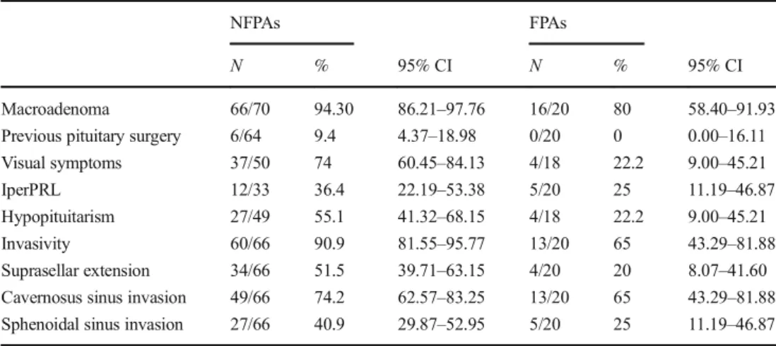
Most of the patients (91.1%) presented with a macroadenoma (maximum diameter mean ± SEM: 27.4 ± 1.1 mm). NFPAs were 77.8% (N = 70) of the total series, while the remaining 22.2% were hormone-secreting adenomas (62.5% GH-, 25% ACTH-, 12.5% PRL-secreting adenomas resistant to medical treatment). Pathological features of these tumors at diagnosis are presented in Table1and are stratified as functioning and non-functioning adenomas. Clinical presentation of NFPAs was related to mass effect as visual impairment in 74.0% of cases. Invasion of the cavernous sinus was present in 74.2% of patients, the invasion of sphenoidal sinus in 40.9%, the suprasellar extension in 51.5% of cases, and at least one type of invasion was present in 90.9% of
NFPAs. As expected, the mean diameter was associated with the risk of visual defects at diagnosis (OR = 4.304,p = 0.025).
Pituitary hormone deficits attributed to the pituitary lesion were detected in 55.1% of cases and replacement therapy ac-cording to the clinical guidelines [14] was prescribed, as shown in Fig.1.
Tumor diameter, extrasellar extension, and sinus invasions were statistically associated with the presence of hypopituita-rism at diagnosis (OR = 3.688, p = 0.05; OR = 3.365, p = 0.005, respectively).
Surgical complications
The most frequent surgical complication was diabetes insipidus (12.2%), 11.1 and 1.1% transient and permanent, respectively, followed by cerebral hemorrhage (4.4%), cerebra-spinal fluid (CSF) rhinorrhea (4.4%), syndrome of inappropriate antidiuresis (SIAD) (2.2%), and epistaxis (2.2%) (Table 2). Risk of intraoperative complications was not statistically asso-ciated with tumor size (OR = 0.588,p = 0.443).
Early postoperative results
Visual function improved in 70.6% of patients who presented visual defects at diagnosis, while recovery of normal gonadal, thyroid, or adrenal function occurred in 48.1% of cases.
Postoperative hypopituitarism was present in 55.7% of cases, as shown in Fig. 2. Considering only patients with normal pituitary function before surgery, postoperative wors-ening of gonadal, thyroid, or adrenal function occurred in 29.2% of patients.
The risk of hypopituitarism was statistically associated with invasive tumor (OR = 3.869, p = 0.042), but not with tumor size and not with the presence of surgery complications (OR = 2.087,p = 0.201).
Table 1 Preoperative pathological features of NFPA and FPA patients
NFPAs FPAs
N % 95% CI N % 95% CI
Macroadenoma 66/70 94.30 86.21–97.76 16/20 80 58.40–91.93
Previous pituitary surgery 6/64 9.4 4.37–18.98 0/20 0 0.00–16.11
Visual symptoms 37/50 74 60.45–84.13 4/18 22.2 9.00–45.21
IperPRL 12/33 36.4 22.19–53.38 5/20 25 11.19–46.87
Hypopituitarism 27/49 55.1 41.32–68.15 4/18 22.2 9.00–45.21
Invasivity 60/66 90.9 81.55–95.77 13/20 65 43.29–81.88
Suprasellar extension 34/66 51.5 39.71–63.15 4/20 20 8.07–41.60
Cavernosus sinus invasion 49/66 74.2 62.57–83.25 13/20 65 43.29–81.88
Sphenoidal sinus invasion 27/66 40.9 29.87–52.95 5/20 25 11.19–46.87
Data are presented as absolute and relative (%) frequencies
NFPAs, non-functioning pituitary adenomas; FPAs, functioning pituitary adenomas; PRL, prolactinemia; CI, confidence interval
Hormones
Surgical outcome
The first postoperative neuroimaging studies demonstrated resid-ual tumor in 72.4% of cases (mean size 10.3 ± 2 mm), whereas no tumor residue was documented in 27.6% of patients.
Multivariate logistic regression analysis showed that tumor invasion of the cavernous sinus (OR = 3.293,p = 0.05) and maximum tumor diameter (OR = 6.857,p = 0.002) were asso-ciated with an unfavorable surgical outcome.
Discussion
The aim of surgical treatment of pituitary adenoma is to re-verse endocrine dysfunction and/or resolve the compressive symptoms, with preservation of normal pituitary function. The introduction of the endoscopic approach improved surgical visualization, leading to a greater extent of resection with im-proved safety [4–6].
In our series of patients who underwent transsphenoidal surgery, low rates of perioperative mortality and major com-plications (4.4%) were reported, except for cerebral hemor-rhage, probably due to the very large size of the treated lesions (4.4%). However, our results are likely similar to those de-scribed by other pituitary surgical teams [9,15].
Postoperative hypopituitarism is a well-known complica-tion of pituitary surgery and many patients require pituitary hormone replacement medications; however, most of these patients are able to discontinue therapy within 2 to 6 months after surgery. The reported incidence of diabetes insipidus is 0.5–15% [16–18], similar to our result (12.2%), while other series reported a lower incidence [2,7], probably because of different definitions of this pathological condition that make it difficult to compare surgical series. Regarding anterior pitui-tary function, new cases of hypopituitarism occurred in ap-proximately 29% of patients in our series; this prevalence is higher by comparison with other series [9], but the short-term follow-up of our study might not permit further validation. On
Table 2 Complications of transsphenoidal pituitary surgery
NFPAs FPAs All
N % 95% CI N % 95% CI N % 95% CI Any complication 18/70 25.7 16.93–37.03 5/20 25 11.19–46.7 23/90 25.5 17.67–35.44 Cerebral hemorrhage 4/70 5.7 2.24–13.79 0/20 0 0.00–16.11 4/90 4.4 1.74–10.88 CSF leaks 4/70 5.7 2.24–13.79 0/20 0 0.00–16.11 4/90 4.4 1.74–10.88 Epistaxis 2/70 2.86 0.79–9.83 0/20 0 0.00–16.11 2/90 2.2 00.61–7.74 SIAD 1/70 1.4 00.25–7.66 1/20 5 0.89–23.61 2/90 2.2 00.61–7.74
Transient diabetes insipidus 7/70 10 4.93–19.23 3/20 15 5.24–36.06 10/90 11.1 6.15–19.26
Permanent diabetes insipidus 1/70 1.4 00.25–7.66 0/20 0 0.00–16.11 1/90 1.1 00.20–6.03
Data are presented as absolute and relative (%) frequencies
NFPs, non-functioning pituitary adenomas; FPAs, functioning pituitary adenomas; CSF, cerebrospinal fluid; SIAD, syndrome of inappropriate
antidiuresis;CI, confidence interval
Fig. 1 Frequency of preoperative hypopituitarism in NFPAs.Multiple:
presence of two or more pituitary deficits, excluding panhypopituitarism
Fig. 2 Postsurgical hypopituitarism. Multiple: presence of two or more pituitary deficits, excluding panhypopituitarism
Hormones
the other hand, in the case of hypopituitarism secondary to the pituitary adenoma, surgical removal could restore pituitary hormonal function [19,20], as confirmed in 48.1% of our patients with impaired preoperative function. Similar results were reported by different authors [9], whereas others have reported either no improvement [16,21] or even a worsening of pituitary function [22]. Some authors speculated that im-proved visualization of the normal pituitary gland during en-doscopic surgery may allow better preservation of the remain-ing gland and lead to improved functional outcomes [3].
It is well known that progressive visual impairment is a hallmark symptom of pituitary adenomas when growth ex-tends into the suprasellar space and causes compression of the optic chiasm. Our findings showed that visual function improved after surgery in 70.6% of our patients, thus confirming the results reported by other authors [19, 23]. This is probably related to the use of the transsphenoidal ap-proach, which causes less trauma to the optic pathway than the transcranial route. However, few cases of postsurgery visual impairment seem to be related to surgical hemorrhagic com-plications [8].
We achieved apparent complete surgical removal of the tumor in approximately 30% of our patients. In a large series by Losa et al., the authors reported complete tumor removal in 63% of 491 patients [9]. Frank et al. reported a total resection of 77% of their 173 cases [2]. The surgical outcomes achieved in smaller series seem less favorable, ranging from 14.3 to 43.1% [22–26], sim-ilar to our findings. The clinical features which seem to negatively affect the surgical outcome in multivariate analysis were cavernous sinus invasion and increasing maximum tumor diameter. Invasion of the cavernous si-nus was also reported as the strongest independent neg-ative predictor of complete surgical removal in another study that used multivariate analysis [26] and was con-firmed by Losa et al. [9].
In conclusion, the rates of gross tumor removal, although as yet not sufficiently high, resolution of hormonal hyperse-cretion, and improvement in visual symptoms confirm the effectiveness of endoscopic pituitary surgery. The safety of the procedure is similarly supported by the low reported inci-dence of complications.
Compliance with ethical standards
The study protocol was approved by local ethics committee and written informed consent was obtained from all subjects.
Conflict of interest The authors declare that they have no conflict of
interest.
Publisher’s Note Springer Nature remains neutral with regard to juris-dictional claims in published maps and institutional affiliations.
References
1. Kovacs K, Scheithauer BW, Horvath E, Lloyd RV (1996) The
World Health Organization classification of adenohypophysial
neo-plasms. A proposed five-tier scheme. Cancer 78:502–510
2. Frank G, Pasquini E, Farneti G et al (2006) The endoscopic versus
the traditional approach in pituitary surgery. Neuroendocrinology 83(3–4):240–248
3. Dallapiazza RF, Jane JA Jr 2015. Outcomes of endoscopic
transsphenoidal pituitary surgery. Endocrinol Metab Clin N Am44(1):105–115
4. Jho HD, Carrau RL (1997) Endoscopic endonasal transsphenoidal
surgery: experience with 50 patients. J Neurosurg 87:44–51
5. Cappabianca P, Alfieri A, de Divitiis E (1998) Endoscopic
endonasal transsphenoidal approach to the sella: towards functional endoscopic pituitary surgery (FEPS). Minim Invas Neurosurg 41:
66–73
6. de Divitiis E, Cappabianca P, Cavallo M (2003) Endoscopic
endonasal transsphenoidal approach to the sellar region. In: de Divitiis E, Cappabianca P (eds) Endoscopic endonasal
transsphenoidal surgery. Springer, Wien, pp 91–130
7. Dehdashti AR, Ganna A, Karabatsou K et al (2008) Pure
endoscop-ic endonasal approach for pituitary adenomas: early surgendoscop-ical results in 200 patients and comparison with previous microsurgical series. Neurosurgery 62(5):1006–1015
8. Cappabianca P, Cavallo LM, Colao A et al (2002) Surgical
compli-cations associated with the endoscopic endonasal transsphenoidal approach for pituitary adenomas. J Neurosurg 97(2):293–298
9. Losa M, Mortini P, Barzaghi R et al (2008) Early results of surgery
in patients with nonfunctioning pituitary adenoma and analysis of
the risk of tumor recurrence. J Neurosurg 108(3):525–532
10. Schneider HJ, Aimaretti G, Kreitschmann-Andermahr I, Stalla GK,
Ghigo E (2007) Hypopituitarism. Lancet 369(9571):1461–1470
11. Molitch ME, Clemmons DR, Malozowski S, Merriam GR, Vance
ML (2011) Evaluation and treatment of adult growth hormone de-ficiency: an Endocrine Society clinical practice guideline. J Clin
Endocrinol Metab 96:1587–1609
12. Tanriverdi F, Kelestimur F (2017) Classical and non-classical
causes of GH deficiency in adults. Best Pract Res Clin Endocrinol Metab 31(1):3–11
13. Knosp E, Steiner E, Kitz K, Matula C (1993) Pituitary adenomas
with invasion of the cavernous sinus space: a magnetic resonance imaging classification compared with surgical findings.
Neurosurgery 33:610–618
14. Fleseriu M, Hashim IA, Karavitaki N et al (2016) Hormonal
re-placement in hypopituitarism in adults: an Endocrine Society
clin-ical practice guideline. J Clin Endocrinol Metab 101(11):3888–
3921
15. Shou XF, Li SQ, Wang YF, Zhao Y, Jia PF, Zhou LF (2005)
Treatment of pituitary adenomas with a transsphenoidal approach.
Neurosurgery 56:249–256
16. Black PM, Zervas NT, Candia GL (1987) Incidence and
manage-ment of complications of transsphenoidal operation for pituitary adenomas. Neurosurgery 20:920–924
17. Ciric I, Ragin A, Baumgartner C, Pierce D (1997) Complications of
transsphenoidal surgery: results of a national survey, review of the literature, and personal experience. Neurosurgery 40:225–236
18. Barker FG II, Klibanski A, Swearingen B (2003) Transsphenoidal
surgery for pituitary tumors in the United States, 1996-2000: mor-tality, morbidity, and the effects of hospital and surgeon volume. J
Clin Endocrinol Metab 88:4709–4719
19. Arafah BM (1986) Reversible hypopituitarism in patients with large
nonfunctioning pituitary adenomas. J Clin Endocrinol Metab 62:
1173–1179
Hormones
20. Webb SM, Rigla M, Wagner A, Oliver B, Bartumeus F (1999) Recovery of hypopituitarism after neurosurgical treatment of
pitu-itary adenomas. J Clin Endocrinol Metab 84:3696–3700
21. Wichers-Rother M, Hoven S, Kristof RA, Bliesener N,
Stoffel-Wagner B (2004) Non-functioning pituitary adenomas: endocrino-logical and clinical outcome after transsphenoidal and transcranial
surgery. Exp Clin Endocrinol Diabetes 112:323–327
22. Dekkers OM, Pereira AM, Roelfsema F et al (2006) Observation
alone after transsphenoidal surgery for nonfunctioning pituitary
macroadenoma. J Clin Endocrinol Metab 91:1796–1180
23. Colao A, Cerbone G, Cappabianca P et al (1998) Effect of surgery
and radiotherapy on visual and endocrine function in
nonfunction-ing pituitary adenomas. J Endocrinol Investig 21:284–290
24. Woollons AC, Hunn MK, Rajapakse YR et al (2000)
Non-functioning pituitary adenomas: indications for postoperative
radio-therapy. Clin Endocrinol 53:713–717
25. Soto-Ares G, Cortet-Rudelli C, Assaker R et al (2002) MRI
proto-col technique in the optimal therapeutic strategy of non-functioning
pituitary adenomas. Eur J Endocrinol 146:179–186
26. Greenman Y, Ouaknine G, Veshchev I, Reider-Groswasser II,
Segev Y, Stern N (2003) Postoperative surveillance of clinically nonfunctioning pituitary macroadenomas: markers of tumor
quies-cence and regrowth. Clin Endocrinol 58:763–769
Hormones