ORIGINAL ARTICLE
Prognostic and biologic signi
ficance of DNMT3B expression in
older patients with cytogenetically normal primary acute
myeloid leukemia
C Niederwieser
1, J Kohlschmidt
1,2, S Volinia
1, SP Whitman
1, KH Metzeler
1, A-K Eisfeld
1, K Maharry
1,2, P Yan
1, D Frankhouser
1, H Becker
1,
S Schwind
1, AJ Carroll
3, D Nicolet
1,2, JH Mendler
1, JP Curfman
1, Y-Z Wu
1, MR Baer
4, BL Powell
5, JE Kolitz
6, JO Moore
7, TH Carter
8,
R Bundschuh
9, RA Larson
10, RM Stone
11, K Mrózek
1,12, G Marcucci
1,12and CD Bloom
field
1,12DNMT3B encodes a DNA methyltransferase implicated in aberrant epigenetic changes contributing to leukemogenesis.
We tested whether DNMT3B expression, measured by NanoString nCounter assay, associates with outcome, gene and microRNA
expression and DNA methylation pro
files in 210 older (⩾60 years) adults with primary, cytogenetically normal acute myeloid
leukemia (CN-AML). Patients were dichotomized into high versus low expressers using median cut. Outcomes were assessed in
the context of known CN-AML prognosticators. Gene and microRNA expression, and DNA methylation pro
files were analyzed
using microarrays and MethylCap-sequencing, respectively. High DNMT3B expressers had fewer complete remissions (CR;
P = 0.002) and shorter disease-free (DFS; P = 0.02) and overall (OS; P
o0.001) survival. In multivariable analyses, high DNMT3B
expression remained an independent predictor of lower CR rates (P = 0.04) and shorter DFS (P = 0.04) and OS (P = 0.001). High
DNMT3B expression associated with a gene expression pro
file comprising 363 genes involved in differentiation, proliferation
and survival pathways, but with only four differentially expressed microRNAs (miR-133b, miR-148a, miR-122, miR-409-3p) and no
differential DNA methylation regions. We conclude that high DNMT3B expression independently associates with adverse
outcome in older CN-AML patients. Gene expression analyses suggest that DNMT3B is involved in the modulation of several
genes, although the regulatory mechanisms remain to be investigated to devise therapeutic approaches speci
fic for these
patients.
Leukemia (2015)
29, 567–575; doi:10.1038/leu.2014.267
INTRODUCTION
Acute myeloid leukemia (AML) is a heterogeneous disease
presenting with a wide spectrum of prognostically relevant
cytogenetic aberrations, gene mutations and abnormal expression
of genes and microRNAs. Cytogenetically normal AML (CN-AML)
patients, constituting 40 to 50% of all AML patients,
1are the
largest and molecularly best characterized cytogenetic subset in
primary (de novo) AML.
1–3Although leukemic blasts of these
patients do not contain microscopically detectable chromosome
abnormalities, they harbor prognostically relevant mutations and
aberrantly expressed genes and microRNAs.
2–16In addition to
these genetic alterations, epigenetic changes have recently been
shown to participate in myeloid leukemogenesis and be
pharmacologically targetable.
17,18Notably, some genes whose
mutations are prognostic in CN-AML encode proteins that are
implicated in epigenetic regulation of gene transcription, namely
IDH2, ASXL1 and DNMT3A. The latter is among the most frequently
mutated genes in primary CN-AML patients, being found mutated
in 29 to 34% of the patients.
9,19DNMT3A
encodes
DNA
methyltransferase
3A
(DNMT3A),
which is involved in epigenetic gene silencing through DNA
hypermethylation.
20In addition to DNMT3A, DNMT1 and DNMT3B
also mediate DNA methylation in normal and malignant cells, and
may represent potential therapeutic targets in cancer and
leukemia.
21–24However, in contrast to DNMT3A, no recurrent
mutations of DNMT1 and DNMT3B genes have been reported in
AML.
25Instead, one study has indicated that higher expression of
DNMT3B is associated with worse outcome in AML.
26However, the
patient cohort analyzed was cytogenetically diverse and
hetero-geneous for clinical features and treatment received. Thus, it is
1The Ohio State University Comprehensive Cancer Center, Columbus, OH, USA;2
Alliance for Clinical Trials in Oncology Statistics and Data Center, Mayo Clinic, Rochester, MN, USA; 3
Department of Genetics, University of Alabama at Birmingham, Birmingham, AL, USA;4
Department of Medicine and Greenebaum Cancer Center, University of Maryland, Baltimore, MD, USA;5
Comprehensive Cancer Center of Wake Forest University, Winston-Salem, NC, USA;6
Monter Cancer Center, Hofstra North Shore-Long Island Jewish School of Medicine, Lake Success, NY, USA;7
Department of Medicine, Duke University Medical Center, Durham, NC, USA;8
Department of Internal Medicine, University of Iowa, Iowa City, IA, USA;9
Departments of Physics and Chemistry and Biochemistry, The Ohio State University, Columbus, OH, USA;10
Department of Medicine, University of Chicago Medical Center, Chicago, IL, USA and11Department of Medical Oncology, Dana Farber Cancer Institute, Boston, MA, USA. Correspondence: Dr K Mrózek, The Ohio State University Comprehensive Cancer Center, 1232 A James Cancer Hospital, 300 West Tenth Avenue, Columbus, OH 43210-1228, USA or Dr G Marcucci, The Ohio State University Comprehensive Cancer Center, 410 Biomedical Research Tower, 460 West Twelfth Avenue, Columbus, OH 43210, USA or Dr CD Bloomfield, The Ohio State University Comprehensive Cancer Center, 1216 James Cancer Hospital, 300 West Tenth Avenue, Columbus, OH 43210-1228, USA.
E-mail: [email protected] or [email protected] or clara.bloomfi[email protected] 12
These senior authors contributed equally to this work.
Presented in part at the 18th Congress of the European Hematology Association, Stockholm, Sweden, 13–16 June 2013, and published in abstract form: Niederwieser C, Kohlschmidt J, Maharry K, Mrózek K, Metzeler K, Volinia S et al. High expression of DNMT3B negatively impacts on clinical outcome of older patients (pts) with primary cytogenetically normal (CN) acute myeloid leukemia (AML) (CALGB 20202 (Alliance)). Haematologica 2013;98(suppl 1): 484 (abstract S1168).
Received 13 May 2014; revised 26 August 2014; accepted 29 August 2014; accepted article preview online 10 September 2014; advance online publication, 7 October 2014
unknown whether DNMT3B expression is an independent
prog-nostic factor and can be used for strati
fication guidance in CN-AML.
Thus, we analyzed the clinical signi
ficance of DNMT3B
expres-sion in the context of a comprehensive panel of molecular
prognosticators in a relatively large cohort of older (aged
⩾ 60
years) patients with CN-AML who were similarly treated on
cytarabine/daunorubicin-based
protocols.
To
gain
biologic
insights, we also derived genome-wide DNMT3B-associated gene
and microRNA expression and DNA methylation pro
files. We
studied older patients because both the incidence of AML and the
role of epigenetics increase with age. Moreover, we have recently
reported a favorable clinical response to hypomethylating agents
in this age group of AML patients.
27PATIENTS AND METHODS
Patients, treatment and cytogenetic studies
Pretreatment bone marrow or blood samples were obtained from 210 patients with primary CN-AML aged 60 to 83 years (median, 68 years) who received intensive first-line therapy on Cancer and Leukemia Group B (CALGB) trials.28–32All patients received cytarabine–daunorubicin-based induction chemotherapy, and no patient received allogeneic hematopoietic stem cell transplantation duringfirst complete remission (CR). For details regarding treatment protocols and sample collection, see Supplementary Information. All patients were enrolled on companion CALGB/Alliance protocols: 8461 (cytogenetic analyses), 9665 (tissue banking) and 20 202 (molecular analyses).
Cytogenetic analyses were performed in institutional CALGB/Alliance cytogenetics laboratories. For the patient’s karyotype to be considered normal,⩾ 20 metaphase cells from short-term cultures of pretreatment bone marrow specimens had to have been analyzed and the normal result confirmed by central karyotype review.33 All patients provided written informed consent for participation in these studies; study protocols were in accordance with the Declaration of Helsinki and approved by local Institutional Review Boards.
Single-gene expression analyses
The expression of DNMT3B transcript was assessed by NanoString nCounter assays (NanoString Technologies, Seattle, WA, USA; Supple-mentary Information).34These assays measured global expression of the
DNMT3B gene, and did not allow for quantification of isoform-specific expression of DNMT3B. DNMT3B expression levels were normalized using ABL as an internal control. We also used NanoString nCounter assays to measure expression of BAALC, ERG and miR-155, and real-time reverse transcription-PCR to measure miR-3151 expression, all of which have been previously shown to affect prognosis of older CN-AML patients.10,15,16
Mutational analyses
The presence or absence of FLT3 internal tandem duplication (FLT3-ITD),35,36FLT3 tyrosine kinase domain mutations (FLT3-TKD),37MLL partial
tandem duplication (MLL-PTD),38and mutations in the NPM1,5CEBPA,39 WT1,40IDH1 and IDH2,7TET2,41ASXL1,8DNMT3A9and RUNX142genes were
determined centrally as previously described.
Gene and microRNA expression pro
filing
The gene and microRNA expression profiling were assessed using the Affymetrix U133 plus 2.0 array (Affymetrix, Santa Clara, CA, USA) and The Ohio State University custom microRNA array (OSU_CCC Version 4.0, The Ohio State University, Columbus, OH, USA), respectively, as previously reported,5,43and detailed in the Supplementary Information.
For DNMT3B, the Affymetrix U133 plus 2.0 arrays measured global DNMT3B expression levels, and did not quantify expression of the individual DNMT3B isoforms. For the gene and microRNA expression profiling, summary measures of gene and microRNA expression were computed, normalized and filtered (Supplementary Information). A DNMT3B expression-associated signature (see Supplementary Information for details) was derived by comparing gene expression between high and low DNMT3B expressers in the CALGB/Alliance cohort and in two additional sets of CN-AML patients with microarray and RNAseq gene expression data publicly available (German AML Cooperative Group (AMLCG)44and The
Cancer Genome Atlas (TCGA)25). For comparison of the high DNMT3B
expression signature with the FLT3-ITD signature we used gene set enrichment analysis (for details see Supplementary Information). For the microRNA expression signature, only the CALGB/Alliance patients were used. Univariable significance levels of Po0.001 (false discovery rates o0.01) were used to select genes and microRNAs that constituted the signatures. To assess enrichment of genes in the DNMT3B gene expression-associated signature in distinct biologic processes, a Gene Ontology analysis was performed using the Database for Annotation, Visualization and Integrated Discovery (DAVID).45We identified as statistically significant ‘annotation clusters’ those clusters of Gene Ontology terms with enrichment scores of 42.0, P-values ⩽ 0.001 and Benjamini corrected P-values⩽ 0.05. All molecular analyses were performed centrally at The Ohio State University.
DNA methylation
Genome-wide DNA methylation and levels of DNA methylation across the genome’s functional regions (that is, genomic features) were measured using the MethylCap-seq assay as previously reported.18
Statistical analyses
The patients were dichotomized into high and low expressers using the median cut. This cut was supported by significant results of the trend test applied to outcome of patients divided into quartiles by DNMT3B expression (P⩽ 0.001). We compared pretreatment features and outcome between patients with high and low DNMT3B expression. Definitions of clinical end points (that is, CR rates, disease-free (DFS) and overall (OS) survival) are provided in the Supplementary Information. Baseline characteristics between high and low DNMT3B expressers were compared using the Fisher’s exact test for categorical and the Wilcoxon rank-sum test for continuous variables.46The categorical variables included the European
LeukemiaNet (ELN) Genetic Groups.47The ELN guidelines classify CN-AML patients within the Favorable or Intermediate-I Genetic Groups based on CEBPA, NPM1 and FLT3 mutational status. The ELN Favorable Genetic Group consists of CN-AML patients with CEBPA mutation and/or NPM1 mutation without FLT3-ITD, whereas the Intermediate-I Genetic Group is comprised of patients with wild-type CEBPA and FLT3-ITD with or without NPM1 mutation, or wild-type NPM1 without FLT3-ITD.47
For time-to-event analyses, we calculated survival estimates using the Kaplan–Meier method, and compared groups by the log rank test.46 In order to provide the odds ratios and hazard ratios and associated confidence intervals, logistic regression and Cox proportional hazards models were generated to compare outcomes between high and low DNMT3B expressers for CR and survival end points (DFS, OS), respectively, and P-values from the Wald test are reported. We constructed multi-variable logistic regression models to analyze factors associated with the achievement of CR, and multivariable Cox proportional hazards models for factors associated with survival end points,46 the details of which are provided in the Supplementary Information. All analyses were performed by the Alliance for Clinical Trials in Oncology Statistics and Data Center.
RESULTS
Associations of DNMT3B expression with pretreatment clinical and
molecular characteristics
At diagnosis, high DNMT3B expressers had higher white blood
counts (WBC; P = 0.004), and percentages of blood (P = 0.004) and
marrow (P = 0.02) blasts than low DNMT3B expressers. Concerning
molecular features, high DNMT3B expressers were more often
FLT3-ITD-positive (P
o0.001) and classified in the ELN
Intermedi-ate-I Genetic Group (P = 0.02). IDH2-R140 mutations were less
frequent in high DNMT3B expressers, whereas six of seven
IDH2-R172 mutations were detected in this patient group. High DNMT3B
expressers also had higher ERG (P
o0.001), BAALC (P = 0.002)
and miR-155 (P = 0.006) expression than low expressers (Table 1,
Supplementary Figure S1).
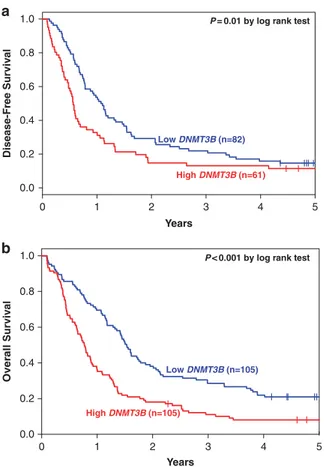
Associations of DNMT3B expression with clinical outcome in the
entire patient cohort
With a median follow-up of 5.1 years (range, 2.3
–11.6 years) for
patients who are alive, high DNMT3B expressers had lower CR
568
rates (P = 0.002, Wald test; 58% vs 78%), and shorter DFS (P = 0.02,
Wald test) and OS (P
o0.001, Wald test) than DNMT3B low
expressers (Table 2, Figure 1).
In a multivariable model for CR, DNMT3B expression remained
prognostic (P = 0.04), after adjustment for BAALC expression status
(P
o0.001), WBC (P = 0.007) and age (P = 0.02) (Table 3). High
DNMT3B expressers were half as likely to achieve a CR as low
expressers. In multivariable analysis for DFS, high DNMT3B
expression associated with shorter DFS (P = 0.04), once adjusted
for BAALC expression (P = 0.004), DNMT3A-R882 mutation status
(P = 0.009) and ELN Genetic Groups (P = 0.03). The risk of
experiencing relapse or death was 46% higher for high DNMT3B
Table 1. Comparison of clinical and molecular characteristics of patients with cytogenetically normal acute myeloid leukemia with high versus low DNMT3B expression
Characteristic High DNMT3B (n = 105) Low DNMT3B (n = 105) P-value Age, years 0.82 Median 68 68 Range 60–83 60–81 Sex,n (%) 0.58 Male 58 (55) 53 (50) Female 47 (45) 52 (50) Race,n (%) 0.48 White 96 (92) 92 (89) Nonwhite 8 (8) 11 (11) Hemoglobin, g/dl 0.71 Median 9.4 9.3 Range 6.5–12.4 5.4–15.0 Platelet count, x 109/l 0.87 Median 68 71 Range 4–850 11–510 WBC, x 109/l 0.004 Median 43.7 21.8 Range 1.0–450.0 0.8–249.3 Blood blasts, % 0.004 Median 64 40 Range 0–99 0–97
Bone marrow blasts, % 0.02
Median 72 64 Range 21–97 4–97 Extramedullary involvement,n (%) 27 (27) 24 (23) 0.63 NPM1,n (%) 0.31 Mutated 67 (66) 60 (59) Wild type 34 (34) 42 (41) FLT3-ITD,n (%) o0.001 Present 54 (53) 21 (21) Absent 48 (47) 81 (79) CEBPA,n (%) 0.83a Mutated 13 (13) 12 (12) Single mutated 10 5 Double mutated 3 7 Wild type 88 (87) 90 (88)
ELN Genetic Group, n (%)b 0.02 Modified Favorable 38 (38) 56 (55)
Intermediate-I 63 (62) 45 (45) FLT3-TKD,n (%) 0.83 Present 12 (12) 11 (11) Absent 89 (88) 91 (89) WT1,n (%) 0.41 Mutated 8 (8) 5 (5) Wild type 93 (92) 97 (95) TET2,n (%) 1.00 Mutated 32 (32) 31 (32) Wild type 68 (68) 67 (68) MLL-PTD,n (%) 1.00 Present 5 (6) 5 (6) Absent 74 (94) 81 (94) IDH1,n (%) 0.18 Mutated 14 (14) 8 (8) Wild type 86 (86) 94 (92) Table 1. (Continued ) Characteristic High DNMT3B (n = 105) Low DNMT3B (n = 105) P-value IDH2,n (%) 0.05 Mutated 18 (18) 31 (30) R140 12 30 0.005c R172 6 1 0.13d Wild type 82 (82) 71 (70) RUNX1,n (%) 0.31 Mutated 17 (18) 11 (12) Wild type 79 (82) 81 (88) ASXL1,n (%) 0.69 Mutated 13 (13) 15 (15) Wild type 87 (87) 84 (85) DNMT3A, n (%) 0.65 Mutated 35 (35) 31 (32) R882 19 20 1.00e Non-R882 16 11 0.40f Wild type 64 (65) 66 (68)
ERG expression group, n (%)g,h o0.001
High 65 (62) 40 (38) Low 40 (38) 65 (62)
BAALC expression group,n (%)g,h 0.002
High 64 (61) 41 (39) Low 41 (39) 64 (61)
miR-155 expression group, n (%)g,h 0.006 High 63 (60) 42 (40)
Low 42 (40) 63 (60)
miR-3151 expression group, n (%)g,i 0.76 High 42 (49) 39 (46)
Low 43 (51) 46 (54)
Abbreviations: ELN, European LeukemiaNet; FLT3-ITD, internal tandem duplication of the FLT3 gene; FLT3-TKD, tyrosine kinase domain mutation in the FLT3 gene; MLL-PTD, partial tandem duplication of the MLL gene; n, number; WBC, white blood count.aThe P-value pertains to a comparison of
frequencies of CEBPA mutations (single and double combined) versus CEBPA wild-type between high and low DNMT3B expressers. bThe ELN modified Favorable Genetic Group is defined as CN-AML patients with mutated CEBPA and/or mutated NPM1 without FLT3-ITD. All remaining CN-AML patients (that is, those with wild-type CEBPA and wild-type NPM1 with or without FLT3-ITD, or mutated NPM1 with FLT3-ITD) belong to the ELN Intermediate-I Genetic Group.47 cThe P-value pertains to a comparison of
frequencies of IDH2-R140 mutations versus IDH2 wild-type between high and low DNMT3B expressers.dThe P-value pertains to a comparison of
frequencies of IDH2-R172 mutations versus IDH2 wild-type between high and low DNMT3B expressers.eThe P-value pertains to a comparison of frequencies of DNMT3A-R882 mutations versus DNMT3A wild-type between high and low DNMT3B expressers.fThe P-value pertains to a comparison of frequencies of DNMT3A non-R882 mutations versus DNMT3A wild-type between high and low DNMT3B expressers.gThe median expression value was used as a cut point. hData was assessed by the NanoString
nCounter assay.iData was assessed by real-time reverse transcription-PCR.
569
expressers than for low expressers. DNMT3B expression also
remained prognostic for OS (P = 0.001), after adjustment for BAALC
(P
o0.001), miR-3151 (P = 0.02) and miR-155 (P = 0.02) expression.
The risk of death was 72% higher for high DNMT3B expressers
compared with low expressers (Table 3).
Associations of DNMT3B expression with clinical outcome in ELN
Genetic Groups
We analyzed the associations of DNMT3B expression with outcome
separately within the ELN Favorable and Intermediate-I Genetic
Groups. Within the Favorable Group (n = 94), there was no
signi
ficant difference in CR rates (71% vs 84%, P = 0.14, Wald test)
or DFS (P = 0.10, Wald test) between high and low DNMT3B
expressers. However, high expressers had shorter OS (P = 0.002,
Wald test) than low expressers (Table 2, Figures 2a and b). In
multivariable analyses for the ELN Favorable Genetic Group
(Table 3), DNMT3B expression remained significant for OS
(P = 0.003) after adjustment for BAALC expression (P = 0.01). High
DNMT3B expressers were twice as likely to die as low expressers.
In the Intermediate-I Group (n = 108), high DNMT3B expressers
had a lower CR rate (49% vs 73%, P = 0.01, Wald test) and shorter
OS (P = 0.03, Wald test) than low DNMT3B expressers, but there
was no signi
ficant difference in DFS between the groups (Table 2,
Figures 2c and d). In multivariable analyses within the ELN
Intermediate-I Genetic Group (Table 3), DNMT3B expression was
significant for OS (P = 0.02), after adjustment for BAALC expression
(P = 0.03), miR-3151 expression (P = 0.03) and WBC (P = 0.03). High
DNMT3B expressers were 1.7 times more likely to die than low
expressers.
Genome-wide gene expression pro
files associated with DNMT3B
expression
To gain biologic insights into the role of DNMT3B, we derived a
DNMT3B-associated gene expression pro
file using three
indepen-dent sets of CN-AML patients, that is, CALGB/Alliance (n = 177),
AMLCG (n = 75) and The Cancer Genome Atlas (n = 88). We
identi
fied 195 upregulated genes and 168 downregulated genes
that were signi
ficantly associated with higher DNMT3B expression
in each of the three cohorts (Supplementary Table S1). As high
DNMT3B expression was associated with the presence of FLT3-ITD
(Table 1), we performed gene set enrichment analysis to test
whether a set of 195 genes that are upregulated in high DNMT3B
expressers was associated with a set of genes differentially
expressed between patients who harbored FLT3-ITD versus those
who did not (Supplementary Information). We found a signi
ficant
correlation between the high DNMT3B expression and FLT3-ITD
signatures (P = 0.006; false discovery rate = 0.006; Supplementary
Figure S2). Among the genes upregulated in high DNMT3B
expressers, we noted a variety of genes previously involved in
AML including CDK6 and WT1 that encode cyclin kinase and
transcription factor proteins, respectively. Among the
down-regulated genes, we noted genes involved with both normal
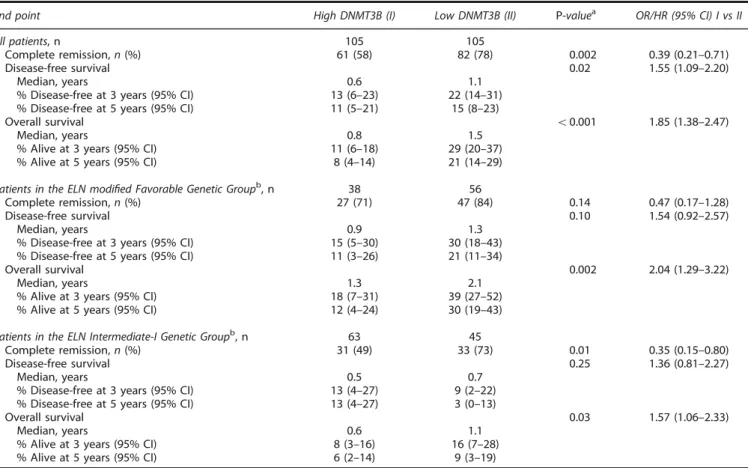
Table 2. Outcomes of patients with cytogenetically normal acute myeloid leukemia according to DNMT3B expression status
End point High DNMT3B (I) Low DNMT3B (II) P-valuea OR/HR (95% CI) I vs II
All patients, n 105 105
Complete remission, n (%) 61 (58) 82 (78) 0.002 0.39 (0.21–0.71) Disease-free survival 0.02 1.55 (1.09–2.20)
Median, years 0.6 1.1
% Disease-free at 3 years (95% CI) 13 (6–23) 22 (14–31) % Disease-free at 5 years (95% CI) 11 (5–21) 15 (8–23)
Overall survival o0.001 1.85 (1.38–2.47)
Median, years 0.8 1.5
% Alive at 3 years (95% CI) 11 (6–18) 29 (20–37) % Alive at 5 years (95% CI) 8 (4–14) 21 (14–29) Patients in the ELN modified Favorable Genetic Groupb, n 38 56
Complete remission, n (%) 27 (71) 47 (84) 0.14 0.47 (0.17–1.28) Disease-free survival 0.10 1.54 (0.92–2.57)
Median, years 0.9 1.3
% Disease-free at 3 years (95% CI) 15 (5–30) 30 (18–43) % Disease-free at 5 years (95% CI) 11 (3–26) 21 (11–34)
Overall survival 0.002 2.04 (1.29–3.22)
Median, years 1.3 2.1
% Alive at 3 years (95% CI) 18 (7–31) 39 (27–52) % Alive at 5 years (95% CI) 12 (4–24) 30 (19–43) Patients in the ELN Intermediate-I Genetic Groupb, n 63 45
Complete remission, n (%) 31 (49) 33 (73) 0.01 0.35 (0.15–0.80) Disease-free survival 0.25 1.36 (0.81–2.27)
Median, years 0.5 0.7
% Disease-free at 3 years (95% CI) 13 (4–27) 9 (2–22) % Disease-free at 5 years (95% CI) 13 (4–27) 3 (0–13)
Overall survival 0.03 1.57 (1.06–2.33)
Median, years 0.6 1.1
% Alive at 3 years (95% CI) 8 (3–16) 16 (7–28) % Alive at 5 years (95% CI) 6 (2–14) 9 (3–19)
Abbreviations: CI, confidence interval; ELN, European Leukemia Net; HR, hazard ratio; n, number; OR, odds ratio.aP-values provided are generated by logistic
regression and Cox proportional hazards models to compare outcome of patients for CR and survival end points (DFS, OS), respectively, using the Wald test.
bThe ELN modified Favorable Genetic Group is defined as CN-AML patients with mutated CEBPA and/or mutated NPM1 without FLT3-ITD. All remaining
CN-AML patients (that is, those with wild-type CEBPA and wild-type NPM1 with or without FLT3-ITD, or mutated NPM1 with FLT3-ITD) belong to the ELN Intermediate-I Genetic Group.47
monocyte/macrophage differentiation and immune function
including CD14, TLR4, CEBPB and TLR8.
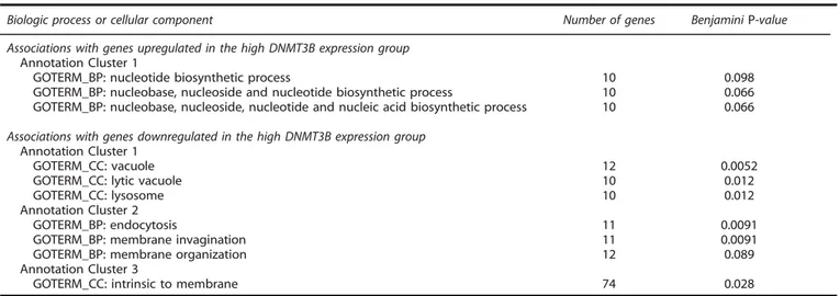
Gene Ontology was used to assess the biologic features of the
DNMT3B expression pro
file (Table 4). For DNMT3B-associated
upregulated genes, there were three Gene Ontology terms
comprising genes involved in nucleotide biosynthetic processes
and metabolism and included in annotation cluster 1 that had a
trend for statistical signi
ficance (Benjamini P-value o0.1). For
DNMT3B-associated downregulated genes, cellular processes
included lysosome biology, endocytosis and membrane signaling.
These results may be interpreted as consistent with the previously
noted dysregulated genes involved in monocyte/macrophage
differentiation and activity.
Genome-wide microRNA pro
files associated with DNMT3B
expression
The in
fluence of DNMT3B expression on microRNA genome-wide
pro
files could be evaluated in 162 patients. In contrast to coding
genes, only four microRNAs were differentially expressed between
high and low DNMT3B expressers (P
⩽ 0.001). High DNMT3B
expression was associated with miR-133b upregulation, and
miR-148a, miR-122 and miR-409-3p downregulation. miR-133b
upregulation in high DNMT3B expressers was somewhat surprising
as this microRNA was reported to have tumor suppressor activity
in other cancers.
48,49However, consistent with the downregulated
gene expression pro
file as discussed above, miR-133b has recently
been
shown
to
target
granulocyte-macrophage
colony-stimulating factor (GM-CSF), a cytokine involved in granulocyte
–
monocyte/macrophage differentiation.
50Among the downregulated
microRNAs, miR-148a was reported to target DNMT3B and to be
itself a target of aberrant hypermethylation in cancer.
51,52Lower
expression of miR-122 has been associated with aggressive
hepatocellular carcinoma and miR-409-3p with cell invasion and
metastasis in gastric cancer
53–56; however, a role for these
microRNAs in AML is currently unknown.
Genome-wide methylation pro
filing associated with DNMT3B
expression
As DNMT3B encodes a methyltransferase that mediates de novo
DNA methylation, we assessed whether high and low DNMT3B
expressers differed in DNA methylation patterns. Surprisingly, we
found no signi
ficant differences in genome-wide DNA methylation
levels or in the numbers of differentially methylated regions
18in
distinct functional genomic regions (for example, gene promoters)
when high versus low DNMT3B expressers were compared.
DISCUSSION
In this study, we report that high DNMT3B expression associates
with lower CR rates and shorter DFS and OS in
chemotherapy-treated CN-AML patients aged
⩾ 60 years. High DNMT3B
expres-sion was associated with such adverse prognostic factors as
FLT3-ITD, high ERG, BAALC and miR-155 expression and the ELN
Intermediate-I Genetic Group; nevertheless the association of
DNMT3B expression with clinical outcome is independent from the
aforementioned and other established molecular and clinical
prognosticators for all outcome end points studied.
Our
findings are consistent to some extent with the only, to our
knowledge, previous study that assessed the prognostic value of
DNMT3B expression.
26Although, in the subset of 93 CN-AML
patients, Hayette et al.
26did not
find significant differences in
event-free survival or OS between high and low DNMT3B
expressers, high DNMT3B expressers had a shorter event-free
survival than low DNMT3B expressers in the whole cytogenetically
diverse cohort of 191 AML patients analyzed. It is dif
ficult to
directly compare their results with ours as approximately one-half
of the patients analyzed by Hayette et al.
26had various abnormal
karyotypes, more than two-thirds of the patients were younger
than 60 years and a quarter underwent allogeneic hematopoietic
stem cell transplantation in
first CR. Thus, although the two studies
are not comparable, they both conclude that higher DNMT3B
expression is associated with worse outcome in AML.
Recently, the ELN reporting system,
47which for CN-AML is
based on only three molecular markers (that is, FLT3-ITD, CEBPA
and NPM1 mutations), was shown to provide important prognostic
information in AML.
57However, we and others have shown that
additional molecular markers, such as TET2,
41ASXL1,
8RUNX1
42and
DNMT3A
58mutations and expression of MN1,
12miR-155
15and
miR-3151,
16may re
fine outcome prediction of CN-AML patients
within the ELN Genetic Groups. Hence, in the current study, we
investigated whether considering DNMT3B expression as a novel
prognosticator could alter patient classification within the ELN
Genetic Groups. In the Favorable Group, we found that low
DNMT3B expression identi
fied a subset of CN-AML patients with a
signi
ficantly longer OS, thus making DNMT3B expression the third
molecular marker, in addition to ASXL1 mutations
8and miR-155
expression,
15capable of re
fining prognostication of older patients
in this ELN Genetic Groups. We also observed a signi
ficant
difference in OS between high and low DNMT3B expressers
classi
fied in the ELN Intermediate-I Genetic Group. Previously,
RUNX1 mutations
42and expression levels of MN1,
12miR-155
15and
miR-3151
16were demonstrated to add prognostic information in
this ELN Genetic Group.
We report the
first, to our knowledge, DNMT3B-associated gene
and microRNA expression and DNA methylation profiles in
CN-AML. We were able to derive a strong gene expression profile
0 1 2 3 4 5 0.0 0.2 0.4 0.6 0.8 1.0
a
Low DNMT3B (n=82) High DNMT3B (n=61) Years Disease-Fr e e Surviv alP = 0.01 by log rank test
0 1 2 3 4 5 0.0 0.2 0.4 0.6 0.8 1.0 Low DNMT3B (n=105) High DNMT3B (n=105) Over all Surviv a l
P < 0.001 by log rank test
Years
b
Figure 1. Clinical outcome of CN-AML patients with high and low DNMT3Bexpression. Kaplan–Meier survival curves for (a) disease-free survival and (b) overall survival. P-values presented are from the log rank test.
comprising 363 genes by overlapping the microarray results from
three independent sets of patients. The pro
file was quite
heterogeneous, comprising genes encoding for proteins involved
in multiple biologic processes that play a role in leukemia cell
differentiation, proliferation and survival. Among the
down-regulated genes, we noted enrichment of genes involved in
monocyte/macrophage differentiation and activity, suggesting a
role of DNMT3B in impairing differentiation of the leukemic blasts
into cells with normal innate immunity activity. Using gene set
enrichment analysis, we found a significant correlation between
the high DNMT3B expression and FLT3-ITD signatures (Supplementary
Figure S2). This, along with the increased frequency of FLT3-ITD in
high DNMT3B expressers (Table 1), suggests the existence of a
functional association between high expression of the DNMT3B gene
and FLT3-ITD.
In contrast, the DNMT3B-associated microRNA profile was relatively
weak, comprising only four microRNAs that were differentially
expressed in high versus low DNMT3B expressers. Nevertheless, the
unique upregulation of expression of miR-133b, recently reported to
target GM-CSF,
50in DNMT3B high expressers was somewhat
consistent with the enrichment of the gene expression profile in
multiple downregulated genes involved in the differentiation and
activity of hematopoietic cells participating in innate immunity.
Surprisingly, despite the fact that DNMT3B encodes a DNA
methyltransferase, we observed no significant association of high
DNMT3B levels and DNA methylation changes. No difference in
global DNA methylation levels and number of differentially
methylated regions could be identi
fied between DNMT3B high
and low expressers. Our results are reminiscent of a recent report
showing that changes in DNMT3B expression did not affect
methylation levels of putative DNMT3B target genes.
59Moreover,
Russler–Germain et al.
60have recently demonstrated that DNA
methylation levels in leukemic blasts from CN-AML patients are
not influenced by DNMT3B expression as mainly inactive splice
variants of DNMT3B are expressed in these cells. Overall,
therefore, these data may suggest that although overexpressed
DNMT3B is a potentially valuable predictive marker for response
to conventional chemotherapy, it does not necessarily identify
subsets of older AML patients characterized by aberrant DNA
methylation who might be responsive to hypomethylating
azanucleosides.
In summary, we have demonstrated that DNMT3B expression
constitutes an independent prognostic factor in older CN-AML
patients
treated
intensively,
and
could
also
refine the
ELN classification. Furthermore, we have provided some
insights into the biologic activity of DNMT3B in CN-AML, which
is
seemingly
independent
from
mechanisms
of
DNA
hypermethylation and/or microRNA-dependent gene repression.
Further studies focused on gaining more clinical and mechanistic
insights into the leukemogenic role of DNMT3B expression are
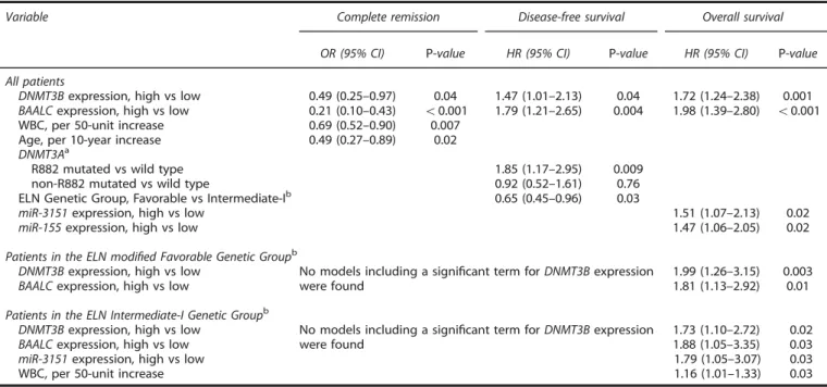
Table 3. Multivariable analyses of CN-AML patients according to DNMT3B expression status
Variable Complete remission Disease-free survival Overall survival OR (95% CI) P-value HR (95% CI) P-value HR (95% CI) P-value All patients
DNMT3Bexpression, high vs low 0.49 (0.25–0.97) 0.04 1.47 (1.01–2.13) 0.04 1.72 (1.24–2.38) 0.001 BAALCexpression, high vs low 0.21 (0.10–0.43) o0.001 1.79 (1.21–2.65) 0.004 1.98 (1.39–2.80) o0.001 WBC, per 50-unit increase 0.69 (0.52–0.90) 0.007
Age, per 10-year increase 0.49 (0.27–0.89) 0.02 DNMT3Aa
R882 mutated vs wild type 1.85 (1.17–2.95) 0.009 non-R882 mutated vs wild type 0.92 (0.52–1.61) 0.76 ELN Genetic Group, Favorable vs Intermediate-Ib 0.65 (0.45
–0.96) 0.03
miR-3151expression, high vs low 1.51 (1.07–2.13) 0.02 miR-155expression, high vs low 1.47 (1.06–2.05) 0.02 Patients in the ELN modified Favorable Genetic Groupb
DNMT3Bexpression, high vs low BAALCexpression, high vs low
No models including a significant term for DNMT3B expression were found
1.99 (1.26–3.15) 1.81 (1.13–2.92)
0.003 0.01 Patients in the ELN Intermediate-I Genetic Groupb
DNMT3Bexpression, high vs low BAALCexpression, high vs low
No models including a significant term for DNMT3B expression were found
1.73 (1.10–2.72) 1.88 (1.05–3.35)
0.02 0.03 miR-3151expression, high vs low 1.79 (1.05–3.07) 0.03 WBC, per 50-unit increase 1.16 (1.01–1.33) 0.03 Abbreviations: CI, confidence interval; ELN, European LeukemiaNet; HR, hazard ratio; OR, odds ratio; WBC, white blood count. An odds ratio o1 means a lower CR rate for the higher values of the continuous variables and thefirst category listed for the categorical variables. A hazard ratio 41 (o1) corresponds to a higher (lower) risk of an event for higher values of continuous variables and thefirst category listed of a dichotomous variable. Variables were considered for inclusion in the multivariable models if they had a univariable P-value of⩽ 0.20. See the Supplementary Information for a full list of variables evaluated in univariable analyses. As NPM1, FLT3-ITD and CEBPA mutations are integrated in the ELN genetic classification, they were not additionally considered as individual variables. In the entire patient cohort, variables considered for inclusion in the model for achievement of CR were DNMT3B, ERG, BAALC, miR-155 and miR-3151expression, ELN Genetic Groups, WT1 and ASXL1 mutation status, WBC, age and extramedullary involvement. In the model for DFS, we considered DNMT3B, ERG, BAALC and miR-3151 expression, ELN Genetic Groups, FLT3-TKD, ASXL1, DNMT3A-R882 and DNMT3A non-R882 mutation status and extramedullary involvement; and in the model for OS, DNMT3B, ERG, BAALC, miR-155 and miR-3151 expression, ELN Genetic Groups, MLL-PTD, WT1, ASXL1, DNMT3A-R882 and DNMT3Anon-R882 mutation status, WBC and extramedullary involvement. For patients in the ELN modified Favorable Genetic Group, variables considered for inclusion in the model for OS were DNMT3B, ERG, BAALC and miR-155 expression, ASXL1 and TET2 mutation status and extramedullary involvement. For patients in the ELN Intermediate-I Genetic Group, variables considered for inclusion in the model for OS were DNMT3B, ERG, BAALC, miR-155 and miR-3151 expression, RUNX1, IDH1, DNMT3A-R882 and DNMT3A non-R882 mutation status, and WBC and hemoglobin.aThe types of DNMT3A mutations detected in our cohort are
provided in Supplementary Table S2.bThe ELN modified Favorable Genetic Group is defined as CN-AML patients with mutated CEBPA and/or mutated NPM1 without FLT3-ITD. All remaining CN-AML patients (that is, those with wild-type CEBPA and wild-type NPM1 with or without FLT3-ITD, or mutated NPM1 with FLT3-ITD) belong to the ELN Intermediate-I Genetic Group.47
warranted to design active therapeutic strategies for high
DNMT3B expressers in CN-AML.
CONFLICT OF INTEREST
The authors declare no conflict of interest.ACKNOWLEDGEMENTS
The Cancer and Leukemia Group B institutions, and their principal investigators participating in this study are provided in the Supplementary Information. We thank
Donna Bucci and the CALGB/Alliance Leukemia Tissue Bank at The Ohio State University Comprehensive Cancer Center, Columbus, OH, USA for sample processing and storage services; Lisa J. Sterling and Chris Finks for data management; and The Ohio State University Comprehensive Cancer Center’s Nucleic Acid and Microarray Shared Resources for technical support. This work was supported in part by the National Cancer Institute (grants CA101140, CA114725, CA140158, CA31946, CA33601, CA16058, CA77658 and CA129657), the Coleman Leukemia Research Foundation, the Pelotonia Fellowship Program (A-KE), the Conquer Cancer Foundation (JHM) and the Deutsche Krebshilfe–Dr Mildred Scheel Cancer Foundation (HB). 0 1 2 3 4 5 0.0 0.2 0.4 0.6 0.8 1.0
a
Low DNMT3B (n=47) High DNMT3B (n=27) Years Disease-Fr e e Surviv al Disease-Fr e e Surviv alP = 0.10 by log rank test
0 1 2 3 4 5 0.0 0.2 0.4 0.6 0.8 1.0 Low DNMT3B (n=56) High DNMT3B (n=38) Years Over all Surviv a l
P = 0.002 by log rank test
0 1 2 3 4 5 0.0 0.2 0.4 0.6 0.8 1.0 High DNMT3B (n=31) Low DNMT3B (n=33) Years
P = 0.24 by log rank test
0 1 2 3 4 5 0.0 0.2 0.4 0.6 0.8 1.0 High DNMT3B (n=63) Low DNMT3B (n=45) Years Over all Surviv a l
P = 0.02 by log rank test
b
d
c
Figure 2. Clinical outcome of CN-AML patients with high and low DNMT3B expression classified into ELN Genetic Groups. Kaplan–Meier survival curves for (a) disease-free survival and (b) overall survival of patients in the ELN modified Favorable Genetic Group; (c) disease-free survival and (d) overall survival of patients in the ELN Intermediate-I Genetic Group. P-values presented are from the log rank test.
Table 4. Gene Ontology terms associated with differentially expressed genes in the high DNMT3B expression group
Biologic process or cellular component Number of genes BenjaminiP-value Associations with genes upregulated in the high DNMT3B expression group
Annotation Cluster 1
GOTERM_BP: nucleotide biosynthetic process 10 0.098 GOTERM_BP: nucleobase, nucleoside and nucleotide biosynthetic process 10 0.066 GOTERM_BP: nucleobase, nucleoside, nucleotide and nucleic acid biosynthetic process 10 0.066 Associations with genes downregulated in the high DNMT3B expression group
Annotation Cluster 1
GOTERM_CC: vacuole 12 0.0052
GOTERM_CC: lytic vacuole 10 0.012
GOTERM_CC: lysosome 10 0.012
Annotation Cluster 2
GOTERM_BP: endocytosis 11 0.0091
GOTERM_BP: membrane invagination 11 0.0091 GOTERM_BP: membrane organization 12 0.089 Annotation Cluster 3
GOTERM_CC: intrinsic to membrane 74 0.028 Abbreviations: BP, biologic process; CC, cellular component; GO, Gene Ontology, see also Huang da et al.45
AUTHOR CONTRIBUTIONS
CN, JK, SV, KMr, GM and CDB designed the study, analyzed the data and wrote the manuscript, and all authors agreed on thefinal version; SPW, KHM, A-KE, PY, DF, HB, SS, JHM, JPC, Y-ZW, and RB carried out laboratory-based research; JK, KMa, SV and DN performed statistical analyses; and AJC, MRB, BLP, JEK, JOM, THC, RAL, RMS, KMr, GM and CDB were involved directly or indirectly in the care of patients and/or sample procurement.
REFERENCES
1 Mrózek K, Heerema NA, Bloomfield CD. Cytogenetics in acute leukemia. Blood Rev 2004;18: 115–136.
2 Mrózek K, Marcucci G, Paschka P, Whitman SP, Bloomfield CD. Clinical relevance of mutations and gene-expression changes in adult acute myeloid leukemia with normal cytogenetics: are we ready for a prognostically prioritized molecular classification?. Blood 2007; 109: 431–448.
3 Walker A, Marcucci G. Molecular prognostic factors in cytogenetically normal acute myeloid leukemia. Expert Rev Hematol 2012;5: 547–558.
4 Whitman SP, Maharry K, Radmacher MD, Becker H, Mrózek K, Margeson D et al. FLT3 internal tandem duplication associates with adverse outcome and gene- and microRNA-expression signatures in patients 60 years of age or older with primary cytogenetically normal acute myeloid leukemia: a Cancer and Leukemia Group B study. Blood 2010;116: 3622–3626.
5 Becker H, Marcucci G, Maharry K, Radmacher MD, Mrózek K, Margeson D et al. Favorable prognostic impact of NPM1 mutations in older patients with cytogen-etically normal de novo acute myeloid leukemia and associated gene- and microRNA-expression signatures: a Cancer and Leukemia Group B study. J Clin Oncol 2010;28: 596–604.
6 Becker H, Marcucci G, Maharry K, Radmacher MD, Mrózek K, Margeson D et al. Mutations of the Wilms tumor 1 gene (WT1) in older patients with primary cytogenetically normal acute myeloid leukemia: a Cancer and Leukemia Group B study. Blood 2010;116: 788–792.
7 Marcucci G, Maharry K, Wu Y-Z, Radmacher MD, Mrózek K, Margeson D et al. IDH1 and IDH2 gene mutations identify novel molecular subsets within de novo cytogenetically normal acute myeloid leukemia: a Cancer and Leukemia Group B study. J Clin Oncol 2010;28: 2348–2355.
8 Metzeler KH, Becker H, Maharry K, Radmacher MD, Kohlschmidt J, Mrózek K et al. ASXL1 mutations identify a high-risk subgroup of older patients with primary cytogenetically normal AML within the ELN Favorable genetic category. Blood 2011;118: 6920–6929.
9 Marcucci G, Metzeler KH, Schwind S, Becker H, Maharry K, Mrózek K et al. Age-related prognostic impact of different types of DNMT3A mutations in adults with primary cytogenetically normal acute myeloid leukemia. J Clin Oncol 2012; 30: 742–750.
10 Schwind S, Marcucci G, Maharry K, Radmacher MD, Mrózek K, Holland KB et al. BAALC and ERG expression levels are associated with outcome and distinct gene and microRNA expression profiles in older patients with de novo cytogenetically normal acute myeloid leukemia: a Cancer and Leukemia Group B study. Blood 2010;116: 5660–5669.
11 Heuser M, Beutel G, Krauter J, Döhner K, von Neuhoff N, Schlegelberger B et al. High meningioma 1 (MN1) expression as a predictor for poor outcome in acute myeloid leukemia with normal cytogenetics. Blood 2006;108: 3898–3905. 12 Schwind S, Marcucci G, Kohlschmidt J, Radmacher MD, Mrózek K, Maharry K et al.
Low expression of MN1 associates with better treatment response in older patients with de novo cytogenetically normal acute myeloid leukemia. Blood 2011; 118: 4188–4198.
13 Marcucci G, Mrózek K, Radmacher MD, Garzon R, Bloomfield CD. The prognostic and functional role of microRNAs in acute myeloid leukemia. Blood 2011;117: 1121–1129.
14 Schwind S, Maharry K, Radmacher MD, Mrózek K, Holland KB, Margeson D et al. Prognostic significance of expression of a single microRNA, miR-181a, in cyto-genetically normal acute myeloid leukemia: a Cancer and Leukemia Group B study. J Clin Oncol 2010;28: 5257–5264.
15 Marcucci G, Maharry KS, Metzeler KH, Volinia S, Wu Y-Z, Mrózek K et al. Clinical role of microRNAs in cytogenetically normal acute myeloid leukemia: miR-155 upregulation independently identifies high-risk patients. J Clin Oncol 2013; 31: 2086–2093.
16 Eisfeld AK, Marcucci G, Maharry K, Schwind S, Radmacher MD, Nicolet D et al. miR-3151 interplays with its host gene BAALC and independently affects outcome of patients with cytogenetically normal acute myeloid leukemia. Blood 2012;120: 249–258.
17 Marcucci G, Yan P, Maharry K, Frankhouser D, Nicolet D, Metzeler KH et al. Epigenetics meets genetics in acute myeloid leukemia: clinical impact of a novel seven-gene score. J Clin Oncol 2014;32: 548–556.
18 Yan P, Frankhouser D, Murphy M, Tam HH, Rodriguez B, Curfman J et al. Genome-wide methylation profiling in decitabine-treated patients with acute myeloid leukemia. Blood 2012;120: 2466–2474.
19 Renneville A, Boissel N, Nibourel O, Berthon C, Helevaut N, Gardin C et al. Prognostic significance of DNA methyltransferase 3A mutations in cytogenetically normal acute myeloid leukemia: a study by the Acute Leukemia French Association. Leukemia 2012;26: 1247–1254.
20 Hervouet E, Vallette FM, Cartron PF. Dnmt3/transcription factor interactions as crucial players in targeted DNA methylation. Epigenetics 2009;4: 487–499. 21 Trowbridge JJ, Sinha AU, Zhu N, Li M, Armstrong SA, Orkin SH. Haploinsufficiency
of Dnmt1 impairs leukemia stem cell function through derepression of bivalent chromatin domains. Genes Dev 2012;26: 344–349.
22 Li KK, Luo LF, Shen Y, Xu J, Chen Z, Chen SJ. DNA methyltransferases in hema-tologic malignancies. Semin Hematol 2013;50: 48–60.
23 Wang J, Walsh G, Liu DD, Lee JJ, Mao L. Expression ofΔDNMT3B variants and its association with promoter methylation of p16 and RASSF1A in primary non-small cell lung cancer. Cancer Res 2006;66: 8361–8366.
24 Hlady RA, Novakova S, Opavska J, Klinkebiel D, Peters SL, Bies J et al. Loss of Dnmt3b function upregulates the tumor modifier Ment and accelerates mouse lymphomagenesis. J Clin Invest 2012;122: 163–177.
25 Cancer Genome Atlas Research Network Genomic and epigenomic landscapes of adult de novo acute myeloid leukemia. New Engl J Med 2013; 368: 2059–2074.
26 Hayette S, Thomas X, Jallades L, Chabane K, Charlot C, Tigaud I et al. High DNA methyltransferase DNMT3B levels: a poor prognostic marker in acute myeloid leukemia. PLoS One 2012;7: e51527.
27 Blum W, Garzon R, Klisovic RB, Schwind S, Walker A, Geyer S et al. Clinical response and miR-29b predictive significance in older AML patients treated with a 10-day schedule of decitabine. Proc Natl Acad Sci USA 2010;107: 7473–7478. 28 Mayer RJ, Davis RB, Schiffer CA, Berg DT, Powell BL, Schulman P et al. Intensive
postremission chemotherapy in adults with acute myeloid leukemia. N Engl J Med 1994;331: 896–903.
29 Stone RM, Berg DT, George SL, Dodge RK, Paciucci PA, Schulman P et al. Granulocyte-macrophage colony-stimulating factor after initial chemotherapy for elderly patients with primary acute myelogenous leukemia. N Engl J Med 1995; 332: 1671–1677.
30 Lee EJ, George SL, Caligiuri M, Szatrowski TP, Powell BL, Lemke S et al. Parallel phase I studies of daunorubicin given with cytarabine and etoposide with or without the multidrug resistance modulator PSC-833 in previously untreated patients 60 years of age or older with acute myeloid leukemia: results of Cancer and Leukemia Group B study 9420. J Clin Oncol 1999;17: 2831–2839. 31 Baer MR, George SL, Sanford BL, Mrózek K, Kolitz JE, Moore JO et al. Escalation of
daunorubicin and addition of etoposide in the ADE regimen in acute myeloid leukemia patients aged 60 years and older: Cancer and Leukemia Group B study 9720. Leukemia 2011;25: 800–807.
32 Marcucci G, Moser B, Blum W, Stock W, Wetzler M, Kolitz JE et al. A phase III randomized trial of intensive induction and consolidation chemotherapy ± oblimersen, a pro-apoptotic Bcl-2 antisense oligonucleotide in untreated acute myeloid leukemia patients 460 years old. J Clin Oncol 2007; 25 (suppl): 360s (abstract 7012).
33 Mrózek K, Carroll AJ, Maharry K, Rao KW, Patil SR, Pettenati MJ et al. Central review of cytogenetics is necessary for cooperative group correlative and clinical studies of adult acute leukemia: the Cancer and Leukemia Group B experience. Int J Oncol 2008;33: 239–244.
34 Payton JE, Grieselhuber NR, Chang LW, Murakami M, Geiss GK, Link DC et al. High throughput digital quantification of mRNA abundance in primary human acute myeloid leukemia samples. J Clin Invest 2009;119: 1714–1726.
35 Whitman SP, Archer KJ, Feng L, Baldus C, Becknell B, Carlson BD et al. Absence of the wild-type allele predicts poor prognosis in adult de novo acute myeloid leukemia with normal cytogenetics and the internal tandem duplication of FLT3: a Cancer and Leukemia Group B study. Cancer Res 2001;61: 7233–7239. 36 Thiede C, Steudel C, Mohr B, Schaich M, Schäkel U, Platzbecker U et al. Analysis of
FLT3-activating mutations in 979 patients with acute myelogenous leukemia: association with FAB subtypes and identification of subgroups with poor prog-nosis. Blood 2002;99: 4326–4335.
37 Yamamoto Y, Kiyoi H, Nakano Y, Suzuki R, Kodera Y, Miyawaki S et al. Activating mutation of D835 within the activation loop of FLT3 in human hematologic malignancies. Blood 2001;97: 2434–2439.
38 Whitman SP, Ruppert AS, Marcucci G, Mrózek K, Paschka P, Langer C et al. Long-term disease-free survivors with cytogenetically normal acute myeloid leukemia and MLL partial tandem duplication: a Cancer and Leukemia Group B study. Blood 2007;109: 5164–5167.
39 Marcucci G, Maharry K, Radmacher MD, Mrózek K, Vukosavljevic T, Paschka P et al. Prognostic significance of, and gene and microRNA expression signatures asso-ciated with, CEBPA mutations in cytogenetically normal acute myeloid leukemia
574
with high-risk molecular features: a Cancer and Leukemia Group B study. J Clin Oncol 2008;26: 5078–5087.
40 Paschka P, Marcucci G, Ruppert AS, Whitman SP, Mrózek K, Maharry K et al. Wilms’ tumor 1 gene mutations independently predict poor outcome in adults with cytogenetically normal acute myeloid leukemia: a Cancer and Leukemia Group B study. J Clin Oncol 2008;26: 4595–4602.
41 Metzeler KH, Maharry K, Radmacher MD, Mrózek K, Margeson D, Becker H et al. TET2 mutations improve the new European LeukemiaNet risk classification of acute myeloid leukemia: a Cancer and Leukemia Group B study. J Clin Oncol 2011; 29: 1373–1381.
42 Mendler JH, Maharry K, Radmacher MD, Mrózek K, Becker H, Metzeler KH et al. RUNX1 mutations are associated with poor outcome in younger and older patients with cytogenetically normal acute myeloid leukemia and with distinct gene and microRNA expression signatures. J Clin Oncol 2012;30: 3109–3118. 43 Marcucci G, Radmacher MD, Maharry K, Mrózek K, Ruppert AS, Paschka P et al.
MicroRNA expression in cytogenetically normal acute myeloid leukemia. N Engl J Med 2008;358: 1919–1928.
44 Metzeler KH, Hummel M, Bloomfield CD, Spiekermann K, Braess J, Sauerland MC et al. An 86-probe-set gene-expression signature predicts survival in cytogeneti-cally normal acute myeloid leukemia. Blood 2008;112: 4193–4201.
45 Huang da W, Sherman BT, Lempicki RA. Systematic and integrative analysis of large gene lists using DAVID bioinformatics resources. Nat Protoc 2009;4: 44–57. 46 Vittinghoff E, Glidden DV, Shiboski SC, McCulloch CE. Regression Methods in Biostatistics: Linear, Logistic, Survival and Repeated Measures Models. Springer: New York, NY, USA, 2005.
47 Döhner H, Estey EH, Amadori S, Appelbaum FR, Büchner T, Burnett AK et al. Diagnosis and management of acute myeloid leukemia in adults: recommenda-tions from an international expert panel, on behalf of the European LeukemiaNet. Blood 2010;115: 453–474.
48 Zhao H, Li M, Li L, Yang X, Lan G, Zhang Y. MiR-133b is down-regulated in human osteosarcoma and inhibits osteosarcoma cells proliferation, migration and inva-sion, and promotes apoptosis. PLoS One 2013;8: e83571.
49 Duan F-T, Qian F, Fang K, Lin K-Y, Wang W-T, Chen Y-Q. miR-133b, a muscle-specific microRNA, is a novel prognostic marker that participates in the progression of human colorectal cancer via regulation of CXCR4 expression. Mol Cancer 2013;12: 164.
50 Sturrock A, Mir-Kasimov M, Baker J, Rowley J, Paine R 3rd. Key role of microRNA in the regulation of granulocyte macrophage colony-stimulating factor expression in murine alveolar epithelial cells during oxidative stress. J Biol Chem 2014;289: 4095–4105.
51 Duursma AM, Kedde M, Schrier M, le Sage C, Agami R. miR-148 targets human DNMT3b protein coding region. RNA 2008;14: 872–877.
52 Zhu A, Xia J, Zuo J, Jin S, Zhou H, Yao L et al. MicroRNA-148a is silenced by hypermethylation and interacts with DNA methyltransferase 1 in gastric cancer. Med Oncol 2012;29: 2701–2709.
53 Hsu SH, Wang B, Kota J, Yu J, Costinean S, Kutay H et al. Essential metabolic, anti-inflammatory, and anti-tumorigenic functions of miR-122 in liver. J Clin Invest 2012;122: 2871–2883.
54 Köberle V, Kronenberger B, Pleli T, Trojan J, Imelmann E, Peveling-Oberhag J et al. Serum microRNA-1 and microRNA-122 are prognostic markers in patients with hepatocellular carcinoma. Eur J Cancer 2013;49: 3442–3449.
55 Li A, Song W, Qian J, Li Y, He J, Zhang Q et al. MiR-122 modulates type I interferon expression through blocking suppressor of cytokine signaling 1. Int J Biochem Cell Biol 2013;45: 858–865.
56 Zheng B, Liang L, Huang S, Zha R, Liu L, Jia D et al. MicroRNA-409 suppresses tumour cell invasion and metastasis by directly targeting radixin in gastric cancers. Oncogene 2012;31: 4509–4516.
57 Mrózek K, Marcucci G, Nicolet D, Maharry KS, Becker H, Whitman SP et al. Prognostic significance of the European LeukemiaNet standardized system for reporting cytogenetic and molecular alterations in adults with acute myeloid leukemia. J Clin Oncol 2012;30: 4515–4523.
58 Gaidzik VI, Schlenk RF, Paschka P, Stölzle A, Späth D, Kuendgen A et al. Clinical impact of DNMT3A mutations in younger adult patients with acute myeloid leukemia: results of the AML Study Group (AMLSG). Blood 2013;121: 4769–4777.
59 Hagemann S, Kuck D, Stresemann C, Prinz F, Brueckner B, Mund C et al. Antiproliferative effects of DNA methyltransferase 3B depletion are not associated with DNA demethylation. PLoS One 2012;7: e36125.
60 Russler-Germain DA, Spencer DH, Young MA, Lamprecht TL, Miller CA, Fulton R et al. The R882H DNMT3A mutation associated with AML dominantly inhibits wild-type DNMT3A by blocking its ability to form active tetramers. Cancer Cell 2014;25: 442–454.