Scuola Normale Superiore
Pisa
Functional interactions of DNA topoisomerases
with a human replication origin
Thesis submitted for the Degree of Doctor Philosophiae
(Perfezionamento in Genetica Molecolare e Biotecnologie)
Academic Year 2005-2006
Candidate: Sorina Radulescu
CONTENTS
Acknowledgements……….page 4 Abstract……….………..page 5 1. Introduction……….…page 6 1.1 DNA Topology and replication……… ………...….page 6 1.2 Reconciling the structure of DNA with its biological functions……..….page 9 1.2.1 The double helix………....….page 9 1.2.2 Nuclear architecture, chromatin and the scaffold………….…page 12 1.2.3 DNA structure and biological processes……….….page 18 1.3 DNA topoisomerases are modulators of DNA topology.……...……….page 20
1.3.1 Classification of DNA topoisomerases.………..….page 20 1.3.2 Human DNA topoisomerases and their roles in the cell.…….page 33 1.3.3 Topoisomerase inhibitors.………page 40 1.3.4 Topoisomerases in DNA replication.………...………page 44 1.4 The human lamin B2 origin - a tool for studying the role of DNA topology in DNA replication.………...…page 48 2. Materials and methods……….…………..……page 51
2.1 Cell culture and synchronization.………page 51 2.2 Mapping of topoisomerase I in vivo………page 51 2.3 Mapping of topoisomerase II in vivo.………..……page 55 2.4 UV laser photo-footprinting.………...…page 56 2.5 BrdU labeling………..…page 57 2.6 Nascent DNA isolation and competitive PCR………page 57 2.7 Trichostatin A (TSA) treatment.……….…page 58 2.8 In vivo crosslinking……….…page 58 2.8.1 Topoisomerase I………...……page 58 2.8.2 Topoisomerase II………..……page 60 2.9 In vitro DMS treatment………...……page 60 2.10 Western blot analyses………page 60
2.11 Mapping of topo I in vitro……….……page 61 2.12 Mapping of topo II in vitro………page 61 2.13 In vitro complex formation.……...……page 62 2.14 Construction of mutated PCR fragments………...……page 63 3. Results………...……page 65
3.1 Mapping of the interaction of topoisomerases I and II with the lamin B2 origin in asynchronous cells.………page 65 3.1.1 Topoisomerase I………...……page 65 3.1.2 Topoisomerase II………..…page 71 3.2 Mapping of the interaction of topoisomerases I and II with the lamin B2 origin in synchronized cells……….…page 76 3.2.1 Topoisomerase I………...………page 76 3.2.2 Topoisomerase II………..……page 79 3.3 Topoisomerase – ORC interactions at the lamin B2 origin………page 83 3.3.1 Topoisomerase I………...…page 83 3.3.2 Topoisomerase II……….…page 88 3.4 The role of topoisomerase I in lamin B2 origin activation…….………page 92 3.5 Mapping of the interaction of topoisomerases I with the lamin B2 origin in vitro………...…page 98 3.6 Mapping of the interaction of topoisomerases II with the lamin B2 origin in vitro……….…page 104 3.7 The effect of the histone deacetylase inhibitor trichostatin A on the
topoisomerase I and II cleavages at the lamin B2 origin………page 111 4. Discussion……….………..……page 117 5. Conclusions and future directions………...……page 125 References………...……page 127
ACKNOWLEDGEMENTS
First and foremost I would like to thank Dr. Gulnara Abdurashidova for being
more than a mentor and a role-model, a friend. You have made me a scientist.
A special thank you goes to Elena Stubel for efficiently replacing the boss three
times per week.
I would also like to thank my family (mimi, tata, isis and ile) for being far away
but ever so close and my old and new friends: Catinca, Ruxi, Ezat, Zahra, Tara and Letitia.
A huge thank you goes to my second family, the Daytona Athletics Gym, whom
have helped me when I needed it the most (Max e Denis: vi voglio tanto, tanto bene!).
Last but not least I would like to thank Ann Crum for all the good advice and
ABSTRACT
The mechanism by which certain DNA sequences are chosen to function as DNA replication origins, in metazoan genomes, is currently not understood. However, a comparison of the DNA loci identified as replicators so far points towards a role for DNA topology in this process. DNA topoisomerases are the modulators of DNA topology inside the cell, their activity being essential in all living organisms, nevertheless, the involvement of topoisomerases in metazoan DNA replication is poorly characterized. In this study, the role of topological modulation of the origin DNA was investigated by mapping the interaction of human topoisomerase I and II with a human origin. The lamin B2 DNA replication origin, located on human chromosome 19, interacts with the DNA topoisomerases I and II in a cell cycle modulated fashion. The topoisomerases interact in vivo with precise bonds ahead of the start sites of bidirectional replication, within the pre-replicative complex region. Topoisomerase I introduces two single stranded cleavages, on the origin upper and lower strand respectively, in M, early G1 and at late G1 - G1/S border, with topoisomerase II introducing also two single stranded cleavages, on the origin upper and lower strand respectively, in M and middle of G1 phase of the cell cycle. At the origin, topoisomerase II interacts with Orc2p during the assembly of the pre-replicative complex in the middle of G1 phase of the cell cycle. Furthermore, topoisomerase I interacts with Orc2p in late G1 - G1/S border as part of the origin initiation complex and inhibition of topoisomerase I activity abolishes origin firing. The two topoisomerases also compete for the same sites bound by the Orc2 protein, in different moments of the cell cycle. In vitro, human recombinant DNA topoisomerase I is able to distinguish the same sites on origin DNA as the in vivo cleaved ones, with some additional cuts on the upper strand. In contrast, human recombinant DNA topoisomerase II, alone, cannot introduce the same precise origin cleavages, but as part of an in vitro origin specific multi-protein complex it can recognize and cut exactly the same sites as in vivo. Thus, the two topoisomerases are members of the replicative complexes with DNA topology playing an important functional role for origin activation.
1. INTRODUCTION
1.1 DNA Topology and replication
In both prokaryotic and eukaryotic organisms DNA replication is the process upon which the cell relies to duplicate its DNA and ensure that at each cell division both daughter cells contain exactly the same genetic information. A process which deals not only with the problem of faithfully copying the entire genome in a considerably short time but also with managing the chromosomes, long polymers which require a higher or lower degree of packaging at any given moment.
When in 1953 James Watson and Francis Crick proposed their model for the structure of DNA in the Nature journal they had in fact guessed correctly the mechanism by which the double helix could be duplicated using the specificity of the base pairing. Melting the duplex allowed for semi-conservative DNA replication assuring that the two new DNA molecules were identical (Watson and Crick, 1953a). Nevertheless they also recognized a problem innate to this process: the DNA overwinding (Watson and Crick, 1953b). If DNA had a straight zipper-like structure denaturing the duplex would be easy enough, instead its helical conformation means trying to separate the two strands will create torsional stress outside the unwound region. They proposed the so-called ‘speedometer model’, where DNA molecules during replication can rotate around their axis much like a car speedometer.
Soon afterwards it was the turn of Max Delbrück to make the correct speculation that, in order to remove the stress of overwinding, DNA could be transiently broken, allowed to rotate around its axis and religated (Delbrück, 1954). Still it was only in 1971 that James Wang first described an enzyme which could catalyze this reaction, the Escherichia coli ω protein (later renamed topoisomerase I).
Topoisomerases are enzymes which can relax or supercoil DNA, decatenate chromosomes and catenate or decatenate circular plasmid DNA. All these reactions involve a single or double stranded cleavage of the substrate with concomitant covalent crosslink of the enzyme to the DNA. This feature has made topoisomerases targets for an ever growing collection of antimicrobial and anti-cancer drugs, which inhibit the
religation step of the catalytic cycle of the enzyme. These therapeutic agents act by ‘poisoning’ the enzyme, transforming it into a DNA-damaging agent due to the persistence of enzyme induced nicks or double stranded breaks.
Today we know that topoisomerases are enzymes essential in any organism, which deal with the topological requirements of not only DNA replication, but also transcription, repair, recombination and many other processes. Nevertheless many of the functions of these enzymes are not well characterized. Although DNA topoisomerases are absolutely required for DNA replication, a detailed characterization of which enzymes are involved in different steps or an investigation of the possible functional overlap between the many types of topoisomerases found in the cell is still to be described. Another interesting question is whether other enzymes might be able to replace DNA topoisomerases during this process.
The effectiveness of the aforementioned antimicrobial and anti-cancer drugs is thought to be replication-dependent: collision of a replication fork with a topoisomerase induced DNA break would generate a double stranded DNA break, a situation called ‘replication run-off’, which in turn would give rise to illegitimate DNA recombination and ultimately apoptosis if the DNA damage is extensive. A better understanding of the topoisomerase function in DNA replication would also help in developing better topoisomerase poisons.
Initiation of DNA replication is a complex process which requires the assembly of a large pre-replicative complex over a chromosomal region which will be used in S phase as an origin of DNA replication. The choice of DNA fragments which will function as replication origins is a poorly understood process. In metazoans, the few origins which have been characterized in detail exhibit no sequence similarity and, so far, no proteins have been discovered which bind specifically only to origin DNA. There are, however, a series of factors which seem to facilitate the ability of a region to function as an origin, like an open chromatin structure, bent DNA structures, gene promoters close-by, binding sites for sequence specific proteins or asymmetric AT-rich stretches. These clues hint at a role for DNA topology in establishing an origin of DNA replication. A particular DNA conformation could be the key to the high affinity binding of the first pre-replicative complex during G1. Furthermore, throughout the G1 phase of the cell cycle, the
pre-replicative complex undergoes a dynamic re-organisation process while bound on the DNA. Since topoisomerases are able to alter the topology of a DNA region, they could play a determining role in origin specification and regulation.
The present work concentrates on the role of human DNA topoisomerases I and II at a human origin of DNA replication: the lamin B2 origin. Currently the best characterized origin of DNA replication in humans, the lamin B2 offers a unique tool for the investigation of the importance of DNA topology regulation for origin function.
1.2 Reconciling the structure of DNA with its biological functions.
In order to understand how the cell deals with the complex process of faithful genome replication before mitosis, the way in which chromosomes are packed and maintained inside the nucleus should be considered. There is a huge discrepancy between the length of the chromosomes and the size of the nucleus and it is obvious that chromosomes are tightly packed inside the cell. Nevertheless, the possible need at any given moment for the production of a particular protein, in response to environmental or developmental cues (just to give one example of a process requiring chromosome decondensation), signifies the unpacking and transcription of a particular chromatin domain. Hence the need for a tightly regulated yet flexible DNA packing-unpacking regime inside the nucleus. DNA replication, transcription, recombination, mitosis and meiosis are all events which rely heavily on the ability of the cell to manage its genome.
1.2.1 The double helix
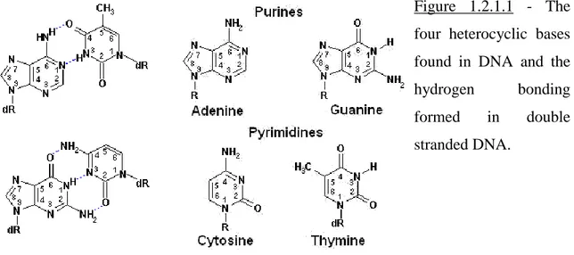
The genetic material inside all organisms is the deoxyribonucleic acid – DNA. It is a long double polymer made out of two chains of polynucleotides which run in an anti-parallel sense. A nucleotide is composed of one of four possible heterocyclic bases linked to a sugar which, in turn, is linked to a phosphate group. The polymerization reaction involves the formation of a phosphodiester bond between two sugar groups, with the base as a side chain. Figure 1.2.1.1 illustrates the building blocks of DNA.
The two polynucleotide chains are kept together by hydrogen bonds formed in between the heterocyclic bases, which are positioned, towards the inside of the DNA structure, roughly perpendicular on the backbone (the bases are stacked). Out of the four possible bases found in DNA: adenine, guanine, thymine and cytosine, the hydrogen bonding is possible only between an adenine and a thymine (2 hydrogen bonds per pair) or a guanine and a cytosine (three hydrogen bonds per pair). These normally weak hydrogen bond interactions give the strength of the polynucleotide-polynucleotide interaction by their sheer number. In fact, the longer the piece of DNA the harder it is to denature it, namely to physically detach one polynucleotide chain from its complement.
Because the base-base interaction in DNA is not random, the two polynucleotide polymers which bind each other are complimentary.
Figure 1.2.1.1 - The four heterocyclic bases found in DNA and the
hydrogen bonding
formed in double
stranded DNA.
The polynucleotide chain has also a polarity stemming from the fact that, when linking the nucleotide groups, two different positions are used to form the bond: the 3’ hydroxyl end of one sugar group with the phosphate found at the 5’ end of another sugar group. As mentioned previously, the DNA is formed of two polynucleotide chains (also called DNA strands) which are anti-parallel. Furthermore, this double stranded structure was shown to adopt a right-handed helical conformation. This allows for the formation of a hydrophobic core, formed of the hydrogen bonded bases, and a hydrophilic exterior composed of the backbone sugar-phosphate groups. This helical structure derives from the fact that the building blocks of DNA are asymmetric (four possible nucleotides based on the four possible heterocyclic bases) and, as a result, the stacking of the hydrogen bonded bases is slightly staggered.
The overall conformation of the double helix depends not only on the sterical hindrance of the bonds within the molecule but also on the physical properties of the solvent. The classical DNA double helix structure proposed by Watson and Crick corresponds to B-form DNA which is thought to correspond to the majority of the cellular DNA. Nevertheless, two other types of DNA have also been described: the A-form and the Z-A-form. In the B-A-form DNA, the helix is right-handed and the bases are stacked almost perpendicularly on the backbone. The helicity of the molecule is not uniform and the DNA has a major groove (wide and deep) and a minor groove (narrow
and deep). The number of base-pairs per helical turn (also called the DNA helical repeat)
was determined to be 10.5 and the diameter of the double helix was measured to be 20Å
(Feughelman et al., 1955).
In A-form DNA the helix is still right-handed but the bases are tilted to form angles much bigger or smaller than 90° with the sugar-phosphate backbone. This tilt results from a different conformation of the furanose ring of the sugar. The number of
base-pairs per helical turn is 11 and the diameter of the double helix is 26Å. As a result
the major groove is narrow and deep, while the minor groove is wide and shallow (Feughelman et al., 1955).
In Z-form DNA the helix is, surprisingly, left-handed and the bases are alternatively perpendicular and tilted as in B-form and A-form DNA. This alternating pattern corresponds to the alternating conformation of the sugar, which is either C2’-endo (B-form) or C3’-endo (A-form). As a result the sugar-phosphate backbone has a zigzagged pattern, the major groove is completely flat while the minor groove is narrow and deep. The number of base-pairs per helical turn is 12 but the diameter of the double
helix is much smaller, 18Å. This particular form of DNA is typical of sequences
composed of alternating GC tracts and is conditioned by the solvent composition (high-salt apparently promotes the formation of Z-form DNA) (Herbert and Rich, 1996).
The existence of A-form and Z-form DNA in vivo is still questioned but there is some evidence that at least in particular situations (e.g. gene activation, supercoiling) DNA may adopt a different conformation (Fairall et al., 1989, Singleton et al., 1982 and Liu et al., 2001) .
Being a flexible polymer DNA can be forced into a more compact or looser double helix, this property referring to the degree of supercoiling of a DNA molecule. In its relaxed state the B-form double helix has a helical repeat of 10.5 nucleotides. If a DNA molecule has this helical repeat for all its helices it is considered to be in its relaxed state (not supercoiled). If parts of the molecule or the whole of it has a higher or lower number of base-pairs per helical turn than this value, it is considered to be, respectively, negatively or positively supercoiled. Negative supercoiling leads eventually to the separation of the two DNA strands, while positive supercoiling tightens the helix and
ultimately makes it coil upon itself. This feature of DNA obviously stems from its helical nature and plays a central role in many aspects of DNA metabolism.
For a circular DNA molecule, or a linear one whose ends cannot rotate freely, a measure of the degree of DNA supercoiling can be obtained from the linking number. This value represents the number of double-helical turns the DNA molecule contains and it describes the topology of DNA. Two DNA molecules which differ only in the linking number are known as topological isomers or topoisomers. Therefore a change in DNA supercoiling implies a change in DNA topology.
All the characteristics here described for the double helix refer to biochemical and biophysical studies of DNA alone in different solutions. This is a good starting point for understanding the nature of DNA but is insufficient for appreciating its behavior in complex situations, as one would expect to find in vivo. Many factors affect the DNA conformation inside the cell, from the ionic strength, to the availability of different cations, to the interaction of the DNA with proteins. This is why it is important to understand the properties of the DNA molecule inside the living organism.
1.2.2 Nuclear architecture, chromatin and the scaffold.
In both prokaryotes and eukaryotes the chromosomes are packed within a much smaller space than the length of the extended DNA molecules. Just to give one example in eukaryotes the estimated packing ratio inside the interphase nucleus is that of 250 fold, while during mitosis it reaches 10000 fold. In spite of the almost one hundred years of research into how DNA is packed in vivo, the exact process by which this tremendous chromosome condensation is achieved remains poorly understood.
There is a striking difference between the way eukaryotes and prokaryotes deal with chromosomes inside the cell. Prokaryotic organisms often contain just one chromosome, for the most circular, which is much smaller in length compared to the eukaryotic counterpart. Inside the cell there is no specific membrane-bound structure (e.g. the nucleus in eukaryotes) which separates the genome from the rest of the cell, instead the chromosome can be detected by electron microscopy as an irregular dispersed structure called the nucleoid. There is evidence that proteins are associated with these
chromosomes for packing purposes but their exact role is not known. Still, the E.coli
chromosome (4.6kb of circular DNA) is compacted roughly 1000 fold in the cell.
Eukaryotes on the other hand, posses an array of chromosomes which are located inside a specific organelle, the nucleus. They also have more than one chromosomal set at least at one stage during their life cycle. The chromosomes are found in a packed state generally referred to as ‘chromatin’ and this term comprises all the different DNA packing stages from the least condensed one during interphase to the structures seen in mitosis. Chromatin inside the nucleus is usually of two types: euchromatin, a low-packing actively transcribed form, and heterochromatin, tightly packed regions which associate with a gene repression status.
The structure of chromatin is characterized well at its basic level of assembly but the mechanism concerning higher levels of packing remains strictly hypothetical. Eukaryotes have evolved a subset of nuclear proteins which associate specifically with DNA, called histones. These are small, highly basic proteins (rich in lysine and arginine) which are extremely well conserved across the whole eukaryotic domain. The histone family contains five members: H1, H2A, H2B, H3 and H4.
Histones H2A, H2B, H3 and H4 form an octameric subunit comprising 4 heterodimers: two H2A- H2B and two H3-H4. The DNA is wrapped 1.7 times around this octamer in a left-handed manner and the resulting structure is called a nucleosome,
the smallest structural subunit of chromatin. Each histone heterodimer binds 30bp of
DNA and a nucleosome was found to contain 147bp of DNA. The nucleosome is a stable structure, with the ability to self assemble in vitro. The fifth histone, H1 binds on the outside of the nucleosome, possibly close to the entry/exit point of the DNA double strand, and has a stabilizing role for the DNA-histone octamer interaction. Together the nucleosome and histone H1 bind 167bp of DNA and the resulting structure is called a chromatosome.
The overall reduction in DNA length through the assembly of nucleosomes is
only of 7 fold and therefore highly insufficient for the requirements of the cell. A
second step in DNA packaging involves the assembly of the 30nm fiber. Chromatosomes can be assembled onto DNA with a spacing of 0-80bp (this represents the length of the ‘linker DNA’). In turn this structure can be further assembled into a solenoid by
compacting the chromatosomes into a tight left handed helix with a diameter of 30nm. The exact way in which this compaction is achieved is still a matter of debate with several models proposed, nevertheless all models agree on a tight chromatosome assembly with the linker DNA completely folded inside the solenoid as a way of shortening the double helix.
It is thought that the majority of the DNA in interphase is in the solenoid state, with different regions being less packed. Actively transcribed regions still have nucleosomes but histone H1 is lost and higher order condensation is usually absent. In general, chromatin is a dynamic and highly regulated structure, with histone modifications altering the nucleosome stability and performing the so called ‘chromatin remodeling’.
Higher levels of chromatin condensation beyond the 30nm fiber are currently just at the speculation step with a range of scientific data pointing towards the assembly of the solenoid into a set of DNA loops attached to a nuclear scaffold. The chromosome size in mitosis makes it obvious that higher degrees of packing must exist and considering that the assembly of the basic unit of chromatin, the nucleosome, is based on protein-DNA interactions, it is logical to assume that further chromatin compaction is also protein dependent.
In a critical experiment it was shown that if the nuclei are isolated, histones are removed with 2M NaCl (along with the majority of the proteins) and DNA degradation is inhibited by inactivating nucleases, the DNA can be seen by electron microscopy as a halo surrounding the remnants of the nucleus (nucleoid). Surprisingly if this DNA preparation is treated with ethidium bromide, an intercalating dye which unwinds the double helix by inserting itself in between the bases, the DNA filaments become less diffused (McCready et al., 1979). This apparent paradox means that the local unwinding caused by the dye has in turn induced DNA supercoiling. This outcome is typical of circular DNA and is only possible in a linear DNA molecule if the ends are unable to freely rotate. Thus came the hypothesis that DNA inside the nucleus is organized into loops which are anchored to a nuclear structure.
Another experiment involving mitotic chromosomes reached the same conclusion using a completely different approach: using electron microscopy, metaphase
chromosomes depleted of histones show an electron-dense core, termed the nuclear scaffold, to which DNA loops are attached (Paulson and Laemmli, 1977). These loops are
70m in size and probably correspond to the next chromatin compaction level after the
30nm fiber. The nature of the nuclear structure which tethers the DNA loops (also called scaffold or matrix) remains an opened question. Problems stemming from the lack of methods for the isolation a ‘bona fide’ nuclear scaffold, with the high probability of many artifacts being present as a result of protein aggregation, made it impossible to progress further in this direction. If the existence of DNA loops is generally accepted, the nuclear structure isolating these loops is an incognita (Turner, 2001).
However this long struggle to characterize the nuclear scaffold has allowed the discovery of a series of important features of the nuclear architecture like the nuclear lamina. This proteinaceous network is formed of two families: A-type lamins (e.g. lamin A and C) and B-type lamins (e.g. lamin B). These proteins form a meshwork at the inner surface of the nuclear envelope and possibly also throughout the nucleoplasm (Hozak et al., 1995). The lamin family of proteins has not yet been found in lower eukaryotes or in plants but is conserved in metazoans. The role of the lamins is not well understood but protein-protein interactions show that they might bridge the gap between the nuclear envelope and chromatin binding proteins and could also function in mechanotransduction signaling inside the cell. Nevertheless, the nuclear lamina is unlikely to correspond to the nuclear scaffold since the high salt extraction method used to isolate chromosome loops removes the nucleoplasm lamin network.
In parallel, studies of scaffold proteins associated with metaphase chromosomes have led to the discovery of another important family of proteins, the SMC (Stability of Minichromosomes) family. The SMCs are proteins required for the correct chromosome condensation and segregation in yeast and homologues have been found in higher eukaryotes as well. They associate with chromosomes during mitosis and their similarity to motor proteins (e.g. kinesin) points towards a role in chromosome movement. Another component of the metaphase chromosome scaffold is topoisomerase II (Gasser et al., 1986). This enzyme is essential in eukaryotes due to its crucial role in chromosome decatenation and mitosis (Nitiss, 1998). If topoisomerase II is thought to be always
present in the nuclear scaffold, the SMC proteins are more likely to be associated with chromosome during mitosis only.
Out of all the proteins studied so far, topoisomerase II is probably the most likely ‘bona fide’ scaffold protein. It is considered the most abundant protein inside the scaffold and many searches of DNA scaffold/matrix attachment sites (SAR/MAR) were based on the affinity of topoisomerase II towards a piece of DNA (Razin et al., 1991). Yet, as an exact description of the scaffold (or safe method to isolate it) does not currently exist, all these data should be treated with a fair amount of skepticism.
However, if DNA loops exist, then they must be somehow kept in place, attached to a fairly rigid structure. If so, certain DNA regions will be indeed SARs/MARs. Whether these DNA regions are bound due to their sequence or at random, by simple charge interactions (as in the case of the histones), is not yet clear. The electron microscopy experiments described previously for both interphase and metaphase chromosomes suggest a very strong attachment of SARs/MARs to the scaffold, so why are these interactions so hard to reproduce in vitro?
A final piece of evidence in favor of DNA loops comes from chromosome staining techniques. The classic Giemsa chromosome staining, used for many years in karyotyping, reveals a species and chromosome-specific reproducible banding pattern of alternating dark bands (also called G-bands) and light bands. The opposite staining pattern (G-bands unstained and the light bands darkly stained) can be obtained by a reverse staining procedure (the dark colored bands resulting in this case are called R-bands). The crucial feature of these bands is that they can be obtained with minimum manipulation of the cells (as opposed to the lengthy and delicate procedures used to obtain the chromosomes and deplete histones) and that they seem to be reproducible with different staining techniques in both mitotic and meiotic chromosomes. Use of different dyes specific for AT-rich or GC-rich DNA has shown that G-bands correspond to chromatin domains containing very AT-rich regions, while R-bands correspond to chromatin domains containing very GC-rich regions. Based on the observation that G-bands have somewhat of a coiled structure in less condensed chromosomes, a new model was proposed for the organisation of DNA loops. Since AT-rich regions were found to be preferentially associated to nuclear scaffold preparations, the coiled structure of G-bands
would be a consequence of the high-density SARs/MARs nature of this DNA. Further condensation of these small loops would cause supercoiling. Instead the R-bands, GC-rich, would contain a much smaller number of SARs/MARs, resulting in much bigger DNA loops (Saitoh and Laemmli, 1994).
If DNA packing is an essential requirement for managing the eukaryotic chromosome complement, tight regulation of nuclear compartments is also necessary for efficient nuclear function. The organization of the interphase nucleus is not well characterized yet but some key features have emerged.
The most important nuclear compartment, the nucleolus, is the place of ribosomal DNA (rDNA) transcription and ribosome assembly. It contains a fibrillar centre, where both active and inactive rDNA genes are found, surrounded by a dense fibrillar component specialized in processing and assembly of rDNA, in turn surrounded by a granular component responsible for ribosome maturation (Shaw and Jordan, 1995). In humans five different pairs of chromosomes contain rDNA genes and all these loci cluster inside nucleoli.
Clustering seems to be a common mechanism by which different biological functions are regulated inside the nucleus. DNA loci used in the same process tend to cluster and form large foci. It is the case of active DNA replication (Nakamura et al., 1986). In mammalians centromeric heterochromatin tends to cluster in pericentric foci (Haaf and Schmid, 1991). Homology-dependent gene silencing which involves the down-regulation of a gene due to the presence of a transgene might function via clustering and physical interaction of the two genes (Fransz et al., 2002). DNA insulator elements which can block the effect of an enhancer or protect from a silencer are also thought to function by tethering a chromatin domain (possibly a DNA loop) to a particular structure (e.g. nuclear pore) (Blanton et al., 2003).
In addition, whole chromosomes seem to occupy individual territories inside the nucleus and rarely a chromosome will invade the territory of another in interphase. This is not a species specific organization, instead is more likely to reflect chromosome decondensation after mitosis. Still, chromosome movement is restricted inside the cell. One obvious reason is the sheer size of the DNA molecule. Quantitation of DNA movement in yeast has shown a difference between heterochromatic and euchromatic
loci, with the active chromatin domains moving more, presumably due to enzymatic events (Heun et al., 2001).
1.2.3 DNA structure and biological processes.
As mentioned in Section 2.1, supercoiling is a very important DNA feature and all nuclear processes involving DNA manipulations have to overcome the consequences arising from the helical nature of DNA. The chromosome is highly packed inside the cell and due to these topological constraints any form of supercoiling will affect heavily a chromatin domain (isolated by an anchored chromatin loop) without the possibility of distributing the strain over a much longer DNA molecule. Hence the need for topoisomerases, enzymes specialized in altering the topological state of DNA.
The chromatin organization inside the nucleus endows DNA with negative supercoiling. During nucleosome assembly positive supercoiling is accumulated on the DNA and is released by the action of topoisomerases. As a result when the nucleosome disassembles the DNA will be left negatively supercoiled. This ‘stored’ negative supercoiling can be used for different biological processes like DNA transcription and replication.
DNA replication initiation requires the formation of a bubble at the origin. This is obviously a process which requires a large amount of negative supercoiling to be introduced in the DNA in order to obtain the separation of the two strands, with resulting accumulation of positive supercoiling on both sides of the origin. Once the replication fork has been assembled and DNA synthesis has started the fork movement along the DNA will continue to unwind the two parental strands and even more positive supercoiling will accumulate ahead of the fork.
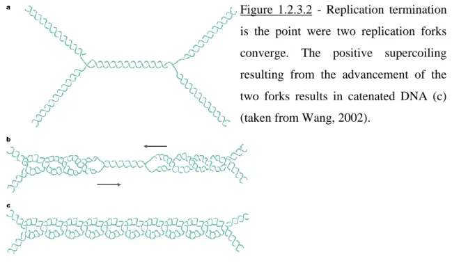
If the replication fork complex is able to rotate around the DNA axis (not a likely possibility due to the sheer size of the replisome), the positive supercoiling will be distributed both ahead and behind the fork. If this positive supercoiling is not removed the DNA will begin to coil upon itself much like an over-twisted rope. At the point of termination of DNA replication where two replication forks converge, due to the accumulated positive supercoiling, the two daughter molecules will be intertwines, i.e.
catenates are formed. Figures 1.2.3.1 and 1.2.3.2 illustrate the type of topological constraints encountered during DNA replication.
Nonetheless, all these considerations about DNA topology during replication are just logical assumptions, what truly happens inside the cell and how the various in vivo protein-DNA interactions help or complicate topological problems is unknown.
DNA transcription is another process which shares a few of the topological difficulties of DNA replication. The first step in transcription is also the separation of the two DNA strands in order to allow access of RNA polymerase to its template with resulting positive supercoiling in the surrounding region. Furthermore during active transcription it has been shown that the RNA polymerase (often coupled with ribosomes) does not rotate around the DNA axis and as a result positive supercoiling accumulates ahead of the enzyme, while negative supercoiling will build behind it (Figueroua and Bossi, 1988 and Tsao et al., 1989).
Figure 1.2.3.1 - Replication
elongation creates an
accumulation of positive
supercoiling ahead of the
replication fork (a). If the fork complex can rotate around the
DNA axis the positive
supercoiling will be distributed both ahead and behind the fork, with resulting formation of pre-catenates in the newly replicated region (b) (taken from Wang, 2002).
Figure 1.2.3.2 - Replication termination is the point were two replication forks converge. The positive supercoiling resulting from the advancement of the two forks results in catenated DNA (c) (taken from Wang, 2002).
1.3 DNA topoisomerases are modulators of DNA topology.
1.3.1 Classification of DNA topoisomerases.
In the cell, the only enzymes capable of modifying the degree of supercoiling of DNA (the so-called “linking number” of a DNA molecule) are the DNA topoisomerases. All topoisomerases identified so far regulate the degree of supercoiling of DNA via cleavage and formation of a transient covalent phosphor-tyrosine enzyme/DNA adduct. Moreover a particular subset of topoisomerases can also catenate/decatenate knot/unknot plasmid DNA. The catalytic cycle of DNA topoisomerases involves 4 steps:
1. binding of the DNA substrate.
2. cleavage of the DNA with concerted covalent attachment of the enzyme to the 3’ or 5’ DNA end via a nucleophilic attach of an enzyme tyrosine residue on a DNA phosphate.
3. rotation and/or strand passage of the substrate in order to remove/introduce positive or negative supercoiling or catenate/decatenate, knot/unknot DNA molecules.
4. religation of the gap via a reversal of the transesterification reaction and release of the DNA substrate.
An example of a topoisomerase cleavage reaction is given in Figure 1.3.1.1. Under physiological conditions the cleavage, rotation and religation are extremely quick and the covalent enzyme-DNA complex (also called the “cleavable complex”) is only a short lived intermediate. Nevertheless under specific conditions the equilibrium of the cleavage reaction can be shifted towards the cleavable complex state and the enzyme can be physically trapped on the DNA. Due to this particular property, the topoisomerases have long been recognized as potential DNA damaging agents and they are targets for a wide range of drugs. Currently topoisomerase inhibitors are used as antimicrobial and antitumor drugs.
Figure 1.3.1.1 -
Eukaryotic topoisomerase I is a type IB enzyme and
becomes covalently
attached to the 3’ end of its DNA substrate during
the catalytic cycle.
Topoisomerases are usually specialized in one particular topological reaction (e.g. removing positive supercoiling as opposed to introducing it) and as a result it is common
to find more than one type of enzyme in each organism. At least one gene coding for a DNA topoisomerase has been identified in all organisms studied and it is generally assumed that topoisomerases are essential enzymes for cell growth due to their unique DNA decatenation activity (Champoux, 2001 and Wang, 2002). Based on the type of DNA cut they introduce, topoisomerases can fall into two classes: type I or type II. Type I enzymes can bind single or double stranded DNA and introduce single-stranded nicks, while type II cleave both DNA strands of a double helix. Further division within type I and type II into subtype A and subtype B is based on structural and catalytical properties. Table 3.1.1 illustrates the topoisomerase classification and enzyme properties.
Table 3.1.1 The topoisomerase classification.
Domain of life Type Structure Notes
Topoisomerase Class IA
The enzyme cleaves just one strand of the DNA substrate with concerted covalent attachment to the 5’ end of the cut. The DNA substrate is always negatively supercoiled with the enzyme requiring an exposed single-stranded region for catalytical activity. The relaxation reaction does not go to completion (i.e. the DNA will still be negatively supercoiled at the end of the reaction). The enzyme is also able to catenate/decatenate and knot/unknot single stranded DNA circles or
nicked duplex circles. The catalytical activity is dependent on Mg2+ ions.
Archaea
Reverse DNA gyrase Monomer
Reverse gyrase is the only enzyme capable of introducing positive
supercoiling into its DNA
substrate. All archaeal
hyperthermophiles studied so far seem to have an active reverse gyrase, most probably important in neutralizing the effects of growth at high temperature.
Topoisomerase III Monomer Relative sequence homology to
few organisms so far.
Bacteria
Topoisomerase I Monomer Seems to be present in all bacterial
genomes fully sequenced so far.
Topoisomerase III Monomer Some bacteria lack this type of
enzyme.
Topoisomerase IIIβ Monomer
Identified only in Bacillus cereus, has distinct featured from all other type IA enzymes.
Reverse DNA gyrase
Monomer
This enzyme has only been
reported in hyperthermophilic
bacteria.
Heterodimer
The only reverse gyrase with a
heterodimeric structure was
identified in the bacterial
hyperthermophile Methanopyrus kandleri.
Eukarya
Yeasts Topoisomerase III Monomer
Saccharomyces cerevisiae topo III is dispensable for cell growth
while Schizosaccharomyces
pombe topo III is essential.
Flies
Topoisomerase IIIα Monomer Essential during development in
Drosophila melanogaster.
Topoisomerase IIIβ Monomer Not essential in D. melanogaster.
Plants None reported so far.
Mammals
Topoisomerase IIIα Monomer Essential for embryogenesis in
Mus musculus.
Topoisomerase Class IB
The enzyme cleaves just one strand of the DNA substrate with concerted covalent attachment to the 3’ end of the cut. The enzyme can relax positively or negatively supercoiled DNA substrates and the relaxation reaction goes to completion (i.e. the DNA will be in a relaxed state at the end of the reaction). The catalytical activity is not dependent on metal ions.
Archaea None reported so far.
Bacteria Topoisomerase V Monomer
Present only in Methanopyrus kandleri and structurally distinct from all other topoisomerases. Eukarya
Yeasts Topoisomerase I Monomer Not essential for growth.
Flies Topoisomerase I Monomer Essential in D. melanogaster.
Plants Topoisomerase I Monomer
Essential in Arabidopsis thaliana. The Pisum sativum enzyme also exhibits reverse gyrase activity.
Mammals Topoisomerase I Monomer Essential in M. musculus.
Topoisomerase Class IIA
The enzyme cleaves both strands of the DNA substrate (with a 4bp stagger) with concerted covalent attachment to the two 5’ ends of the cut. The enzyme has the ability to bind and pass another DNA double stranded region or molecule through the gap resulting from the DNA cleavage. This process results in DNA relaxation, knotting/unknotting or catenation/decatenation
events. The catalytical activity is dependent on Mg2+ ions and ATP. However, it should be noted
that different members of this class have very different efficiencies for relaxation, knotting/unknotting or catenation/decatenation reactions.
Archaea DNA gyrase Heterotetramer Not very common in
archaebacteria.
Bacteria
DNA gyrase Heterotetramer Found in all bacteria studied so
far.
Eukarya
Yeasts Topoisomerase II Homodimer Essential for growth.
Flies Topoisomerase II Homodimer Essential in D. melanogaster.
Plants Topoisomerase II Homodimer
Mammals
Topoisomerase IIα Homodimer In M. musculus essential for early
embryogenesis.
Topoisomerase IIβ Homodimer In M. musculus essential for late
embryogenesis. Topoisomerase Class IIB
The only member of this class of topoisomerases, topoisomerase VI, has the same catalytical properties of class IIA enzymes but shows very little sequence similarity with them. Topo VI is
a A2B2 heterotetramer and the A subunit is homologous to the yeast Spo11 protein involved in
meiotic recombination.
Archaea Topoisomerase VI Heterotetramer Found in all archaebacteria studies
so far.
Bacteria None reported so far.
Eukarya
Yeasts None reported so far.
Flies None reported so far.
Plants Topoisomerase VI Heterotetramer Essential for endoreduplication in
A. thaliana.
Mammals None reported so far.
As mentioned before DNA topoisomerases are encoded in the genome of every genetic organism. Because topoisomerases participate in every aspect of DNA metabolism it is not surprising that during evolution they have acquired specialization for a particular topological reaction. At the same time prokaryotic and eukaryotic
topoisomerases seem to have evolved divergently and their characteristics show less similarities between these two taxonomical domains than expected.
In prokaryotes the first topoisomerase discovered was the E.coli ω protein, now known as topoisomerase I (Wang, 1971). This is a 97 kDa monomer which exhibits a type IA catalytical reaction. The eukaryotic topoisomerase I (which has a reasonably conserved sequence from yeast to man) is a ~100 kDa monomer with a type IB catalysis. It should be noted that these enzymes were both called topoisomerase I assuming that they were orthologs and performed the same function within the cell. Nevertheless they have been shown not only to have no structural or sequence similarities but also to differ in the range of topological actions they can perform. Just to give an example eukaryotic topo I can relax both positive and negative supercoiling while E.coli topo I cannot relax positive supercoils.
Type I topoisomerases act as monomers, contain only one catalytic tyrosine residues (as they introduce single stranded breaks) and do not normally require an energy cofactor (i.e. ATP). Type IA enzymes can relax only negative supercoils, require a single-stranded stretch in the DNA substrate, become covalently attached to the 5’ end of
the DNA cut and cannot function in the absence of Mg2+ ions. Members of this class
include prokaryotic topoisomerase I, prokaryotic and eukaryotic topoisomerase III and prokaryotic reverse gyrase. Apart from removing negative supercoiling, type IA enzymes can also perform knotting/unknotting and catenation/decatenation reactions. James Wang has postulated in his latest review about topoisomerases that “the minimal requirement for DNA topoisomerases in living organisms is probably one type IA and one type II enzyme” (Wang, 2002).
E.coli topo I is efficient in removing negative supercoils but inefficient as a DNA decatenase. It plays a critical role in maintaining the optimum DNA supercoiling for all cellular functions and its role is particularly important during transcription (Drolet et al., 1995). A topo I enzyme seems to be present in all bacteria investigated so far, however this enzyme is absent from archaebacteria (Champoux, 2001). E.coli topo III is related to the E.coli topo I and able to perform the same relaxation reaction in the presence of negative supercoiling. Nonetheless, it requires that the DNA substrate should be hypernegatively supercoiled and it is much more efficient in catenation/decatenation
reactions than topo I (DiGate and Marians, 1988 and Hiasa et al., 1994). Topo III is thought to be involved in the elongation and termination/decatenation events of DNA replication even though these roles would overlap with those of the E.coli type II topoisomerases (Hiasa and Marians, 1994 (b) and Hiasa et al., 1994). More evidence for this apparent functional redundancy comes from the fact that a number of other bacteria lack a topo III homologue (Champoux, 2001).
Interestingly, in the bacterium Bacillus cereus two topo III enzymes have been identified, designated topo IIIα and topo IIIβ (Li et al., 2006). The B.cereus topo IIIα has the same catalytical properties as the E.coli topo III, while topo IIIβ can only partially relax negative supercoils and lacks decatenase activity. Furthermore, topo IIIβ cannot compensate for the lack of E.coli topo III in vivo (Li et al., 2006). The B.cereus topo IIIβ seems to be distinct from all other type IA topoisomerases and it remains to be seen if it performs an essential function in vivo.
An archaeal topo III enzyme, related to the bacterial topo III, has also been reported for the hyperthermophilic archaeon Sulfolobus solfataricus (Dai et al., 2003).
Eukaryotic topoisomerases III have been described in yeast, Drosophila, mouse and humans (Wallis et al., 1989, Wilson et al., 2000, Seki et al., 1998 and Hanai et al., 1996). They have all the main features of the bacterial topo III, they can relax only
negative supercoiling, need a single stranded stretch of DNA and Mg2+ ions in order to
function. Even though Saccharomyces cerevisiae topo III is not absolutely required for cell growth, mutants for the gene exhibit slow growth, increased mitotic recombination and a defect in meiosis resulting in failed sporulation (Gangloff et al., 1999). Instead, deletion of the topo III enzyme is lethal in the yeast Schizosaccharomyces pombe and its presence is essential for accurate nuclear division (Maftahi et al., 1999 and Goodwin et
al., 1999). Higher eukaryotes have two topo III isoforms: and . In Drosophila topo
III is essential during development, while topo III appears to be dispensable for
viability in spite of having a peak of expression during the first 6h of embryogenesis
(Wilson et al., 2000 and Plank et al., 2005). Knockout mice for topo III die early during
embryogenesis, while topo III knockouts have a reduced life-span (Li and Wang, 1998
and Kwan and Wang, 2001). Topo III seems to interact with DNA helicases of the RecQ family at least in yeast and humans and possibly functions in the same processes which
require these helicases (Bennett et al., 2000, Wu et al., 2000 and Shimamoto et al., 2000).
Topo III together with BLM, Bloom’s syndrome helicase, seem to be involved in sister
chromatid dissolution in mitosis (Seki et al., 2006). Interestingly, E.coli topo III also interacts with E.coli RecQ helicase (Harmon et al., 1999).
Reverse gyrase is an enzyme found only in hyperthermophilic organisms including archaea and eubacteria and is the only type IA enzyme which requires ATP as a cofactor. The role of reverse gyrase is that of introducing positive supercoiling in a DNA substrate (Declais et al., 2001). Considering the special type of environment in which hyperthermophilic organisms grow in, it has been postulated that this enzyme helps protect the genome from the DNA denaturing effect of the extreme temperature (Charbonnier and Forterre, 1994).
It is worth mentioning that genes coding for type IA topoisomerases were also found in several plasmids from Gram-positive and Gram-negative bacteria (Champoux, 2001). Interestingly in one case it has been reported that initiation of plasmid replication by DNA polymerase I creates a substrate specific for a plasmid encoded topoisomerase (Bidnenko et al., 1998).
If type IA enzymes are found in every organism, type IB are typical of eukaryotes with just one exception, topoisomerase V, which is present in a hyperthermophilic bacterium, Methanopyrus kandleri.
Type IB topoisomerases can relax both positive and negative supercoiling, become covalently attached to the 3’ end of the DNA cut and do not require any type of metal ion or energy cofactor (Pommier et al., 1998). Their action is inhibited by single-stranded DNA in contrast to type IA enzymes (Been and Champoux, 1984). In contrast to type IA enzymes, they are not capable of catenation/decatenation or knotting/unknotting reactions. Eukaryotic topoisomerase I, the vaccinia virus topoisomerase and Methanopyrus kandleri topoisomerase V are type IB enzymes.
A topo I has been reported in all eukaryotic model genetic organisms including yeasts, Caenorhabditis elegans, Drosophila melanogaster, Xenopus laevis, wheat germ, mouse and human (Goto and Wang, 1985, Uemura et al., 1987a, Kim et al., 1996, Hsieh et al 1992, Pandit et al., 1996, Dynan et al., 1981, Koiwai et al., 1993 and Kunze et al., 1989). In the yeasts S.cerevisiae and S.pombe topo I is not essential for growth, while in
higher eukaryotes, like Drosophila and mouse, absence of this enzyme is lethal (Thrash et al., 1984, Uemura and Yanagida, 1984, Lee et al., 1993 and Morham et al., 1996). Topo I is involved in many aspects of DNA metabolism including DNA replication, transcription, damage repair and possibly chromosome condensation in mitosis (Pommier et al., 1998). Along with the nuclear topoisomerase I, a mitochondrial topo I was identified in Xenopus, calf thymus and humans (Brun et al., 1981, Lazarus et al., 1987 and Zhang et al., 2001).
Unexpectedly, a topo I enzyme isolated from Pisum sativum (pea) showed, besides the typical type IB catalytic reaction, the ability to introduce positive supercoiling
in the presence of Mg2+ ions, a property of reverse gyrases found only in
hyperthermophiles (Reddy et al., 1998). This is a unique feature, not detected so far in any other eukaryotic topo I.
Until recently it was thought that type IB enzymes were exclusive to eukaryotes, considering the complete absence of these enzymes in classic model prokaryotes (e.g. E.coli, Bacillus subtilis). In spite of this, the bacterium M.kandleri, a hyperthermophilic methanogen, contains in its complement of topoisomerases a type IB enzyme, named topoisomerase V (Slesarev et al., 1993). Considering that topo V is dissimilar structurally from all other topoisomerases there it has been proposed that it should be classified apart from other enzymes as type IC (Forterre, 2006). Topo V can relax both positive and negative supercoiling and becomes covalently attached to the 3’ end of the DNA cut, still, the exact role of this enzyme is not currently understood, since other hyperthermophilic bacteria do not seem to require one. M.kandleri is also unusual in the fact that it has a novel heterodimeric reverse gyrase, while all the previously discovered reverse gyrases are monomeric (Krah et al., 1996).
The vaccinia virus (poxvirus) is one of the few viruses known to encode its own topoisomerase (Bauer et al., 1977). Coded by the E6 gene, this topoisomerase is a lot smaller compared to the eukaryotic type IB enzymes and was shown to be essential for viral proliferation in cell culture (Shchelkunov et al., 1993).
Type II topoisomerases differ from type I enzymes in that, when bound to the double helix, they introduce double stranded cleavages (usually four base pair staggered). They can be homodimers or heterotetramers, contain two catalytic tyrosine residues (as
they introduce double stranded breaks) and become covalently attached to the 5’ end of the DNA break. They can also catenate or decatenate (knot or unknot) DNA molecules, by passing an intact DNA molecule through the double stranded break created, this strand passing activity involving ATP hydrolysis. All organisms seem to require a type II topoisomerase since these are the only type of enzymes capable of decatenating double stranded DNA molecules (i.e. newly replicated DNA) (Champoux, 2001 and Wang, 2002). Type II enzymes are also divided into two subcategories, A and B, a distinction based on structural characteristics. The recent discovery of an atypical topoisomerase in the genome of the archebacterium Sulfolobus shibatae, named topoisomerase VI, has prompted the classification of this type II enzyme apart from all others, as type IIB (Bergerat et al., 1997).
The type IIA enzymes are DNA gyrase and topoisomerase IV, found in eubacteria and topoisomerase II, found only in eukaryotes. DNA gyrase was first identified in E.coli as an enzyme capable of transforming a relaxed DNA molecule into a negative supercoiled one (Gellert et al., 1976). Indeed DNA gyrase, a heterotetramer, is the only enzyme known so far able to introduce negative supercoiling. This property plays an essential role in the initiation of DNA replication at the E.coli oriC (Fairweather et al., 1980, Filutowicz, 1980 and Baker and Kornberg, 1988). DNA gyrase can also relax negatively or positively supercoiled DNA but in a much more inefficient manner, while its decatenation activity is almost absent in vivo (Levine et al., 1998). In the cell this enzyme has a role in maintaining the overall DNA superhelicity, facilitating replication elongation and transcription and possibly decatenation of chromosomes after DNA replication (Levine et al., 1998). Even though DNA gyrases are found predominantly in bacteria, there have been several reports of archaebacteria which also posses this type of enzyme (Gadelle et al., 2003). Recently, a genome analysis in the model plant A.thaliana revealed four putative gyrase genes and knockout studies show that all four genes are essential for plant development (Wall et al., 2004). Furthermore, cloning of the A.thaliana putative gyrase genes in E.coli gyrase temperature-sensitive strains lead to phenotype rescue (Wall et al., 2004). However, detailed biochemical analysis of the gene products is still missing for the time being.
Topoisomerase IV is the other main type II enzyme found in eubacteria. Topo IV is also a heterotetramer and a potent decatenase responsible for decatenating daughter chromosomes at the end of DNA replication. Like DNA gyrase, this enzyme can also relax negatively or positively supercoiled DNA but at a much slower rate than decatenation and its roles in the cell include maintaining the overall DNA superhelicity, facilitating replication elongation and transcription (Champoux, 2001).
Topoisomerase II acts as a homodimer and it is the only type II topoisomerase found in eukaryotes (except for the newly discovered topo VI in plants). When the work concerning topoisomerases was just at the beginning, the identification of a type II enzyme in eukaryotes led to the logical assumption that its topological activity would be very similar to the prokaryotic type II enzymes. As it was discovered later, eukaryotic topoisomerase II, in spite of being homologous to both DNA gyrase and topo IV, has a behavior more similar to topo IV.
Topo II can relax both negative and positive supercoiled DNA but it cannot perform the opposite reaction of introducing either type of supercoiling. This characteristic leaves the puzzling question of whether there is a yet undiscovered enzyme that can function as a gyrase or reverse gyrase in eukaryotes or if during evolution eukaryotes have lost the need for this topological reaction. In addition to the DNA relaxation activity, topo II is able to decatenate DNA, an enzymatic reaction essential in de-tangling sister chromatids at the end of DNA replication. Topo II action requires ATP
and Mg2+ ions (Champoux, 2001). The topo II decatenation reaction is illustrated in
figure 1.3.1.2.
In the yeasts S.cerevisiae and S.pombe topo II is absolutely required for the decatenation of daughter chromosomes and chromosome condensation before cell division (diNardo et al., 1984, Holm et al., 1985 and Uemura et al., 1987b). In Drosophila topo II is essential for anaphase sister chromatid separation and does have a partial role in chromosome condensation (Chang et al., 2003). Higher eukaryotes (with
the exception of Drosophila) have two isoforms of this enzyme: topo II and topo II
(Champoux, 2001). The two isozymes have a different expression pattern: topo II is
preferentially expressed in proliferating cells while topo II apparently has the same level
seems to correspond to the yeast and Drosophila topo II, being required for chromosome
decatenation and condensation, while topo II is not required for cell division (Nitiss,
1998). Knockout mice for topo II die at the 4- or 8-cell stage of embryonic
development, while knockout mice for the topo II isoform die shortly before birth and
show abnormal neural development (Akimitsu et al., 2003a and Yang et al., 2000). So in
spite of the fact that topo II is not essential for cell growth, it plays an important role
during development. Furthermore, both murine embryos and HeLa cells lacking topo II,
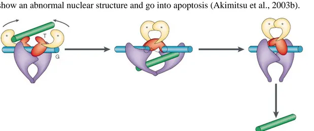
show an abnormal nuclear structure and go into apoptosis (Akimitsu et al., 2003b).
Figure 1.3.1.2 - Topoisomerase II can pass a second DNA double helix through a induced DNA double strand break. The green DNA segment (T) represents the intact double helix, while the blue DNA segment (G) represents the cleaved double helix. The asterisks represent ATP binding sites (taken from Wang, 2002).
Apart from these critical functions, topo II and topo II probably share with
topo I a functional redundancy, since both type of topoisomerases seem to have a role in the elongation step of DNA replication, DNA transcription and genome stability (Nitiss, 1998).
Aside from the topoisomerases described so far, a type IIA enzyme was also found in the genome of three viruses: bacteriophage T4, African swine fever virus and paramecium Bursaria chlorella virus (Liu et al., 1979, Garcia-Beato et al., 1992 and Lavrukhin et al., 2000).
As mentioned before, the discovery of a novel type II topoisomerase in Archaea has provoked the split of type II enzyme into two subcategories: A and B. This was due
to the fact that the S.shibatae topoisomerase VI, a heterotetrameric A2B2 enzyme, was not
homologous to any other topoisomerase, but one of its subunits (subunit A) showed homology to Spo11p, an enzyme involved in meiotic recombination (Malone et al., 1991 and Bergerat et al., 1997). It seems that Spo11 cleaves the DNA in order to produce the double stranded break required for meiotic recombination initiation. Furthermore it was shown that Spo11 is covalently attached to the DNA substrate much like a regular topoisomerase (Keeney et al., 1997). It remains to be seen if indeed Spo11 and its homologues in higher eukaryotes can function indeed as topoisomerases.
Topoisomerase VI is also found in plants, as revealed from the sequencing of the A.thaliana genome. Since plants are normally polyploid it is thought that this enzyme plays a role in endoreduplication of the genome, a common event in plant development (Sugimoto-Shirasu et al., 2002).
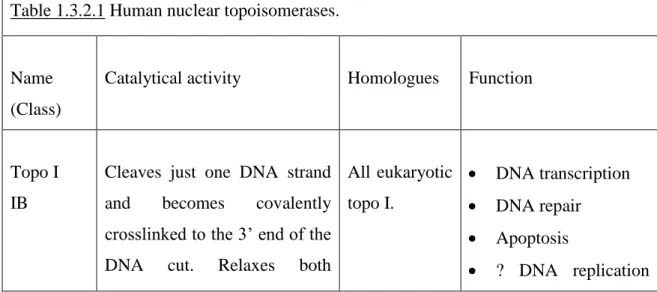
1.3.2 Human DNA topoisomerases and their roles in the cell.
The human topoisomerase complement contains 6 enzymes: a nuclear topoisomerase I (topo I), a mitochondrial topoisomerase I (topo Imt), two topoisomerases
II, and , and two topoisomerases III, and (Kunze et al., 1989, Zhang et al., 2001,
Tsai-Pflugfelder et al., 1988, Austin et al., 1993, Hanai et al., 1996 and Ng et al., 1999). Table 1.3.2.1 presents the human topoisomerases and their key features.
Table 1.3.2.1 Human nuclear topoisomerases.
Name (Class)
Catalytical activity Homologues Function
Topo I IB
Cleaves just one DNA strand
and becomes covalently
crosslinked to the 3’ end of the
DNA cut. Relaxes both
All eukaryotic topo I. DNA transcription DNA repair Apoptosis ? DNA replication
positive and negative
supercoiling. Cannot
catenate/decatenate,
knot/unknot DNA molecules. Cannot introduce negative or positive supercoiling.
elongation.
Topo IIα IIA
Cleaves both DNA strands with a 4bp stagger and
becomes covalently
crosslinked to the 5’ end of the
DNA cut. Relaxes both
positive and negative
supercoils.
Catenates/decatenates,
knots/unknots DNA molecules. Cannot introduce negative or positive supercoiling.
Yeasts and
Drosophila topo II and topo IIα from higher eukaryotes. Nuclear scaffold. Chromosome condensation. Chromosome segregation. DNA replication termination (decatenation). ? DNA replication elongation. Topo II IIA
Cleaves both DNA strands with a 4bp stagger and
becomes covalently
crosslinked to the 5’ end of the
DNA cut. Relaxes both
positive and negative
supercoils.
Catenates/decatenates,
knots/unknots DNA molecules. Cannot introduce negative or positive supercoiling.
Yeasts and
Drosophila topo II and topo IIβ from higher eukaryotes. Signal dependent transcriptional activation ? Mitosis. ? DNA replication.
Topo IIIα IA
Cleaves just one DNA strand
and becomes covalently
crosslinked to the 5’ end of the
DNA cut. Relaxes only
negative supercoiling and
requires a region of single
stranded DNA. The
catenation/decatenation,
knotting/unknotting activity
has not been tested yet. Cannot introduce negative or positive supercoiling.
Prokaryotic and yeast topo III, Drosophila and mouse topo IIIα. ? Topo IIIβ IA No biochemical characterization is available yet. Prokaryotic and yeast topo III,
Drosophila
and mouse
topo IIIβ.
?
The mitochondrial topoisomerase I is 601 amino acids (aa) long and highly homologous to the nuclear topoisomerase I. Topo Imt is a type IB topoisomerase and
requires Ca2+ or Mg2+ and alkaline pH for optimum activity (Zhang et al., 2001).
Human nuclear topo I is 765aa long, has a predicted molecular weight of 91kDa and presents four structural domains: a N-terminal domain, non-essential for catalytical activity but involved in protein-protein interactions, a highly-conserved core domain which is in contact with DNA, a positively charged linker domain and a conserved C-terminal domain (Stewart et al., 1996 and Champoux, 2001). It has a type IB catalysis