Neonatal therapy with clenbuterol and salmeterol restores
spinogenesis and dendritic complexity in the dentate gyrus of the
Ts65Dn model of Down syndrome
Marco Emili, Fiorenza Stagni, Maria Elisa Salvalai, Beatrice
Uguagliati, Andrea Giacomini, Christelle Albach, Marie-Claude
Potier, Mariagrazia Grilli, Renata Bartesaghi, Sandra Guidi
PII:
S0969-9961(20)30149-2
DOI:
https://doi.org/10.1016/j.nbd.2020.104874
Reference:
YNBDI 104874
To appear in:
Neurobiology of Disease
Received date:
13 January 2020
Revised date:
9 April 2020
Accepted date:
19 April 2020
Please cite this article as: M. Emili, F. Stagni, M.E. Salvalai, et al., Neonatal therapy
with clenbuterol and salmeterol restores spinogenesis and dendritic complexity in the
dentate gyrus of the Ts65Dn model of Down syndrome, Neurobiology of Disease (2019),
https://doi.org/10.1016/j.nbd.2020.104874
This is a PDF file of an article that has undergone enhancements after acceptance, such
as the addition of a cover page and metadata, and formatting for readability, but it is
not yet the definitive version of record. This version will undergo additional copyediting,
typesetting and review before it is published in its final form, but we are providing this
version to give early visibility of the article. Please note that, during the production
process, errors may be discovered which could affect the content, and all legal disclaimers
that apply to the journal pertain.
Neonatal therapy with clenbuterol and salmeterol restores spinogenesis and dendritic complexity in the dentate gyrus of the Ts65Dn model of Down syndrome
Marco Emili1,1 , Fiorenza Stagni2,1 , Maria Elisa Salvalai3, Beatrice Uguagliati1, Andrea Giacomini1,
Christelle Albach4, Marie-Claude Potier4, Mariagrazia Grilli3, Renata Bartesaghi1,**
[email protected], Sandra Guidi1,* [email protected]
1Department of Biomedical and Neuromotor Sciences, University of Bologna, Bologna, Italy 2Department for Life Quality Studies, University of Bologna, Rimini, Italy
3Department of Pharmaceutical Sciences, University of Piemonte Orientale, Italy
4Institut du Cerveau et de la Moelle- CNRS UMR7225 - INSERM U1127 – Sorbonne University, Hôpital de
la Pitié-Salpêtrière, Paris, France
*Corresponding author. **Co-Corresponding author.
ABSTRACT
Down syndrome (DS), a neurodevelopmental disorder caused by triplication of chromosome 21, is characterized by intellectual disability. In DS, defective neurogenesis causes an overall reduction in the number of neurons populating the brain and defective neuron maturation causes dendritic hypotrophy and reduction in the density of dendritic spines. No effective therapy currently exists for the improvement of brain development in individuals with DS. Drug repurposing is a strategy for identifying new medical use for approved drugs. A drug screening campaign showed that the β2-adrenergic receptor (β2-AR) agonists clenbuterol hydrochloride (CLEN) and salmeterol xinafoate (SALM) increase the proliferation rate of neural progenitor cells from the Ts65Dn model of DS. The goal of the current study was to establish their efficacy in vivo, in the Ts65Dn model. We found that, at variance with the in vitro experiments, treatment with CLEN or SALM did not restore neurogenesis in the hippocampus of Ts65Dn mice treated during the postnatal (P) period P3-P15. In Ts65Dn mice treated with CLEN or SALM, however, dendritic spine density and dendritic arborization of the hippocampal granule cells were restored and the lowest dose tested here (0.01 mg/kg/day) was sufficient to elicit these effects. CLEN and SALM are used in children as therapy for asthma and, importantly, they pass the blood-brain barrier. Our study suggests that treatment with these β2-AR agonists may be a therapy of choice in order to correct dendritic development in DS but is not suitable to rescue neurogenesis.
Keywords: Down syndrome; Ts65Dn model; dendritic pathology; spine density; dendritic complexity; pharmacotherapy
1 The Authors indicated with an asterisk contributed equally to this work.
1. INTRODUCTION
Down syndrome (DS) is a neurodevelopmental disorder caused by triplication of chromosome 21. The gene burden causes various clinical problems, not all of which occur in all people with DS. Intellectual disability (ID), however, is consistently present in DS. Although the severity of ID may vary across individuals, the general outcome is the lack of an autonomous life. The prevalence of live DS births is relatively high, ranging from 1:1000 to 1.2:1000, depending on world area (de Graaf et al., 2015) (Web site Link 4). Thanks to an improvement in medical care, people with DS now live longer and may outlive their parents. Parents of people with DS and researchers interested in DS, therefore, feel the urgent need to identify means to correct the neurodevelopmental alterations that characterize DS.
The brain of people with DS is smaller in comparison with that of the general population due to two major reasons. Defective neurogenesis causes an overall reduction in the number of neurons populating the brain and defective neuron maturation causes dendritic hypotrophy and reduction in the density of dendritic spines (Bartesaghi et al., 2011). Intense efforts are currently underway to establish whether it is possible to pharmacologically improve the developmental defects of DS, thereby improving cognitive performance. The molecular causes of brain developmental alteration in DS are extremely complex due to the large number of triplicated genes. Yet, alteration of certain cellular pathways seems to play a prominent role (Creau, 2012, Vacca et al., 2019). Based on this knowledge, a variety of agents acting on these pathways have been used in DS mouse models (Stagni et al., 2015a, Vacca et al., 2019). These studies show that it is possible to pharmacologically improve or even correct one or more trisomy-linked brain defects in DS mouse models. Some of these studies, however, remain at the proof-of-principle level because the agents used and/or the doses of the agents may raise concern for human use due to possible side effects. Consequently, no effective therapy currently exists for the improvement of brain development in individuals with DS.
Drug repurposing is a strategy for identifying new medical indications for approved drugs that are outside the scope of the original medical indication. One of the advantages of this strategy is that repurposed drugs have well-characterized tolerability and safety profiles, along with established pharmacokinetic properties. Therefore, at least in principle, approved drugs that result effective in DS mouse models might be more easily proposed for clinical trials in individuals with DS. Based on these considerations, we recently carried out a drug screening campaign in order to identify among FDA/EMA approved drugs, compounds
which could increase the neurogenic potential of trisomic neural precursor cells and their differentiation. The general idea was to test the effects of chemical libraries of drugs on neural progenitor cells (NPCs) obtained from the Ts65Dn model of DS, and then to test the more promising hits in vivo, in the same model.
Among the FDA/EMA approved drugs we focused our attention on -adrenergic drugs. These molecules are particularly interesting because data in the literature point to the importance of noradrenergic signaling in DS and other neurodevelopmental disorders (Nadel, 2003, Murchison et al., 2004, Salehi et al., 2009, Sallee, 2010, Mellios et al., 2014, Garcia-Font et al., 2019). The β2-adrenergic receptor (β2-AR) agonists clenbuterol hydrochloride (CLEN) and salmeterol xinafoate (SALM) were identified in vitro as effective proliferation and neuronal differentiation enhancers of trisomic neural precursor cells. These two compounds are used as medication in children and adults for the treatment of asthma. Both CLEN and SALM are able to pass the blood-brain barrier (BBB) and belong to the class of Long-Acting β2-ARs (LABAs) (Billington et al., 2017). Due to its higher liposolubility, however, CLEN has a greater BBB permeability than SALM; in addition CLEN, has a longer plasma half-life (Web site Link 1, Web site Link 2), (Yamamoto et al., 1985, Fitzpatrick et al., 1990, Manchee et al., 1993, Fenton and Keating, 2004, Daley-Yates et al., 2014, Anwar et al., 2015, Yang et al., 2015, Kirjavainen et al., 2018). On the other hand, SALM has higher affinity and selectivity for 2-AR compared to CLEN (Cohen et al., 1982, Baker, 2010). The first goal of the current study was to establish whether treatment with CLEN during the first two postnatal weeks, i.e., the period of maximum hippocampal neurogenesis in rodents (Altman and Bayer, 1975), is able to rescue neurogenesis in Ts65Dn mice, and whether this effect is accompanied by the rescue of neuronal maturation. The second goal of our study was to establish whether treatment with SALM is able to elicit the same effects as CLEN.
2. METHODS 2.1 COLONY
Ts65Dn mice were generated by mating B6EiC3Sn a/A-Ts(17^16)65Dn females with C57BL/6JEiJ x C3H/HeSnJ (B6EiC3Sn) F1 hybrid males. This parental generation was provided by Jackson Laboratories (Bar Harbor, ME, USA). To maintain the original genetic background, the mice used here were of the first generation. Animals were genotyped as previously described (Reinholdt et al., 2011). The day of birth was designated postnatal day zero (P0). The animals’ health and comfort were controlled by the veterinary service. The animals had access to water and food ad libitum and lived in a room with a 12:12 h light/dark cycle.
Experiments were performed in accordance with the European Communities Council Directive of 24 November 1986 (86/609/EEC) for the use of experimental animals and were approved by the Italian Ministry of Public Health (205/2019-PR) and the French Ministry of Agriculture (authorization number 75-2138). In this study, all efforts were made to minimize animal suffering and to keep the number of animals used to a minimum.
2.2 IN VITRO EXPERIMENTS
2.2.1 Isolation and Culture of SVZ neural progenitor cells
Cells were isolated from the subventricular zone (SVZ) of the lateral ventricle of newborn (age 1-2 days) Ts65Dn mice, as previously described (Stagni et al., 2017a). Briefly, brains were removed, the SVZ region was isolated and collected in ice-cold PIPES buffer pH 7.4. After centrifugation, tissue was digested for 10 min at 37°C using Trypsin/EDTA 0.25% (Life Technologies) aided by gentle mechanical dissociation. Cell suspensions from individual mice were pooled and plated onto 25 cm2 cell-culture flasks (Thermo Fisher
Scientific) and cultured as floating neurospheres in medium containing basic fibroblast growth factor (bFGF, 10 ng/ml; Peprotech) and epidermal growth factor (EGF, 20 ng/ml; Peprotech) using a previously well-established protocol (Valente et al., 2015). Primary (Passage 1, P1) neurospheres were dissociated using Stempro Accutase (Life Technologies) after 7 days in vitro (DIV); thereafter neurospheres were passaged every 5 DIV. For further in vitro studies cells at P3-P12 were used.
2.2.2 Neural Progenitor Cell Proliferation and Differentiation
In order to evaluate cell proliferation of neural progenitor cells (NPCs), neurospheres from the SVZ of Ts65Dn mice were dissociated into a single cell suspension and plated onto NunclonTM Delta Surface 96-well plate (Thermo Fisher Scientific) at a density of 4×10³ cells per well in DMEM/F-12 medium supplemented with B27, GlutamaxTM, heparin sodium salt (4 μg/ml; ACROS Organics), bFGF (10 ng/ml) and 100 U/100 μg/ml Penicillin/Streptomycin (Life Technologies) in presence of CLEN (0.1-1000 nM, Santa Cruz Biotechnology), or SALM (3-1000 nM, MedChem Express) or vehicle (DMSO 0.05%) for 96 h. Cell proliferation was quantified as relative luminescence unit (RLU) values using CellTiter-Glo viability assay reagent (Promega) on a Victor3-V plate reader (PerkinElmer). For differentiation experiments neurospheres from the SVZ of Ts65Dn mice were dissociated into single cells and plated onto laminin-coated Lab-Tek 8-well permanox chamber slides (Thermo Fisher Scientific) at a density of 35×103 per well in differentiation medium
F12 supplemented with B27, 2 mM Glutamax and 100 U/100 mg/ml penicillin/streptomycin). NPCs were treated in presence of CLEN (1–1000 nM) or SALM (3-1000 nM) or vehicle (DMSO 0.05 %) for 96 h. Thereafter, cells were fixed for 20 min at room temperature using 4% paraformaldehyde. Phenotypic characterization of NPC-derived cells was carried out as previously described (Cvijetic et al., 2017) by immunolocalization for MAP2 (rabbit polyclonal, 1:50000; Abcam) and Nestin (chicken monoclonal, 1:2500; Neuromics). Secondary antibodies were used as follows: Alexa Fluor555-conjugated goat anti rabbit (1:1400; MolecularProbes), and Alexa Fluor488-conjugated goat anti chicken (1:1400; Molecular Probes). Nuclei were counterstained with 0.8 ng/ml Hoechst (Thermo Fisher Scientific) diluted in PBS. In each experiment, five fields/well (corresponding to about 150–200 cells/well) were counted with a 60X objective by a Leica DMIRB inverted fluorescence microscope. The number of immunoreactive cells was expressed as percentage over total number of viable cells. All experiments were run in triplicate.
2.3 IN VIVO EXPERIMENTS 2.3.1 Experimental protocol
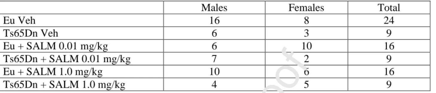
A first group of mice (euploid: n=69; Ts65Dn: n=30), received a daily subcutaneous injection (at 9-10 am) of different doses of CLEN (Santa Cruz Biotechnologies, 0.01, 0.5, 1.0, and 2.0 mg/kg in vehicle) from postnatal day 3 (P3) to P15. Age-matched euploid (n=22) and Ts65Dn (n=11) mice were injected with vehicle (PBS). A second group of mice (euploid: n=32, Ts65Dn: n=18), received a daily subcutaneous injection (at 9-10 am) of different doses of SALM (Sigma, 0.01 and 1.0 mg/kg in vehicle) from P3 to P15. Age-matched euploid (n=24) and Ts65Dn (n=9) mice were injected with vehicle (PBS). The number and sex of mice of the first and second groups are summarized in Table 1 and Table 2, respectively. Mice that received different doses of CLEN or SALM will hereafter be called “treated mice” whereas mice that received the vehicle will be called “untreated mice”. On P15, mice of all experimental groups received a subcutaneous injection (150 g/g body weight) of BrdU (5-bromo-2-deoxyuridine; Sigma Aldrich), in TrisHCl 50 mM 2 h before being killed. The body weight was recorded prior to sacrifice. The brains were excised and cut along the midline. The left hemispheres were fixed by immersion in PFA 4%, frozen and used for BrdU immunohistochemistry and Hoechst-staining. The right hemispheres were processed for Golgi staining. The number of animals used for each experimental procedures is specified in the figure legends.
2.3.2 Histological procedures
The left hemispheres were cut with a freezing microtome into 30-m-thick coronal sections that were serially collected in anti-freezing solution (30% glycerol; 30% ethylen-glycol; 10% PBS10X; sodium azide 0.02%; MilliQ to volume) and used for BrdU immunohistochemistry and Hoechst staining, as described below. BrdU immunohistochemistry. Immunohistochemistry was carried out as previously described (Contestabile et al., 2007, Guidi et al., 2013). Briefly, one out of six free-floating sections (n=15-19 sections) from the hippocampal formation was incubated with rat anti-BrdU antibody (diluted 1:200; Biorad). Detection of BrdU positive-cells following treatment with either CLEN or SALM was performed with a Cy3-conjugated anti rat-secondary antibody (diluted 1:200; Jackson Immunoresearch) or with a HRP-conjugated anti-rat rat-secondary antibody (dilution 1:200; Jackson Immunoresearch) and DAB kit (Vector Laboratories), respectively. Cy3 immunofluorescent sections were mounted on slides whereas DAB-stained sections were counterstained with hematoxylin and then mounted on slides.
Hoechst-staining. One out of six free-floating sections taken from the beginning to the end of the hippocampal formation (n=15-19 sections) were mounted on slides and stained with Hoechst nuclear dye (2 mg/ml in PBS). Golgi staining. The right hemispheres were immersed in the impregnation solution of the FD Rapid Golgi Stain TM Kit (FD Neuro Technologies, Inc.) containing mercuric chloride, potassium dichromate and potassium
chromate and were stored at room temperature in the dark for 2 weeks. Hemispheres were then transferred into Solution C of the same kit and stored at room temperature in the dark for at least 72 h. After these steps, hemispheres were cut with a microtome into 90-μm-thick coronal sections. Sections were mounted on gelatin-coated slides, and were air dried at room temperature in the dark for at least one day. After drying, sections were rinsed with distilled water and subsequently stained in a developing solution (FD Rapid Golgi Stain Kit).
2.3.3 Equipment
For fluorescence microscopy, we used the Nikon Eclipse TE 2000-S inverted microscope (Nikon Corp., Kawasaki, Japan) equipped with a Nikon digital camera DS-Qi2, and for light microscopy we used the microscope Nanozoomer 2.0 RS (HAMAMATSU) or a Leitz Diaplan microscope (Ernst Leitz Wetzlar GmbH) equipped with a motorized stage and focus control system and a Coolsnap-Pro color digital camera (Media Cybernetics). All measurements were carried out using the software Image Pro Plus (Media Cybernetics).
2.3.4 Measurements
Number of BrdU-positive cells. BrdU-positive cells in the hippocampal dentate gyrus (DG) were detected in
Journal Pre-proof
images acquired using a fluorescence microscope (Nikon Eclipse; objective: x 20, 0.5 NA) or a light microscope (Nanozoomer 2.0 RS; objective: 20x; 0.75 NA). Quantification of BrdU-labeled nuclei was conducted in 1 out of 6 sections using a modified unbiased stereology protocol that has previously been reported to successfully quantify BrdU labeling (Malberg et al., 2000, Kempermann and Gage, 2002, Tozuka et al., 2005). All BrdU-labeled cells located in the granule cell and subgranular layers were counted in their entire z axis (1 µm steps) in each section. To avoid oversampling errors, nuclei intersecting the uppermost focal plane were excluded. The total number of BrdU-labeled cells per animal was determined and multiplied by six to obtain the total estimated number of cells per DG.
Stereology of the DG. Unbiased stereology was performed on Hoechst-stained sections. The optical disector method was used to obtain cell density, and the Cavalieri principle was used to estimate volume, as previously described (Giacomini et al., 2015).
Spine density. Spines of granule cells of the DG were counted live in Golgi-stained sections using a 100x oil immersion objective lens (Leitz microscope and objective with 1.4 NA). Spine density values were evaluated in dendritic segments located in the inner (proximal dendrites) and outer (distal dendrites) half of the molecular layer. For each neuron, 3 proximal and 3 distal segments were analyzed. For each animal, spines were counted in at least 4/5 neurons. The length of each sampled dendritic segment was determined by tracing its profile and the number of spines was counted manually. The linear spine density was calculated by dividing the total number of spines by the length of the dendritic segment. Spine density was expressed as number of spines per 20 m dendrite.
Measurement of the dendritic tree. The dendritic tree of granule cells was traced in Golgi-stained sections with a dedicated software custom-designed for dendritic reconstruction (Immagini Computer di Maurizio Abbate & C. SNC, Bareggio, MI, Italy) interfaced with Image Pro Plus. The dendritic tree was traced live, at a final magnification of 500x, by focusing into the depth of the section. The operator starts with branches emerging from the cell soma and after having drawn the first parent branch goes on with all daughter branches of the next order in a centrifugal direction. At the end of tracing, the program reconstructs the total dendritic length, the number of branches of each order, and the mean length of branches of each order. The dendritic tree was reconstructed in 7-10 neurons per animal. These data were averaged in order to obtain the mean value for each animal.
2.4 STATISTICAL ANALYSIS
Results are presented as mean ± standard error of the mean (SE). Data were analyzed with the IBM SPSS 22.0 software. Before running statistical analyses, we checked data distribution and homogeneity of variances for each variable using the Shapiro-Wilk test and Levene’s test, respectively. If the data were normally distributed and variance was homogeneous, statistical analysis was carried out using either a one-way ANOVA or a two-way ANOVA with genotype (euploid, Ts65Dn) and treatment (saline, CLEN, SALM), as factors. Post hoc multiple comparisons were then carried out using Fisher’s least significant difference (LSD) test. If the data were not normally distributed and variance was heterogeneous, transformations were made to achieve normality. If the transformed data did not achieve normality, statistical analysis was carried out using the Kruskal-Wallis test followed by the Mann–Whitney U test. Based on the “Box plot” tool available in SPSS Descriptive Statistics, in each analysis we excluded the extremes, i.e., values that were larger than 3 times the IQ range [x ≥ Q3 + 3 * (IQ); x ≤ Q1 – 3 * (IQ)]. The number of mice included in (and excluded from, if any) individual analyses is reported in the legends of figures. A probability level of p ≤ 0.05 was considered to be statistically significant.
3. RESULTS
3.1 CHARACTERIZATION OF THE EFFECTS OF CLEN AND SALM ON PROLIFERATION AND DIFFERENTIATION IN CULTURES OF TRISOMIC NPCs
NPCs from the SVZ of neonate Ts65Dn mice are characterized by impairment of the proliferation rate (Trazzi et al., 2011, Stagni et al., 2017a) as well as in the acquisition of a neuronal phenotype (Trazzi et al., 2013). In a series of in vitro experiments, we sought to better characterize the effects of CLEN and SALM on the proliferation and differentiation of trisomic NPCs from the SVZ.
The effects of CLEN on proliferation and differentiation of trisomic NPCs were tested under wide range of concentrations (0.1-1000 nM). A one-way ANOVA showed a significant effect of treatment [F(5,42) =59.644, p < 0.001]. A post hoc Fisher’s LSD test showed that concentrations of 10-1000 nM significantly increased the proliferation rate (Fig. 1A). Cell fate characterization of trisomic NPC progeny under differentiative conditions was performed according to a previously validated assay based on double
Nestin immunolocalization and identification of four distinct subpopulations (Cvijetic et al., 2017). More specifically, we evaluated over total viable cell number the percentage of cells that were: i) immunopositive for MAP2 (a marker of cells with a neuronal phenotype) and immunonegative for Nestin (a marker of undifferentiated NPCs); ii) double immunopositive for MAP2 and Nestin, i.e., neuroblasts; iii) immunonegative for MAP2 and Nestin, i.e., cells putatively of non-neuronal lineage; iv) immunonegative for MAP2 and immunopositive for Nestin, i.e., undifferentiated NPCs. A one-way ANOVA on the percentage of MAP2+/Nestin- cells showed a significant effect of treatment [F(6,14) = 7.084, p = 0.001]. A post hoc Fisher’s LSD showed that drug concentrations of 100 and 500 nM caused a significant increase in the percentage of MAP2+/Nestin- cells in comparison with vehicle- treated cultures (Fig. 1B), suggesting that CLEN favors the acquisition of a neuronal phenotype. A one-way ANOVA on the percentage of MAP2+/Nestin+ cells showed no effect of treatment (Fig. 1C), suggesting that treatment does not affect the population of proliferating neuroblasts. An evaluation of the percentage of double-negative cells (MAP2-/Nestin-) showed a significant effect of treatment [F(6,14) = 2.972, p = 0.044]. A post hoc Fisher’s LSD test showed that the concentration of 100 nM caused a reduction in the percentage of MAP2-/Nestin- cells (Fig. 1D). Since cells that are immunonegative for both MAP2 and Nestin mainly represent cells committed to non-neuronal lineages (Cvijetic et al., 2017), these results may suggest that CLEN promotes neuronal differentiation of trisomic NPCs at the expense of their commitment toward non-neuronal lineages. An evaluation of the percentage of MAP2-/Nestin+ cells showed an effect of treatment at the concentration of 50 nM only (Fig. 1E).
The effects of SALM on proliferation and differentiation of trisomic NPCs were also examined under a wide range of concentrations (3-1000 nM). Regarding the effect of SALM on proliferation, a one-way ANOVA showed a significant effect of treatment [F(4,15) =24.140, p < 0.001]. A post hoc Fisher’s LSD test showed that concentrations of 3-300 nM caused a significant increase in NPC proliferation in comparison with cultures treated with vehicle (Fig. 1F). A one-way ANOVA on the percentage of MAP2+/Nestin- cells showed a significant effect of treatment [F(4,10) = 12.787, p = 0.001]. A post hoc Fisher’s LSD test showed that drug concentrations of 30-1000 nM caused a significant increase in the percentage of MAP2+/Nestin- cells in comparison with cultures treated with vehicle (Fig. 1G), indicating that SALM favors the acquisition of a neuronal phenotype. A one-way ANOVA on the percentage of cells that were MAP2+/Nestin+ showed a significant effect of treatment [F(4,10) = 3.530, p = 0.048]. A post hoc Fisher’s LSD test showed that the
concentrations of 300 and 1000 nM caused a significant increase in the percentage of MAP2+/Nestin+ cells in comparison with cultures treated with vehicle (Fig. 1H), suggesting that SALM affects the population of in vitro generated neuroblasts. An evaluation of the percentage of MAP2-/Nestin- cells showed a significant effect of treatment [F(4,10) = 6.403, p = 0.008]. A post hoc Fisher’s LSD test showed that the concentrations of 300 and 1000 nM caused a significant reduction in the percentage of MAP2-/Nestin- cells in comparisons with cultures treated with vehicle (Fig. 1I). These results suggest that SALM favors the acquisition of a neuronal phenotype at the expense of non-neuronal lineages. A one way ANOVA on the percentage of MAP2-/Nestin+ cells showed no significant effect of treatment (Fig. 1J).
Taken together, these findings suggest that both CLEN and SALM increase the proliferation rate of trisomic NPCs and favor their neuronal differentiation at the expense of their commitment to non-neuronal lineages.
3.2 EFFECTS OF CLEN IN VIVO
3.2.1 Effect of CLEN on neural precursor proliferation in the dentate gyrus of Ts65Dn mice
A severe reduction of hippocampal neurogenesis underlies the deficits in long-term memory that characterize DS (Bartesaghi et al., 2011). Results obtained in vitro showed that CLEN is able to increase the proliferation rate of trisomic NPCs. In order to establish whether CLEN enhances the proliferation rate in the hippocampal dentate gyrus (DG) of Ts65Dn mice, trisomic and euploid pups were injected daily with different doses of CLEN (0.01, 0.5, 1.0, and 2.0 mg/kg) from postnatal day 3 (P3) to P15. We used here the same timing protocol as in previous studies (Bianchi et al., 2010, Giacomini et al., 2015, Stagni et al., 2016, Stagni et al., 2019), because the first two postnatal weeks represent a critical time window for neurogenesis in the DG (Altman and Bayer, 1975). On P15, mice received an intraperitoneal injection of BrdU, a marker of cells in the S-phase of the cell cycle (Nowakowski et al., 1989), 2 h before being killed. The NPCs giving origin to granule cells of the DG are mainly located in the subgranular zone (SGZ), but NPCs can also be found in the granule cell layer. Therefore, we evaluated the total number of BrdU-positive cells in the SGZ and granular layer of the DG.
A Kruskal-Wallis test on the number of BrdU-positive cells revealed a significant difference between groups [χ2(2) = 18.671, p < 0.028]. The Mann-Whitney test showed that both in euploid and Ts65Dn mice
treatment with CLEN had no effect on the total number of BrdU-positive cells in comparison with their untreated counterparts (Fig. 2C). Ts65Dn mice treated with CLEN 2.0 mg/kg, however, had significantly more cells in comparison with Ts65Dn mice treated with 0.01 mg/kg CLEN (Fig. 2C: symbol #). It must be noted, in addition, that in absolute terms Ts65Dn mice treated with 1.0 and 2.0 mg/kg CLEN had more BrdU-positive cells in comparison with untreated Ts65Dn mice (untreated Ts65Dn mice: 6673±407 cells; Ts65Dn mice treated with 1.0 mg/kg CLEN: 7454±1104 cells; Ts65Dn mice treated with 2.0 mg/kg CLEN: 7939±471 cells). A comparison between Ts65Dn mice and untreated euploid mice showed that Ts65Dn mice treated with vehicle and 0.01 mg/kg CLEN had fewer BrdU-positive cells in comparison with untreated euploid mice (Fig. 2C). In contrast, no difference emerged between Ts65Dn mice treated with 0.5, 1.0, and 2.0 mg/kg CLEN and untreated euploid mice although, in absolute terms, in Ts65Dn mice the number of BrdU-positive cells did not reach the value of untreated euploid mice (8533±163; Fig. 2C). Taken together these findings suggests that in Ts65Dn mice treatment with high doses of CLEN moderately increases the proliferation rate of granule precursors, thereby mitigating the differences in comparison with euploid mice.
3.2.2 Effect of CLEN on the stereology of the dentate gyrus of Ts65Dn mice
In view of the increase in the proliferation potency in the DG of Ts65Dn mice treated with 2.0 mg/kg of CLEN, we wondered whether this effect led to an improvement/restoration of the defective cellularity that characterizes the DG of trisomic mice. To clarify this issue, we stereologically evaluated the total number of granule cells in treated and untreated mice. The Kruskal-Wallis test on the volume of the granule cell layer showed a significant difference between groups [χ2 (3) = 9.096, p = 0.028]. The Mann-Whitney test showed a reduced volume of the granule cell layer in untreated Ts65Dn mice compared to untreated euploid mice (U = 1.000, p = 0.010) and demonstrated that treatment did not increase the volume of the granule cell layer (Fig. 3B). A two-way ANOVA on the granule cell density showed a genotype x treatment interaction [F(1,18) = 17.924, p < 0.001], and a main effect of genotype [F(1,18) = 27.736, p < 0.001] and of treatment [F(1,18) = 12.314, p = 0.003]. A post-hoc Fisher’s LSD test showed that the density of the granule cells of untreated Ts65Dn mice was reduced in comparison with that of untreated euploid mice (Fig. 3C). In treated Ts65Dn mice, the cell density underwent an increase and became similar to that of untreated euploid mice (Fig. 3C). A two-way ANOVA on total number of granule cells showed no genotype x treatment interaction and no effect
of treatment, but did show a main effect of genotype [F(1,18) = 21.749, p < 0.001]. A post hoc Fisher’s LSD test showed that the granule cell number of untreated Ts65Dn mice was reduced in comparison with untreated euploid mice and that treatment did not increase total granule cell number (Fig. 3D).
The restoration of granule cell density induced by CLEN in Ts65Dn mice is consistent with the moderate pro-proliferative effect of treatment with the dose of 2.0 mg/kg. However, in treated Ts65Dn mice the volume of the granule cell layer remained lower than in euploid mice, which indicates that the effect of treatment was not sufficient to lead to restoration of total granule cell number.
3.2.3 Effect of CLEN on dendritic spine density in the dentate gyrus of Ts65Dn mice
Spine density reduction is a typical feature of the trisomic brain (Benavides-Piccione et al., 2004, Guidi et al., 2013, Stagni et al., 2017b) that, in conjunction with neurogenesis impairment, is thought to be a critical determinant of intellectual disability. In order to establish whether CLEN has a positive effect on spine density, we evaluated spine density in the dendritic arbor of the granule neurons in Golgi-stained brains of mice treated with different doses of CLEN (0.01, 0.5, 1.0, and 2.0 mg/kg). Since the inputs to the dendritic tree of granule cells are organized in a laminar manner, we deemed it of interest to separately evaluate spine density in dendritic branches harbored in the inner half (proximal spines) and in the outer half (distal spines) of the molecular layer.
We carried out a two-way ANOVA on spine density with genotype (euploid and Ts65Dn mice) and treatment (vehicle, 0.01, 0.5, 1.0, and 2.0 mg/kg of CLEN) as factors. A two-way ANOVA on the proximal spines showed a genotype x treatment interaction [F(4,26) = 5.465, p = 0.002], no main effect of genotype, but a main effect of treatment [F(4,26) = 32.113, p < 0.001]. A two-way ANOVA on the distal spines showed no genotype x treatment interaction, no main effect of genotype, but a main effect of treatment [F(4,26) = 35.868, p < 0.001]. We then carried out a post hoc Fisher’s LSD in order to establish differences across groups. Confirming previous evidence (Stagni et al., 2019), spine density of untreated Ts65Dn mice was significantly reduced both in the proximal (-24%) and distal (-15%) dendrites in comparison with that of untreated euploid mice (Fig. 4C,D). After treatment with any dose of CLEN the spine density of Ts65Dn mice underwent a notable increment and became even larger both in the proximal (+14%-+22%) and distal (+15%-+28%) dendrites in comparison with that of untreated euploid mice (Fig. 4C,D). In euploid mice, treatment with any
dose of CLEN increased proximal and distal spine density in comparison with euploid mice treated with vehicle, save for the dose of 0.5 mg/kg that had no effect on the proximal spines (Fig. 4C,D).
Taken together these data show that the tested doses of CLEN (from 0.01 to 2.0 mg/kg) enhance the process of spinogenesis along the whole extent of the dendritic tree of the granule cells in both euploid and Ts65Dn mice. Interestingly, in Ts65Dn mice, all tested dose of CLEN were able to increase spine density to values above those of untreated euploid mice. Cognitive impairment in Down syndrome has been linked to increased GABAergic inhibition (Zorrilla de San Martin et al., 2018). Since dendritic spines are the target of excitatory inputs, a treatment-induced increase in spine density in Ts65Dn mice above control levels may increase the weight of excitation thereby correcting the excitation/inhibition imbalance.
3.2.4 Effect of CLEN on the dendritic architecture of the granule neurons of Ts65Dn mice
Spine density reduction in DS is worsened by dendritic hypotrophy (Bartesaghi et al., 2011, Guidi et al., 2013, Dang et al., 2014, Stagni et al., 2015b). In view of the positive effects of CLEN on dendritic spine density in Ts65Dn mice, we wondered whether treatment was also able to foster dendritic development. To this purpose, we examined the effect of CLEN (0.01 mg/kg) on the dendritic tree of granule neurons already present in the DG at birth (i.e. neurons with soma located in the outer portion of the granule cell layer) because they represent a homogeneous population that was subjected to the effect of CLEN for the whole period of treatment. We first examined the number of segments and the total length of the dendritic tree. The Kruskal-Wallis test on the number of segments revealed a difference between groups [χ2 (3) = 14.621, p= 0.002]. The Mann-Whitney tests showed a reduced number of segments in untreated Ts65Dn mice (-34%) in comparison with untreated euploid mice (Fig. 5B). In Ts65Dn mice treated with CLEN the number of segments underwent a notable increment (+96%) and became even larger than that of untreated euploid mice (Fig. 5B). In euploid mice treated with CLEN the number of segments became significantly larger (+46%) than that of their untreated counterparts (Fig. 5B). The Kruskal-Wallis test on the total length of the dendritic tree showed a significant difference between groups [χ2 (3) = 11.458, p= 0.009]. The Mann-Whitney test showed that untreated Ts65Dn mice had a shorter total dendritic length (-25%) in comparison with untreated euploid mice (Fig. 5C). In Ts65Dn mice treated with CLEN the dendritic length underwent an increase (+46%) and became
similar to that of untreated euploid mice (Fig. 5C). In euploid mice treated with CLEN the dendritic length underwent an increase (+28%) and became significantly larger than that of untreated euploid mice (Fig. 5C).
In order to dissect the effect of treatment on details of the dendritic architecture we examined each dendritic order separately. Regarding the number of branches of individual orders, data of orders 1-5 were subjected to two-way ANOVA followed by post hoc Fisher’s LSD test and data of orders 6 and 7 were subjected to Kruskal-Wallis test followed by the Mann–Whitney test. Results showed that untreated Ts65Dn mice had a similar number of branches of orders 1 and 2 as untreated euploid mice but fewer branches of orders 3–5. In Ts65Dn mice treated with CLEN the number of branches of orders 3-5 became similar or even larger (in the case of order 4) in comparison with that of untreated euploid mice. Unlike euploid mice, untreated Ts65Dn mice lacked branches of order 6 (Fig. 5D,E black arrow). Importantly, this defect was restored by treatment. Euploid mice treated with CLEN underwent an increase in the number of branches of order 3-6 that became larger in comparison with their untreated counterparts (Fig. 5D,E). Although data of order 7 could not be statistically compared because the Kruskal-Wallis test did not show a significant effect, it is interesting to note that both treated euploid and Ts65Dn mice acquired branches of order 7 (Fig. 5D). Regarding the mean length of dendritic branches of individual orders, a post hoc Fisher’s LSD test (after two-way ANOVA) showed that in untreated Ts65Dn mice the branches of order 1 and 2 were notably longer in comparison with untreated euploid mice (Fig. 5E). This is consistent with previous evidence in adult mice (Guidi et al., 2013, Stagni et al., 2015b). No difference was found in the mean branch length of orders 3-5 (Fig. 5E). Importantly, Ts65Dn mice treated with CLEN underwent a reduction in the excessive length of branches of order 1 and 2 (Fig. 5E). In euploid mice treated with CLEN the mean length of branches of order 2 and 3 underwent a reduction in comparison with that of untreated euploid mice.
Taken together, these results indicate that treatment with CLEN rescues total dendritic length of Ts65Dn mice. This effect is due to a notable increase in the number of intermediate order branches and the appearance de novo of branches of orders 6 and 7. An increase in total dendritic length also takes place in treated euploid mice and is mainly attributable to an increase in the number of branches of orders 3-6. The overall effects of genotype and treatment on the dendritic architecture of the granule cells are summarized in the dendrograms of Fig. 5F.
3.2.5 Effects of CLEN on the body weight
We evaluated the body weight of P15 mice that received vehicle or different doses of CLEN (0.01, 0.5, 1.0 and 2.0 mg/kg) in order to establish possible adverse effects of treatment. Confirming previous evidence (Bianchi et al., 2010, Stagni et al., 2019) untreated Ts65Dn had a reduced body weight in comparison with untreated euploid mice (p = 0.01; two-tailed t-test, Table 3). A one-way ANOVA on the body weight of Ts65Dn mice revealed no significant effect of treatment. However, a post hoc Fisher’s LSD test showed that while the doses of 0.01, 0.5, and 1.0 mg/kg had no effect on the body weight, the dose of 2.0 mg/kg caused a significant weight reduction (-23%) in comparison with untreated Ts65Dn mice (Table 3). A one-way ANOVA and the Fisher’s LSD test revealed no effect of treatment in euploid mice. These data show that a dose of 2.0 mg/kg of CLEN has a negative impact on the growth of Ts65Dn mice.
3.3 EFFECTS OF SALM IN VIVO
3.3.1 Effect of SALM on neural precursor proliferation in the dentate gyrus of Ts65Dn mice
The results of Section 3.2.1 showed that a high dose of CLEN (2.0 mg/kg) was necessary to increase the number of proliferating cells in the DG of Ts65Dn mice. However, this dose had a negative effect on the body weight (see Section 3.2.5.). For this reason, we decided to treat mice with doses of SALM lower than 2.0 mg/kg. A two-way ANOVA on the total number of BrdU-positive cells in the DG of untreated euploid and Ts65Dn mice and euploid and Ts65Dn mice treated with SALM 1.0 mg/kg showed no genotype x treatment interaction, a main effect of genotype [F(1,16) = 33.937, p < 0.001], and no main effect of treatment. A post hoc Fisher's LSD test showed that untreated Ts65Dn mice had fewer BrdU-positive cells in comparison with untreated euploid mice (Fig. 6B). In treated Ts65Dn mice the number of proliferating cells did not undergo any increase and remained similar that of untreated Ts65Dn mice (Fig. 6B). This is in line with the absence of pro-proliferative effects observed with doses of CLEN lower than 2.0 mg/kg.
3.3.2 Effect of SALM on dendritic spine density in the dentate gyrus of Ts65Dn mice
The results of Section 3.2.3 showed that all the tested doses of CLEN restored the density of dendritic spines in Ts65Dn mice and that the lowest dose (0.01 mg/kg) had the same positive effects as higher doses.
Based on this evidence, we examined the effects of a low (0.01 mg/kg) and high dose (1.0 mg/kg) of SALM on spine density in the dendritic tree of the granule cells of euploid and Ts65Dn mice.
We carried out a two-way ANOVA on spine density with genotype (euploid and Ts65Dn mice) and treatment (vehicle, 0.01 mg/kg, and 1.0 mg/kg of SALM) as factors. A two-way ANOVA on spine density in the proximal dendrites showed a genotype x treatment interaction [F(2,24) = 46.863, p < 0.001], a main effect of genotype [F(1,24) = 101.481, p < 0.001], and a main effect of treatment [F(2,24) = 76.629, p < 0.001]. A two-way ANOVA on spine density in the distal dendrites showed a genotype x treatment interaction [F(2,24) = 74.371, p .<0.001], a main effect of genotype [F(1,24) = 117.266, p < 0.001], and a main effect of treatment [F(2,24) = 96.938, p < 0.001]. We then carried out a post hoc Fisher’s LSD in order to establish differences across groups. We found that in Ts65Dn mice spine density was significantly reduced in comparison with that of untreated euploid mice both in the proximal (-20%) and distal (-22%) dendrites (Fig. 7C,D). In Ts65Dn mice both doses of SALM caused a notable increment in spine density that became similar to that of untreated euploid mice, both in the proximal and distal dendrites (Fig. 7C,D). Unlike in Ts65Dn mice, in euploid mice treatment with SALM had no effect on spine density (Fig. 7C,D). Taken together these findings show that both the tested doses of SALM (0.01 and 1.0 mg/kg) are able to fully restore spine density along the whole extent of the dendritic tree of the granule cells of Ts65Dn mice.
3.3.3 Effect of SALM on the dendritic architecture of the granule neurons in the dentate gyrus of Ts65Dn mice
The results of Section 3.2.4. showed that the dose of 0.01 mg/kg of CLEN restores the dendritic length of Ts65Dn mice. Based on this evidence, we examined the effects of the same dose of SALM in euploid and Ts65Dn mice. A two-way ANOVA on the number of dendritic segments showed no genotype x treatment interaction, a main effect of genotype [F(1,14) = 20.718, p < 0.001], and a main effect of treatment [F(1,14) = 20.267, p < 0.001]. A post hoc Fisher’s LSD test showed that in Ts65Dn mice treated with SALM the number of segments underwent an increase (+50%) and became similar to that of untreated euploid mice (Fig. 8B). In euploid mice treated with SALM the number of segments became slightly larger (+15%) than that of their untreated counterparts (Fig. 8B). A two-way ANOVA on the total dendritic length showed no genotype x treatment interaction, a main effect of genotype [F(1,14) = 16.088, p = 0.001], and a main effect of treatment
[F(1,14) = 12.329, p = 0.003]. A post hoc Fisher’s LSD test showed that in Ts65Dn mice treated with SALM, the dendritic length underwent an increase (+31%) and became similar to that of untreated euploid mice (Fig. 8C).
We next examined the effect of treatment on each dendritic order separately. Regarding the number of branches of individual order, data of orders 1-5 were subjected to two-way ANOVA followed by post hoc Fisher’s LSD test and data of orders 6 and 7 were subjected to Kruskal-Wallis test followed by the Mann– Whitney test. Results showed that Ts65Dn mice treated with SALM underwent a significant increase in the number of branches of order 3 and 4 that became similar of that of untreated euploid mice. Although data of order 6 and 7 could not be statistically compared because the Kruskal-Wallis test did not show a significant effect, it is interesting to note that in treated Ts65Dn mice there was the de novo appearance of branches of order 6 (Fig. 8D). In treated euploid mice, there was an increase in the number of branches of order 5 in comparison with untreated euploid mice, although the difference was not statistically significant, and the de novo appearance of branches of order 7. Regarding the mean branch length of each individual order, a post hoc Fisher’s LSD test (after two-way ANOVA) showed that in treated Ts65Dn mice the excessive length of branches of order 1 and 2 underwent a significant reduction and became similar to that of untreated euploid mice (Fig. 8E).
Taken together these findings indicate that in Ts65Dn mice a dose of SALM of 0.01 mg/kg is able to restore the total dendritic length by restoring the number of intermediate order branches and inducing the de novo appearance of branches of order 6. In contrast, in euploid mice, treatment with SALM had only a moderate effect on the dendritic architecture. The overall effects of genotype and treatment on the dendritic architecture of the granule cells are summarized in the dendrograms of Fig. 8F.
3.3.4 Effects of SALM on the body weight
We evaluated the body weight of P15 mice that received vehicle or SALM (0.01 and 1.0 mg/kg) in order to establish the outcome of treatment on somatic growth. A one-way ANOVA on the body weight of Ts65Dn mice revealed a significant effect of treatment [F(2,24) = 5.695, p = 0.009]. A post hoc Fisher’s LSD test showed that the dose of 0.01 mg/kg caused a significant increase (+29%) in the body weight in comparison with untreated Ts65Dn mice, while the higher dose (1.0 mg/kg) had no effect on the body weight (Table 4).
The Kruskal-Wallis test on the body weight of euploid mice revealed a significant effect of treatment [χ2(2) = 23.502, p < .001]. The Mann-Whitney tests showed an increase in the body weight with both doses of SALM (Table 4). These results indicate that unlike CLEN, SALM may have a positive impact on the body weight.
3.4 COMPARISON OF THE EFFECTS OF CLEN AND SALM ON SPINOGENESIS AND DENDRITOGENESIS
The results reported above show that a dose of CLEN and SALM of 0.01 mg/kg was sufficient to restore spine density and dendritic complexity in Ts65Dn mice. However, from the observation of Figs. 4, 5, 7, 8 it appears that CLEN exerts larger effects in comparison with SALM. In order to gain information on this issue, we deemed it of interest to compare the magnitude of the effects of the two drugs in both genotypes.
A Kruskal-Wallis test on the dendritic spines of euploid and Ts65Dn mice that received vehicle, CLEN, or SALM revealed a significant effect of genotype x treatment both for the proximal [χ2(2) = 31.143, p < 0.001] and distal [χ2(2) = 29.153, p < 0.001] spines. The Mann-Whitney test showed that in euploid mice treatment with CLEN, but not with SALM, increased proximal and distal spine density in comparison with their vehicle-treated counterparts (Fig. 9A,B: asterisks within the white bars). In Ts65Dn mice, treatment with CLEN and SALM increased proximal and distal spine density in comparison with their vehicle-treated counterparts (Fig. 9A,B: symbol # within the grey bars), but the magnitude of the effects of CLEN was larger in comparison with that of SALM (Fig. 9A,B: asterisks above the grey bars).
A Kruskal-Wallis test on the dendritic tree of euploid and Ts65Dn mice that received vehicle, CLEN, or SALM revealed a significant effect of genotype x treatment both for the total dendritic length [χ2(2) = 15.409, p < 0.009] and the total number of branches [χ2(2) = 20.987, p < 0.001]. The Mann-Whitney test showed that in euploid mice treatment with CLEN, but not with SALM, increased the dendritic length and the number of branches in comparison with their vehicle-treated counterparts (Fig. 9C,D: asterisks within the white bars). Thus, euploid mice treated with CLEN had a total dendritic length larger than that of euploid mice treated with SALM (Fig. 9C: asterisks above the white bars). In Ts65Dn mice, treatment with CLEN and SALM increased the total dendritic length in comparison with their vehicle-treated counterparts (Fig. 9C: symbol # within the grey bars), with no difference in the magnitude of the effects of CLEN and SALM. In Ts65Dn mice treatment with CLEN and SALM increased the total number of branches in comparison with their vehicle
treated counterparts (Fig. 9D: symbol # within the grey bars) but the magnitude of the effects of CLEN was larger in comparison with that of SALM (Fig. 9D: asterisk above the grey bars).
Taken together, these results indicate that a dose of CLEN of 0.01 mg/kg exerts larger effects on spine density as well as number of dendritic branches in comparison with the same dose of SALM, both in euploid and Ts65Dn mice.
4. DISCUSSION
4.1 Neonatal treatment with the β2-AR agonists CLEN or SALM does not restore hippocampal neurogenesis in the Ts65Dn mouse
The role of the noradrenergic system on the proliferation rate of the granule cells has been established by several studies and recent evidence shows that progenitor cells in the adult dentate gyrus express α1, α2, β1 and β2 receptors (Masuda et al., 2012, Meneghini et al., 2014, Bortolotto et al., 2019). In cell cultures from the adult rat dentate gyrus, noradrenaline promotes proliferation of the pool of the 2a early progenitor cells through the β2-AR (Masuda et al., 2012), while the β1-AR selective partial agonist xamoterol does not, indicating a key role of the β2-AR in the promotion of neurogenesis mediated by the noradrenergic system.
Neurogenesis impairment is one of the key determinants of brain malfunctioning in DS. Hence, it is of importance to find treatments that counteract this defect. The current study shows that although the dose of 2.0 mg/kg CLEN attenuated the differences in the proliferation rate of granule cell precursors between Ts65Dn and euploid mice this effect did not translate into an increase in total granule cell number. This is in agreement with evidence that administration of 2.0 mg/kg/day of another β2-AR agonist, formoterol, restores the number of BrdU-positive cells in the dentate gyrus of adult Ts65Dn mice but had no effect on hippocampal neurogenesis (DCX-positive cells) (Dang et al., 2014). However, since neurogenesis in the dentate gyrus starts before birth, it is possible that a treatment with CLEN (or SALM) that starts prenatally and is continued postnatally is able to restore total granule cell number.
The absence/paucity of pro-proliferative effects of CLEN and SALM in vivo is at variance with the large pro-proliferative effects of CLEN and SALM observed here in cultures of trisomic NPCs with a wide spectrum of doses. Both CLEN and SALM can cross the BBB (see section 4.3 for details) and, a dose of 0.01
mg/kg of CLEN and SALM was sufficient to restore spine density and dendritic length in Ts65Dn mice, suggesting that both drugs cross the BBB in an amount sufficient to exert biological actions. Taken together these findings suggest that the cellular machinery underlying dendritic development is more sensitive to the action of the β2-AR than the machinery that regulates the cell cycle. An alternative possibility is that, unlike postmitotic granule cells, the progenitors of the granule cells in the neonatal hippocampus have a low expression of the β2-AR and that this obstacle may be overcome in vitro due to constant exposure to the agonist.
4.2 Neonatal treatment with the β2-AR agonists CLEN or SALM restores dendritic development of the granule cells in the Ts65Dn mouse
The current study provides novel evidence that a brief pharmacological therapy with two β2-AR agonists, CLEN or SALM, in the early postnatal period (P3-P15) fully rescues dendritic pathology in the hippocampal dentate gyrus of the Ts65Dn mouse model of DS. In particular, treatment was able to restore both the length and number of dendritic branches as well as dendritic spine density. The effects on spine density took place with all doses tested here and the lowest dose (0.01 mg/kg) elicited effects of the same magnitude as the larger doses. These findings suggest that the β2-AR may play a prominent role in the regulation of dendritic development in the trisomic brain.
There is evidence that exposure of neurons to conditioned media from noradrenaline-stimulated glial cells increases dendritic complexity and that conditioned media from glial cells treated with the β2-AR agonists SALM and CLEN, but not the β1-AR agonist xamoterol, mimicked the ability of noradrenaline to increase neuronal complexity. In addition, noradrenaline induced the expression of a range of growth factors, including BDNF (Day et al., 2014). This suggests that the effects of β2-AR agonists on dendritic complexity may be both cell-autonomous and mediated by soluble factors released by astrocytes. BDNF is one of the master regulators of dendritic development and spine density production/maturation (De Vincenti et al., 2019) and its levels are reduced both in fetuses with DS and in the Ts65Dn model (see (Rueda et al., 2012)). The β2-ARs activate extracellular signal-regulated kinases (ERK/MAPKs), that represent key steps in the activation of CREB (Hagena et al., 2016). While the question regarding the effects of CREB on neurogenesis is not completely settled, it is clear that CREB plays a key role in the process of neuron maturation (Merz et al.,
2011). This effect is mediated by the transcription of many proteins, including BDNF (Wang et al., 2018). Thus, the effects of treatment with CLEN and SALM on dendritic development may be largely mediated by BDNF. This conclusion is supported by demonstration that CLEN increases the levels of BDNF in the kainic model of excitotoxicity (Gleeson et al., 2010) and in a mouse model of Rett syndrome (Mellios et al., 2014).
It is interesting to note that in Ts65Dn mice the restoring effects of CLEN and SALM took place at all levels of the dendritic tree of the granule cells. The major extrinsic input to the dentate gyrus, which takes its origin from the medial and lateral divisions of the entorhinal cortex is laminarly organized (Amaral, 1995). The medial perforant pathway terminates on the middle third of the dendritic tree of the granule cells, while the lateral perforant pathway terminates on the outer third. Both inputs are fundamental for the participation of the hippocampal formation in long-term memory functions. The effects of CLEN and SALM on the whole dendritic tree suggests restoration of connectivity from both divisions of the entorhinal cortex. Thus, treatment with CLEN and SALM may lead to improvement/restoration of the learning and memory defects that characterize the trisomic brain.
4.3 CLEN exerts larger effects on dendritic development in comparison with SALM
Results show that both CLEN and SALM enhance dendritic development of the granule cells, although some differences must be underlined. 1) The doses of 0.01 and 1.0 mg/kg of SALM enhance spine density in the proximal and distal dendrites of Ts65Dn mice, but their effect is significantly smaller in comparison with the effects elicited by the same doses of CLEN. 2) The doses of 0.01 and 1.0 mg/kg of SALM do not enhance spine density in the proximal and distal dendrites of the granule cells of euploid mice. In contrast, the same doses of CLEN largely enhance spine density in euploid mice. 3) The dose of 0.01 mg/kg of SALM increases the number of dendritic branches of the granule cells of Ts65Dn mice, but this effect is smaller in comparison with that elicited by the same dose of CLEN.
Although all β2-AR agonists have as final target the β2-ARs, their efficacy may largely vary in relation to their pharmacokinetics and their BBB permeability. Based on the duration of their effects, the β2-AR have been classified as SABAs (Short-Acting β2-ARs), LABAs (Long-Acting β2-ARs) and ultra LABAs (ultra Long-Acting β2-ARs) (Billington et al., 2017). Both CLEN and SALM belong to the class of LABAs. CLEN has a large permeability across the BBB (Web site Link 1) and a half-life of 27-35 h in humans and 30 h in
rats after oral administration (Yamamoto et al., 1985, Yang et al., 2015). SALM is able to cross the BBB in trace amounts (Fitzpatrick et al., 1990, Manchee et al., 1993) (Web site link 2). It has a half-life of 2 h in mice and of 5.5-7.0 h in humans after oral administration (Fenton and Keating, 2004, Daley-Yates et al., 2014, Anwar et al., 2015).
The large BBB permeability of CLEN in conjunction with its long half-life implies that it reaches larger brain levels than SALM and for a more prolonged time. This, in turn, means that CLEN can exert larger and more prolonged biological effects on the brain in comparison with SALM. Taken together, these data can account for the larger effects of CLEN on the development of the dendritic tree of the granule cells observed in the current study.
4.4 Neonatal treatment with CLEN and SALM affects the body mass of the Ts65Dn mouse in an opposite manner
Unlike in euploid mice, in Ts65Dn mice the highest tested dose (2.0 mg/kg) of CLEN caused a reduction in the body weight. There is evidence that CLEN increases the skeletal muscle mass (Carter et al., 1991, Hinkle et al., 2002) but concomitantly decreases the body fat content. The latter effect is associated with a lipolytic effect on adipocytes (Kim et al., 2010) and a reduced expression of PPAR (Li et al., 2015), an adipocytokine that controls the steroid synthetase expression of adipocytes. Body builders and athletes use CLEN in order to improve the muscle mass, although this use is illegal due to cardiotoxic effects (Spiller et al., 2013). Consistently with evidence that SALM has anabolic effects (Moore et al., 1994), treatment with SALM caused a body weight increase in Ts65Dn and euploid mice. This effect may be related to activation of peripheral β2-ARs in the muscle cells and/or other body organs, with a consequent enhanced anabolic response (Ryall et al., 2006). CLEN and SALM are selective β2-AR agonists, although they can also act on β1-ARs and β3-ARs with much lower affinity and efficacy (Baker, 2010). The β3-AR are involved in the regulation of thermogenesis and lipolysis (Yang and Tao, 2019). It has been shown that the efficacy of CLEN on β3-AR is larger in comparison with that of SALM (Baker, 2010). An enhancement of lipolysis induced by high doses of CLEN may explain the observation that treatment with CLEN but not with SALM causes a reduction in body weight in Ts65Dn mice. Ts65Dn mice are constitutively smaller and more fragile in comparison with
euploid mice (Reeves, 1995). This may explain the body weight reduction that took place in Ts65Dn but not euploid mice with the highest tested dose of CLEN.
4.5 Use of CLEN or SALM as therapy for DS?
We found here that both CLEN and SALM restored the dendritic length and the density of dendritic spines in neonate Ts65Dn mice and that this effect was elicited even with the smallest dose tested here (0.01 mg/kg). This is of obvious relevance considering that a low dose of a given drug reduces the probability of unwanted side effects. Indeed, although CLEN and SALM are selective β2-AR agonist, the fact that high doses might also activate β1 receptors, thereby affecting the heart function, cannot be overlooked.
A critical issue in animal studies aimed at exploring the effects of different drugs to combat different diseases regards the translation of the tested doses from animals to humans. Based on the article by Reagan-Shaw (Reagan-Reagan-Shaw et al., 2008) we calculated the human doses equivalent to the doses of CLEN and SALM used here (Table 5). A dose of 0.01 mg/kg of either agonist in mice corresponds to a dose of 0.0012 mg/kg in children and a dose of 2.0 mg/kg in mice corresponds to a dose of 0.24 mg/kg, in children. CLEN and SALM are used as therapeutic treatment for asthma at the typical doses reported in Table 6, column A. From these values, it can be calculated that for a child weighing 20 kg the daily inhaled doses range between 0.0025 and 0.050 (Table 6, column B). Although drug bioavailability is different according to the route of administration, the current results show that in Ts65Dn mice subcutaneous injections of CLEN and SALM at doses that are in the range of the therapeutic doses used in children for the treatment of asthma are able to rescue dendritic pathology.
Based on results obtained here with doses of CLEN and SALM that are practicable in children we believe it may not be unreasonable to envisage a therapy with either CLEN or SALM for the improvement of brain development in children with DS. It must be noted that CLEN is no longer approved by FDA since the year 2006 (web site link 1). The FDA banned its use because it was concerned by potential adverse effects in people consuming food from livestock illegally treated with CLEN in order to increase their muscle development and decrease fat deposition. CLEN, however, is still available in Europe and Latin America as a bronchodilator in humans (Spiller et al., 2013) (web site link 3). SALM is currently an approved drug by both
FDA and EMA. Thus, although CLEN has larger effects than SALM a therapy with CLEN for DS may be proposed in some countries only while a therapy with SALM may be proposed for clinical trial worldwide.
Neurogenesis impairment and dendritic pathology are the major developmental defects of DS. The current results suggest that treatment with β2-AR agonists may be a therapy of choice in order to correct dendritic development but is not suitable to rescue neurogenesis. It can be envisaged that combined therapy with CLEN or SALM and a drug than restores neurogenesis may represent a potential strategy for the overall restoration of brain development in DS. Although the Ts65Dn mouse recapitulates many phenotypes of DS, it must be noted that it also carries 50 protein-coding triplicated genes that are not orthologs of Hsa21 genes (Gardiner, 2015). The contribution of these extra genes in determining the drug response cannot be ruled out. We hope that our study will prompt additional investigations aimed at testing the effects of CLEN and SALM in other mouse model of DS and that the gained knowledge may pave the way to clinical trials in children/adults with DS.
ACKNOWLEDGMENT
This work was supported by grants to R. B. from “Fondazione Generali e Assicurazione Generali”, Italy, “Fondazione del Monte”, Italy, and the programme ‘Investissements d'avenir’ ANR-10-IAIHU-06 to MCP. The assistance of Melissa Stott in the revision of the language is gratefully acknowledged.
CONFLICT OF INTEREST
The authors declare that they have no conflict of interest.
Credit Author Statement
Marco Emili: carried out treatment with clenbuterol, evaluated spine density in mice treated with salmeterol, and analyzed the dendritic architecture of mice treated with clenbuterol and salmeterol.
Fiorenza Stagni: carried out treatment with clenbuterol, immunohistochemistry in mice treated with clenbuterol, and wrote the manuscript.
Maria Elisa Salvalai: carried out the in vitro experiments.
Beatrice Uguagliati: carried out genotyping and evaluated spine density in mice treated with clenbuterol. Andrea Giacomini: carried out statistical analysis of in vivo and in vitro experiments.
Christelle Albach: carried out treatment with salmeterol and immunohistochemistry in mice treated with salmeterol.
Marie-Claude Potier: supervised the experiments of mice treated with salmeterol and reviewed the paper. Mariagrazia Grilli: supervised the in vitro experiments and reviewed the paper.
Renata Bartesaghi: conceived and designed the experiments and wrote the manuscript.
Sandra Guidi: supervised the experiments of mice treated with clenbuterol and wrote the manuscript.
REFERENCES
Altman J, Bayer S (1975) Postnatal development of the hippocampal dentate gyrus under normal and experimental conditions. In: Isaacson RL and Pribram KH, editors. The hippocampus, Vol 1. Plenum Press, New York and London. p 95-122. 95-122.
Amaral DG, Witter, M.P. (1995) Hippocampal formation: The rat nervous system. Academic Press Pages 443-493.
Anwar MM, El-Haggar RS, Zaghary WA (2015) Salmeterol Xinafoate. Profiles Drug Subst Excip Relat Methodol 40:321-369.
Baker JG (2010) The selectivity of beta-adrenoceptor agonists at human beta1-, beta2- and beta3-adrenoceptors. Br J Pharmacol 160:1048-1061.
Bartesaghi R, Guidi S, Ciani E (2011) Is it possible to improve neurodevelopmental abnormalities in Down syndrome? Rev Neurosci 22:419-455.
Benavides-Piccione R, Ballesteros-Yanez I, de Lagran MM, Elston G, Estivill X, Fillat C, Defelipe J, Dierssen M (2004) On dendrites in Down syndrome and DS murine models: a spiny way to learn. Prog Neurobiol 74:111-126.
Bianchi P, Ciani E, Guidi S, Trazzi S, Felice D, Grossi G, Fernandez M, Giuliani A, Calza L, Bartesaghi R (2010) Early pharmacotherapy restores neurogenesis and cognitive performance in the Ts65Dn mouse model for Down syndrome. J Neurosci 30:8769-8779.
Billington CK, Penn RB, Hall IP (2017) beta2 Agonists. Handb Exp Pharmacol 237:23-40.
Bortolotto V, Bondi H, Cuccurazzu B, Rinaldi M, Canonico PL, Grilli M (2019) Salmeterol, a beta2 Adrenergic Agonist, Promotes Adult Hippocampal Neurogenesis in a Region-Specific Manner. Front Pharmacol 10:1000.
Brusasco V, Crimi E, Mangini S, Vibelli C (1980) A clinical trial of oral clenbuterol (NAB 365) in chronic airways obstruction. Current medical research and opinion 6:449-455.
Carter WJ, Dang AQ, Faas FH, Lynch ME (1991) Effects of clenbuterol on skeletal muscle mass, body composition, and recovery from surgical stress in senescent rats. Metabolism 40:855-860.
Chapman KR, Ringdal N, Backer V, Palmqvist M, Saarelainen S, Briggs M (1999) Salmeterol and fluticasone propionate (50/250 microg) administered via combination Diskus inhaler: as effective as when given via separate Diskus inhalers. Can Respir J 6:45-51.
Cohen ML, Wiley KS, Bemis KG (1982) Analysis of the beta 1 and beta 2 adrenoceptor interactions of the partial agonist, clenbuterol (NAB365), in the rat jugular vein and atria. Naunyn Schmiedebergs Arch Pharmacol 320:145-151.
Contestabile A, Fila T, Ceccarelli C, Bonasoni P, Bonapace L, Santini D, Bartesaghi R, Ciani E (2007) Cell cycle alteration and decreased cell proliferation in the hippocampal dentate gyrus and in the neocortical germinal matrix of fetuses with Down syndrome and in Ts65Dn mice. Hippocampus 17:665-678. Creau N (2012) Molecular and cellular alterations in Down syndrome: toward the identification of targets for
therapeutics. Neural Plast 2012:171639.
Cvijetic S, Bortolotto V, Manfredi M, Ranzato E, Marengo E, Salem R, Canonico PL, Grilli M (2017) Cell autonomous and noncell-autonomous role of NF-kappaB p50 in astrocyte-mediated fate specification of adult neural progenitor cells. Glia 65:169-181.
Daley-Yates PT, Mehta R, Chan RH, Despa SX, Louey MD (2014) Pharmacokinetics and pharmacodynamics of fluticasone propionate and salmeterol delivered as a combination dry powder from a capsule-based inhaler and a multidose inhaler in asthma and COPD patients. Journal of aerosol medicine and pulmonary drug delivery 27:279-289.
Dang V, Medina B, Das D, Moghadam S, Martin KJ, Lin B, Naik P, Patel D, Nosheny R, Wesson Ashford J, Salehi A (2014) Formoterol, a long-acting beta2 adrenergic agonist, improves cognitive function and promotes dendritic complexity in a mouse model of Down syndrome. Biol Psychiatry 75:179-188. Day JS, O'Neill E, Cawley C, Aretz NK, Kilroy D, Gibney SM, Harkin A, Connor TJ (2014) Noradrenaline
acting on astrocytic beta(2)-adrenoceptors induces neurite outgrowth in primary cortical neurons. Neuropharmacology 77:234-248.
de Graaf G, Buckley F, Skotko BG (2015) Estimates of the live births, natural losses, and elective terminations with Down syndrome in the United States. Am J Med Genet A 167A:756-767.
De Vincenti AP, Rios AS, Paratcha G, Ledda F (2019) Mechanisms That Modulate and Diversify BDNF Functions: Implications for Hippocampal Synaptic Plasticity. Front Cell Neurosci 13:135.
Fenton C, Keating GM (2004) Inhaled salmeterol/fluticasone propionate: a review of its use in chronic obstructive pulmonary disease. Drugs 64:1975-1996.
Fitzpatrick MF, Mackay T, Driver H, Douglas NJ (1990) Salmeterol in nocturnal asthma: a double blind, placebo controlled trial of a long acting inhaled beta 2 agonist. BMJ 301:1365-1368.
Garcia-Font N, Martin R, Torres M, Oset-Gasque MJ, Sanchez-Prieto J (2019) The loss of beta adrenergic receptor mediated release potentiation in a mouse model of fragile X syndrome. Neurobiol Dis 130:104482.
Gardiner KJ (2015) Pharmacological approaches to improving cognitive function in Down syndrome: current status and considerations. Drug Des Devel Ther 9:103-125.