Introduction
Ligament injuries of the lateral compartment of the ankle occur with high frequency following sports and / or work activities. The chronicization of these injuries involves, in some cases, a chronic instability of the ankle that manifests itself with pain, lameness and swelling when walking on uneven ground and some-times also an important limitation of daily activities. Failure of conservative treatment often leads the pa-tient to seek adequate surgical treatment.Various ligament reconstruction techniques have been pro-posed using autologous transplants (tendons - semitendinous, plantar gracil, patellar, fascia lata, perios-teum), allogeneic, xenogenic or synthetic.In our experience we enumerate both reconstructions with autologous and allogenic material, following a failure of the arthroscopic method or by choice of the pa-tient. In this retrospective study authors present the cases
treated with allograft transplantation for liga-ment reconstruction in chronic ankle instability.
Materials and method
From January 1999 to December 2016, 72 patients were operated , including 29 women and 43 men, aged between 22 and 47 (average 29.5). 17 patients practiced sporting activities at a competitive and / or ama-teur level: 7 football, 4 running, 3 basketball, 2 volleyball, 1 tennis.
All reported in a history repeated ankle sprain episodes occurred during the performance of sports activi-ties or simple daily activities (walking on uneven ground, descent of the stairs), neglected or partially treated. Among the most frequently accused symptoms the following were reported: the feeling of in-Abstract
Introduction: In this retrospective study authors present a serie of cases treated with allograft transplantation for
liga-ment reconstruction in chronic ankle instability.
Methods: From January 1999 to December 2016, 72 patients were operated , including 29 women and 43 men, aged
between 22 and 47 (average 29.5). Of the 72 patients treated, 61 were evaluated in the follow up (12 months-12 years, with an average of 59 months) with clinical and radiographic evaluation, AFASS score (American Foot and Ankle Society Scoring System) and grading according to Good et al.
Results: There were no cases of transplant infection or rejection. Based on the criteria of Good et al., the results were
excellent or good in the 57 patients operated with semitendinosus or frozen fresh gracil, sufficient in the 3 transplant cases of freeze-dried semitendinosus and 1 case of bad result.
Conclusions: Transplantation with allograft in chronic ankle instability represents an “innovative” technique that
has achieved good results even in young patients with sporting needs.
Level of evidence: III. Retrospective study.
Keywords: Polydactyly, hexadactyly, hexadactylism.
The allograft in chronic lesions of the lateral
compartment of the ankle
Luigi Di Palma1, Cecilia Pasquali2, Riccardo Compagnoni3,4, Pietro Simone
Randelli3,4, Alberto Branca1.
1) Orthopaedic Department Sondrio hospital , ASST Valtellina ed Alto Lario, Italy; 2) Orthopaedic Department ASST-Valle Olona Busto Arsizio Varese, Italy;
3) Laboratory of Applied Biomechanics, Department of Biomedical Sciences for Health, Università degli Studi di Milano, Via Mangiagalli 31, 20133 Milan, Italy; 4) 1° Clinica Ortopedica, ASST Centro Specialistico Ortopedico Traumatologico Gaetano Pini-CTO, Piazza Car-dinal Ferrari 1, 20122 Milan, Italy
Corresponding author: Luigi di Palma - Via Pini,3 20122 Milano - [email protected]
Archivesof MedicineAnd surgery ofthe universityof Mil An
Attribution-NonCommercial-NoDerivatives 4.0 International (CC BY-NC-ND 4.0)
stability of the ankle during a light run, the descent of steps and walking on rough terrain and the appear-ance of pain and lateral and/or medial perimalleolar swelling.
The period between initial trauma and allograft transplantation was from 10 to 24 months. In this time range, 61 patients underwent ankle arthroscopy for the evaluation and possible treatment of associated injuries (removal of mobile bodies, shaving and microfractures of cartilage lesions, removal of intra-articular adhesions) and of these 5 patients also practiced an arthroscopic ligament repair of the PAA with thermal shrinkage, however reporting little benefit and persistence of joint sagging. The remaining pa-tients immediately chose allograf ligament reconstruction as the first surgical treatment.
Of the 72 patients treated, 61 were evaluated in the follow up (12 months-12 years, with an average of 59 months) with clinical and radiographic evaluation, AFASS score (American Foot and Ankle Society Scoring System) and grading according to Good et al. [ 1]. Allogeneic transplant. Donor explanting is performed only after passing the medical social questionnaire and analyzing the blood samples that are collected immediately before tissue removal. Blood samples are tested exclusively with kits approved by the FDA for HIV 1 and 2, Hepatitis B Virus, Hepatitis C Virus, Human Lymphoma Virus I and II, Cytomegalovirus, Syphilis.
Washing with the “Biocleanse” system is completely automatic and uses materials used in the pharma-ceutical industry and allows you to remove blood and fat, kill bacteria and fungi, inactivate viral loads, maintain tissue integrity, maintain biocompatibility, and not “cross contamination”. Transmission risks are 1 <1,650,000. We initially used the freeze-dried semitendinosus, later replaced by the gracile or the semitendinosus frozen fresh from the Bank of Florida in the early years and, later, from the Collection Center of the Rizzoli Orthopedic Institute in Bologna and from the
G. Pini Orthopedic Insti-tute of Milan.
3 freeze-dried semitendinosus, 29 frozen fresh gracili, 40 frozen fresh semitendinosus were transplanted. Frozen fresh gracil tendon has been used preferably in women, considering the female physical constitution as opposed to the male one. [Fig. 1]
Transplant preparation. The tendon is thawed at the time of surgery, placing it in physiological solution enriched with rifampicin, at room temperature, for 10-20 minutes. It is then prepared by tubulising the ends with non-absorbable Mercylene 1 thread, so as to obtain a diameter of 4.5 mm.
Surgical technique. The patient lies supine, with tourniquet positioned at the level of the thigh at 250-300 mmHg. The skin incision, with reference to the lateral malleolus, extends from the distal 1/3 of the fibula to the calcano-cuboidal joint, passing L along the lateral surface of the calcaneus. In this way it is possible to obtain a good exposure of the peroneal malleolus, the peroneal tendons, the ankle and subtalar joint, the anterior astragalic peroneal ligaments (PAA) and calcaneal peroneal (PC). The calcaneal and peroneal surfaces are skeletal and the bone tunnels are prepared at the level of the calcaneal neck (in the plantar-dorsal and latero-medial direction) and of the peroneal malleolus (in the antero-posterior, proximal-distal direction) with
Figure 1 - The allograft before the implant.
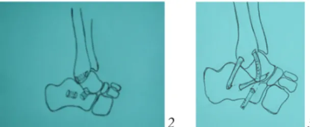
Figure 3 - Example of bone tunnel in calcaneus and fibula with the graft
Figure 2 - Example of bone tunnel in calcaneus and fibula
4.5mm drill mm in diameter. [Fig. 2]
The transplant suitably prepared (as already described), is inserted into the calcaneal tunnel in the plan-tar-dorsal direction, then through the peroneal tunnel in the anteroposterior direction, to then bring it back to the heel along the course of the PC ligament, in the back tendon with respect to the peroneal tendons. In correspondence with the heel insertion of this ligament, the transplant is fixed with a double anchor or, more recently, with a bioresorbable interference screw 5.5 mm x 16mm, after tensioning it by placing the foot in flexion at 90 ° [Fig. 3].
The ends are sutured with non-absorbable thread on the transplant itself along its calcaneal-peroneal course, deeply to the peroneal.The articularity of the ankle and subtalar joint is checked. Accurate hemo-stasis and layered suturing are then performed after placement of a suction drain. Finally, a pinstripe drain boot is packaged and the patient will have to keep it for 4 weeks. After this period of immobilization the plaster is removed and a bivalve brace is placed which must be worn for 8-12 weeks. 4 weeks after surgery, progressive tolerance load is granted with two crutches and the physiokinesitherapy program begins, consisting of proprioceptive recovery, muscle strengthening, hydrotherapy, exercise bike. Sports can be resumed about 4-5 months after the intervention.
Results
There were no cases of transplant infection or rejection. Based on the criteria of Good et al., the results were excellent or good in the 57 patients operated with semitendinosus or frozen fresh gracil, sufficient in the 3 transplant cases of freeze-dried semitendinosus and bad results in 1 case [2,3]. This patient com-plained of medial and lateral pain under stress, when sport was resumed. In no case were there any joint limitations. There were no signs of ligamentous instability in clinical
trials, although the 3 patients with ly-ophilized semitendinosus transplantation reported a lower percentage improvement. Of the 17 patients practicing sports, 15 have completely resumed it, while the rest have changed sports. Discussion
Ankle sprains, if not properly treated at their first presentation, can develop into painful syndromes caused by instability of the ankle joint. As reported by other authors, the treatment of acute trauma (RICE, Rest, Ice, Compression, Elevation, joint discharge, proprioceptive rehabilitation) can be decisive in many cases if applied promptly [3]. Neglected or insufficiently treated sprains often result in ligamentous laxity, with consequent repeated episodes of pain, lameness, swelling. The functional limitation can ex-tend from performing sports to simple daily gestures.The patients who came to our observation com-plained of such ailments after repeated traumas, untreated, or only partially cured. In addition to liga-mentous instability, we also considered necessary to treat the associated joint injuries (mobile bodies, osteochondral lesions of the astragalus domus, fibrous adhesions, ...) as they are considered responsible for a good component of the pain symptomatology accused.The ligamentous reconstruction of the lateral compartment of the tibiotarsal has seen the application of different techniques and materials over time, among which, in the past, tenodesis with the tendon of the autologous short peroneum according to the Evans technique has enjoyed greater popularity , Watson-Jones or Chrisman-Snook which provides for the simple reconstruction of the PAA ligament alone, or twofold, of the PAA and PC [3,4,5].
Following the revision of the patients operated with these techniques, a more careful re-evaluation was carried out: the restriction of the articularity of the subtalar joint, with painful limitation of the inversion and progressive
arthritic degeneration, prompted the search for more “anatomical” and functional liga-ment reconstructions with free or pedunculated autologous tendon transplants (short peroneal tendon, third peroneal, plantar, delicate, patellar of fascia lata or periosteum [6-14]. Despite the good results, these procedures still involve a weakening of the anatomical structures considered and, sometimes, also a biomechanical alteration. The use of the short peroneal tendon, for example, subtracts a component from the eversion force which opposes the inversion mechanism typical of distorting trauma.
From these considerations, and on the basis of the successes found in the reconstruction of the anterior cruciate ligament, the idea was born of the use of allogeneic transplantation for ligament reconstruction of the ankle [15,16]. Many authors have presented a good percentage of successes with the use of this technique: Nakata reports 60% and 35%, Horibe 69% and 31% of excellent and good results, with good ligamentous and articular stability and excellent recovery of recreational and sports activities [2,17,18].
The choice of allograft transplantation, documented by us here, finds in our opinion valid indications, as it does not weaken the anatomical structures and does not alter the subject’s podalic support (unlike the short peroneal tendon), it allows the choice of transplantation according to the physical constitution (frail or semimembranous) and has adequate length for the execution of the different surgical steps. Unlike some techniques, moreover, the one adopted does not involve joint limitations of the subtalar: the choice of the calcaneal and peroneal bone tunnels does not confer rigidity to the joint, allowing good joint stability.
Respect for the biology of the transplant (slow biomechanical response, with a weakness around 3-4 months), however, this treatment requires longer times for start again sport activity which cannot be granted before 4/5
months from the intervention. The risk of transmission of infectious diseases (HIV, HBV, HCV) is currently clearly decreasing, thanks to the techniques of donor selection, explantation, steri-lization and conservation of the transplant. The economic cost has also decreased in recent years thanks to the increase in donations and the better organization of the withdrawal Centers.
Conclusion
Transplantation with allograft in chronic ankle instability represents an “innovative” technique that has achieved good results even in young patients with sporting needs.
Declaration of interests: None declared. Funding: No funding to declare.
References
1. Good CJ, Jones MA, Livingstone BN.(1975) Reconstruction of the lateral ligament of the ankle. In-jury 7(63-65).
2. Nakata K, Shino K, Horibe S, Natsu-Ume T, Mae T, Ochi T. (2000) Reconstruction of the lateral lig-aments of the ankle using solvent-dried and gamma-irradiated allogeneic fascia lata. JBJS [Br] 82(4): 579-582. 3. Evans DL. (1953) Recurrent instability of the ankle joint: a method of surgical treatment. Proc R Soc Med ; 46(343-4).
4. Watson-Jones R. (1952) Recurrent forward dislocation of the ankle joint JBJS [Br] 34-B: 519. 5. Chrisman OD, Snook GA. (1969) Reconstruction of lateral ligament tears of the ankle: an experi-mental study and clinical evaluation of seven patients treated by a new modification of the Elmslie proce-dure. JBJS 51-A: 904-12.
6. Liu SH, Baker CL. (1993) Comparison of lateral ankle ligamentous reconstruction procedures. AM J Sports Med; 22(313-7)
7. Hollis JM, Blasier RD, Flahiff CM, Hofmann OE. (1995) Biomechanical comparison of reconstruction techniques in simulated laterale ankle ligament injury.
Am J Sports Med; 23(678-82)
8. Solakoglu C, Kiral A, Pehlivan O, Akmaz I, Arpacioglu MO, Kaplan H. (2003) Late-term reconstruc-tion pf lateral ankle ligaments using a split peroneus brevis tendon graft (Colville’s technique) in patients with chronic lateral instability of the ankle. Int Orthop 27(4):223-7.
9. Mabit C, Pécout C, Arnaud JP.(2008) La ligamentoplastie au troisième fibulaire (peroneus tertius) dans les laxities latérales de la cheville. Rev Chir Orthop; 82.
10. Palladino SJ, Smith SB, Jackson JL. (1991) Plantaris tendon reconstruction of the lateral ankle liga-ments. J Foot Surg; 30(4):406-13.
11. Coughlin MJ, Schenck RC, Grebing BR, Treme G. (2004) Comprehensive reconstruction of the lat-eral ankle for chronic instability using a free gracilis graft. Foot Ankle Int; 25(4):231-41.
12. Coughlin MJ, Matt V, Schenck RC. (2002) Augmented lateral ankle reconstruction using a free gra-cilis graft. Orthopedics; 25(1):31-5.
13. Sugimoto K, Takakura Y, Kumai T, Iwai M, Tanaka Y. (2002) Reconstruction of the lateral ankle lig-aments with bone-patellar tendon graft in patients with chronic ankle instability: a preliminary report. Am J Sports Med 30(3): 340-6.
14. Roy-Camille R, Sailant G, Gagna G, Benazet JP, Feray C. (1986) Les laxités externes chroniques de cheville. Cure chirurgicale par une ligamentoplastie au périoste. Rev Chir Orthop; 72(121-126)
15. Shino K, Kimura T, Hirose H, Inoue M, Ono K.(1986) Reconstruction of the anterior cruciate liga-mentby allogeneic tendon graft: an operation for chronic ligamentous insufficiency. JBJS [Br] 68-B:739-46. 16. Indelicato PA, Linton RC, Huegel M. (1992) The results of fresh-frozen patellar tendon allografts for chronic anterior cruciate ligament deficiency of the knee. Am J Sports Med; 20(118-21)
17. Su EP, Healey JH. (2003) Salvage reconstruction for lateral ankle instability using a tendon allograft. Clin Orthop; 415:232-8.
18. Horibe S, Shino K, Taga I, Inoue M, Ono K. (1991) Reconstruction of lateral ligaments of the ankle with allogeneic tendon grafts. JBJS [Br]; 73-B(802-5)