a.a. 2010/2013
Università degli Studi di Catania
Scuola Superiore di Catania
International PhD
in
Translational Biomedicine
XXVI cycle
Diet, genetic and epigenetic signatures in women of
childbearing age from a Mediterranean population:
perspectives for public health
Annalisa Quattrocchi
Coordinator of PhD
Prof. Daniele Condorelli
Tutor of PhD
Prof. Antonella Agodi
2
INDEX
1. INTRODUCTION 4
1. 1 Human nutrition: a global perspective 4
1.2 Obesity 5
1.2.1 Relationship between inflammation, obesity and lipid metabolism 7
1.3 Dietary fatty acids (FAs) 8
1.4 Tumor necrosis factor-α 9
1.4.1 TNFA gene variants, obesity, serum lipids and TNFα signaling 10
1.4.2 TNFA and dietary FAs 11
1.5 Mediterranean Diet 12
1.5.1 Obesity and Mediterranean diet (MD) 14
1.6 Role of folate in 1-carbon metabolism 15
1.6.1 MTHFR polymorphisms 16
1.6.2 Folate and DNA stability 17
1.6.3 Folate and women’s health 18
1.7 Epigenetic and epigenomic 19
1.7.1 DNA methylation 21
1.7.2 Gene-environment versus epigene-environment 22
1.7.3 Environmental effects on the epigenome 23
1.7.4 Effects of bioactive food components on DNA methylation: the field of nutriepigenomic
24
1.8 Cancer epigenetic 25
1.8.1 DNA hypermethylation and hypomethylation in cancer 26
1.9 The human mobilome 27
1.9.1 LINE-1 methylation in peripheral blood as potential biomarker for cancer molecular epidemiology
28 1.9.2 DNA methylation analysis with nutritional applications 30
1.9.3 DNA methylation, cancer and folate 31
2. RATIONALE AND AIMS OF THE STUDY 36
3. METHODS 40
3.1 Study design 40
3.2 Dietary Assessment 40
3.3 MDS and adherence to MD 41
3
3.4.1 DNA extraction 42
3.4.2 DNA quantification 43
3.4.3Analysis of TNF A -308 G>A polymorphism 43
3.4.4 Analysis of MTHFR C6777T polymorphisms 45
3.4.5 Real-time PCR analysis of mtDNA deletions 45
3.5 DNA methylation technologies 45
3.5.1 Pyrosequencing 46
3.5.2 LINE-1 methylation analysis 46
3.6 Statistical analyses 51
4. RESULTS 54
4.1 Distribution of the MTHFR C677T polymorphism in relation to women’s year of birth, folate intake and folic acid supplementation
54 4.2 Dietary Folate Intake and Blood Biomarkers Reveal High-Risk Groups in a
Mediterranean Population of Healthy Women of Childbearing Potential
55 4.3 Levels of mtDNA 4977-bp deletion in lymphocytes of healthy young women, folate intake, RBC folate levels, and distribution of the MTHFR C677T polymorphism
57 4.4 Interactions between TNFA -308 G>A polymorphism and adherence to MD pattern or FA intakes, on overweight/obesity risk
59 4.5 Association between MD pattern, folate intake and LINE-1 methylation level 61
5. CONCLUSIONS AND DISCUSSION 65
5.1 Distribution of the MTHFR C677T polymorphism in relation to women’s year of birth, folate intake and folic acid supplementation
66 5.2 Dietary Folate Intake and Blood Biomarkers Reveal High-Risk Groups in a Mediterranean Population of Healthy Women of Childbearing Potential
68 5.3 Levels of mtDNA 4977-bp deletion in lymphocytes of healthy young women, folate intake, RBC folate levels, and distribution of the MTHFR C677T polymorphism
73 5.4 Interactions between TNFA -308 G>A polymorphism and adherence to MD pattern or FA intakes, on overweight/obesity risk
76 5.5 Association between MD pattern, folate intake and LINE-1 methylation level 80
6. FURTHER PERSPECTIVES 91
7. REFERENCES 94
4
INTRODUCTION 1. Human nutrition: a global perspective
Diet constitutes one of the major environmental factors that exert a profound effect on many aspects of health and disease risk (Jiménez-Chillarón et al., 2012), playing an important role in the prevention and causation of multifactorial diseases, which account for more than half of the deaths worldwide, having a huge impact on national economies (World Health Organization, 2003).
The nutritional quality and quantity of foods, and therefore nutritional status, are major modifiable factors in promoting health and well-being, in preventing and treating diseases, as well as in improving quality of life.
It was only in the second half of the eighteenth century that nutrition started to experience its first renaissance with the observation that intakes of certain foods, later called nutrients, and eventually other substances not yet classified as nutrients, influence the function of the body, protect against diseases, restore health, and determine people’s response to changes in the environment. It is now accepted that nutritional status influences health and risk of both infectious and non-communicable diseases (NCDs).
The NCDs that are related to diet and nutrient intakes are obesity, hypertension, atherosclerosis, ischemic heart disease, myocardial infarction, cerebrovascular disease, stroke, diabetes mellitus (type 2), and nutrition-induced cancers of the breast, colon, and stomach.
Together with tobacco use, alcohol abuse and physical inactivity, an unhealthy or inappropriate diet is an important modifiable risk factor for NCDs. There are solid evidences, coming from ecological, numerous epidemiological studies and interventions placebo-controlled trials that established the associations between diet and specific nutrient deficiencies and/or excesses with the devolplment of NDCs, which may therefore be used in their prevention and treatment. Last, molecular and genetic research has elucidated many mechanisms through which diet and nutrients affect genetic mutation and expression, increasing our knowledge of how nutrition influences NCD development (Gilbeny et al., 2009).
This body of knowledge has led to several sets of international dietary recommendations and guidelines to reduce the burden of nutrition-related NCDs (i.e. guidelines from the WHO), to be used as the basis for the development of country-specific strategies and food-based guidelines for dietary prevention of NCDs.
5 On a genetic level it is now accepted that nutrients dictate phenotypic expression of an individual’s genotype by influencing the processes of transcription, translation, or post- translational reactions. Nutrients can directly influence DNA expression and also the synthesis of structural and functional proteins.
Nutrients also act as substrates and cofactors in all of the metabolic reactions in cells necessary for the growth and maintenance of structure and function.
The study of human nutrition therefore seeks to understand the complexities of both social and biological factors on how individuals and populations maintain optimal function and health, how the quality, quantity and balance of the food supply are influenced and the way that diet affects health and well-being.
We now have the opportunity to obtain a much better understanding of how specific genes interact with nutritional intake and other lifestyle factors to influence gene expression in individual cells and tissues and this integrated approach has led to a better understanding of the relationship between nutrition and health.
At the population level, molecular epidemiology is opening up much more incisive approaches to understanding the role of particular dietary patterns in disease causation. The sequencing of the human genome has highlighted the narrower range of genes controlling human biology, emphasising the critically important role of the environment including diet in human health. Moreover, we now recognize the important role that diet plays in interacting with our genome both in utero and in the immediate period of postnatal development (Gilbeny et al., 2009).
1.2 Obesity
Obesity constitutes a major public health problem that, in current years, evolved into a worldwide epidemic (WHO, 2000; WHO, 2003). A recent study (von Ruesten et al., 2011) conducted in the Diogenes cohort (Diet, Obesity and Genes Dietary Study European), shows an increase in obesity prevalence since the 1990s and predicts a further increase in European populations of about 30% in 2015.
It is a multifactorial disorder, reflecting complex interactions of genes, environment and lifestyle (Newel et al., 2007), associated with a high risk of chronic diseases such as diabetes, cardiovascular disease and certain cancers (Couto et al., 2011).
A number of candidate genes have been implicated in the pathogenesis of obesity in humans and screenings of those candidate regions as well as genome-wide scans have helped to
6 identify Single Nucleotide Polymorphisms (SNPs) that increase the risk of overweight or of obesity (Peeters et al., 2009).
General recommendations for obesity related dietary factors are available, but these diseases affect individuals and at risk subsets (i.e., minorities, elderly) of the population differently. Some of this variability is explained by genetic variation, and in this regard the genetics of obesity, have been explored by several studies, but results are inconsistent and heritability was only partially explained. Part of that inconsistency and unexplained heritability could be attributable to complex gene-environment and particularly to gene-diet interactions (Corella D and Ordovas JM, 2009). While most environmental factors are discretionary and transitory (e.g. smoking and exercise), nourishment is a necessary, lifelong and universal environmental factor. Gene-diet interactions reflect the fact that genetic variations can predispose individuals to disease while diet can decrease or exacerbate this risk (Ordovás Muñoz JM).
Nutrigenetics is the emerging discipline studying the different physiological responses to diet depending on the genotype(s) of each individual. From a nutrition research standpoint, gene-diet interactions likely explain some of the inconsistencies of the diet-disease associations reported in different populations. From a genetic research standpoint, a meaningful gene-diet interaction can neutralize genetic effects, resulting in a null genetic effect. From a public health standpoint, it is critical to distinguish between genetic susceptibility, diet impact, gene-diet interactions, and to be able to quantify their relative importance as risk factors for morbidity and mortality in an aging population. The proportion of the excess incidence of disease risk that can be reduced by altering the environmental (i.e., dietary) agent can then be estimated and acted upon. Therefore, nutrigenetics could reveal risks and benefits of specific diets or dietary components to the individual and thus assist the development of personalized dietary recommendations instead of generalized ones. Nutrigenetics offers substantial and prudent direction in the translation of nutrition research into public health recommendations by contributing to the definition of optimal dietary and behavioral (i.e., physical activity and biorhythms) recommendations aimed at preventing disease and promoting optimal health and aging (Ordovás Muñoz JM, 2013).
In addition, other lifestyle factors also have an important role in modifying the onset, development and severity of obesity and obesity-related complications. The observed clinical and molecular heterogeneity in the obese phenotype that can vary both within and across populations stems from genetic, lifestyle and behavioral factors (Stryjecki and Mutch, 2011).
7
1.2.1 Relationship between inflammation, obesity and lipid metabolism
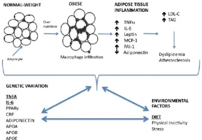
Obesity is characterized by the accumulation of lipid in white adipose tissue (AT). The AT is a specialized connective tissue composed of a number of different cell types each with specific functions. AT can be roughly divided into two fractions: the adipocyte fraction (AF) and the stroma vascular fraction (SVF). The AF is composed of mature adipocyte cells that are primarily involved in lipid storage, thereby having a major role in the regulation of whole-body energy homeostasis (Di Girolamo et al., 1998). In contrast to the cellular homogeneity of the AF, the SVF is composed of numerous cell types, including preadipocytes, mesenchymal stem cells, macrophages, endothelial cells and fibroblasts - all of which can play a role in regulating adipose tissue inflammation (Nair et al., 2005).
Indeed, obese AT was previously shown to be infiltrated by numerous types of inflammatory cells, and may promote the increase in extracellular components and fibrotic regions seen in adipose tissue biopsies from obese patients (Mutch et al., 2009; Divoux et al., 2010). Furthermore, it is now appreciated that AT is a metabolically active endocrine organ, in which both the AF and SVF secrete molecules, generically described as “adipokines” (Wang et al., 2008), influencing a wide range of biological functions, such as immunity, insulin sensitivity, inflammation, blood pressure, lipid metabolism, energy homeostasis and appetite (Trayhurn and Wood, 2004). Therefore, the identification of factors that regulate their secretion has become a primary interest in obesity research (Wang et al., 2008).
The macrophages present in the SVF are the primary source of obesity-induced inflammation (Gutierrez et al., 2009; Galic et al., 2010). The number of macrophages present in the SVF is directly correlated with the level of adiposity and adipocyte size (Xu et al., 2003). Adipocyte hypertrophy, results in increased chemokine secretion and subsequent increased infiltration of macrophages, which in turn secrete cytokines such as IL-6 and TNFα. Since AT expansion is characterized by increased macrophage infiltration, these cells are responsible for almost all TNFα and significant amounts of IL-6 secreted by AT (Weisberg et al., 2003 ).
Genetic sequence variants within the promoter region of inflammatory genes influence gene transcription, altering protein production. Functional polymorphisms have been reported in the TNFA, IL-1, IL-6 and lymphotoxin-a (LTA) inflammatory genes, altering cytokine production (Burzotta et al., 2001; Santtila et al., 1998; Wilson et al., 1997).
Figure 1 illustrates the development of obesity-associated low-grade inflammation and the impact of diet–gene interactions on obesity and dyslipidemia.
8
1.3 Dietary fatty acids (FAs)
Dietary FAs have received considerable attention for their ability to regulate inflammatory gene expression and secretion. It has been proposed that dietary FAs affect inflammatory processes through the modulation of transcription factors such as kappa-light-chain-enhancer of activated B cells (NFκB) and peroxisome proliferator-activated receptor gamma (PPARγ) (Calder P.C 2003 and 2006). PPARγ inhibits NFκB, and both transcription factors are sensitive to dietary FAs (Van den Berghe et al., 2003).
Although the molecular mechanisms by which dietary fats regulate adipokine production remain unclear, one proposed link between dietary FA and inflammation may be via the toll-like receptor 4 (TLR4) pathway. The TLR4 pathway is expressed in both SAT and VAT, and has been shown to be activated by saturated fatty acids (SFAs), inducing inflammatory cytokine production and signaling (Fessler et al., 2009). This results in a localized inflammation in adipose tissue that propagates an overall systemic inflammation (Shi et al., 2006; Poulain-Godefroy et al., 2010).
The n-6 polyunsaturated fatty acid (PUFA), linoleic acid (LA), constitutes the majority of PUFA intake in the western diet. LA is the precursor of the n-6 PUFA arachidonic acid (AA). A high LA intake is considered proinflammatory, however the evidences are contradictory and not conclusive (Baum et al., 2012; Bjermo et al., 2012). AA intake in the diet is low relative to LA intake, its metabolic precursor. However, AA is the most prevalent
n-6 PUFA in inflammatory cell membranes and is the substrate for the synthesis of the
proinflammatory eicosanoids, including prostaglandin E2 (PGE) and 4-series leukotrienes, associated with inflammatory processes (Calder et al., 2011). Despite this, studies investigating the impact of AA on inflammatory markers are inconclusive, and few human intervention studies have reported on the effect of dietary intake of AA on low-grade inflammation (Thies et al., 2001a; Thies et al., 2001b).
The n-3 PUFA, alpha-linolenic acid (ALA) is an essential FA common in canola, soybean oil and some nuts, but in greatest concentrations in flax seed and flax seed oil (Erkkila et al., 2008). ALA is elongated and desaturated to eicosapentanoic acid (EPA) and further to decosahexanoic acid (DHA), however the efficiency of this conversion has been debated (Williams et al., 2006). Association studies between dietary intake of ALA and inflammatory markers suggest a modest anti-inflammatory effect of ALA (Stulnig T.M, 2003; Zhao et al., 2007).
The long chain n-3 PUFAs, EPA and DHA are found in seafood, especially oily fish and in some algal oils. Is it proposed that n-3 PUFAs affect inflammation mainly through altered
9 eicosanoid production, but potentially also impacting cell signaling and gene expression (Calder P.C, 2011; Stulnig T.M, 2003). When EPA and DHA are incorporated into human inflammatory cells, this is partly at the expense of AA, providing less substrate for eicosanoid production (Calder P.C, 2011). Culture systems, animal models and human intervention studies have been generally consistent in recognizing the anti-inflammatory actions of n-3 PUFAs (Calder P.C, 2009; Itariu et al., 2012).
Emerging research shows that dietary FAs and SNPs in the genes encoding TNF-A, IL-6 and adiponectin can interact to regulate their production and secretion, thus adding an additional level of complexity in the study of obesity. Taken together, nutrigenomics research can provide a more thorough understanding of how changes in dietary FAs intake may contribute to the inter-individual variability in inflammatory status (Stryjecki and Mutch, 2011 ; Yu et al., 2011).
1.4 Tumor necrosis factor-α
TNFα is a pro-inflammatory cytokine whose expression and circulating levels are increased with obesity and decreased with weight loss (Maury and Brichard, 2010). TNFα acts in a paracrine manner, suggesting that circulating TNFα levels may not be indicative of actual TNFα levels (Suganami et al., 2005). The primary cell type responsible for the production of TNFα are macrophages in the SVF (Trayhurn and Wood, 2004). It is hypothesized that TNFα is produced by macrophages in response to chemoattractant signals released by dying adipocytes. TNFα has numerous effects in adipose tissue, including the regulation of apoptosis, adipogenesis, lipid metabolism and insulin signalling (Galic et al., 2010). The effects of TNFα on lipid metabolism occur in different cells, tissues, and organs and include a number of metabolic processes. TNFα induces lipolysis, increasing free fatty acid (FFA) production. In addition, TNFα regulates cholesterol metabolism and other adipocyte-derived adipokines such as leptin and adiponectin, which may also alter lipid metabolism (Chen et al., 2009).
Evidence suggests that TNFα triggers a signalling cascade that induces cell apoptosis, which may be one mechanism by which TNFα regulates adipose tissue mass (Cawthorn et al., 2007).
An increase in TNFα promotes the secretion of other pro-inflammatory cytokines and reduces anti-inflammatory cytokines, resulting in an overall pro-inflammatory state.
A study conducted by Wang and Trayhurn (2006) found that treating human adipocytes with TNFα for 24 h led to significant decreases in adiponectin expression and increases in IL-6
10 and TNFα expression, showing that TNFα is a powerful regulator of inflammatory molecules, favoring an overall inflammatory state (Wang and Trayhurn, 2006).
1.4.1 TNFA gene variants, obesity, serum lipids and TNFα signaling
Recent evidence suggests that individual SNPs in the genes encoding TNFα, TNFα receptor 2 and TNFα converting enzyme can modify an individual’s risk for obesity and obesity-related complications.
One aspect that has not yet been studied is whether a haplotype consisting of SNPs in the three aforementioned genes will have an additive or synergistic effect on these complications.
Several SNPs have been identified in the promoter region of the TNFA gene, however the
TNFA –308 G>A (rs1800629) and –238 G>A (rs361525) SNPs are most commonly
associated with measures of adiposity, obesity risk and serum lipids. The A allele of the functional –308 G>A SNP results in a 2-fold increase in TNFA transcription, with a subsequent increase in TNFα production (Wilson et al., 1997).
Many studies have reported that carriers of the pro-inflammatory –308 A allele (AA and GA genotypes) reported a higher body mass index (BMI) and/or percent body fat than those with the GG genotype (Brand et al., 2001; Nieters etr al., 2002; Pihlajamaki et al., 2003). In two large recent meta-analyses it was shown that the –308 GA + AA genotypes were associated with an increased risk of obesity (Yu et al., 2011; Sookoian et al., 2005).
In comparison to the –308 G>A SNP, only ew studies have investigated the association between the –238 G>A SNP and obesity, reporting an association with body fat, insulin resistance and circulating free FA (Fontaine-Bisson et al., 2007). Joffe and collegues (2012) found that black South African women with the –238 A allele had greater body fat % than those with the GG genotype.
However, not all studies have shown the interaction between the –308 G > A and –238 G > A SNPs and obesity (Joffe et al., 2010; Joffe et al., 2011; Hedayati et al., 2011).
Only two papers have found an independent association between the –308 G > A SNP and serum lipid concentrations. In Caucasian men, the –308 A allele was associated with increased triglycerides (Dalziel et al., 2002), and in Polish Caucasian men and women the AA genotype was associated with lower high-density lipoprotein cholesterol (HDL-C) concentrations compared to the GG genotype (Wybranska et al., 2003).
Further, no independent associations have been reported between the –238 G > A SNP and serum lipid concentrations. While the A allele of the –308 G > A and –238 G > A SNPs
11 appear to be associated with the obese phenotype and serum lipid concentrations, it is highly likely that genetic variation in the TNFA gene may provide only a partial explanation with regards the inter-individual variability observed and the heterogeneity of the results in these studies. Other variables such as ethnicity, gender, diet, lifestyle and environmental factors may modulate these associations and contribute to the different results observed.
Although the mechanisms by which these promoter variants affect circulating TNFα levels remain unclear, their significant association with obesity and insulin levels reinforce the importance of conducting further studies (Joffe et al., 2013).
1.4.2 TNFA and dietary FAs
The TNFA –308 G > A and –238 G > A SNPs have been shown to modulate the relationship between dietary fat intake on obesity and serum lipid profiles in different populations.
Several studies have investigated the effect of dietary FAs on TNFα concentrations and
TNFA gene expression in cell, animal and human models. Lifestyle factors, including dietary
components, such as FAs, interact with genetic variants to regulate the development and progression of obesity and its comorbidities and these interactions may explain the differences observed across populations (Joffe et al., 2013).
The A allele of -308 G > A SNP has been associated with obesity, obesity-related insulin resistance, and altered serum lipid concentrations in some Caucasian populations (Fontaine-Bisson et al., 2007; Brand et al., 2001), but not all (Um et al., 2003; Sookoian et al., 2005). In addition, in some populations the TNFA -308 G>A polymorphism changes the relationship between fatty acids (FA) intake and the risk of obesity (Stryjecki and Mutch, 2011).
Indeed, palmitic acid (saturated FA) was found to increase TNFA gene expression and protein secretion in a dose-dependent manner. In contrast, incubation with the n-9 monounsaturated fatty acid (MUFA) oleic acid (C18:1) and n-3 PUFA DHA had no affect on TNFα expression or secretion (Bradley et al., 2008).
In rodent studies, mice that consumed a high fat diet for 5 weeks showed increases in TNFα expression; however, the concomitant administration of EPA prevented this increase (Perez-Matute et al., 2007). In another study, mice fed menhaden fish oil showed a reduced expression of TNFα in kidneys (Chandrasekar and Fernandes, 1994). When considered altogether, these studies indicate that SFA increase and n-3 PUFA decrease TNFα expression and/or secretion.
12 Importantly, these murine results appear to translate to human beings. Subjects consuming a fish oil supplement showed a significant decrease in TNFα production; however, levels of TNFα returned to baseline upon cessation of the supplements (Endres et al., 1989). This was confirmed in another study where subjects who ingested ALA for 4 weeks had a 30% reduction in TNFα production. When these subjects were further supplemented with fish oil, TNFα synthesis was inhibited by up to 70% (Caughey et al., 1996). Finally, men who consumed fish oil supplements for 12 weeks showed differential decreases in TNFα production in accordance with their pre-supplementation levels (Grimble et al., 2002). In addition to the independent influences of genetic variation and different FA on the TNFα signalling pathway, research discovering diet–gene interactions offers a further explanation for the inter-individual variability observed with obesity-related inflammatory status. Grimble et al. (2002) found that men with one A allele at the -308 SNP and who were in the highest tertile for TNFα production had the most significant reduction in TNFα levels after supplementation with fish oil in comparison to homozygotic G/G men. No conclusions could be made about -308A/A individuals owing to the small sample size.
In another study by Fontaine-Bisson and El-Sohemy (2008), a low n-6 PUFA intake in subjects who were homozygous for G at the -238 SNP and had at least one A (that is, GA or AA) at the -308 SNP had an increased risk of developing obesity; however, Nieters et al. (2002) found that German Caucasian men and women with the –308 A allele, who were in the ighest tertile for intake of the n-6 PUFAs LA and AA (%E), had an increased risk of developing obesity. More recently, it has been reported that the odds of obesity for black South African women with the –308 A allele increased with total dietary fat intake (%E) (Joffe et al., 2011). This interaction was not observed in white South African women (Joffe et al., 2012).
Considered together, these newly discovered diet-gene interactions that affect TNFα signalling are an important point to consider in future intervention studies targeting obesity-related inflammation.
1.5 Mediterranean Diet
Since the early 1990s, growing evidence indicates that the Mediterranean diet (MD), a concept first proposed by Keys in the mid1980s (Keys et al, 1986), has a beneficial influence on health, preventing NCDs, and longevity (Trichopoulou et al, 1995, 2004, 2005, Sofi et al, 2008, 2010).
13 The Mediterranean-style diet is not a specific diet, but rather a collection of eating habits traditionally followed by people in the different countries bordering the Mediterranean Sea. The diet refers to a dietary profile commonly available in the early 1960s in the Mediterranean regions and characterized by a high consumption of fruit, vegetables, legumes, and complex carbohydrates, with a moderate consumption of fish, and the consumption of olive oil as the main source of fats and a low-to-moderate amount of red wine during meals.
A great deal of attention has been given to tools that estimate the adherence of individuals to the MD because of the usefulness of these tools to identify the whole dietary pattern instead of single foods or nutrients (Bach et al., 2006). Hence, computational scores have been created and used in several large epidemiologic studies to seek whether they could be useful to estimate the risk of disease in the general population (Kourlaba and Panagiotakos, 2009). Some large epidemiologic studies conducted in different cohorts evidenced an association between a greater adherence to MD, a reduced risk of mortality, and the incidence of major chronic diseases (Mitrou et al., 2007). In 2008, a meta-analysis performed by Sofi et al, that included all cohort prospective studies that investigated this issue, observed that a 2-point increase of adherence to MD conferred a significant protection against mortality, the occurrence of cardiovascular diseases, and major chronic degenerative diseases (Sofi et al., 2008).
Despite the widely proven benefits of the MD the Southern European countries in which MD originated are rapidly withdrawing from this eating pattern orienting their food choices towards products typical of the Western diet which is rich in refined grains, saturated fats, sugars, red and processed meat (Laccetti et al., 2012).
The reasons why people keep on drifting from one dietary regimen to another remain open to several hypotheses (Bonaccio et al., 2012).
Social changes appear to have contributed to radical reversal in dietary habits in Western and Southern European societies although developing countries are slightly turning to westernised diets as well (Prentice, 2006).
The cost of MD seem to have led people to give up this eating pattern in favour of less-expensive products which allow to save money, but are definitively unhealthy (Lopez et al., 2009). Many studies suggest that diet quality follows a socioeconomic gradient highlighting how disadvantaged people present higher rates of obesity, diabetes, CVD and some types of cancer (Darmon and Drewnowski, 2008).
14 The abandoning of MD is also considered as a possible cause of the increasing obesity pandemic (Esposito et al., 2011). Several studies took a step forward to see whether there is an association between diet cost and obesity, and found that a higher adherence to healthy dietary patterns is linked to higher monetary costs and is inversely associated with BMI and obesity (Schröder et al., 2006).
In 2010 UNESCO has recognized the MD as an Intangible Cultural Heritage of Humanity (UNESCO, 2010). A number of epidemiological studies have shown that greater adherence to the traditional MD, is associated with a significant reduction in total mortality, and particularly of death due to coronary heart disease and to cancer (Trichopoulou et al., 2003), could reduce overall cancer risk (Couto et al., 2011), and provide a consistent protection for the occurrence of major chronic degenerative diseases (Sofi et al., 2010).
1.5.1 Obesity and Mediterranean diet (MD)
The health consequences of obesity in developed countries represent an economic load of between 2 and 7% of the total health cost, a substantial proportion of the national health cost. Obesity is not a single disorder, but a complex multifactorial disease involving environmental and genetic factors. Among the environmental factors, diet appears to be an important contributor to the development of obesity. Epidemiologic evidence on the association of nutrients, particularly fat, with obesity remains controversial. Because of several short-comings in traditional, nutrient-based diet and disease analysis, the focus has shifted from this type of analysis to one describing food intake patterns. Such analysis takes into account the complex combination of foods in a diet. The effect of such food-based defined dietary patterns might be more closely related to obesity than a single nutrient or food. Hence, food-based dietary patterns may be more useful than nutrient based methods for dietary counseling and in public health efforts (Schröder et al., 2004).
Research interest over the past years has been focused on estimating adherence to the whole MD rather than analyzing the individual components of the dietary pattern in order to consider important interactions between components of the diet.
Several epidemiologic studies examined the association of dietary patterns and excessive weight (Togo et al., 2001; Newby et al., 2003). Identifying palatable dietary patterns that prevent weight gain is an important task for health policy in view of the social and economic burden of obesity.
The MD is an eating pattern that successfully combines pleasant taste with positive health effects. The MD does not stand for a homogeneous exclusive model throughout the
15 Mediterranean basin; rather, it represents a set of healthy dietary habits including high consumption of vegetables and fresh fruits, with olive oil as the main source of fat. However, whether adherence to this healthy dietary pattern might be protective against weight gain remains unclear (Schröder et al., 2004).
Furthermore, independently of energy and macronutrient quantity intakes, a better adherence to the MD, is associated with lower obesity risk (Mozaffarian et al., 2011; Martìnez-Gonzàlez et al., 2012).
1.6 Role of folate in 1-carbon metabolism
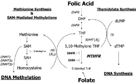
Folate is an essential water-soluble vitamin occurring naturally in select foods as well as in the synthetic form (folic acid) used in supplements and in food fortification programs (Crider et al., 2012). According to chemical nomenclature, the difference between folate and folic acid is just one proton. However, the term folic acid is in general applied to the synthetic form of this B-vitamin, which is also the most stable form (Blom et al. 2006; Pitkin RM 2007). There are many critical cellular pathways dependent on folate as a 1-carbon source including DNA, RNA, and protein methylation as well as DNA synthesis and maintenance. Folate can be a limiting factor in all these reactions (figure 2).
Under normal dietary conditions, absorbed folate is metabolized to 5-methyltetrahydrofolate (5-methylTHF) in the intestine and/or liver. 5-MethylTHF is the primary folate constituent taken up by nonhepatic tissues, which then must be polyglutamated for cellular retention and 1 carbon cycle coenzyme function.
Tetrahydrofolate (THF) is the most effective substrate for polyglutamate synthetase; therefore, 5-methylTHF must be converted to THF via the methionine synthase reaction. When folic acid is consumed in fortified foods or supplements, it is metabolized primarily to 5-methylTHF during intestinal absorption and/or first pass in the liver, after which it behaves identically to natural dietary folate. Folic acid is normally first reduced to dihydrofolate by dihydrofolate reductase and subsequently to THF to enter the folate pool. Once the THF coenzyme is formed from either folic acid or dietary folate, it is first converted to 5,10-methyleneTHF by the vitamin B-6–dependent enzyme serine hydroxymethyltransferase and subsequently irreversibly reduced to 5-methylTHF by 5,10 methylenetetrahydrofolate reductase (MTHFR). This reaction is key to maintaining the flux of methyl groups for the remethylation of homocysteine (Hcy) to methionine via the vitamin B-12–dependent methionine synthase reaction. Methionine is the substrate for SAM or AdoMet, a cofactor and methyl group donor for numerous methylation reactions including the methylation of
16 DNA, RNA, neurotransmitters, and other small molecules, phospholipids, and proteins, including histones (Stover PJ, 2009). A number of SAM-dependent reactions have regulatory roles by affecting both genome stability and gene transcription, localization of protein, and small molecule degradation (Winter-Vann et al., 2003; Stead et al., 2004). In addition to folate, a number of other dietary nutrients are required to maintain 1 carbon flux, ensuring normal Hcy remethylation, SAM formation, and DNA methylation. These nutrients include vitamin B-6, riboflavin, vitamin B-12 and choline (Shin et al., 2010). The 1-carbon pathway, and thus DNA methylation, functions under tight regulatory controls. SAM is the major regulator of folate-dependent Hcy remethylation because it is a potent inhibitor of MTHFR. Under the condition of high SAM concentration, MTHFR is inhibited, which reduces the synthesis of 5-methylTHF and hence remethylation of Hcy. Conversely, when SAM concentrations are low, remethylation of Hcy is favored (Crider et al., 2012).
1.6.1 MTHFR polymorphisms
MTHFR activity and thus 5-methylTHF formation may be modified by several SNPs. The
MTHFR gene maps to chromosome 1p36.3, and plays a central role in folate metabolism,
together with other enzymes by irreversibly catalyzing the conversion of 5, 10-methylenetetrahydrofolate to 5-methyltetrahydrofolate, the primary circulating form of folate and a cosubstrate for Hcy methylation to methionine (Goyette et al., 1994). Many rare mutations of the MTHFR gene have been described in individuals, resulting in very low enzymatic activity, whereas the most common polymorphism is a C to T mutation in exon 4 at nucleotide 677, leading to Ala222Val and presenting in healthy individuals with lower enzyme activity (Frosst et al., 1995).
Individuals with the MTHFR 677TT genotype have been shown to have only 30% of in vitro MTHFR enzyme activity, which reduces plasma folate levels and elevates plasma Hcy levels, compared with the wild type, whereas those with the heterozygous CT genotype have 60% of wild-type MTHFR enzyme activity (Frosst et al., 1995). Reduction of the MTHFR enzyme activity can increase the pool of 5, 10-methylene-THF at the expense of the pool of 5-methyl-THF and impair the DNA methylation (Guo, 2012).
Because the DNA methylation plays a critical role in regulation of gene expression and maintenance of genomic stability, the aberrations in normal methylation patterns have been associated with the development of cancer by impairing the DNA methylation (Cheng et al., 1997). More importantly, the homozygous variant genotype MTHFR 677TT has been
17 associated with risk for many different types of cancer, including colorectal cancer, gastric cancer and breast cancer (Taioli et al., 2009; Dong et al., 2010; Zacho et al., 2011).
1.6.2 Folate and DNA stability
Biomarkers that are influenced by nutritional factors offer a potentially important contribution to public health programs because of their direct relevance to explain the effects of diet on the risk of disease, and their potential role in monitoring the effectiveness of preventive programs (Boffetta P. 2010). Biomarkers of early events should be related to, and ideally critical for, the development of the adverse effect that is the basis for the risk assessment (Renwick et al., 2003, Renwick and Walton, 2001).
Low folate status (as defined by various measures including blood folate concentrations, folate and/or folic acid intake) has been associated with an increased risk of cardiovascular disease, multiple cancers, and neural tube defects (NTDs) (Erickson JD, 2002; Lamprecht and Lipkin, 2003).
The mechanisms by which low folate status contributes to these disorders remain unclear. During DNA replication, folate depletion can have destabilizing consequences. Inadequate folate availability during cell division can result in the compromised production of thymidine, such that uracil may be substituted in the DNA sequence. This mutagenic event may trigger attempts to repair the defect and increases the frequency of chromosomal breaks. In tissue cultures, it has been demostrated that low folic acid results in the formation of micronuclei (indicative of chromosome breakage) and that the MTHFR TT genotype leads to increased micronuclei formation under low folate conditions (Kimura et al., 2004; Crider et al., 2012).
Mitochondrial DNA (mtDNA) is more susceptible to oxidative damage than nuclear DNA since it is not protected by histones and it reveals limited capacity for DNA repair (Yakes and Van Houten, 1997). Alterations in mtDNA both qualitatively (mutations) and quantitatively (mtDNA copy number) have been associated with many human diseases including neurodegenerative diseases, metabolic diseases and various types of cancer (Chen et al., 2011; Nie et al., 2013). Among several mutations that have been reported to be associated with various diseases, a 4977-bp deletion, occurring between two 13-bp direct repeats at positions 13447-13459 and 8470-8482, has attracted great interests since it has also been shown to accumulate in many tissues during aging and it has been used as a mtDNA damage biomarker (Meissner et al., 2008). The mtDNA 4977-bp deletion has been detected in fast replicating cells such as blood leukocytes (Ross et al., 2002; von
Wurmb-18 Schwark et al., 2010), but little is known about the relationship between this mutation and folate status (Chou and Huang, 2009).
In an experimental study in rodents, a folate-dependent accumulation of mtDNA deletions in lymphocytes has been reported after folate deprivation, suggesting that accumulated lymphocytic mtDNA deletions may serve as a molecular biomarker responding to dietary folate deprivation (Chou and Huang, 2009). Furthermore, mtDNA deletions and low folate status, proposed characteristics of carcinogenesis, have been suggested in relation to human cancer susceptibility, in a case-control study, an increased frequency of lymphocytic mtDNA 4977-bp deletion has been associated with human hepatocellular carcinoma risk; moreover, in the same study, a high frequency of lymphocytic mtDNA 4977-bp deletion was associated with low levels of lymphocytic folate (Wu et al., 2009).
As suggested from experimental studies in rodents (Chou and Huang, 2009), it has been hypothesised that folate intake and folate status may have an impact on lymphocytic mtDNA 4977-bp deletion levels in healthy humans and thus on mitochondrial genomic instability.
1.6.3 Folate and women’s health
The effect of folate status on pregnancy outcomes has long been recognized. Folate is now viewed not only simply as a nutrient needed to prevent megaloblastic anemia in pregnancy but also as a vitamin essential for reproductive health, disease prevention, and health maintenance (Tamura and Picciano, 2006). Adequate maternal nutrition during the periconceptional period as well as in pregnancy are key focus of attention in public health because of the increased needs and greater vulnerability of pregnant women to the effects of micronutrient deficiency or imbalance (Ortiz-Andrellucchi et al., 2009).
Since NTDs are caused by the failure of fusion of the neural tube 22–28 days after conception, the public health goal is that each woman could begin her pregnancy with an optimal folate status, estimated to be a red blood cell (RBC) folate concentration >906 nmol/l (Daly et al., 1995). Blood folate levels are directly correlated with intake and, when low, are not only associated with NTDs but also with megaloblastic anemia and high blood Hcy levels (Dary O, 2009).
The crucial role of folate in the conversion of Hcy to methionine and, subsequently, to S-adenosylmethionine, has been already demonstrated. Maternal total Hcy (tHcy) concentrations have been linked to a wide range of adverse pregnancy outcomes, including growth retardation in utero (Hogeveen et al., 2012), preeclampsia, abruptio placentae, low birth weight, and other maternal or fetal complications (Ubeda et al., 2011).
19 Plasma Hcy levels are determined by both dietary and genetic factors, namely the common SNP MTHFR C 677T. Individuals with the MTHFR 677 TT genotype have been shown to have higher plasma Hcy concentrations and to be at higher risk of coronary heart disease and pregnancy complications particularly in the setting of a low folate status (Fekete et al., 2010; Klerk et al., 2002).
Besides, cigarette smoking has been related to increased levels of Hcy, and smokers have been shown to develop hypofolatemia (Das et al., 2010; Haj Mouhamed et al., 2011).
There is a growing body of epidemiologic evidence that suggests folate deficiency contributes to cancer risk at several sites (Flatley et al., 2009). Epidemiologic and molecular studies on cervical cancer have shown a causal relationship between infection with high-risk (HR) human papillomaviruses (HPV) and this cancer. Nutritional status and food consumption may be important HPV cofactors that increase risk of persistence and progression to cervical intraepithelial neoplasia (CIN); however, there is insufficient evidence to support the association between nutritional status and cervical carcinogenesis (Garcia-Closas R, 2005). Previous research has shown that circulating folate status significantly influences the natural history of infections with HR-HPV and lower the likelihood of developing HR-HPV–associated CIN 2+ (CIN of grade 2 or higher) (Piyathilake et al., 2009). The apparent role of folate in carcinogenesis in several tissues has stimulated investigations on MTHFR SNPs. The MTHFR C677T SNP can lead to abnormal DNA methylation and DNA synthesis, possibly resulting in an increased risk of cancer. However, the effect of MTHFR SNPs on cancer susceptibility remains controversial. In a population of women in Catania, Italy, with high prevalence of HR-HPV infection (Agodi et al 2009), a decreased risk for CIN of individuals homozygous for the MTHFR T allele has been previously reported (Agodi et al., 2010). However, some studies have supported the existence of gene-folate status interactions in the etiology of cervical cancer. Specifically it has been reported that the MTHFR T allele and reduced dietary folate may increase the risk for cervical squamous intraephitelial lesions (Goodman et al, 2001), and on the contrary, a study conducted after folate fortification reported that MTHFR SNP is associated with reduced risk of CIN 2 or 3 (Henao et al, 2005). The above scenario suggests a possible role of folate as a modulating factor on the risk of cervical cancer.
1.7 Epigenetic and epigenomic
Classic genetics alone cannot explain the diversity of phenotypes within a population. Nor does classic genetics explain how, despite their identical DNA sequences, monozygotic
20 twins or cloned animals can have different phenotypes and different susceptibilities to a disease (Fraga et al. 2005; Humpherys etal. 2001). The concept of epigenetics offers a partial explanation of these phenomena (Esteller, 2008).
The term epigenetics, first intoduced in 1942 by Conrad H Waddington, to name “the causal interactions between genes and their products, which bring the phenotype into being” (Waddington, 1939), was later defined as heritable changes in gene expression and chromatin organization that are not attributable to any alteration in the DNA sequence (Holliday, 1987).
Until recently epigenomics was considered a black box. Like genetics, epigenetic traits can be mitotically and meiotically inherited (translational inheritance), but unlike genetics they are not conferred by the sequence of bases defining the genetic code. Recently it has been used the analogy of computer hardware and software to describe genetic and epigenetic mechanisms (Brower, 2011). Thus, while the genetic code can be considered the hardware for life, the epigenetic code is considered the software that determines how the hardware behaves. Fundamentally, ‘faulty’ software can potentially be re-written. This notion in particular has led to a frenzy of research activity in the field of onco-epigenetics, as exemplified by a substantial increase in the number of scientific publications regarding cancer and epigenetics over the past decade (Widschwendter et al., 2013).
Presently, epigenetics is highlighted in many other fields, such as aging, embryo development, inflammation, obesity, type 2 diabetes mellitus, cardiovascular - neurodegenerative - and immune diseases (Choi and Friso, 2010).
The predominant epigenetic mechanisms are DNA methylation, modifications to chromatin structure, loss of imprinting, and noncoding RNA (Gibney and Nolan, 2010).
DNA methylation is described as the covalent attachment of a methyl group to specific nucleotide residues within the DNA sequence, and occurs frequently, though not exclusively, at cytosine residues within a cytosine–guanosine (CpG) dinucleotide context (Jones and Baylin, 2007). CpG dinucleotides are unequally distributed throughout the genome but tend to cluster within and around gene promoter regions. These clusters are referred to as CpG islands, which vary in size from 500 up to 2000 base pairs in length. CpG islands usually remain unmethylated, with the exception of those occurring in or close to genes required for normal embryonic development, including X-chromosomeinactivation and genomic imprinting (Baylin and Jones, 2011).
Histones are proteins that package DNA into structural units or nucleosomes to enable higher-order chromatin formation. Protein modifications to the histone ‘tails’ that are
21 exposed outside of the nucleosome affect gene accessibility by influencing an ‘open’ or ‘closed’ chromatin formation (Greer and Shi, 2012).
Imprinted genes are monoallelically expressed from either the maternally or paternally inherited allele. The crucial nature of this regulation is reflected in phenotypic manifestations in individuals exhibiting improper imprinted gene expression. The maintenance of proper parent of origin gene expression is often aberrant in cancer leading to loss of imprinting. This alteration is observed in many different types of cancer cells and frequently influences the timing of key developmental events or potentially the loss of imprinting in stem cells leading to the genesis of cancer.
Non-coding RNAs are RNA transcripts that are not translated into protein but can demonstrate functional regulation of gene expression by blocking transcription or enhancing mRNA degradation (Esteller, 2011).
1.7.1 DNA methylation
Regarding nucleic acid modification, DNA methylation of cytosine nucleotide at the carbon 5 position of a cytosine (5-mC) is a common epigenetic mark involved in epigenetic regulation of gene expression in many eukaryotes and is often found in regions of the genome rich in sequences of a cytosine preceding a guanine (CpG islands), or in CpHpG (H: A, T, C) (Lister et al., 2009, Mansego et al., 2013).
CpG islands exist in the promoter regions of approximately half of all genes and are usually unmethylated in normal differentiated cells, while CpG islands located in intergenic regions are methylated (Esteller, 2007; Taby and Issa, 2010). Promoter methylation is typically associated with repression, whereas genic methylation correlates with transcriptional activity. In the particular case of cancer, promoter CpG islands of numerous tumor suppressor genes (TGSs) are found to be densely methylated, resulting in transcriptional silencing, with these “epimutations” being cancer type-specific and tumor stagespecific (Rodriguez-Paredes and Esteller, 2011).
DNA methylation has many roles in various cellular processes and may impact the transcription of genes by preventing the binding of key transcriptional factors (Altaf et al., 2007). Transcriptional silencing due to DNA methylation is thought to occur by the recruitment of methyl CpG-binding transcriptional repressors and by interfering with the DNA binding of transcriptional activators, which results in a condensed chromatin state. In addition, methylated DNA may be bound by methyl-CpG-binding domain proteins, which are essential for binding to 5-methylcytosine (Meeran et al., 2010).
22 DNA methylation is catalyzed by enzymes known as DNA methyltransferases (DNMTs), using the cofactor S-adenosylmethionine (SAM) (Gibney and Nolan, 2010) and hypermethylation of CpG dinucleotides or CpG islands by DNMTs usually results in transcriptional gene silencing and gene inactivation (Herceg, 2007). The human genome contains four DNMT genes, DNMT1, DNMT2, DNMT3A and DNMT3B (Stresemann et al., 2006). Altered DNMT expression and activity is seen in numerous diseases including autism, cardiovascular diseases, obesity, Type-2 diabetes and cancer (Grafodatskaya et al., 2010; Milagro et al., 2011; Maier and Olek, 2002, Chowdhury et al., 2003). Finally, global hypomethylation is associated with nearly all human cancers (Irizarry et al., 2009; Tollefsbol T, 2008).
1.7.2 Gene-environment versus epigene-environment
The interplay between the environment and human genome has been traditionally presented under the framework of gene-environment interactions (also indicated as genotype-environment) (Baccarelli and Bollati, 2009).
In gene-environment interactions, the genetic polymorphisms that modify the effects of environmental exposures are transmitted transgenerationally according to Mendelian genetics, and the trait determining effect modifications is generally assumed to follow the same genetic model (dominant, co-dominant, recessive) as that of the levels of expression or function of the protein coded by the locus of concern. A second wellestablished area of interplay includes the direct effects of environmental exposures on the genome, e.g., DNA damage and/or mutations induced by environmental exposures.
In environmental health, the recognition that exposures could produce DNA mutations represented a major landmark for risk assessment and prevention. Consequently, genotoxic agents have been categorized according to their capability to alter DNA sequence and thus increase disease risk (Siemiatycki et al, 2004). Such information has been fundamental to determine environmental risks and shape current regulatory efforts for exposure reduction. Specifically, potential carcinogenic agents have been carefully tested in in-vitro and in-vivo models of mutagenicity. In human subjects, some of these molecular events may represent early events along the pathways linking carcinogen exposure to cancer.
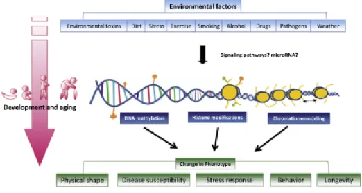
In principle, the effect-modification model should apply to epigene-environment interactions as well as to gene-environment interactions (figure 3). Similarly to the effect modifications demonstrated or postulated for genetic polymorphisms, epigenetic differences determining disease risk could make individuals less or more vulnerable to environmental.
23 In environmental studies, the flexibility of epigenetic states has generated a growing interest in evaluating the direct alterations that environmental exposures may produce on epigenetic states, including changes in DNA methylation and histone modifications (Bollati and Baccrelli, 2010).
1.7.3 Environmental effects on the epigenome
Supporting evidence implicates the epigenome as the interface between the genome and the environment (Feil and Fraga, 2011; Jirtle and Skinner, 2007) a broad definition of the environment includes nutrition, lifestyle, external environmental exposures and reproductive history. For this reason epigenetics is now considered an important mechanism in the unknown etiology of many environment-associated diseases, with growing interest in associating epigenetic regulation with lifestyle and risk of disease (Choi and Friso, 2010). Numerous environmental exposures have linked altering epigenetic patterns during a lifetime, and subsequent risk of disease.
Environmentally triggered epigenetic alterations are often tissue-specific and are established during a phase of epigenetic ‘hypersensitivity’ during development, but can also accumulate as a function of age. The epigenome can be thought as a mechanism of cellular memory that records environmental exposures which accumulate during life-time and which potentially explain predisposition to late-onset disease such as cancer. Such induced epigenetic changes can be inherited during cell division, resulting in permanent maintenance of the acquired phenotype (Widschwendter et al., 2013). Moreover, the reversibility of epigenetic alterations stimulates the development of novel therapeutic approaches with an open field for development of early cancer detection and prevention, namely through chemoprevention (Nogueira da Costa and Herceg, 2012).
In the nutritional field, epigenetics is exceptionally important because nutrients and bioactive food components can modify epigenetic phenomena and alter the expression of genes at the transcriptional level (Choi and Friso, 2010) (figure 4).
An interesting study showed how diverse environmental exposures may alter the epigenome by looking at the methylation and histone modifications in monozygotic twins at different stages in life. It was demostrated that, early in life, their epigenetic profiles were nearly identical, but, by age 50, considerable changes were detected (Feinberg AP, 2008). These studies indicate that inherited genetic factors make only a minor contribution to susceptibility in most types of neoplasms, indicating that the environment has a principal role in influencing disease predisposition (Lichtenstein et al., 2000).
24 In general terms, environmental and dietary carcinogens, also known as epimutagens, are capable of inducing tumoral development by deregulating the epigenenome and can be divided into two groups: (a) those that induce, both directly or indirectly, changes to genomic DNA and (b) those that affect key cellular processes such as gene transcription, DNA damage and repair, cell cycle control and apoptosis (Herceg, 2007; Herceg and Vaissiere, 2011).
Examples of these epimutagens include tobacco smoke, alcohol, viruses and bacteria and dietary contaminants such as aflatoxin B1 (Lambert et al., 2011; Zhang et al., 2012).
Although extensive and novel studies were conducted in the past half a decade, there is still no clear-cut casual relationship between epimutagens and changes to epigenetic signatures (Bollati and Baccarelli, 2010; Herceg and Vaissiere, 2011). The main limitation of these studies still reside in the fact that the epigenetic changes are thought of as being mostly subtle, cumulative and requiring a long “timeframe” until full manifestation is detectable.
1.7.4 Effects of bioactive food components on DNA methylation: the field of
nutriepigenomic
Bioactive dietary components consumed by ingesting natural products, including fruits and vegetables, can act as sources of vitamins and minerals. While this is an invaluable role, these agents have high potential for application to oncogenesis, partially owing to their anticarcinogenic properties (Shu et al., 2010; Link et al., 2010).
A growing body of evidence suggests that dietary agents as well as non-nutrient components of fruits and vegetables can affect epigenetic processes and are involved in processes, including the reactivation of TGS, the initiation of apoptosis, the repression of cancer-related genes and the activation of cell survival proteins in different cancers (Hardy and Tollefsbol, 2011).
Folate, vitamin B-12, methionine, choline, and betaine can affect DNA methylation and histone methylation through altering 1-carbon metabolism. Two metabolites of 1-carbon metabolism can affect methylation of DNA and histones: SAM , which is a methyl donor for methylation reactions, and S-adenosylhomocysteine (SAH), which is a product inhibitor of methyltransferases. Thus, theoretically, any nutrient, bioactive component or condition that can affect SAM or SAH levels in the tissue, can alter the methylation of DNA and histones. Other water-soluble B vitamins like biotin, niacin, and pantothenic acid also play important roles in histone modifications. Biotin is a substrate of histone biotinylation. Niacin is involved in histone ADPribosylation as a substrate of poly (ADP-ribose) polymerase as well
25 as histone acetylation as a substrate of Sirt1, which functions as histone deacetylase (HDAC).
Pantothenic acid is a part of CoA to form acetyl-CoA, which is the source of acetyl group in histone acetylation. Bioactive food components directly affect enzymes involved in epigenetic mechanisms. For instance, genistein and tea catechin affects DNMT. Resveratrol, butyrate, sulforaphane, and diallyl sulfide inhibit HDAC and curcumin inhibits histone acetyltransferases (HAT). Altered enzyme activity by these compounds may affect physiologic and pathologic processes during our lifetime by altering gene expression (Choi and Friso, 2010).
While most natural dietary products have shown beneficial effects on the epigenome, not all dietary components share this characteristic. In fact, alcohol consumption is associated with harmful epigenetic modifications as well as the development/progression of several human cancers: colorectal cancer patients with high alcohol consumption had a prevalence of promoter hypermethylation of numerous genes when compared with patients with low alcohol consumption (Van Engeland et al., 2003; Giovannucci et al., 1995).
Interestingly, one of the most widely used models of diet-induced obesity in animals is the intake of a high-fat diet, and various reseraches have analyzed the epigenetic modifications induced by this dietary approach in rodents (Lomba et al., 2010). However, it is still unknown the capacity of the different types of FAs to induce epigenetic modifications. Few studies have evidenced the role of n-3 and n-6 PUFA on DNA methylation, although there are examples concerning effects of EPA (Ceccarelli et al., 2011), DHA (Kulkarni et al., 2011) and AA (Kiec-Wilk et al., 2011) FAs on DNA methylation.
It has been also reported that MUFA can modulate other epigenetic mechanisms such as histone acetylation. Thus, more studies must be designed as it is well known that the dietary FA composition is one of the main factors in the development of obesity and metabolic syndrome (Milagro et al., 2013); particularly in relation to the beneficial effects associated to high long-chain n-3 PUFA intake, whose anti-inflammatory properties are being studied to reduce obesity-related low-grade inflammatory condition (Calder et al., 2011).
1.8 Cancer epigenetic
Epigenetic modifications are often involved in transcriptional regulation and have been implicated both in tumor development and progression (Hardy and Tollesfbol., 2011).
These modifications, causing transcriptional deregulation, may result in the inappropriate expression or activation of transcription factors associated with oncogenes and/or the failure
26 to express genes responsible for tumor suppression. In fact, cancer cells have genomewide aberrations in a number of epigenetic markers, including global hypomethylation, global downregulation of miRNAs, promoter-specific hypermethylation, histone deacetylation and upregulation of epigenetic machinery (Taby and Issa, 2010).
In tumorigenesis, epigenetic aberrations are believed to play key roles in TSG inactivation, oncogene activation, and chromosomal instability, all of which are involved in the deregulation of critical cellular pathways and steps of carcinogenesis including tumor initiation, invasion and plasticity (Carmona and Esteller, 2010; Jones, 2012; Sincic and Herceg, 2011).
The most studied epigenetic changes is the DNA methylation, which occurs primarily in CpG dinucleotides and is often altered in cancer cells. Generally, tumours are characterized by sporadic gene-specific hypermethylation and global DNA hypomethylation, which increases according to tumour progression.
Gene-specific hypermethylation can affect many different types of cancers and is often mediated through the silencing of TSG, whereas hypomethylation can contribute to genomic instability frequently observed in cancer, activation of oncogenes, or loss of imprinting which are also causes of oncogenesis (Tollefsbol T, 2009).
Histone modifications are also important in cancer and result in dramatic changes in chromatin structure as well as the accessibility of DNA to transcription factors that mediate gene expression. Histone acetylation has been associated with an increase in gene activity whereas histone deacetylation normally prevents transcription. Other histone modifications such as histone methylation can have varied effects and taken together the many types of histone alterations seen in cancer have a major impact not only on the initiation of cancer processes but also on its progression potentially to malignant cells.
1.8.1 DNA hypermethylation and hypomethylation in cancer
Hypermethylation is characterized by the addition of methyl groups and, if highly specific to the CpG islands in the promoter region of a particular gene, may lead to transcriptional silencing of the gene, with subsequent loss of protein expression.
This mechanism is currently recognized as an alternative to mutations or allelic loss for gene-silencing in TSGs (Herman and Baylin, 2003). A number of TSGs and other cancer-related genes (i.e. the retinoblastoma gene, RASSF1A, VHL, MLH1, CDH1, LKB1, p16 gene, GSTP1 and MGMT), were found to be silenced by promoter hypermethylation (Feinberg and Tycko, 2004).
27 Hypermethylation of genes has been implicated in carcinogenesis due to its involvement in cell cycle, DNA repair, angiogenesis, metabolism of carcinogens, apoptosis, and cell-cell interaction. Thus, methylation patterns can be considered as biomarkers for early detection, diagnosis, prognosis, prediction and monitoring of therapy response. The identification of these cancer-associated methylation signatures may also provide the foundations for cancer prevention strategies, with DNA hypermethylation being proposed as a source of potential early event biomarkers in carcinogenesis that may precede the neoplastic process (Taby and Issa, 2010).
Hypomethylation is characterized as a genome-wide decrease in methylation and is frequent in CpG sites in all classes of repeated sequence, intragenic and single-copy intergenic sequences, the inactive X chromosome, and imprinted regions, as well as a subset of promoters or CpG islands that show tissue-specific methylation (Jones, 2012).
Measurements of the total level of 5meC initially identified that the genomes of cancer cells and tissues were frequently hypomethylated relative to the DNA of healthy tissues. While hypomethylation at specific sites of individual genes was shown in some instances, it soon became apparent that hypomethylation of repeat DNA sequences was the dominant factor in the overall reduction in methylation levels. Studies of DNA methylation over the past 20 years have led to the prevailing view that development of cancer is accompanied by widespread epigenetic changes involving global hypomethylation, particularly of repeat DNA sequences, accompanied by focal hypermethylation of multiple CpG island gene regulatory regions.
Compared with gene-specific hypermethylation, the role of hypomethylation is less well understood and characterized (Tollefsbol T, 2009). It is proposed that hypomethylation in coding regions of genes is associated with carcinogenesis through the favoring of mitotic recombination which may lead to deletions, translocations, chromosomal rearrangements as well as alterations in mRNA levels. Also, hypomethylation is associated with alterations to signaling cascades influencing proto-oncogenes, such as c-Jun, c-Myc, and c-H-Ras (Calvisi et al., 2007). In addition, repetitive elements spread across the genome and while normally heavily methylated tend to loose methylation (Roberts and Gores, 2005).
1.9 The human mobilome
Our 3 billion base pairs are overwhelmingly non-coding, and 50% or more are recognizable as repetitive sequences. Most of the repeats are interspersed, meaning that they occur discontinuously as singularly scattered copies in the genome (Babatz and Bruns, 2013).