UNIVERSITÀ DEGLI STUDI DI CATANIA
FACOLTÀ DI FARMACIA
DOTTORATO DI RICERCA IN BIOTECNOLOGIE
XXIV CICLO
Dott.ssa Silvia Nicolosi
New phytopharmaceutical anti-breast
cancer formulations: immunoliposomes
containing extra virgin olive oil polyphenols
Coordinatore e Tutor:
Chiar.mo Prof. Federico Cicirata
Supervisor:
Chiar.mo Prof. Vicente Micol
Index
Introduction……….1
1. The cancer burden………2
2. Breast cancer………3
2.1 Breast Cancer Current Statistics ……….3
2.2. The normal breast………...5
2.3. Breast Cancer Types………...6
2.4 Breast Cancer Risk Factors……….6
2.5 Stages of breast cancer………..10
2.6. Tumor Markers ………11
2.7. The importance of HER2 in breast cancer………...12
2.8. Trastuzumab Monotherapy ……….15
2.8.1. The mechanisms of action………16
2.8.1.1 Immune-mediated response………16
2.8.1.2 Inhibition of angiogenesis………..16
2.8.1.3 Inhibition of HER2 extracellular cleavage…….16
2.8.1.4 Inhibition of PI3K pathway………17
2.8.2. The resistance to trastuzumab……….17
3. Mediterranean diet and health status..………...18
4. Health benefits of olive oil………..21
4.1 Phenolic compounds content of olive oils……….22
4.2 Health Benefits from phenolic compounds of EVOO………..23
4.3 Olive oil polyphenols and breast cancer risk………27
4.4 Limits in the use of bioactive natural compounds……….29
5. Drug Delivery System ………...30
5.1 Liposomes………31
5. 1. 1 Classification of liposomes.………..32
5.2.1 Clearance of immunoliposomes from the circulation……35
6. Cellular membrane………..37
6.1 Lipid composition of membrane ………..39
6.2 Membrane alterations and cancer ……….42
6.3 The Fluidity of membranes………...43
.6.4 Lipidic polymorphism...46
Research’s Aim……….49
Materials And Methods………51
1. Olive Oil………...52
2. Breast Cancer Cell Lines and Culture Conditions………...52
2.1 Thawing of JIMT1 and MCF7 cells………53
2.2 Passaging of JIMT1 and MCF7 cells………..53
3. Metabolic Status Assessment (MTT-Based Cell Viability Assays)54
3.1 Experimental procedure………...55
4. Quantification and identification of EVOO polyphenols compounds
……….56
4.1 RRLC-MS Analysis………..56
4.1.1 Experimental protocol………..59
4.2 Determination of total phenols by Folin-Ciocalteu
technique………...60
4.2.1 Experimental protocol………...60
5. Protein expression and analysis………..61
5.1 Preparation of cell lysates………..61
5.2 Determination of protein concentration……….61
5.3 Separation of proteins by polyacrylamide gel electrophoresis
(SDSPAGE)………61
5.4 Western blotting……… 62
5.5 Enhanced chemiluminescence detection………62
6. Flow cytometer analysis………..63
6.1 Principle of FACS analysis………..63
6.2 Analysis of HER2 expression………...63
7. LIPOSOMES ………64
7.1 Chemicals………...64
7.2 Liposomes preparation: thin film method………64
7.3 Size Reduction of Liposomes………..65
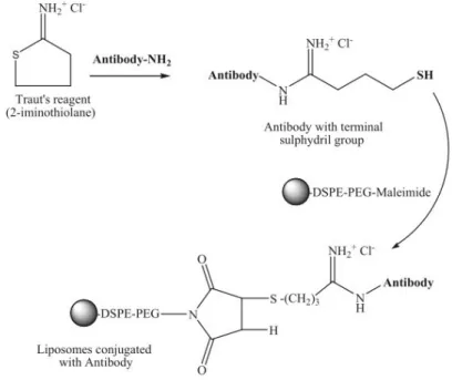
7.4 Immunoliposomes preparation: antibody derivatization and
conjugation to liposomes………...65
7.4.1 Experimental protocol………66
7.5 Separation of no-encapsulated compounds and unbound antibody
from liposomes………...66
7.5.1 Separation of no-encapsulated compounds from liposomes
by Ultrafiltration………...67
7.5.2 Separation of no-encapsulated compounds and unbound
antibody from immunoliposomes by Size Exclusion
Chromatography………67
7.6 Quantitative analysis of lipid concentration………....68
7.7 Size determination of liposomes………...69
8. Pinoresinol’s effects on phospholipid model membrane…………..69
8.1 Chemicals ………69
8.2 Determination of pinoresinol partition coefficient in model
membranes (K
P)………70
8.2.1 Spectroscopic measurements ………..70
8.2.2 Determination of capacity factor k’
IAMby IAM-HPLC....71
8.2.2.1 Experiemental procedure ………..73
8.3 Differential Scanning Calorimetry………..75
8.3.1 Experimental procedure ………77
8.4 Steady-state fluorescence anisotropy………....78
8.4.1 Experimental procedure ………...80
1. Polyphenols’ quantification by Folin-Ciocalteu technique………...82
2. Quantification of drugs’ content by HPLC analysis……….83
3. Cytotoxic effects of free Evoo A and free Evoo B on the viability of
JIMT1 and MCF7………..86
4.Cytotoxic effect of EVOO A and EVOO B incorporated into
liposomes………...89
5. Optimization of drugs’ encapsulation into liposomes...97
6. Determination of liposome’s size………..99
7. Expression of HER2 on breast cancer cell lines………..101
7.1 Detection of HER2 by Western blot……….101
7.2 Detection of membrane surface HER2 by Flow Cytometry….102
8. Comparative effect of the three delivery systems: free compounds,
liposomes and immunoliposomes...103
9. Spectroscopic measurement and determination of Pinoresinol
partition coefficient……….107
10. Partition value of Pinoresinol into phospholipid bilayers studied by
IAM-HPLC………..109
11. Effect of Pinoresinol on the thermotropic behaviour of neutral
charged phospholipids model membrane...110
12. Effect of Pinoresinol on the thermotropic behaviour of negatively
charged phospholipids model membranes...113
13. Steady-state fluorescence anisotropy ………...115
Discussion………...116
Conclusions………117
List of Abbreviations……….119
Acknowledgement……….127
1. The cancer burden
Every year 12 million people worldwide die of the results of atherosclerosis, heart infarctions, and strokes. These are the most common causes of death of our time. The second largest common disease is cancer. Coronary disease and cancer together are responsible for over 80% of all deaths in industrialized countries. In many Third World countries cancer incidence appears much lower, most likely because of the higher death rates due to infectious disease or injury . Based on the most recent incidence and mortality data available, there were 10.1 million new cases, 6.2 million deaths and 22.4 million persons living with cancer in the year 2000. In terms of incidence, the most common cancers worldwide (excluding non-melanoma skin cancers) are lung (12.3% of all cancers), breast (10.4%) and colorectum (9.4%) (Fig.1). Lung cancer is the largest single cause of deaths from cancer in the world (1.1 million annually), since it is almost invariably associated with poor prognosis. On the other hand, appropriate intervention is often effective in avoiding a fatal outcome following diagnosis of breast cancer. Hence this particular cancer, which ranks second in terms of incidence, is not among the top three causes of death from cancer, which are respectively cancers of the lung (17.8% of all cancer deaths), stomach (10.4%) and liver (8.8%) ( Fig. 1) .
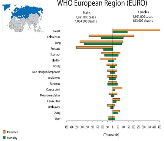
About the distribution of cancer between the sexes, in the European Region (EURO), as in the rest of the world, the commonest incident cancer for men is lung cancer followed by prostate, colorectal, bladder and stomach cancer. They are also the commonest forms of cancer death in men (Fig. 2). Among women breast cancer is the commonest form of cancer and it is also the main cancer cause of death in women, followed by colorectal cancer, lung cancer and stomach cancer (Fig. 2).
Figure 2 Cancer Incidence and Mortality in World Health Organization European Region (EURO) (Data from World Cancer Report 2008)
2.Breast cancer
2.1 Breast Cancer Current Statistics
Breast cancer is the formation of a malignant tumor that has developed from cells in the breast. A malignant tumor is a group of cancer cells that may grow into (invade) surrounding tissues or spread (metastasize) to distant areas of the body. The disease occurs almost entirely in women, but men can get it, too.
It is the most commonly occurring cancer in women, comprising almost one third of all malignancies in females. It is second only to lung cancer as a cause of cancer mortality, and it is the leading cause of death for American women between the ages of 40 and 55 .
The lifetime risk of a woman developing invasive breast cancer is 12.6 % - one out of 8 females in the United States will develop breast cancer at some point in her life . The American Cancer Society's most recent estimates for breast cancer in the United States are for 2012:
• About 226,870 new cases of invasive breast cancer will be diagnosed in women.
• About 63,300 new cases of carcinoma in situ (CIS) will be diagnosed (CIS is non-invasive and is the earliest form of breast cancer).
• About 39,510 women will die from breast cancer
From the 1940's until recently, the rate of new cases of breast cancer in the United States increased by a little over one percent a year. In the 1980's, the rate of new cases rose markedly (likely due to increased screening) and during the 1990's, the rate of new cases leveled off. Since 2003 there has been a marked decline in the rate of new breast cancer cases . This large decrease was thought to be due to the decline in use of hormone therapy after menopause that occurred after the results of the Women's Health Initiative were published in 2002. This study linked the use of hormone therapy to an increased risk of breast cancer and heart diseases. These decreases are believed to be, also, the result of earlier detection through screening and increased awareness, as well as improved treatment. Incidence rates have been stable since 2004. Rates of breast cancer around the world vary a great deal, with industrialized countries generally having higher rates than non-industrialized countries (Fig. 3). And, although all the factors responsible for this variation aren't known, differences between such countries in lifestyle and reproductive factors are thought to play a large role .
Fi gure 3 Breast cancer incidence (Data from American Cancer Society 2008).
2.2 The normal breast
In adults, each mammary gland is composed of fifteen to twenty sections called lobes, divided by adipose tissue. Each lobe is subdivided into lobules, which contain the glandular alveoli that secrete the milk into a series of secondary tubules.
These tubules converge to form a series of mammary ducts, which in turn converge to form a lactiferous duct that drains at the tip of the nipple (Fig. 4). Fatty tissue and connective tissue surrounding the ducts and lobules, blood vessels, and lymphatic vessels (stroma) .
Figure 4 Anatomy of the breast
2.3 Breast Cancer Types
The most common type of breast cancer is ductal cancer. It is found in the cells of the ducts. Cancer that starts in lobes or lobules is called lobular cancer. It is more often found in both breasts than other types of breast cancer. Cancers also are classified as non - invasive (in situ) and invasive (infiltrating). The term in situ refers to cancer that has not spread past the area where it initially developed. Invasive breast cancer has a tendency to spread (metastasize) to other tissues of the breast and/or other regions of the body. A less common type of breast cancer is inflammatory breast cancer characterized by general inflammation of the breast.
Other rare types of breast cancer are medullary carcinoma (an invasive breast cancer that forms a distinct boundary between tumor tissue and normal tissue), mucinous carcinoma (formed by the mucus-producing cancer cells), tubular carcinoma, etc .
2.4 Breast Cancer Risk Factors
Every woman is at risk for developing breast cancer. Several relatively strong risk factors for breast cancer that affect large proportions of the general population have been known for some time. However, the vast majority of breast cancer cases occur in women who have no identifiable risk factors other than their gender .
The “established” risk factors for breast cancer are female gender, age, previous breast cancer, benign breast disease, hereditary factors (family history of breast cancer), early age at menarche, late age at menopause, late age at first full-term pregnancy, post
menopausal obesity, low physical activity, race/ethnicity and high-dose exposure to ionizing radiation early in life.
The “speculated” risk factors for breast cancer include never having been pregnant, having only one pregnancy rather than many, not breast feeding after pregnancy, use of postmenopausal estrogen replacement therapy or postmenopausal hormone (estrogen/progestin) replacement therapy, use of oral contraceptives, certain specific dietary practices (high intake of fat and low intakes of fiber, fruits, and vegetables, low intake of phytoestrogens), alcohol consumption, tobacco smoking, and abortion.
Although men can and do develop breast cancer, the disease is 100 times more likely to occur in a woman than in a man . Women are at a higher risk of breast cancer because they have much more breast tissue than men do. Also, estrogen promotes the development of breast cancer. The risk of breast cancer is higher in middle-aged and elderly women than in young women . This risk increases as a woman ages, rising sharply after the age of 40. In 6 the United States, more than three-fourths of all breast cancers occur in women aged 50 or older. A woman who has previously had breast cancer has a three- to four-fold increased risk of developing a new cancer in the other breast. Women who have had benign breast problems are also at increased risk but to a lesser extent .
The risk of breast cancer is higher among women who have a close blood relative (mother, sister, or daughter) who have had the disease. The increase in risk is especially high if the relative developed breast cancer before the age of 50 or in both breasts .
However, most women who get breast cancer (approximately 80 percent) have no such family history of the disease . The effect of family history on breast cancer risk is believed to be due primarily to genetic factors. As much as 5–10 percent of all breast cancer cases are attributable to specific inherited single-gene mutations, and many other cases have some genetic component. The evidence from individual families in which breast cancer occurs very frequently and from large epidemiological studies has shown that some women have a familial predisposition to breast cancer. Epidemiological studies have shown that in women with a family history of breast cancer, the risk of breast cancer is increased two- to threefold. Studies have also shown that there are families in which breast cancer risk is inherited in an autosomal-dominant fashion (‘hereditary breast cancer’). Recently, it has been shown that germline mutations in the BRCA1 and BRCA2 genes account for a large proportion of cases of hereditary breast cancer . Histopathological findings and careful autopsy examinations have played a major role in the recognition of many familial cancer syndromes .
In addition mutations in the BRCA1 and BRCA2 genes, there are as yet unidentified genetic defects that predispose to breast cancer development , and additional studies may help in identifying these genes in the future. Women who reach menarche at a relatively early age (12 or younger) and those who reach menopause at a relatively late age (55 or older) are slightly more likely than other women to develop breast cancer These relationships are believed to be mediated through estrogen production . During the reproductive years, a woman’s body produces high levels of estrogen. Women who start to menstruate at an early age and/or reach menopause at a late age are exposed to high levels of estrogen for more years than are women who have a late menarche or early menopause. Age at first pregnancy is another aspect of reproductive history that that is associated with breast cancer risk. Women who have their first full-term pregnancy at a relatively early age have a lower risk of breast cancer than those who never have children or those who have their first child relatively late in life .The biologic basis for this relationship is not entirely clear. Obesity has been consistently associated with an increased risk of breast cancer among postmenopausal women. This relationship may be mediated again by estrogen production. Fat cells produce some estrogen and obese postmenopausal women, therefore, tend to have higher blood estrogen levels than lean women. Studies have consistently shown that the risk of breast cancer is lower among physically active premenopausal women than among sedentary women .
Physical activity during adolescence may be especially protective, and the effect of physical activity may be strongest among women who have at least one full-term pregnancy. Studies of racial/ethnic characteristics of breast cancer reveal that non-Hispanic white, Hawaiian, and black women have the highest levels of breast cancer risk. Other Asian/Pacific Islander groups and Hispanic women have lower levels of risk. Some of the lowest levels of risk occur among Korean and Vietnamese women .
Women who were exposed to high doses of radiation, especially during adolescence, have an increased risk of breast cancer. This association has been observed both among atomic bomb survivors and among women who received high-dose radiation for medical purposes .
Parity (having children) and the age of the woman at the birth of her first offspring are other endogenous hormonal factors that influence breast cancer. Women who have never had children (nulliparous) are at greater risk for the development of breast cancer than women who have had children (parous). There is also consistent evidence that first pregnancy completed before age 30-35 lowers risk of breast cancer, and that first full-term pregnancy after age 30-35 raises risk. More limited evidence suggests that women
who have many pregnancies may be less likely to develop breast cancer than those who have only one pregnancy .Some studies have shown that women who breast-feed their babies may be less likely to develop breast cancer than those who have children but do not breast-feed [22]. Other studies, however, indicate that there may be little or no relationship between breast feeding and breast cancer risk. If breast-feeding does protect against breast cancer, it may do so by delaying the resumption of ovulation (with its accompanying high estrogen levels) after pregnancy. The long-term (more than five years) use of postmenopausal estrogen therapy (ERT) or combined estrogen/progestin hormone replacement therapy (HRT) may be associated with an increase in breast cancer risk . The associations between the use of oral contraceptives and breast cancer have been studied. Many studies attempting to link oral contraceptives with increased breast cancer have been inconclusive .
But these studies have shown that oral contraceptives do not have a large effect on breast cancer risk. Whether they have a small effect on risk is less clear. A possible relationship between breast cancer and diet has been suggested due to the variation of breast cancer in societies with different national diets (the high rates in Western industrialized nations and the low rates in Asia, Latin America, and Africa). A comparison of vegetarian versus meat-eating women produced inconclusive results. No relation between breast cancer risk and total fat, saturated fat, or cholesterol was found. Some of the effects that were once attributed to dietary fat intake were probably due to obesity (which is often linked with high fat intake) rather than to fat intake per se. And the effects of fiber, fruits, and vegetables now appear to be small, at best. Diets high in fruits and vegetables and low in fat and calories are healthful for many reasons, and they may indirectly reduce the risk of breast cancer by helping to prevent obesity. Plant substances called isoflavones (sometimes referred to as phytoestrogens) are most commonly found in soy products. It has been speculated that these substances may be 10 protective against breast cancer . They appear to have effects similar to those of estrogen in some tissues while antagonizing the effects of estrogen in other tissues. A positive, but modest association between alcohol use and breast cancer risk is seen in most studies .
There is also some evidence that cigarette smoking may be associated with a small increase in breast cancer risk. However, epidemiological studies have variably shown positive, inverse, or null associations . Among women who have already been diagnosed with breast cancer, smoking may be associated with an increased risk that the cancer will progress more rapidly. In some studies, premature termination of pregnancy appears to increase breast cancer risk .
In incomplete pregnancy, the breast is exposed only to the high estrogen levels of early pregnancy and thus may be responsible for the increased risk seen in these women. However, some other studies found no association between abortions and increased risk of breast cancer .
2.5
Stages of breast cancer
The staging systems currently in use for breast cancer are based on the clinical size and extent of invasion of the primary tumor (T), the clinical absence or presence of palpable axillary lymph nodes and evidence of their local invasion (N), together with the clinical and imaging evidence of distant metastases (M). This is then translated into the TNM classification which has been subdivided into Stage 0 called carcinoma in situ (lobular carcinoma in situ (LCIS) and ductal carcinoma in situ (DCIS) and four broad categories by the Union Internationale Centre Cancer (UICC), which are the following.
Stage I – early stage breast cancer where the tumor is less than 2 cm across and hasn't spread beyond the breast .
Stage II – early stage breast cancer where the tumor is either less than 2 cm across and has spread to the lymph nodes under the arm; or the tumor is between 2 and 5 cm (with or without spread to the lymph nodes under the arm); or the tumor is greater than 5 cm and hasn't spread outside the breast.
Stage III – locally advanced breast cancer where the tumor is greater than 5 cm across and has spread to the lymph nodes under the arm; or the cancer is extensive in the underarm lymph nodes; or the cancer has spread to lymph nodes near the breastbone or to other tissues near the breast.
Stage IV – metastatic breast cancer where the cancer has spread outside the breast to other organs in the body (Figure 5).
2.6 Tumor Markers
A tumor marker is defined as a substance present/overexpressed in or produced by a tumor (tumor-derived), or the host (tumor-associated), that can be used for differentiating neoplastic from normal tissue. Tumor markers are found in cells, tissues, and body fluids such as cerebrospinal fluid, serum, plasma, and milk. Some widely used tumor markers include: AFP, Her2/Neu, beta-HCG, CA 19-9, CA 27.29 (CA 15-3), CA 125, CEA, and PSA. Some tumor markers are associated with many types of cancer; others, with as few as one. Some tumor markers are always elevated in specific cancers; most are less predictable. However, no tumor marker is specific for cancer and most are found in low levels in healthy persons, or can be associated with non-neoplastic diseases as well as cancer. Tumor markers have been categorized as enzymes, isoenzymes, hormones, specific cell membrane proteins, oncofetal and cell-specific antigens, carbohydrate epitopes, oncogene products, genetic changes, etc. There are only a handful of well-established tumor markers that are being used by physicians. Many other potential markers are still being researched. There are many studies now that are trying to find new genes involved in signaling molecules or proteins that “tell” cells to proliferate, invade or metastasize.
HER-2/neu is an oncogene-encoded growth factor receptor (homologue of epidermal growth factor (EGF) receptor), also known as c-erbB-2. It is overexpressed in breast cancers as a result of HER 2 proto-oncogene amplification. It is measured in the tissue from a biopsy either by immunological assays of the protein or PCR. The presence of HER-2/neu is generally associated with a more aggressive growth and poorer prognosis for breast and ovarian cancer .
The human epidermal growth factor receptors (HER), also known as ERbB receptors, are a family of signal transduction proteins, with partial homology that normally regulate cell growth and survival, as well as adhesion, migration, differentiation, and other cellular responses. There are 4 family members in humans, HER1, HER2, HER3, and HER4 composed of a cysteine-rich extracellular region, a lipophilic
transmembrane segment, an
intracellular domain with tyrosine kinase activity and a carboxy terminal domain that is autophosphorylated upon receptor activation (Fig 6). HER-family receptors become active upon interaction with ligand and subsequent dimerization, followed by activation of downstream signaling proteins by phosphorylation. In humans, the 4 HER proteins interact with a range of ligands, although HER2 has no known activating ligands . For example, HER1 interacts with at least 6 ligands: epidermal growth factor (EGF), transforming growth factor α (TGFα), amphiregulin, heparin-binding EGF-like growth factor, betacellulin, and epiregulin . HER2, also referred to as ERBB2, NEU, or HER2/neu, can act as a dimerization partner with the other HER receptor proteins. As there are no known activating ligands for HER2, its activity is a consequence of its dimerization with other HER-family receptors as well as its homodimerization. Upon activation, the tyrosine kinase domain of each of the HER-family receptors can activate downstream signaling molecules, such as those in the PI3K/Akt and RAS/RAF/MEK/MAPK pathways .
The HER2 receptor, a 185 kDa transmembrane glycoprotein, is encoded by the HER2 gene, a protooncogene located on the long arm of chromosome 17q21 . Because of its function as an activator of signaling pathways, HER2 plays a central role in a number of cellular processes, including proliferation, motility, and resistance to apoptosis. As mentioned above, HER2 has no known ligand and can heterodimerize with other HER proteins, thus allowing HER2 to participate in a number of signal transduction pathways in the absence of a specific ligand.
This effect may be enhanced by the overexpression of HER2 in cancer cells, leading to increased cell proliferation and decreased cell death, as well as changes in cell motility. The association of HER2 protein overexpression or HER2 gene amplification with cancer, notably with breast cancer, was reported over 20 years ago.
HER2 is normally expressed at low levels in adult cell types including the breast epithelium, the central nervous system, bone, muscle, skin, heart, lungs and intestinal epithelium. The function of HER2 in adult tissues is not fully understood, however the receptor appears to play a role in the proliferation and differentiation of epithelial cells and in the protection of cardiomyocytes against apoptosis . In fetal tissues, however, HER2 is widely expressed and is critical to normal development. The clinical significance of HER2 in cancer was first discovered in the early 1980s following the identification of the neu onocogene, the mutationally active rat homologue of HER2 . The human homologue was soon identified and found to be overexpressed in a mammary carcinoma cell line [37]. On the basis of these findings, Slamon and co-workers examined HER2 expression in a series of primary human breast tumours and reported a significant association between HER2 overexpression, relapse and patient survival . HER2 is now known to be overexpressed in approximately 20-30% of BCs , and overexpression is also common in ovarian, prostate, lung, gastric and oral cancers .
Overexpression of HER2 is a combined result of increased transcription and protein translation. Indeed, breast cancer cells may have as many as 100copies of the gene per cell compared with two copies of the HER2-gene innormal cells [31]. Moreover, there are approximately 20 thousand receptors per cell on normal cells, but breast cancer cells may contain as many as 500 thousand to 2 million HER2 receptors per cell. At this high level of HER2 receptor expression, the kinase activity of HER2 becomes constitutively activated which appears to exert potent mitogenic and transforming effects on cells (Fig.
7).
This high density of HER2 promotes the formation of HER2 heterodimers as well as the formation of ligand-independent constitutively active HER2 homodimers . HER2-containing dimers persist longer on the cell surface due to their slower rate of internalization, resulting in overactive cell 18 signaling and leading to dysregulated cell growth, proliferation and malignant transformation [37]. Overexpression of the HER2 receptor is associated with poor prognosis in patients with breast cancer, as well as with aggressive tumor growth and metastases. HER2 positivity has also been associated with tumor grade, 1, 6 positive lymph node metastases at presentation, 1 and mitotic count . HER2+ tumors are less likely to be hormone receptor (estrogen receptor [ER] or
progesterone receptor [PgR]) –positive. HER2 status also correlates with relative response to various agents. HER2 positivity may result in increased resistance to endocrine therapy and with a decreased benefit from non-anthracycline, non-taxane– containing chemotherapy. Conversely, HER2-positive patients may exhibit improved response to anthracycline therapy, as well as to paclitaxel .Data surrounding the prognostic and predictive value of HER2 status are continually evolving. In a recent study, it was shown that higher levels of HER2 gene amplification were associated with worse outcomes in patients treated with doxorubicin-based therapy in the adjuvant setting. The association of HER2 gene amplification or HER2 overexpression with some breast cancers has allowed for the development of agents that specifically target HER2, altering the treatment landscape for these cancers. Trastuzumab, which was approved for the treatment of metastatic breast cancer in 1998, for the adjuvant treatment of lymph node–positive breast cancer in 2006, and for the adjuvant treatment of lymph node– negative breast cancer in 2008, is a umanized monoclonal antibody to the HER2 protein. Lapatinib is a selective inhibitor of the tyrosine kinase activity of HER2 and EGFR. Each of these agents has shown efficacy in patients whose tumors are HER2+ .
Figure 7 Amount of
HER2 in normal and tumoral breast cell
2.8. Trastuzumab Monotherapy
Trastuzumab (Herceptin®, Genentech, Inc., San Francisco, CA), is currently the only HER2-targeted therapeutic agent that has received marketing clearance from the
U.S. Food and Drug Administration (FDA) for use in the treatment of patients with HER2- overexpressing breast cancer .Has been used to treat almost a million women worldwide since 1998. Herceptin given for one year delivers high cure rates in women with HER2-positive early breast cancer. It reduces the risk of cancer coming back by half (50%), as consistently shown by five large studies involving over 15,000 patients and it reduces the risk of dying from breast cancer by a third (34%) Herceptin extends survival across all stages of HER2-positive breast cancer. In advanced (metastatic) HER2-2/4 positive breast cancer, it extends survival by up to four to eight additional months of life on average when used with chemotherapy (Taxol [paclitaxel] and Taxotere® [docetaxel]).Continued treatment with Herceptin prolongs survival without cancer progression in women requiring additional treatment after their cancer had progressed, even though they had already received previous Herceptin therapy. A recent trial has indicated that patients with HER2-positive metastatic breast cancer and central nervous system metastases who receive Herceptin and chemotherapy may experience longer survival than those who do not receive Herceptin therapy. Trastuzumab is a humanized mAb that binds specifically to HER2 on the C-terminal portion of the extracellular domain (ECD) near the juxtamembrane region in domain IV of the receptor. Trastuzumab was constructed by grafting the complementary-determining regions (CDRs) from the murine mAb 4D5 into a human kappa IgG1 to avoid eliciting a human anti-mouseantibody (HAMA) response in patients .
2.8.1 The mechanisms of action
The effectiveness of trastuzumab appears to be correlated with the level of HER2 expression in breast cancer, and with the accessibility of tumors to the drug . However, the mechanism by which trastuzumab induces regression of HER2-overexpressing tumors is not completely understood, but several molecular and cellular effects have been observed in experimental in vitro and in vivo models. Several of the proposed mechanisms are described below.
2.8.1.1 Immune-mediated response
One of the proposed mechanisms of trastuzumab anti-tumor action is through antibody-dependent cellular cytotoxicity (ADCC) . Specifically, the natural killer (NK) cells are important for the ADCC response to trastuzumab . These cells express the Fcɣ receptor that binds the Fc domain of the IgG1 trastuzumab antibody, and promotes lyses of trastuzumab-bound cancer cells. The importance of this immunological effect was
revealed by Clynes et al. , who achieved a tumor regression rate of 96% in mice bearing HER2-overexpressing BT-474 xenografts treated with trastuzumab, but only a 26% reduction in tumor growth in mice lacking the Fcɣ receptor.
2.8.1.2 Inhibition of angiogenesis
Both primary and metastatic breast cancer are dependent on angiogenesis for tumor growth . Trastuzumab can modulate different pro- and antiangiogenic factors to achieve angiogenesis suppression .
2.8.1.3 Inhibition of HER2 extracellular cleavage
The ECD of HER2 can be released by proteolytic cleavage from the fulllength receptor, yielding a 110-kDa fragment that can be detected in vitro in cell culture medium, and a 95-kDa amino-terminally truncated membrane associated fragment with increased kinase activity . The HER2-ECD can also be detected in vivo in serum, and is currently measured in the clinic withan FDA approved ELISA-based test to follow-up and monitor patients with metastatic breast cancer .Molina et al. demonstrated in HER2-overexpressing SK-BR-3 and BT-474 human breast cancer cells that trastuzumab can block metalloprotease-mediated cleavage of the HER2- ECD. Moreover, several clinical studies have demonstrated that a decline in serum HER2-ECD during trastuzumab treatment correlates with improved tumor responsiveness and progression-free survival , which indirectly supports the hypothesis that trastuzumab may act by inhibiting HER2 cleavage .
2.8.1.4 Inhibition of PI3K pathway
Overexpression of HER2 receptor tyrosine kinases leads to ligandindependent homodimerization and autophosphorylation of tyrosine residues on the cytoplasmic domain of the receptors. Phosphatidylinositol 3' kinase (PI3K) associates with these phosphorylated tyrosine residues and activates downstream effectors, which ultimately leads to enhanced protein synthesis, cell proliferation, survival and motility [64]. Trastuzumab can inhibit the PI3K pathway.
2.8.2. The resistance to trastuzumab
Despite the fact that trastuzumab-based treatment strategy has established a milestone in the therapy of HER-2 positive breast cancer with attractive clinical benefits, either as a single agent or in combination settings, one of the major drawbacks of the
trastuzumab-therappy is trastuzumab resistance, even in highly selected HER-2 overexpressed patients. In fact, only about 30% of HER-2 positive metastatic breast cancer patients respond to trastuzumab and approximately 70% of patients with overexpressed HER-2 receptor may have primary resistance . Additionally, the majority of those patients who achieve initial efficacy tend to develop secondary trastuzumab resistance within one or two years .
Several mechanisms have been postulated in an attempt to explain both intrinsic and acquired resistance to trastuzumab but it is not completely understood.
a) Cleavage of HER-2 extracellular domain to form the truncated HER-2 receptor and the overexpression of membrane associated mucin MUC4 to mask or block the trastuzumab binding site, which can interrupt the interaction between HER-2 receptor and this antibody .
b) Although trastuzumab reduces HER2-mediated signaling, it might not reduce signaling mediated through other HER receptors . For example, heterodimerisation of HER2 with other receptors of the erbB2-family can be induced by ligands of HER1, c) bypass signalling through the non-EGFR family growth factor receptor insulin-like growth factor-1 receptor (IGF-1R), enables activation of the downstream signal cascades without the participation of HER-2. Therefore, the further understanding of trastuzumab actionand resistance mechanisms highlights the need of novel targeted drugs aiming at HER-2 overexpression .
HER3 and HER4, and in the presence of an excess of ligands, the resulting heterodimers may initiate mitogenic signaling even in the presence of trastuzumab .
Indeed, increased levels of the ErbB family ligands EGF and heregulin blocked trastuzumab-mediated growth inhibition of HER2- overexpresssing breast cancer cell lines, and this inhibition was associated with increased signaling from HER2/HER1 (EGFR) and HER2/HER3 complexes .
Valabrega et al. compared tumor tissue from patients with advanced HER2-positive breast cancer before and after trastuzumab treatment, and observed a strong increase in TGFα production upon disease progression, suggesting a possible role of EGFR signaling by TGFα as mediator of acquired resistance to trastuzumab. Indeed, these authors found that trastuzumab was less efficient at inhibiting the growth of HER2-positive SKBR-3 cells engineered to overexpress Tumor Growth Factor alpha (TGFα compared to the parental cells .
Cardiovascular diseases and cancer are leading cause of morbidity and mortality in most developed countries. Cancer prevention is therefore an absolute priority for public health, and it should be the obvious choice for financial, social, and above all, human reasons . Cancer and cardiovascular diseases are both known to be fairly refractory to curative interventions. In addition, the occurrence of these diseases exhibits a wide between-country variability and is largely determined by environmental factors and lifestyle habits. Primary prevention through diet and lifestyle is today an established reality to prevent coronary heart disease and stroke, and has achieved an important role in reducing the incidence of, and mortality from, cardiovascular diseases . However, attempts at prevention of cancer have thus far had very modest success . One of the potential answers to overcome this underachievement is related to the growing interest in the Mediterranean dietary pattern as a model for healthy eating and for the primary prevention of cancer. The incidence of certain cancers (e.g. breast and colon cancers) is (or it was) lowest in the Mediterranean area (Fig. 8)
Figure 8 (A) Per capita consumption of olive oil in selected countries (Olive Oil Council Data). (B) Rates of coronary heart disease and cancer mortality according to the World Health Organization standard (Cardiovascular Disease Infobase).
The traditional Mediterranean diet (MD) is the heritage of millennia of exchanges of people, cultures and foods of all countries around the Mediterranean basin. Some 20 countries can be thought of as Mediterranean . Their populations vary in culture, ethnicity, religion, economic development and other factors that can influence dietary intake. Variations on the Mediterranean diet exist in parts of Italy, France, Portugal,
Spain, and elsewhere in the Mediterranean basin, including North Africa and the Middle East . The implication that all Mediterranean people have the same diet is therefore a misnomer, since countries around the Mediterranean basin have different diets, religions and cultures. Their diets vary in the amount of total fat and olive oil consumption, wine intake, type of meat and dairy products, as well as fruit and vegetable. For example, the Italian diet includes a high consumption of pasta, whereas pulses are common in Greece and fish consumption is relatively high in Spain . Moreover, considering the same food item, the nutrient content can vary considerably in different countries. Even in the same country, dietary patterns may be different: for example, dietary patterns are different in northern and southern Italy, the average consumption of cereals, fruit and vegetables being higher in southern Italy, and milk and dairy products in northern Italy . But the dietary patterns that prevail in the Mediterranean region have many common characteristics.
The traditional Mediterranean diet may be thought of as having eight components: 1. High ratio of monounsaturated to saturated dietary lipids (mainly olive oil); 2. Moderate ethanol consumption;
3. High consumption of legumes;
4. High consumption of non-refined cereals, including bread; 5. High consumption of fruits;
6. High consumption of vegetables;
7. Low consumption of meat and meat products;
8. Moderate consumption of milk and dairy products (Fig 9).
Figure 9Mediterranean Diet Foods
All these items are important sources of dietary antioxidants. Apart from the most widely known antioxidants contained in fruits and vegetables, other compounds such as
oleuropein, hydroxytyrosol and other polyphenols present in olive oil, and resveratrol, from red wine, possess a marked antioxidant activity and other advantageous biological properties. The role of free radical production in carcinogenesis together with many epidemiologic studies linking antioxidant intake with a reduced incidence of cancer indicates that dietary antioxidants probably play a protective role. Therefore, the highly palatable traditional Mediterranean diet has many options to be the first choice in the dietary prevention of cancer .
Many epidemiological studies, conducted since the 1980s, investigated the role of the Mediterranean diets on the risk of various chronic diseases. Several aspects of the Mediterranean diet have been related to reduced risks of overall mortality , cardiovascular diseases, as well as several common neoplasms . It has been estimated that up to 25% of colorectal, 15% of breast and 10% of prostate, pancreas and endometrial cancer could be prevented by shifting to a healthy Mediterranean diet .
4. Health benefits of olive oil
Food can be regarded as functional if it is satisfactorily demonstrated to affect beneficially one or more target functions in the body, beyond adequate nutrition, in a way that improves health and well-being or reduces the risk of disease. Extra virgin olive oil (EVOO) could be considered as functional food. Its health properties have been discussed extensively in literature . Different countries and regions in the Mediterranean basin have their own dietary traditions, but in all of them olive oil occupies a central position. The relationship between the intake of olive oil and cancer risk has become a controversial issue that could have very important repercussions in human health as it may have a potential role in lowering the risk of some human neoplasms .
Thus, different studies have shown that the consumption of olive oil have a potential protective effect towards several malignancies, especially breast cancer (stomach, ovary, colon and endometrium cancer too) .
Virgin olive oil (VOO) is a vegetable oil, which can be obtained directly from olive fruit using only mechanical extraction and which can be consumed without further treatments.
The chemical composition of olive oil is principally glycerols representing more than 98% of the total weight. Minor components amount to about 2% of the total oil weight and include, among others, aliphatic and triterpenic alcohols, sterols, hydrocarbons, volatile compounds, and several antioxidants .
Phenolic compounds represent one of these families of antioxidants which are present in olive oil.
They are of fundamental importance for their nutritional properties, sensory characteristics, and the shelf life of virgin olive oil, since they have high antioxidative properties, can confer a marked bitter taste or a sweet taste typical of some virgin olive oils. They also play an important role in human nutrition as preventative agents against several diseases .
4.1 Phenolic compounds content of olive oils
Among olive oil compounds, the phenolic fraction has received considerable attention in recent years. Evidence from several studies have revealed that the protective effects of EVOO against chronic diseases such as atherosclerosis, cancer, obesity, diabetes, and coronary diseases are related to the phenolic compounds. The pharmaceutical interest in olive oil phenolic compounds due to their bioactivity on different cancer cells is also well known and has stimulated multidisciplinary research on the composition of olive biophenols. The bioactivity of the phenolic compounds in these chronic diseases could be related to different properties such as antioxidant and anti-inflamatory although the molecular mechanism of these compounds in relation to many diseases could have different cellular targets. The most important phenolic compounds that have been identified on EVOO may be divided into different groups such as phenolic acids, phenolic alcohols, secoiridoids, lignans, and flavones (Fig. 10).
The main components of the phenolic fraction of EVOO are hydroxytyrosol, tyrosol, and their derivatives linked to the aldehydic and dialdehydic forms of elenolic acid, which are described as secoiridoids (oleuropein, demethyloleuropein, ligstroside). Moreover, significant amounts of lignans, such as pinoresinol and acetoxypinoresinol, are also present, as well as flavonoids (luteolin and apigenin), phenolic acids (such as caffeic, vanillic, syringic, p-coumaric, o-coumaric, protocatechuic, sinapic and p-hydroxybenzoic acid) and hydroxy-isochromans. The exact composition of phenolics compounds in EVOO is related to agronomic and technological aspects .
While the phenolic acids, phenolic alcohols and flavonoids occur in many fruits and vegetables belonging to various botanical families, secoiridoids are present exclusively in plants belonging to the Family of Olearaceae. 3,4-Dihydroxyphenyl- ethanol , or hydroxytyrosol, and p-hydroxyphenyl-ethanol, or tyrosol are the main phenolic alcohols in VOO (Fig. 10).
Figure 10 Structures of some EVOO polyphenols
4.2 Health Benefits from phenolic compounds of EVOO
Research has documented a wide variety of anti-inflammatory mechanisms used by olive oil polyphenols to lower our risk of inflammatory problems. These mechanisms include decreased production of messaging molecules that would otherwise increase inflammation (including TNF-alpha, interleukin 1-beta, thromboxane B2, and leukotriene B4); inhibition of pro-inflammatory enzymes like oxygenase 1 and cyclo-oxygenase 2; and decreased synthesis of the enzyme inducible nitric oxide synthase .
Many different cardiovascular problems - including gradual blocking of the arteries and blood vessels (called atherosclerosis) - have their origin in two unwanted circumstances.
The first of these circumstances is called oxidative stress. Oxidative stress means too much damage (or risk of damage) from the presence of overly reactive oxygen-containing molecules. One of the best ways to help avoid oxidative stress is to consume a diet that is rich in antioxidant nutrients. Many foods contain valuable amounts of antioxidants and anti-inflammatory compounds, but few foods are as rich in these compounds as extra virgin olive oil, and this fact alone accounts for many of the research-based benefits of this culinary oil for health of our cardiovascular system. In terms of antioxidant protection for our blood vessels, olive oil has been shown to lower risk of lipid peroxidation (oxygen damage to fat) in our bloodstream. Many of the fat-containing molecules in our blood - including molecules like LDL - need to be protected from oxygen damage. Oxygen damage to molecules like LDL significantly increases our risk of numerous cardiovascular diseases, including atherosclerosis. Protection of the LDL molecules in our blood from oxygen damage is a major benefit provided by olive oil and its polyphenols .
Several of the polyphenols found in olive oil - including hydroxytyrosol, oleuropein and luteolin - appear to be especially helpful in keeping our blood platelets in check and avoiding problems of too much clumping (called platelet aggregation). There are also two messaging molecules (called plasminogen activator inhibitor-1 and factor VII) that are capable of triggering too much clumping together of the platelets, and the polyphenols in olive oil can help stop overproduction of these molecules .
Benefits of olive oil for the digestive tract were first uncovered in research on diet and cancers of the digestive tract. Numerous studies found lower rates of digestive tract cancers - especially cancers of the upper digestive tract, including the stomach and small intestine - in populations that regularly consumed olive oil. Studies on the Mediterranean Diet were an important part of this initial research on olive oil and the digestive tract. Protection of the lower digestive tract (for example, protection of the colon from colon cancer) is less well-documented in the olive oil research, even though there is some strongly supportive evidence from select laboratory animal studies. Many of these anti-cancer effects in the digestive tract were believed to depend on the polyphenols in olive oil and their antioxidant plus anti-inflammatory properties . One fascinating area of recent research has involved the polyphenols in olive oil and the balance of bacteria in our digestive tract. Numerous polyphenols in olive oil have been shown to slow the growth of unwanted bacteria, including bacteria commonly responsible for digestive tract infections. These polyphenols include oleuropein, hydroxytyrosol, and tyrosol. Some of these same polyphenols - along with other olive oil polyphenols like ligstroside - are
specifically able to inhibit the growth of the Helicobacter pylori bacterium. This effect of the olive oil polyphenols may be especially important, since overpopulation of Helicobacter bacteria coupled with over-attachment of Helicobacter to the stomach lining can lead to stomach ulcer and other unwanted digestive problems .
The polyphenols found in olive oil are a natural for helping us lower our risk of certain cancer types. Many types of cancer only get initiated when cells are overwhelmed by oxidative stress (damage to cell structure and function by overly reactive oxygen-containing molecules) and by chronic excessive inflammation. Since the polyphenols in olive oil act both as antioxidants and anti-inflammatory molecules, they are perfectly suited for lowering our cells' risk of oxidative stress and chronic unwanted inflammation. Research studies have shown that as little as 1-2 tablespoons of olive oil per day can lower our risk of certain cancer types, including cancers of the breast, respiratory tract, upper digestive tract, and to a lesser extent, lower digestive tract (colorectal cancers) . While most of the anti-cancer research on olive oil has focused on its polyphenols and their antioxidant and anti-inflammatory properties, several studies have uncovered other fascinating ways in which olive oil provides its anti-cancer benefits. These other ways include the improvement of cell membrane function in a way that lowers risk of cancer development and the altering gene expression in cells in a way that enhances their antioxidant defense system. A final important mechanism linking olive oil intake to decreased cancer risk involves protection of our DNA. The antioxidants in olive oil appear to have a special ability to protect DNA (deoxyribonucleic acids) - the key chemical component of genetic material in our cells - from oxygen damage. DNA protection from unwanted oxidative stress means better cell function in wide variety of ways and provides a cell with decreased risk of cancer development t. There is also encouraging research on the potential for olive oil to help with control of certain cancers once they have already developed. For example, improvement of breast cancer status has been an area of particular interest in olive oil research. Here some of the research has focused on the secoiridoids in olive oil (especially oleocanthal), and its ability to help keep breast cancer cells from reproducing . Another example involves the ability of hydroxytyrosol (HT) in olive oil to trigger programmed cell death (apoptosis) in colon cancer cells . HT may be able to accomplish this anti-cancer effect by helping block the enzymatic activity of fatty acid synthetase (FAS). These cancer-controlling properties of olive oil and olive oil constituents are generally referred to as the "antiproliferative" properties of olive oil.
In numerous laboratory studies, flavonoids have demonstrated the ability to inhibit aromatase activity and thus lower estrogen biosynthesis and circulating estrogen levels, inhibit tumor cell proliferation, and inhibit the formation of reactive oxygen species, all of which are mechanisms thought to influence breast cancer development. Furthermore, dietary intake of certain flavonoids has been reported to potentially protect humans from developing certain types of cancer, including breast cancer .
Several showed that higher dietary intakes of flavonoids and lignans were associated with a reduction in the risk of postmenopausal breast cancers .
Oleuropein has several pharmacological properties, including antioxidant [2], antiinflammatory [36], anti-atherogenic [37], anti-cancer [38], antimicrobial [39], and antiviral [40], and for these reasons, it is commercially available as food supplement in Mediterranean countries. In addition, oleuropein has been shown to be cardioprotective against acute adriamycin cardiotoxicity [41] and has been shown to exhibit anti-ischemic and hypolipidemic activities .
Lignans have anticancer effects in the breast, lung, skin and colon . Antioxidant and antiviral properties of lignans have been reported, and the similarities in structure between some lignans and estradiol or tamoxifen suggest possible activity against breast cancer .
Fini et al. have demonstrated that pinoresinol-rich EVOO have potent chemopreventive properties and specifically is able to decrease the cell viability, to induce apoptosis and modulate cell cycle dynamics in colorectal cancer cell lines.
4.3 Olive oil polyphenols and breast cancer risk
It has been repeatedly suggested that the ability of Mediterranean diet to significantly reduce the risk of several types of human carcinomas including breast cancer , can be largely attributed to the unique healthy characteristics of EVOO, an integral ingredient of the traditional MD. Although these findings might suggest that, in the future, the use of supplements derived from EVOO will be a useful strategy for the prevention and/or treatment of cancer, both the specific components and the specific molecular mechanisms that exert EVOO-related anti-carcinogenic effects have not yet been thoroughly elucidated.
Hydrophilic phenolics represent the most abundant family of bioactive EVOO compounds. As for many plant-derived phenolics, it has been largely assumed that EVOO-derived complex phenols such as secoiridoids (that include aglycone derivatives of oleuropein, dimethyloleuropein and ligstroside, which are also present in olive fruit)
and lignans (such as pinoresinol and acetoxypinoresinol) provide health benefits mainly because of their antioxidant activity . However, the antioxidant capacity of polyphenols does not directly correlate with their efficacy in terms of anticancer activity. Moreover, plasma concentrations of EVOO polyphenols when provided in the diet are often far lower than the levels required for protection against oxidation. It could be argued that metabolites of EVOO polyphenols can reach EVOO-derived compounds, however, tend to have a decreased antioxidant activity compared to parent compound . A large body of evidence indicates that olive oil polyphenols are capable of significantly affecting the overall process of carcinogenesis due to their abilities to inhibit cell cycle, cell proliferation or oxidative stress, improve the efficacy of detoxification enzymes, induce apoptosis, and stimulate the immune system.
Alternatively to general mechanisms largely related to the antioxidant and/or trapping activity of oxygen radicals commonly observed in many plant-derived phenolics, recent studies have demonstrated that complex polyphenols can exert an anti-carcinogenic effect by directly modulating the activities of various types of receptor tyrosine kinases (RTKs) including several members of the HER family . Results studies support the notion that EVOO-derived complex polyphenols may constitute a previously unrecognized family of clinically valuable anti-cancer phytochemicals that significantly affect breast cancer cell proliferation and survival through a molecular mechanism involving the specific suppression of the activity, expression and signal transduction events of the Type I RTK HER2 .
Menendez et al. investigated the anti HER-2 effects of phenolic fractions directly extracted from commercial extra virgin olive oil (EVOO) in cultured human breast cancer cell lines. They tested for the ability to kill both HER-2 positive and negative tumors. The effects of the EVOO fractions on the expression and activation status of HER-2 oncoprotein were evaluated.
They found that all the major EVOO polyphenols (i.e. secoiridoids and lignans) induced strong tumor killing effects by selectively triggering high levels of apoptotic cell death (programmed cell death) in cells over expressing HER-2. The EVOO polyphenols drastically depleted HER-2 protein and reduced autophosphorylation in a dose and time dependent manner. EVOO polyphenols induced HER-2 downregulation regardless of the molecular mechanism contributing to HER-2 overexpression. (i.e.naturally by gene amplification and ectopically driven by a viral promoter).
The researchers concluded that the ability of EVOO derived polyphenols to inhibit HER-2 activity by promoting the degradation of the HER-HER-2 protein itself, together with the fact
that humans have safely been ingesting polyphenols from olive oil for as long as they have been consuming olives and olive oil, support the notion that EVOO is an excellent and safe treatment targeting HER-2.
EVOO-derived phenolics with strong anti-JIMT-1 activity may underscore innovative cancer molecules with novel therapeutic targets because, in order to elicit tumoricidal effects, they should affect the expression and/or activity of genes and transduction cascades closely involved in enhanced cancer cell survival. Importantly, EVOO polyphenols (i.e. lignans and secoiridoids) – but not monophenols and phenolic acids – strongly suppressed the growth of breast cancer cells bearing high levels of HER2 (erbB2) – one of the most commonly analyzed oncogenes that plays a decisive role in malignant transformation, tumorigenesis andmetastasis in a biologically aggressive subset of human breast carcinomas .
MENENDEZ et al. designed a systematic approach to investigate the tumoricidal and the anti-HER2 effects of EVOO-derived single phenols (i.e. tyrosol and hydroxytyrosol), phenolic acids (i.e. elenolic acid), lignans (pinoresinol and acetoxypinoresinol) and secoiridoids (i.e. deacetoxy oleuropein aglycone -DAOA-, oleuropein aglycone and ligstroside aglycone) directly extracted from EVOO. EVOO-derived lignans and secoiridoids were significantly more effective than EVOO-derived single phenols and phenolic acids to inhibit HER2-associated tyrosine kinase activity strongly suggests that EVOO phenolic compounds with a simple structure (i.e. involving only a single phenol ring) cannot exert inhibitory effects against the tyrosine kinase domain of HER2, and that a more complex structure (i.e. involving two or more phenol rings) is required to efficiently block HER2 tyrosine kinase activity.
Lozano et al. employed crude phenolic extracts (PEs) directly obtained from 14 different monovarieties of EVOOs produced in Spain to preliminary delineate both the biological actions (in terms of cytotoxicity) and the clinical value (in terms of physiologically relevant concentration ranges) of complex multicomponent PEs against HER2 gene-amplified SKBR3 breast cancer cells.
The 14 EVOO varieties had significantly different phenolic compositions, in which secoiridoids were the major phenolic fraction (>90% of total phenolics) in 11 EVOO monovarieties) and lignans were significantly enriched (5-10%of total phenolics) in three EVOO monovarieties. When compared with EVOO PE containing low to undetectable amounts of lignans, their data clearly demonstrated that lignans-enriched EVOO varieties had a relatively weak ability to alter cell viability in the SKBR3 breast cancer model. Thus, the cytotoxic potency of the lignans-negative EVOO-PE 7 (Picual variety from
Cordoba) was found to be 12 times higher than that observed in lignans-enriched EVOO-PE 12 (Arbequina variety from Reus). It should be noted, however, that EVOO-PE exhibiting small differences in their secoiridoid content notably differed in their abilities to significantly decrease breast cancer cell viability. These findings, altogether, strongly suggest that quality rather than quantity of the entire battery of complex phenols present in individual EVOO-PE ultimately dictate their antibreast cancer cytotoxic effects.
4.4 Limits in the use of bioactive natural compounds
The biological activities of plant polyphenols have been examined by various methods in vitro and in vivo for prediction of their ability to prevent human diseases. Unfortunately, these valuable natural compounds’s uses are substantially limited . It is reported that the polyphenol concentrations needed to obtain in vitro efficiency are generally superior to in vivo moderate levels. According to the route of administration, the efficiency of these compounds depends on their bioavailability and integrity. Indeed, a small proportion of molecules administered orally are absorbed, because of insufficient gastric residence time, low permeability and/or low solubility. Their instability during food processing, distribution or storage, or in the gastrointestinal tract (pH, enzymes, presence of other nutrients), limits the activity and the potential health benefits of polyphenols. The topical use of natural polyphenols is also delicate because of their important sensitivity to environmental factors, including physical, chemical, and biological conditions. Unfortunately, they oxidize very quickly, leading to the progressive appearance of a brown color and/or unwanted odors with a considerable loss in activity. Other problems related to polyphenol use in human health have to be solved. A large number of polyphenolic compounds from natural sources are interesting for their properties, however in their free form, they can show limited water solubility. Furthermore, many polyphenols have an unpleasant taste which must be masked before their incorporation in foodstuffs or oral medicines. Therefore, the administration of phenolic compounds requires the formulation of a finished protecting product able to maintain the structural integrity of the polyphenol until the consumption or the administration, mask its taste, increase its water solubility and bioavailability, and convey it precisely towards a physiological target. Among the existing stabilization methods, encapsulation is an interesting means. The use of encapsulated polyphenols instead of free compounds is the source of numerous works.
Drug delivery systems (DDSs) can improve the pharmacological properties of traditional drugs by altering drug pharmacokinetics and biodistribution .
DDS can include liposomes, nanospheres, micelles, dendrimers, as well as various polymeric-based systems . Among the many DDS available, liposomes gained popularity in recent years thanks to their clinical success.
Due to their small size (about 100 nm or less), they readily extravasate from circulation through vascular gaps or defects attributed to ongoing angiogenesis that is typical of tumour sites , which have been reported to be about 200 nm or greater. DDS retention within these sites is generally high due to the poor lymphatic drainage observed within tumors . Furthermore, their lower size limit of diameter ensures that these vehicles do not randomly penetrate normal vessel walls. In cancers, an imbalance in factors that regulate angiogenesis, such as overexpression of, results in both increased vascular permeability and chaotic tumour-vessel architecture. In combination, these effects cause enhanced permeation and retention (EPR), resulting in high local drug concentrations.
5.1 Liposomes
Liposomes are spherical, self-closed structures formed by one or more concentric
lipid bilayers containing an aqueous phase inside and between the bilayers (Fig. 11 A).
The lipids used in the formation of liposomes are usually comprised of a hydrophilic headgroup and two hydrophobic fatty acyl chains. These amphiphilic molecules
spontaneously assemble into aggregates in an aqueous environment. Phosphatidylcholine
(PC), also called lecithin, is a biocompatible phospholipid that exists in plants and animals and used frequently in liposomal preparation. Moreover, there are other molecules widely used in combination with phospholipids, such as cholesterol.
The exact location of a drug in liposomes will depend upon its physicochemical characteristics and the composition of the lipids.
However, as a general rule, the hydrophilic drug molecules can be encapsulated in the aqueous space whereas the hydrophobic and amphiphilic molecules can be incorporated into the lipid bilayer (Fig. 11 B).


![Figure 19 Cleavage of the yellow-colored tetrazolium salt, 3-[4,5- dimethylthiazol-2-yl]-2,5-diphenyl tetrazolium bromide (MTT) into a purple-colored formazan by the mitochondrial enzyme succinatedehydrogenase](https://thumb-eu.123doks.com/thumbv2/123dokorg/4520450.34890/57.892.207.674.210.432/cleavage-tetrazolium-dimethylthiazol-diphenyl-tetrazolium-formazan-mitochondrial-succinatedehydrogenase.webp)